Original Research Article
Treatment outcome of postoperative radiotherapy for squamous cell
carcinoma of the head and neck
H. U. Ghori*, M. Sohaib, Vivek Tiwari, Manish Ahirwar
INTRODUCTION
Head and neck cancer are common in India. It account for 30 percent of cancer in male and 13 percents in female. Nearly 60 percents of cases presents in advance stages. Loco regional failure constitute predominant recurrence pattern and most death result from uncontrolled local or/regional disease. The two important main treatment modalities for patients with squamous cell carcinoma of the head and neck are surgery and radiotherapy (RT). Patients with stage I and II disease are optimally treated with one modality. Unfortunately, a large number of patients present with stage III and IV disease. The organ preservation treatment modalities using RT alone or with concomitant chemotherapy have proven to be acceptable
for patients with favorable low volume malignancies. Despite regimens that permit organ preservation in selected patients with advanced carcinomas of the head and neck, ablative surgical resection and postoperative radiotherapy are required in many patients. Various studies shows that many local or regional disease, recurrence occur in 30 percent of patients, and distant metastases occurs in 25 percents.4 Patients who have two or more regional lymph nodes involved, extracapsular spread of disease, or microscopically involved mucosal margins of resection have particularly high rates of local recurrence (27 to 61 percent) and distant metastases (18 to 21 percent) and a high risk of death (five-year survival rate, 27 to 34 percent).5
ABSTRACT
Background: Though early stage malignancy of head and neck can be cured by surgery or radiotherapy, cure of patients with locally advanced disease continues to pose a therapeutic challenge. Loco regional failure is the major cause of death in head and neck cancer. As the cure of locally advance head and neck cancer is less than expectation, a combined modality approach is generally used in these type of patients. The combination of surgery, radiotherapy and chemotherapy can improve outcome in locally advance head and neck cancer.
Methods: This observational study reviewed cancer patients reported in our hospital with the following inclusion criteria: primary head and neck cancer, treatment with radical surgery, and multiple nodal metastases. The prescribed dose of postoperative radiotherapy was 60–66 Gy. Patient characteristics, treatment parameters and clinical outcome were recorded.
Results: The primary end point was overall survival, and the secondary endpoint was disease status.
Conclusions: Concurrent postoperative administration of chemotherapy and radiotherapy is a way to intensify
treatment for resectable high-risk head and neck tumors.
Keywords: Head and neck cancer, Postoperative, Radiotherapy, Concomitant, Cisplatin
Department of Radiotherapy, Gandhi Medical College, Bhopal, M.P., India
Received: 09 August 2017
Revised: 20 September 2017
Accepted: 21 September 2017
*Correspondence:
Dr. H. U. Ghori,
E-mail: hughori@rediffmail.com
Copyright: © the author(s), publisher and licensee Medip Academy. This is an open-access article distributed under
the terms of the Creative Commons Attribution Non-Commercial License, which permits unrestricted non-commercial use, distribution, and reproduction in any medium, provided the original work is properly cited.
METHODS
This observational study done in the department of Radiotherapy, Gandhi Medical College, Bhopal between July 20015 to December 2016.
Inclusion criteria
Patients with disease involving the oral cavity, oropharynx, larynx, or hypopharynx; with histopathology of squamous-cell carcinoma, treated with macro-scopically complete resection of disease were analysed. Patients having following high-risk characteristics i.e. histologic evidence of invasion of two or more regional lymph nodes, extra capsular extension of nodal disease, and microscopically involved mucosal margins of resection were analysed.
Exclusion criteria
Patient with histopathology other than squamous cell ca., pt. with distant metastasis disease, co morbid condition,
All patients treated with postoperative radiotherapy using a conventional fractionated dose of 2 Gy single fraction every day, 5 days every week using Cobalt 60, upto total dose of 60 to 66 Gy. The initial treatment volume included the primary tumor and the regional cervical lymph nodes. Patients treated using the conventional field arrangement, including a bilateral opposing field. The spinal cord was shielded after 46 Gy. The radiotherapy treatment started immediately after adequate healing had occurred. Normally, this occurs four to six weeks after the surgical procedure. Inj cisplatin was used as concurrent chemotherapy. A continuous course of radiotherapy was maintained. Any interruptions due to adverse effects had to be kept to a minimum. Inj. Cisplatin therapy was with old if the absolute neutrophil count was below 4000 per cubic millimeter or the platelet count was below 1,00,000 per cubic millimeter. All the patients were examined regularly on weekly basis during the treatment. All the patients were evaluated for the response and toxicity at nine weeks, then every three months for the first year. All patients were followed at outpatient department. The statistical analyses of survival and disease-free interval were based on a comparison of Kaplan–Meier curves by the log-rank test. The primary end point was overall survival (OS), and the secondary end points were recurrence-free survival (RFS) and local-regional recurrence-free survival. The survival period was calculated from the date of surgery to the date of the any recurrence or death from the disease.
RESULTS
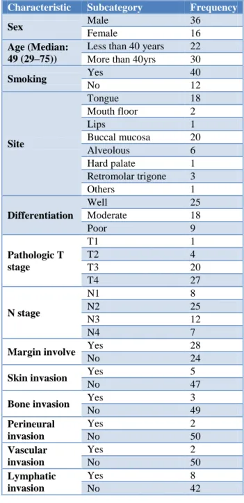
Total 52 number of patients included in the analysis, between the ages from 25 to 65 years old. The median age was 49 years. Thirty six (69.23%) patients were male, and 16 (30.76%) were female. The buccal mucosa was the most common sub site (20, 38.46%), followed by the
tongue (18, 34.61%), alveolous (6, 11.53%), retro molar trigone (3, 5.76%), floor of mouth f (2, 3.84%), hard palate (1, 1.92%), and lips (1, 1.92%). Majority of the patients reported with pathologic stage IVa disease, and their nodal stage was among either N2 or N3. There were 1 (1.92%) patient with stage T1, 4 (7.69%) patients with stage T2, 20 (38.46%) patients with stage T3, and 27 (51.92%) patients with stage T4 disease, respectively. After radical surgery, 18 (34.61%) patients were given postoperative radiotherapy within 6 weeks, and 34 (65.38%) given postoperative radiotherapy within 8 weeks. Fifteen (28.84%) patients given 60 Gy dose of postoperative radiotherapy, and 37 (71.15%) patients given between 60 and 66 Gy dose of radiotherapy. The overall survival rate at 1-year was 85%.
Table 1: Patients characteristics.
Characteristic Subcategory Frequency
Sex Male 36
Female 16
Age (Median: 49 (29–75))
Less than 40 years 22 More than 40yrs 30
Smoking Yes 40
No 12
Site
Tongue 18 Mouth floor 2
Lips 1
Buccal mucosa 20 Alveolous 6 Hard palate 1 Retromolar trigone 3
Others 1
Differentiation
Well 25
Moderate 18
Poor 9
Pathologic T stage
T1 1
T2 4
T3 20
T4 27
N stage
N1 8
N2 25
N3 12
N4 7
Margin involve Yes 28
No 24
Skin invasion Yes 5
No 47
Bone invasion Yes 3
No 49
Perineural invasion
Yes 2
No 50
Vascular invasion
Yes 2
No 50
Lymphatic invasion
Yes 8
Table 2: Parameters of treatment.
Characteristic Subcategory Frequency
RT dose ≤6000 CGy 12
≥6000 CGY 35
RT duration
Incomplete RT 3
≤7 weeks 12
≥7 weeks 35
Time between OP and RT ≤6 weeks 18
≥6 weeks 34
Chemotherapy No chemotherapy 10
Cisplatin 42
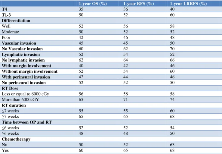
Table 3: Survival and recurrence free survival for patients based on different factors.
1-year OS (%) 1-year RFS (%) 1-year LRRFS (%)
T4 35 36 40
T1-3 50 52 60
Differentiation
Well 52 56 58
Moderate 50 52 52
Poor 42 46 48
Vascular invasion 45 45 50
No Vascular invasion 60 62 70
Lymphatic invasion 52 54 52
No lymphatic invasion 62 64 66
With margin involvement 40 42 46
Without margin involvement 52 54 60
With perineural invasion 42 44 46
No perineural invasion 52 52 50
RT Dose
Less or equal to 6000 cGy 56 58 58
More than 6000cGY 65 71 74
RT duration
≤7 weeks 55 55 60
≥7 weeks 65 65 68
Time between OP and RT
≤6 weeks 52 52 54
≥6 weeks 48 48 50
Chemotherapy
No 50 52 63
Yes 60 65 68
In the analysis, we found that pathologic T4 disease, bone invasion and lymphatic invasion were correlated with poor overall survival. The 1-year recurrence free survival rate was 85%. The most common recurrence was the nodal recurrence, then local recurrence, and then distant meets. In the analysis, we found that pathologicT4 disease, bone invasion, and skin invasion were significantly correlated with poor recurrence free survival (Table 3). Pathologic T4 disease, skin invasion, and bone invasion were significantly correlated with poor local recurrence free survival (Table 3). The effect of radiotherapy with chemotherapy was further evaluated. The OS in the patients received chemotherapy along with
RT, 60% as compare to 50% in cases without chemotherapy.
DISCUSSION
recurrence are the most common form of treatment failure, concurrent chemotherapy along with radiotherapy after a macroscopically complete resection may intensify the control of the disease. Various studies have shown the better result of postoperative concomitant chemotherapy in the treatment head and neck cancers having positive margins or lymph node involvement with extra capsular spreading. In a nonrandomized, phase 2 RTOG 8824 trial, result showed that the risk of local and regional recurrence decrease in high risk patients who have been treated with post op radiotherapy and concomitant chemotherapy.5 The presence of any of the below mentioned pathological features i.e. positive surgical margins, extra nodal spread of disease, perineural disease, or vascular tumor emboli are considered as high risk. In various studies, multiple-node metastases have shown higher tumor recurrence and poor overall survival.8,9 The various pathologic factors are predictive for local– regional recurrence after surgery. These pathological factors i.e. close (less than 5 mm) or positive margins, extra capsular extension through the lymph node capsule, involvement of the soft tissues of the neck, subglottic invasion, two or more positive lymph nodes, perineural invasion and endothelial-lined space invasion are the important parameter.10-13 The presence of multiple-node metastases is also an indication for postoperative radiotherapy for the treatment of head and neck cancers. The patients who present with advanced cancer and have no adverse pathologic factors may have an excellent prognosis with surgery alone. A number of factors are associated with an increased risk of local–regional recurrence after surgery alone. The two most unfavorable parameters are positive margins and extra capsular extension. The adjuvant concomitant radiotherapy reduces the risk of recurrence and result in better outcome. The survivals were improved by concomitant radiotherapy in each categories except patients who had no other pathologic risk factors. Because the chances of control is low after a local–regional recurrence, factors that affect the probability of disease control above the clavicles are also likely to have a negative influence on survival. According to various studies, the presence of multiple-node metastases was correlated with higher tumor recurrence and poor overall survival. A study carried out at the Medical College of Virginia where two groups of surgeons, surgical oncologist and oto-laryngologists, operated on patients with head and neck cancer, general surgical oncologists used surgery alone and radiotherapy for treatment of recurrent disease only, and otolaryngologists routinely sent patients with locally advanced disease for postoperative radiotherapy For patients who are at high-risk for local–regional failure following surgery, postoperative radiotherapy may significantly improve both disease control and survival. Vikram was among the first to report a decreased chances of cure, secondary to delay in initiation of postoperative radiotherapy in patients.14 The interval between surgery and postoperative radiotherapy, and the dose-fractionation schedule have influence on the overall treatment time. Various studies revealed that advanced
T-stage, extra capsular extension, positive margins, perineural invasion and vascular invasion significantly increased the risk of a local–regional failure (Table 3). Extra capsular extension, perineural invasion and advanced T-stage had an adverse impact on the survival. The addition of adjuvant chemotherapy was associated with a significant improvement in local–regional control and disease-free survival. However, overall survival was similar and acute toxicity was more pronounced in those who received chemotherapy. In a literature review, dose escalation through conventional fractionation was effective only for patients with extra capsular spreading.15 Time factors, such as an elapsed time between surgery and postoperative radiotherapy of more than 6 weeks and a duration of postoperative radiotherapy of more than 8 weeks, did not result in inferior overall survival or recurrence free survival. In most studies, time factors play a critical role in tumor control. Delays in starting postoperative treatment and prolonged treatment times were found to be correlated with more local-regional recurrence in various study.15-17
CONCLUSION
In summary, our study shows that certain pathological factors are important factors to decide further treatment with radiotherapy and concomitant chemotherapy after surgery. Concurrent postoperative administration of chemotherapy and radiotherapy is a way to intensify treatment for resectable high-risk head and neck tumors. In head and neck cancer patients with unfavorable factors have increased risk of loco regional failure after surgery, and post-operative radiotherapy have better outcome. Intensification of treatment with weekly cisplatin is now important in patient with high risk of disease. Over all treatment time should be minimized by avoiding delay in treatment between date of surgery and end of radiotherapy
ACKNOWLEDGMENTS
We thank for the help of faculty members of my department, students and our patients.
Funding: No funding sources Conflict of interest: None declared
Ethical approval: The study was approved by the Institutional Ethics Committee
REFERENCES
1. The Department of Veterans Affairs Laryngeal Cancer Study Group. Induction chemotherapy plus radiation compared with surgery plus radiation in patients with advanced laryngeal cancer. N Engl J Med. 1991;324:1685-90.
of Cancer phase III trial. J Natl Cancer Inst. 1996;88:890-9.
3. Forastiere AA, Goepfert H, Maor M, Pajak TF, Weber R, Morrison W, et al. Concurrent chemotherapy and radiotherapy for organ preservation in advanced laryngeal cancer. N Engl J Med. 2003;349:2091-8.
4. Laramore GE, Scott CB, Al-Sarraf M, Haselow RE, Ervin TJ, Wheeler R, et al. Adjuvant chemotherapy for resectable squamous cell carcinomas of the head and neck :report on Intergroup Study 0034. Int J Radiat Oncol Biol Phys. 1992;23:706-13.
5. Cooper JS, Pajak TF, Forastiere A, Jacobs J, Fu KK, Ang KK, et al. Precisely defining high-risk operable head and neck tumors based on RTOG #85-03 and #88-24:targets for postoperative radiochemotherapy? Head Neck. 1998;20:588-94. 6. Bernier J, Domenge C, Ozsahin M, Matuszewska K,
Lefebvre JL. Postoperative irradiation with or without concomitant chemotherapy for locallyadvanced head and neck cancer. N Engl J Med. 2004;350:1945–52.
7. Cooper JS, Pajak TF, Forastiere AA, Jacobs J, Campbell BH. Postoperative concurrent radiotherapy and chemotherapy for high-risksquamous-cell carcinoma of the head and neck. N Engl J Med. 2004;350:1937–44.
8. Liao CT, Chang JT, Wang HM, Ng SH, Hsueh C. Survival insquamous cell carcinoma of the oral cavity: differences between pT4 N0 andother stage IVA categories. Cancer. 2007;110:564–71.
9. Fan KH, Lin CY, Kang CJ, Huang SF, Wang HM. Combined modality treatment for advanced oral tongue squamous cell carcinoma. Int J Radiat Oncol Biol Phys. 2007;67:453–61.
10. Amdur RJ, Parsons JT, Mendenhall WM, Million RR, Stringer SP, Cassisi NJ. Postoperative irradiation for squamous cellcarcinoma of the head and neck:an analysis of treatment results and complications. Int J Radiat Oncol Biol Phys. 1989;16:25-36.
11. Peters LJ, Goepfert H, Ang KK, Byers RM, Maor MH, Guillamondegui O, et al. Evaluation of the
dose for postoperative radiation therapy of head and neck cancer: First report of a prospective randomized trial. Int J Radiat Oncol Biol Phys. 1993;26:3-11.
12. Olsen KD, Caruso M, Foote RL, Stanley RJ, Lewis JE, Buskirk SJ, et al. Primary head and neck cancer. Histopathologic predictors of recurrence after neck dissection in patients with lymph nodeinvolvement. Arch Otolaryngol Head Neck Surg. 1994;120:1370-4.
13. Zelefsky MJ, Harrison LB, Fass DE, Armstrong JG, Shah JP, Strong EW. Postoperative radiation therapy for squamous cell carcinomas of the oral cavity and oropharynx: impact of therapy on patients with positive surgical margins. Int J Radiat Oncol Biol Phys. 1993;25:17-21.
14. Vikram B. Importance of the time interval between surgery and postoperative radiation therapy in the combined management of head & neck cancer. Int J Radiat Oncol Biol Phys. 1979;5:1837-40.
15. Peters LJ, Goepfert H, Ang KK, Byers RM, Maor MH. Evaluation of the dose for postoperative radiation therapy of head and neck cancer: firstreport of a prospective randomized trial. Int J Radiat Oncol Biol Phys. 1993;26:3–11.
16. Ang KK, Trotti A, Brown BW, Garden AS, Foote RL. Randomized trial addressing risk features and time factors of surgery plus radiotherapy inadvanced head-and-neck cancer. Int J Radiat Oncol Biol Phys. 2001;51:571–8.
17. Porceddu SV, Campbell B, Rischin D, Corry J, Weih L, Guerrieri M, et al. Postoperative chemoradiotherapy for high-risk head-and-neck squamous cellcarcinoma. Int J Radiat Oncol Biol Phys. 2004;60:365–73.
Cite this article as: Ghori HU, Sohaib M, Tiwari V,