156
STRUCTURAL ANATOMICAL ASPECTS OF TWO
EUPHORBIA (EUPHORBIACEAE JUSS.) SPECIES LEAVES
DAN RĂZVAN POPOVICIU, RODICA BERCU Faculty of Natural and Agricultural Sciences,”Ovidius” University, Constantza University Alley, No. 1, B, 900470, Constantza
E-mail: [email protected]
Abstract.
The paper presents a study concerning the leaf structure (petiole, as the case) and blade of two Euphorbia species belonging to Euphorbiaceae family: Euphorbia trigona Mill., and E. tirucallli L. In literature is little information concerning the leaf petiole or blade anatomy of species of Euphorbia genus in general and concerning these two species in particular.
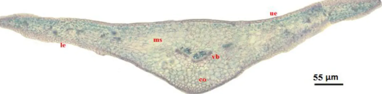
The petiole of Euphorbia trigona possesses one epidermis cells which are more or less rounded in shape, covered by a thin cuticle. The vascular system of the petiole is represented by three closed–collateral vascular bundles. Anatomically, the leaves blade of the studied species are heterogenous and amphistomatic with lacticifers especially presented to the midrib zone, more abundant în Euphorbia tirucalli. The midrib vascular system is more developed in Euphorbia tirucalli than E. trigona.
Keywords: anatomy, leaf, mesophyll, laticifers, Euphorbia
INTRODUCTION
The studied species belong to Euphorbiaceae family, one of the largest in the plant world, sometimes known as spurges. It includes around 300 genera and 7,700 species, mainly non-succulent herbs, shrubs and trees found in temperate, sub-tropical and tropical climates. The spurge family takes its name from the genus Euphorbia. Euphorbia tirucalli L. (pencil plant, milky shrub, pencil cake) is a perennial plant native from Africa where it was very widespread. Currently, it can also be found spontaneously, in restricted areas, in the center, southern and northeast of the African continent. It is a plant included in the IUCN Red List of Species since 2006 and has been threatened with extinction since 2004 (Haevermans, 2004). It is a plant with
a bushy harvested port, with the appearance of green rods and thick branches as a pencil (hence the popular name of the plant). Leaves are rare and small (Fig. 1, A). It develops well in hydroculture’s (Burger and Huft, 1995). It is a very toxic euphoric species, currently used more as an ornamental plant (Miaulane, 2004).
Euphorbia trigona Mill. is a perennial plant, originating from Central Africa, with more popular names as the African milk tree, cactus cathedral (Broschat and Meerow, 1991; Fuller and McClintock, 1986) or cactus -candle (Miaulane, 2004),
The plant has a straight, thick stem, 3-4 lined, slightly branched, dark green with lighter green and V-shaped pattern.
157
Among stems and spines are rare, oblong-lanceolate leaves, with the whole margin, which appear in the spring, placed especially in the top of the stems (Fig. 1, B). In our country it is known as an ornamental plant.
In literature is little information concerning the leaf anatomy of species of Euphorbia genus in general and
concerning these two species in particular.
Mostly researches followed the origin, development and structure of the laticifers. The most ample paper which analyzes the structure of the stem and leaf of spontaneous species and not only, from the Euphorbiaceae family is that of Gaucher (1902). Succinct references on the structure of the leaf as vegetative organ of some Euphorbia species are found in some
general studies concerning the angiosperms anatomy (G. Bonnier and Leclerc du Sablon, 1905; Dilcher, 1974; Esau, 1965; Metcalfe and Chalk, 1950; Napp-Zinn, 1973, 1974).
The Romanian literature there are only a few data on the structure of some vegetative organs (Ivanescu and Toma, 2003) of Euphorbia species; or general mentions in some lectures articles and manuals of Anatomy and Morphology of Plants (Bavaru and Bercu, 2002; Galeş and Toma, 2007; Grințescu, 1985, Niculescu, 2009; Șerbănescu-Jitariu and Toma, 1980; Toma and Gostin, 2000;).
The purpose of this paper is to highlight the anatomical characteritics of those two species. This paper could bring more information about this genus and this large family.
Fig. 1. Euphorbia tirucalli L. (A). Euphorbia trigona Mill. (B) (original).
MATERIAL AND METHODS The mature leaves, of Euphorbia tirucalli and E. trigona were collected from S.C. IRIS S.R.L. Greenhouse and from the Vegetal Morphology laboratory of the faculty.
Small pieces of mature leaves were fixed in FAA (formalin: glacier acetic acid: alcohol 5:5:90). Cross sections of the vegetative organs were performed by manual technique used in vegetal histology (Bercu and Jianu
158
2003). The samples were stained with alum-carmine and iodine green. Anatomical observations and micrographs were performed with a BIOROM–T bright field microscope, equipped with a TOPICA 6001A video camera.
RESULTS AND DISCUSSION The petiole is present only at Euphorbia trigona leaf. In cross-section, the petiole has a triangular contour shaped determinate by the presence of two developed lateral wings (Fig. 2).
Fig. 2. Cross section of Euphorbia trigona petiole: bs- basic tissue, co- collenchyma, e- epidermis vb- vascular bundles.
The epidermis cells are more or less rounded in shape, especially in the vascular bundles area, slightly cutinized and covered by a thin cuticle.
Beneath the epidermis is a hypoderm, represented by a collenchyma tissue with chloroplasts (3-4 layers of cells) with mechanical role (Fig. 2).
Centrally located, in a basic parenchyma, is the vascular system of the petiole, represented by 3 closed – collateral type vascular bundles
The blade. The blade epidermis
of Euphorbia tirucalli is one-layered celled and exhibits to upper and lower epidermis slightly rounded contour, covered with a thin cuticle. The lower epidermis forms a slightly vault. At this species, notable are the lateral parts of the blade that have a sinuous outline, with the margins slightly curved towards the adaxial face (Fig, 3). Among the lower and upper epidermis cells, rare stomata are present (Fig. 4, A).
159
Cross sections of the blade exhibits an upper and lower epidermises, both one-layered and the mesophyll with vascular bundles embedded. The mesophyll is
heterogeneous and amphistomatic with laticifers (Fig. 4, A).
The main vain is represented by a collateral bundle (Raven et al., 1992) formed by xylem and phloem elements. Laticifers are present as well (Fig. 4, B).
Fig. 4. Cross sections of Euphorbia tirucalli blade. Portion with mesophyll (A) Portion with midrib zone (B): co- collenchyma, l –lactiferous tube, ph- phloem, pt- palisade tissue, s- stomata, sc- sub-stomata cavity, sp- spongy tissue, ue- upper epidermis, x- xylem.
The sessile leaf of Euphorbia trigona has one layered upper and lower epidermis, covered by a thin cuticle, followed by a heterogeneous mesophyll with 2 layers of palisade tissue just below the upper epidermis and a number of spongy tissue
between the palisade tissue layers and lower epidermis. The mesophyll is amphistomatic with lacticifers especially presented to the midrib zone (Fig. 5, 6, A). The main vain is represented of a collateral bundle with xylem and phloem elements.
Fig. 5. Cross section of Euphorbia trigona blade - ensemble: co- collenchyma, le- lower epidermis, ms- mesophyll, ue- upper epidermis, vb- vascular bundle.
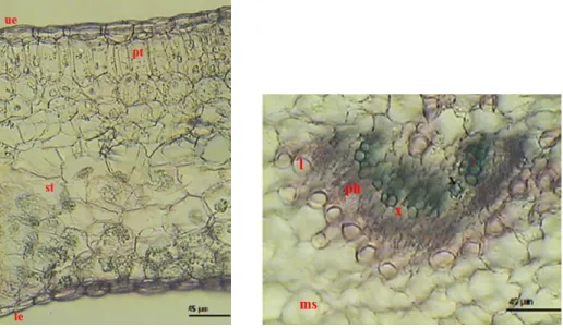
As Batanouny (1992) reported for other Euphorbia species, our
findings are the same for Euphorbia trigona concerning xylem conductive
160
elements which are placed towards the upper epidermis and exhibits a more or less radial arrangement. Phloem
elements, placed towards the lower epidermis, are poor developed than xylem ones (Fig, 6, B).
Fig. 6. Cross sections of Euphorbia trigona blade. Portion with mesophyll (A). Portion with midrib zone (B): l- laticifer tube, le- lower epidermis, ms- mesophyll, ph- phloem, pt- palisade tissue, sp- spongy tissue, x- xylem.
Just like in other studied Euphorbia sp. (Galeş and Toma, 2007) in the mesophyll of these two species, especially in the spongy tissue, the no articulated laticifers are present around the phloem tissue and have cellulosic thin walls, round or polygonal shape. In all studied species laticifers are also present mainly in the phloem aria of the vascular bundles (Fig. 4, A, B: 6, B).
CONCLUSIONS
The petiole of Euphorbia trigona possesses epidermis cells which are more or less rounded in shape, slightly cutinized and covered by a thin cuticle.
The vascular system of the petiole, represented by three closed –collateral type vascular bundles. Cross sections of the leaf blade of the two studied species (Euphorbia trigona și E. tirucalli) exhibits a single layered upper and lower epidermis which differ in shape and size, covered by a more or
less thin cuticle. The continuity of both epidermises is broken by the presence of stomata, more abundant in the lower epidermis. The mesophyll of both species is heterogeneous and amphistomatic with lacticifers especially presented to the midrib zone. The midrib vascular bundle of both species possesses xylem elements to the upper epidermis and phloem elements, placed towards the lower epidermis which are poor developed than xylem one. The secretory elements – laticifers - are placed mainly in the main vein aria, but also in the mesophyll, in all studied species.The mechanical tissue is not very well represented. It occurs such as collenchyma tisuue in Euphorbia trigona petiole and to the lower epidermis for the blade of this species. It is poor developed for Euphorbia tirucalli leaf blade.
161
REFERENCES
Batanouny K.H., Anatomy of Plants, 1992 - University Press of Cairo, Cairo.
Bavaru A., Bercu R., 2002 -
Morfologia şi anatomia plantelor, Ed. Ex Ponto, Constanţa.
Bercu, R., Jianu, D.L., 2003 -
Practicum de Morfologia şi anatomia plantelor, Ed. “Ovidius” University Press, Constanţa.
Bonnier M.G., Leclerc du Sablon M.,
1905 - Cours de Botanique, T.I., Librairie gknkrale de I'enseignement, Paris, pp. 798-814.
Broschat T.K., Meerow A.W., 1991 - Betrock's Reference Guide to Florida Landscape Plants. Betrock Information Systems, Hollywood, Florida, SUA, p. 123.
Burger W., Huft M., 1995 - Family 113
Euphorbiaceae, Fieldiana, Bot., 36: 1– 169.
Dilcher D.L., 1974 - Aproaches to the identification of the angiosperm leaf remains. The Botanical Review (Bot. Garden), New York, 40, 24-103.
Esau K., 1965 - Plant Anatomy, John
Wiley and Sons, New York, London, Sydney, pp. 318-337.
Fuller C.T., McClintock May
Elizabeth, 1986 - Poisonous Plants of California, Berkeley University of California Press, Berkeley, Los Angeles, London, p. 372.
Galeş R. C., Toma C., 2007 -
Researches regarding the morphology, structure and distribution of laticifers in the vegetative organs of some Euphorbia species from Romania’s flora, Analele ştiinţifice ale Universităţii “Al. I. Cuza”, Tomul LIII, s. II a. Biologie vegetală, Iaşi.
Gaucher L., 1902 - Recherches
anatomique sur les Euphorbiacbes, Ann. des Sci. Nat., ser. Bot., 15, pp. 161-309.
Haevermans T., 2004 - Euphorbia tirucalli. In: List of Threatened Species,
Published by IUCN Red List of Threatened Species, IUCN, 2006. Metcalfe C. R., Chalk L., 1950 - Anatomy of the Dicotyledons, 11, Clarendon Press, Oxford.
Miaulane P., 2004 - Enciclopedia
Truffaut-Grădini şi plante de interior, Ed. Enciclopedia Rao, București, p. 126.
Niculescu M., 2009 - Morfología şi anatomia plantelor, vol. I, Edit. Sitech, Craiova.
Niculescu Mariana, 2016 - Diversity, distribution and ecology of the freshwater natural habitats from Southern of Oltenia, ROMANIA SCIENTIFIC PAPERS-SERIES A-AGRONOMY Volume: 59 Pages: 116-121, ISSN- 1222-5339
Niculescu Mariana, 2009 - Metode de
cercetare şi prezentare a florei, Edit.Sitech, Craiova, 119 p
Niculescu Mariana, Alexandru
Tudor, Grecu Florina, 2016 - The
corology, ecology, phytosociology and hierarchical analysis of the bushes plant communities in the Parang Mountains (Southern Carpathians) Romania,International Multidisciplinary Scientific GeoConference: SGEM: Surveying Geology & mining Ecology Management, Vol. 3., Surveying Geology & Mining Ecology Management (SGEM), 371-378 p., Napp-Zinn K., 1984 - Anatomie des Blattes II Angiospermen. In: HJ Braun, S Carlquist, P Ozenda, I Roth, Handbuch der Pflanzenanatomie.
Raven P.H.; Evert F.R., Eichhorn E.S., 1992 - Biology of Plants (5th ed.), Worth Publ. Inc. NY.
Șerbănescu-Jitariu G., Toma C.,
1980 - Morfologia și anatomia plantelor, Ed. Didactică și Pedagogică, București.