GENE EXPRESSION ALTERATIONS IN CHRONIC PERI-‐IMPLANITIS SITES
Nicole Dominique Luedin
A thesis submitted to the faculty to the University of North Carolina at Chapel Hill in partial fulfillment of the requirements for the degree of Master of Science in the School of Dentistry
(Prosthodontics)
Chapel Hill
2015
© 2015
ABSTRACT
Nicole Dominique Luedin: Gene Expression Alterations in Chronic Peri-‐Implantitis Sites (Under the direction of Ingeborg de Kok)
ACKNOWLEDGEMENTS
I am extremely thankful to Ingeborg de Kok for her precious advice throughout the my thesis work. Her suggestions guided me to the right direction in my work. Furthermore, I thank for the interesting and educational talks during the time I spent with her.
My greatest thanks to my graduate prosthodontics program director, Dr. Lyndon Cooper. I would like to express my gratitude for the opportunity to do my studies in such an interesting field, under his supervision. His great support, constant interest, suggestions, critical reading of the manuscript and unwavering trust made it after all possible to bring this study to a conclusion. I got to know him as a generous, modest and extremely helpful human being, which I really appreciated.
I am very thankful to Antonio Moretti for his great help going through the literature with me and help to guide me in the right direction.
I am grateful to Ruiwei Liang, Daniela Mendonça, and Ghadeer Thalji for their immense help with the laboratory work and statistical analysis.
Many thanks go to the patients who donated their tissue and to the residents and UNC faculty who collected it. Without their help this study would not have been possible.
TABLE OF CONTENTS
LIST OF TABLES ... vii
LIST OF FIGURES ... viii
LIST OF ABBREVIATIONS ... ix
Abbreviations for Remodeling and Differentiation ... ix
Abbreviations for Inflammation and Destruction ... xii
CHAPTER 1: LITERATURE REVIEW OF PERI-‐IMPLANTITS ... 1
Bacterial etiology of peri-‐implantitis ... 7
Definitions of peri-‐implant mucositis and peri-‐implantitis ... 9
Diagnosis and monitoring of peri-‐implantitis ... 10
Host response ... 12
Pathogenesis ... 14
Peri-‐implant sulcular fluid molecular markers ... 18
Peri-‐implant Immunohistology ... 23
Genetic markers ... 25
Therapy ... 29
CHAPTER 2: A WITHIN SUBJECT MOLECULAR COMPARISON OF SOFT TISSUES AT IMPLANTS WITH AND WITHOUT PERI-‐IMPLANTITIS ... 31
INTRODUCTION ... 31
MATERIALS AND METHODS ... 33
Clinical Protocol ... 34
RNA Isolation and Gene Profile Data Analyses ... 35
RESULTS ... 35
DISCUSSION ... 37
CONCLUSION ... 42
APPENDIX: TABLES AND FIGURES FOR CHAPTER 2 ... 43
REFERENCES ... 58
LIST OF TABLES
Table 1.1. Prevalence of Peri-‐implantitis………...……….………..……….5
Table 1.2. Review Articles for Peri-‐implantitis Markers………...………...…………...16
Table 1.3. Peri-‐implant Sulcular Fluid Molecular Markers………..……….18
Table 1.4. Peri-‐implant Tissue Biomarkers……….………..……….24
Table 1.5. Genetic Biomarkers………...……….26
Table 2.1. Demographics of study participant………...………...43
Table 2.2.1. Up-‐regulated genes…………...…………..………...………45
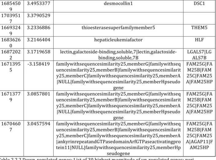
Table 2.2.2. Down-‐regulated genes………..………...………47
Table 3.1. Gene ontology up-‐regulated genes………..…….…...…51
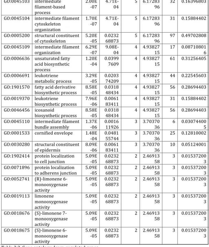
Table 3.2. Gene ontology down-‐regulated genes………..………...……..56
LIST OF FIGURES
Figure 1: PCA analysis………...………..44 Figure 2.1. Up-‐regulated heat map………..……49 Figure 2.2 Down-‐regulated heat map………..…..50
LIST OF ABBREVIATIONS
Abbreviations for Remodeling and Differentiation
Abbreviation Marker Main biological process
BGLAP Gene for osteocalcin Bone remodeling (osteoblast
differentiation)
BMP-‐2 Bone morphogenetic protein 2 Bone development
BMP-‐7 Bone morphogenetic protein 7 Bone development
BRAF erine/threonine-‐protein kinase B-‐Raf Cell differentiation
CD19 B-‐lymphocyte antigen CD19/ Cluster of differentiation 19
Lymphocite differentiation
CD31 Platelet endothelial cell adhesion molecule/
cluster of differentiation 31 Leukocyte transmigration, angiogenesis, integrin activation
cFn Cellular fibronectin Cell differentiation
COL9A1 Gene for Collagen alpha-‐1(IX) chain Collagen IX
DDK-‐1 Dickkopf-‐related protein-‐1 Bone remodeling (osteoblast differentiation antagonist)
FGF18 Fibroblast growth factor 18 Cell growth, tissue repair
GAPDH Glyceralaldehyde-‐3-‐ phosphate dehydrogenase
Reference gene
Hp-‐Hb Haptoglobin-‐Hemoglobin Access for enzymes to
hemoglobin
IL-‐2 Interleukin-‐2 T-‐cell differentiation
IL-‐4 Interleukin-‐4 B-‐cell differentiation
IL-‐5 Interleukin-‐5 B-‐cell growth
IL-‐7 Interleukin-‐7 T-‐cell maturation
IL-‐13 Interleukin-‐13 Anti-‐inflammation
MMP-‐2 Matrix metalloproteinase-‐2 Tissue remodeling
MMP-‐3 Matrix metalloproteinase-‐3 Tissue remodeling
MMP-‐9 Matrix metalloproteinase-‐9 Bone remodeling (osteoclast
differentiation)
OC Osteocalcin Bone remodeling (osteoblast
differentiation)
OPG Osteoprotegerin Bone remodeling (osteoclast
differentiation antagonist)
OPN Osteopontin Bone remodeling (osteoclast
anchoring)
PAI-‐2 Plasminogen activator inhibitor MMP antagonist
PDGFA platelet-‐derived growth factor a Stimulating factor for cell growth
PGE2 Prostaglandin E2 Bone remodeling (osteoblast
differentiation)
PPARγ Peroxisome proliferator-‐activated receptor γ Anti-‐inflammation
PTH Parathyroid hormone Bone remodeling (osteoclast
differentiation)
RANK Receptor activator of NF-‐κB Bone remodeling (osteoclast differentiation)
RANKL Receptor activator of NF-‐κB ligand Bone remodeling (osteoclast differentiation)
RUNX2 Runt-‐related transcription factor 2 Osteoblast differentiation
SPARC Gene for osteonectin Bone formation
SPP1 Gene for osteopontin Bone remodeling (osteoclast
anchoring)
sRANKL soluble receptor activator of NF-‐κB ligand Bone remodeling (osteoclast differentiation)
TGF-‐α Transforming growth factor-‐alpha Epidermal growth factor
TGFβ-‐1 Transforming growth factor-‐beta Fibrogenesis and vascular homeostasis
TIMP-‐1 Tissue inhibitors of metalloproteinases MMP antagonist
Abbreviations for Inflammation and Destruction
Abbreviation Marker Main biological process
ABCC9 ATP-‐binding cassette, sub-‐family C
(CFTR/MRP), member 9
Defence response to virus
AST Aspartate aminotransferase Tissue destruction
CatK Cathepsin K Bone resorption
cC1qR C1q receptors for collagen Pro-‐inflammation
CD3 Cluster of differentiation 3 T-‐cell activation
CD4 Cluster of differentiation 4 Antigen presentation
CD8 Cluster of differentiation 8 Antigen presentation
CD14 Cluster of differentiation 14 Antigen presentation
COLEC12 Collectin sub-‐ family member 12 Innate immune response
CRP C-‐reactive protein Pro-‐inflammation
gC1qR C1q receptors for globular domains Pro-‐inflammation
GM-‐CFS Granulocyte-‐macrophage colony-‐stimulating factor
Immune/inflammatory cascade
HBD1 Human Beta-‐defensin 1 Immune/inflammatory
Cascade
HBD2 Human Beta-‐defensin 2 Immune/inflammatory
Cascade
HCN2 Hyperpolarization-‐activated cyclic nucleotide-‐gated 2
Inflammatory pain
HMGB1 High mobility group chromosomal protein B1
Pro-‐inflammation
HMGN2 High mobility group chromosomal protein N2
Pro-‐inflammation
ICTP C-‐pelopeptide pyridinoline crosslinks of type
I collagen Bone resorption and collagen degradation
IFN-‐γ Interferon γ Immune/inflammatory cascade
IKKI Inhibitor of κB kinase Pro-‐inflammation
IL-‐1α Interleukin-‐1α Pro-‐inflammation
IL-‐1β Interleukin-‐1β Pro-‐inflammation
IL-‐6 Interleukin-‐6 Pro-‐inflammation
IL-‐8 Interleukin-‐8 Neutrophil chemotaxis and
angiogenesis
IL-‐10 Interleukin-‐10 Pro-‐inflammation
IL-‐12 Interleukin-‐12 Pro-‐inflammation
IL-‐17 Interleukin-‐17 Pro-‐inflammation
IL-‐22 Interleukin-‐22 Pro-‐inflammation
IL-‐22R Interleukin-‐22 receptor Pro-‐inflammation
IL-‐23 Interleukin-‐23 Pro-‐inflammation
MCC-‐1 Mast cell chymase Pro-‐inflammation
MCP-‐1 Monocyte chemo-‐ tactic protein Pro-‐inflammation
MCT-‐1 Mast cell tryptase Pro-‐inflammation
MiR146a microRNA 146 Regulation of inflammation
MiR499 microRNA 499 Regulation of inflammation
MMP-‐1 Matrix metalloproteinase-‐1 Pro-‐inflammation
MMP-‐7 Matrix metalloproteinase-‐7 Pro-‐inflammation and tissue remodelling
MMP-‐8 Matrix metalloproteinase-‐8 Pro-‐inflammation and tissue remodelling
MMP-‐13 Matrix metalloproteinase-‐13 Pro-‐inflammation
MMP-‐25 Matrix metalloproteinase-‐25 Pro-‐inflammation
MMP-‐26 Matrix metalloproteinase-‐26 Pro-‐inflammation
PPP2R2B Serine/threonine-‐protein phosphatase 2A 55
kDa regulatory subunit B beta isoform
Cell death
SRGN Serglycerin/ hematopoetic proteoglycan core protein
TANK TRAF family member-‐associated NF-‐κB activator
Pro-‐inflammation
TNC Tenascin-‐C
Pro-‐inflammation TNF-‐α Tumor necrosis factor α
Pro-‐inflammation
TRAP Tartrate-‐resistant acid phos-‐ phatase Bone resorption
CHAPTER 1: LITERATURE REVIEW OF PERI-‐IMPLANTITS
Over a 40-‐year period, clinical development and research efforts established dental implant therapy as a successful treatment modality for tooth replacement. Success was clearly defined in the late 1980s to include features of survival, marginal bone levels and the absence of infection (Albrektsson, Zarb et al. 1986). These guidelines helped to objectively define endosseous implant therapy as a reliable and safe means of tooth replacement for complete and partial edentulism. Since the mid 1980’s, numerous clinical studies, clinical reports and experience extending across millions of individuals treated with dental implants worldwide have provided additional insight into both the success and failure of dental implants.
Recently, the definition of survival was re-‐affirmed as an implant that remains in situ with or without modification during the observation period (Jung, Zembic et al. 2012). The reported survival rate varies from 73.4% to 100%, with a mean of 94.6% after up to 20 years (Moraschini, Poubel et al. 2014). Implant success has most frequently been assessed by survival rate, prosthesis stability, radiographic bone loss and absence of infection. Due to the heterogeneity of the
criteria at the prosthetic level. Finally, to evaluate patient satisfaction, discomfort, paresthesia, satisfaction with appearance and ability to chew and taste were parameters used. Papaspyridakos et al. (Papaspyridakos, Chen et al. 2012) concluded that “success in implant dentistry should ideally evaluate a long-‐term primary outcome of an implant-‐prosthetic complex as a whole.” The success rate ranged from 34.9% to 100%, with a mean success rate of 89.7%, during a mean follow-‐up of 15.7 years (Moraschini, Poubel et al. 2014). Clearly, continued reporting of dental implant outcomes has revealed that among the predominant successes, implant prostheses incur complications and do fail, that marginal bone levels may change over time and that infection is reported. Given that inflammatory disease is known to affect implant success, much more emphasis is now focused on peri-‐implantitis. Peri-‐implantitis is a condition with inflammation in the soft tissue around an implant with loss of bone.
Marginal bone level assessment with radiographs is one of the most important reference criteria for evaluating the long-‐term success of dental implants. Changes in bone occur in the first 6 months after implant placement due to a physiological healing process (Sanz and Chapple 2012, De Bruyn, Vandeweghe et al. 2013). The healing process include re-‐establishment of the junctional epithelium and the supra-‐alveolar connective fibers independent from the surgical technique (Abrahamsson, Berglundh et al. 1999) and the implant system (Abrahamsson, Berglundh et al. 1996). Berglundh and Lindhe stated “that once the implant is exposed to the oral environment and in function, a mucosal attachment of a certain minimum dimension is required to protect
osseointegration” (Berglundh and Lindhe 1996). Hence, it was found that a stabilization of a crestal bone level of 1.5-‐2.0mm below the implant abutment interface normally occurs one year after loading (Cochran, Nummikoski et al. 2009).
over a time period of up to 15.7 years. Marginal bone loss is encountered with some frequency. For example, in full-‐arch implant-‐supported prostheses bone loss ≥ 2mm were found in 16-‐29% of the patients after 12-‐15 years of function (Ravald, Dahlgren et al. 2013). Renvert et al. (Renvert, Lindahl et al. 2012, Renvert, Polyzois et al. 2013) have found that bone loss was greater in the first 7 years of function compared from 7 to 13 years of function. Tomasi et al. (Tomasi, Wennstrom et al. 2008) compared the longevity of teeth and implants. They found that in well-‐maintained patients the survival rate of teeth is greater than the one of implants and with regular maintenance the bone loss is small around teeth and implants. However, comparison between the survival rate of teeth and implants is difficult due to the heterogeneity of the study design and the patient population. Rasperini et al. (Rasperini, Siciliano et al. 2014) compared bone levels on teeth adjacent to implants and found that the bone levels around teeth were more stable than around implants in a 10 year follow-‐up.
While there exist differences among implant systems, a consistent observation has been the stabilization of bone changes relative to the implant abutment interface (Laurell and Lundgren 2011). An important hallmark of peri-‐implantitis is the progression of marginal bone loss beyond these accepted adaptive changes in health.
It has been demonstrated that inflammation is a main cause of alveolar bone loss at dental implants and that the extent of inflammation is associated with marginal bone level changes (Schou, Holmstrup et al. 1992). In a foundational study in the dog model, the abundance of inflammatory cells in the inflammatory cell infiltrate at implants was correlated with the extent of marginal bone loss (Broggini, McManus et al. 2006). In clinical studies, the extent of marginal bone loss at implants was correlated with the degree of clinical inflammation (Kehl, Swierkot et al. 2011). This
implants placed by a two-‐stage procedure were examine after oral exposure following abutment connection of selected implants. Unexposed implants did not demonstrate bone loss during the period of time, while implants with abutments that were exposed to the oral environment did show bone loss (Naert, Gizani et al. 1999). The investigators demonstrated that marginal bone loss at implants was not a result of surgical placement and bone remodeling, but attributable to abutment connection and further biological integration.
As indicated above, the process of marginal bone loss is preceded by peri-‐implant mucosal inflammation. Peri-‐implant mucosal inflammation other biological complications are the most common problem encountered with dental implants. Peri-‐implant mucositis is the number one biological complication reported (Moraschini, Poubel et al. 2014), followed by alveolar bone loss around implants. For example, Jung et al. (Jung, Zembic et al. 2012) found a 5-‐year cumulative peri-‐ implant soft tissue complication rate of 7.1%, and a 5.2% cumulative complication rate of alveolar bone loss ≥2mm around single dental implants. It is commonly observed; this is underscored by a recent systematic review, indicating the prevalence of peri-‐implant mucositis (with varying definitions) was 42.9% (95% CI 32-‐54%) (Derks and Tomasi 2014).
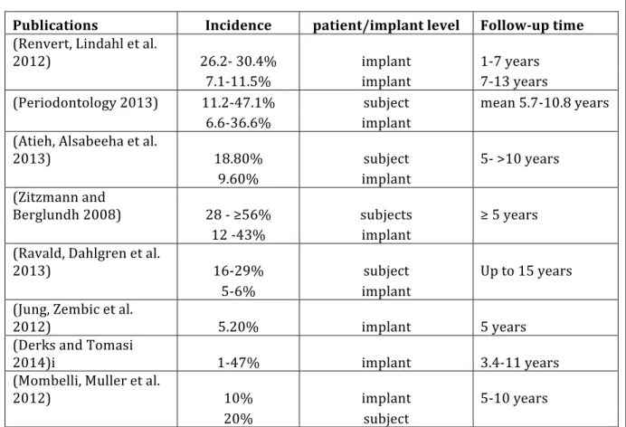
According to Mombelli et al. (Mombelli, Muller et al. 2012) the prevalence of peri-‐ implantitis seemed to be 10% in 5-‐10 years of function. In a recent systematic review, the
there are the different prevalences of peri-‐implantitis summarized. It changes on an implant level between 1% up to 47% and on a subject or patient level up from 11.2% to 56%
Publications Incidence patient/implant level Follow-‐up time (Renvert, Lindahl et al.
2012) 26.2-‐ 30.4% implant 1-‐7 years
7.1-‐11.5% implant 7-‐13 years
(Periodontology 2013) 11.2-‐47.1% subject mean 5.7-‐10.8 years
6.6-‐36.6% implant
(Atieh, Alsabeeha et al.
2013) 18.80% subject 5-‐ >10 years
9.60% implant
(Zitzmann and
Berglundh 2008) 28 -‐ ≥56% subjects ≥ 5 years
12 -‐43% implant
(Ravald, Dahlgren et al.
2013) 16-‐29% subject Up to 15 years
5-‐6% implant
(Jung, Zembic et al.
2012) 5.20% implant 5 years
(Derks and Tomasi
2014)i 1-‐47% implant 3.4-‐11 years
(Mombelli, Muller et al.
2012) 10% implant 5-‐10 years
20% subject
Table 1.1. Prevalence of Peri-‐implantitis
lifestyle, environmental and local factors” and therefore additional and adequately powered research needs to be done.
The effect of history of periodontitis on implant success revealed no differences in terms of survival.; nevertheless, patients with a history of periodontitis had a lower implant success rate (Ramanauskaite, Baseviciene et al. 2014). Systemic disease and a history of periodontitis have been considered risk factors for peri-‐implantitis (Renvert, Polyzois et al. 2013). However, systematic reviews on the effects of periodontitis on dental implant survival showed a great variability (Faggion and Giannakopoulos 2013).
When considering the existing data published and summarized in the present literature regarding peri-‐implantitis, it may be concluded that peri-‐implantitis and the associated profound interfacial bone loss that occurs at dental implants is the result of inflammation. Factors
contributing to the initiation and progression of this inflammation as well as factors that preclude its control or elimination represent risk factors influencing peri-‐implantitis. While the extent to which individuals are susceptible to or harbor one or another risk factor influencing peri-‐
implantitis has not been fully elucidated, it may be further concluded that inflammatory events are likely modulated by local tissue architecture, prosthesis and abutment factors, local and systemic biology that influence the accumulation, nature and response to an adherent biofilm. A biofilm-‐ associated inflammatory response leading to bone loss can progress to implant failure.
knowledge regarding the pathophysiology of peri-‐implantitis. The goal of this review was to identify the present information regarding molecular mediators of inflammatory processes in peri-‐ implantitis and their measurement in clinical therapy.
Bacterial etiology of peri-‐implantitis
Mombelli (Mombelli and Lang 1998) proposed the hypothesis that “microbial colonization of dental implants and infection of the peri-‐implant tissues can cause peri-‐implant bone destruction and may lead to implant failure.” The author based the hypothesis on five lines of evidence, which were (1) human trials showing that peri-‐implant mucositis can be induced by deposition of plaque, (2) the microbiota associated with healthy and infected implants showed qualitative and
quantitative differences, (3) shifts in the microbiota and peri-‐implantitis could be found in animal models with plaque-‐retentive ligatures, (4) clinical status of peri-‐implantitis patients could be improved with antimicrobial therapy and (5) “the level of oral hygiene has an impact on the long-‐ term success of implant therapy.”
Secondarily, Mombelli also assumed that “peri-‐implant infections are amenable to
treatment just as periodontal infections are” (Mombelli 2002). Therefore, the author inferred that the disease is a combination of a bacterial colonization of the implant surfaces and that the immune response causes bone destruction around the implant.
Dental implants provide a target for biofilm formation. The biofilm derives from various micro-‐ecological niches, including the neighboring natural dentition, saliva and mucosa. The microbiota of peri-‐implantitis shares many species found in periodontitis with important
colonized by gram-‐positive facultative anaerobic cocci in significant lesser counts (Mombelli 1997, Mombelli 2002). T. forsythia and T. denticola were increased in peri-‐impantitis and periodontitis. Distinct for peri-‐implantitis, when compared to periodontitis, the microbiota included S. aureus, S.
epidermidis, and the aerobic gram-‐negative bacilli E: aerogenes, E. cloace, E. coli, H. pylori, P. micra,
Pseudonomas ssp and Candida ssp fungi (Koyanagi, Sakamoto et al. 2013, Belibasakis 2014, Smeets,
Henningsen et al. 2014, Belibasakis, Charalampakis et al. 2015). It has been reiterated that peri-‐ implantitis lesions harbor bacteria that are typically found in periodontitis lesions. Staphylococcus
aureus is predominant in peri-‐implantitis and may provide a high positive (80%) and negative
(90%) predictive value (Rasperini, Siciliano et al. 2014, Salvi and Zitzmann 2014, Smeets,
Henningsen et al. 2014). Thus, a unique bacterial infection in peri-‐implantitis may induce unique inflammatory sequelae.
Interestingly, fully edentulous patients had a different composition of the microbiota with a smaller amount of spirochetes and P. gingivalis than partially edentulous patients. That led to the hypothesis that “a transmission of periodontal pathogens and partially edentulous patients with a history of periodontitis are at an elevated risk of developing peri-‐implantitis” (Mombelli 1997) (Mombelli 2002). It is widely accepted that oral bacteria derived biofilm is a primary etiology of peri-‐implantitis.
commonly found in periodontitis. Furthermore, microorganisms unique to peri-‐implantitis have been identified and include S. aureus, S epidermidis, E. aerogenes, E cloace, E. coli, H. pylori, P. micra,
Pseudomonas and Candida spp (Belibasakis 2014).
Initial investigations identified gram-‐negative anaerobic bacteria and S. aureus as potential pathogens (Mombelli, van Oosten et al. 1987). Other periodontal pathogens, F. nucleatum, A.
actinomycetemcomitans, P. gingivalis, and P. intermedia, have been repeatedly found in peri-‐
implantitis. These and other lead bacteria have been implicated in the process however a comprehensive assessment of the microbial population using 16S rRNA based-‐assessment underscored a diverse bacterial population in peri-‐implantitis that is more complex than that formed in periodontitis (Koyanagi, Sakamoto et al. 2013). The microbial diversity associated with peri-‐implantitis continues to be illustrated by molecular assessment of the biofilm (da Silva, Feres et al. 2014).
Definitions of peri-‐implant mucositis and peri-‐implantitis
Peri-‐implantitis is distinct from peri-‐implant mucositis. Withdrawal of oral hygiene with resulting plaque formation on teeth and implants results in a local inflammatory response that is clinically evidenced by redness and bleeding on probing and evidenced histologically by the time-‐ dependent increase of the inflammatory cell infiltrate adjacent to the implant/abutment (Zitzmann, Berglundh et al. 2001). This peri-‐implant mucosal response is termed peri-‐implant mucositis. According to the American Academy of Periodontolgy (AAP) (2013) and the VIII European
defining peri-‐implantitis, as well as for the time point when the bone loss occurred, Sanz et al. (Sanz and Chapple 2012) agreed on the definition for peri-‐implantitis when “changes in the level of crestal bone, presence of bleeding on probing and/or suppuration, with or without concomitant deepening of peri-‐implant pockets are present.” Furthermore, they defined the bone level of 2mm from the expected level in absence of baseline radiographs as a threshold to define peri-‐implantitis. With existing baseline radiographs, they set threshold of bone loss is 1-‐1.5mm for the definition of peri-‐implantitis (Sanz and Chapple 2012).
Diagnosis and monitoring of peri-‐implantitis
There are no single diagnostic parameters for peri-‐implantitis. The diagnosis involves clinical and radiographic parameters. Probing with a traditional periodontal probe with 0.25N for the incidence of bleeding on probing (BoP), suppuration and probing depth (PPD) and their
changes over time is widely recommended (AAP) (Zitzmann and Berglundh 2008, Sanz and Chapple 2012, 2013). Additionally, plaque accumulation indexes (PlI), clinical attachment levels (CAL), and gingival recession (REC) are recommended by other authors (Graziani, Figuero et al. 2012).
BoP has a high negative predictive value, as well as a positive predictive value of a 100%. The absence of BoP is an indicator of peri-‐implant tissue health. However, BoP only diagnoses a peri-‐implant mucositiis and is not specific for peri-‐implantitis. Suppuration indicates an
the formation of a peri-‐implant pocket > 4 mm, bleeding or suppuration on probing, and radiographic evident saucer-‐shaped bone destruction around the implant (Belibasakis 2014).
Mobility is not a useful clinical sign of for peri-‐implantitis, since it represents complete implant failure. When mobility is presented, identification of a mobile abutment should be made, as that mobility and associated inflammation at the implant-‐abutment level lead to alveolar bone loss (2013). Implant mobility can be measured either manually or with a specific device such as the Periotest dental measuring instrument (Siemens, Bensheim Germany) or with the Ostell instrument (Ostell, Gothenburg, Sweden). There is a differential diagnosis for mobility that includes a) mobility of the prosthesis at the abutment interface, b) mobility of the abutment at the implant interface, and c) mobility of the implant itself. Because implants are often splinted and the mobility can be
masked, it is recommended to unscrew the prosthesis and evaluate individual unsplinted implants (Todescan, Lavigne et al. 2012).
detect bone loss (De Bruyn, Vandeweghe et al. 2013). The low predictive nature of these
parameters (BoP, PPD, suppuration, radiograph etc.) by itself makes it necessary to combine them and measure them over time to render the diagnosis of peri-‐implantitis.
Host response
As stated before, while periodontal disease and peri-‐implantitis appear to share common bacterial etiology and both reflect a host response to biofilm, previous reports highlight potentially important differences between dental implants and natural teeth. Biofilm associated inflammation and the innate immune response progresses faster and results in a more extensive and severe tissue destruction at implants than at teeth (Smeets, Henningsen et al. 2014, Belibasakis, Charalampakis et al. 2015). The reasons for this differential host response (beyond yet to be determined differences in biofilm) may reflect local anatomic differences between teeth and
implants. A prominent difference is that Sharpey’s fibers inserting perpendicular into cementum do not exist at the implant surface. The collagen fibers of the submucosal tissue connective tissue are arranged parallel to the surface of the implant, resulting in a deeper peri-‐implant crevice and therefore allows for potentially deeper penetration of the biofilm when compared to teeth. A more pronounced apical extension of the inflammatory cell infiltrate was found in peri-‐implantitis compared to periodontitis (Berglundh, Gislason et al. 2004, Berglundh, Zitzmann et al. 2011, Alani, Kelleher et al. 2014). Furthermore, there is no periodontal ligament at implants. This results in the absence of an important periodontal vascular plexus that differentiates the blood supply between implants and teeth. The peri-‐implant mucosa is often reported to be less vascular than the
connective tissue toward the alveolar bone and a separation of the biofilm from the connective tissue by a pocket epithelium. These compartmentalizations are missing in the peri-‐implantitis lesions (Carcuac and Berglundh 2014).
Regarding the inflammatory infiltrate that exists at both healthy and inflamed implant sites, Broggini et al. (Broggini, McManus et al. 2006) found that the highest concentration of
inflammatory cells were at or immediately coronal to the implant-‐abutment interface. The
predominant peri-‐implantitis cell type was neutrophils. Furthermore, there is a positive correlation with the depth of the interface, and the magnitude of the peri-‐implant inflammation. Carcuac and Berglundh (Carcuac and Berglundh 2014) found that peri-‐implantitis lesions are double the size compared to periodontitis lesions. Peri-‐implantitis sites “contained significantly larger area proportions, numbers and densities of plasma cells, macrophages and PMN cells compared to periodontitis.” PNM cells “indicate that the effector systems of the host response, such as
phagocytosis, are active in peri-‐implantitis” (Berglundh, Gislason et al. 2004). They concluded that in peri-‐implantitis both cells from the innate and the adaptive immune system are playing a role (Carcuac and Berglundh 2014). The innate immune response results in an increased infiltration of neutrophils, macrophages, interstitial dendritic cells, B-‐and T-‐ cells, osteoclasts and a decrease of Langerhans cells. (Berglundh, Zitzmann et al. 2011, Belibasakis 2014, Belibasakis, Charalampakis et al. 2015).
Experimental models were needed to study peri-‐implantitis further. Both dogs and
not loss of osseointegration, while occlusal overload can determine a loss of osseointegration. The use of ligatures resulted in a foreign body reaction and induced a destructive process around the implants and therefore does not represent a good model to study the disease in humans. In the same way, overload studies showed no similarity to bone loss in humans (Pesce, Menini et al. 2014). Berglundh et al. (Berglundh, Zitzmann et al. 2011) concluded that a “self-‐limiting” process of the induced tissue inflammation after removal of the ligature does not occur around implants. They found signs of acute inflammation and large amounts of osteoclasts lining the surface of the bone crest after the removal of the ligature, and therefore a progression of the bone loss. A literature review was done on canine ligature models by Martins et al. (Martins, Ramos et al. 2014). This group found that most of the studies were done with Beagle dogs and implants in the premolar and molar region of the mandible. Cotton or silk ligaments were placed in a submarginal position around implants, however the methods varied widely. Their conclusion was that the defect configuration differs between humans and the dogs and that the ligature placement results in a traumatic foreign body bone loss rather than a natural one. Therefore, an “ideal canine peri-‐ implantitis induction model would be naturally occurring peri-‐implantitis induction without the action of any ligature” (Martins, Ramos et al. 2014). Pesce et al. (Pesce, Menini et al. 2014)
concluded that animal studies reported contrasting results depending on the model employed and are not completely representative of the human disease.
Pathogenesis
that the inflammatory tissue destruction that occurs at implants is more aggressive than that observed at teeth (Belibasakis 2014). The details of the many aspects of peri-‐implantitis pathogenesis remain relatively obscure.
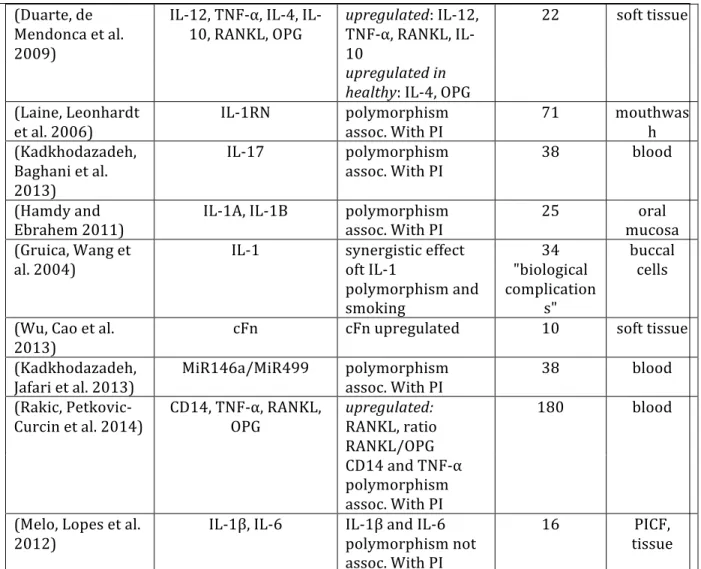
In the pursuit of understanding the pathogenesis of peri-‐implant mucositis and peri-‐ implantitis a significant number of articles has been published. One approach has been to examine the gene polymorphisms associated with peri-‐implantitis. Some studies have identified gene polymorphism associations for OPG, IL-‐6, TNF-‐α, RANKL, MiR146a/ MiR499 with peri-‐implantitis (Cury, Horewicz et al. 2009, Kadkhodazadeh, Tabari et al. 2012, Slotte, Lenneras et al. 2012, Casado, Villas-‐Boas et al. 2013, Kadkhodazadeh, Ebadian et al. 2013, Kadkhodazadeh, Jafari et al. 2013). Those genes are associated with inflammatory diseases and bone resorption. Some papers showed contradictory results of IL-‐1 gene polymorphism, which is a general marker of inflammation and has been implicated as a genetic marker for periodontal disease. IL-‐1 polymorphism has been associated with peri-‐implantitis (Laine, Leonhardt et al. 2006, Hamdy and Ebrahem 2011, Casado, Villas-‐Boas et al. 2013), associated in combination with smoking (Gruica, Wang et al. 2004) or not associated at all (Lachmann, Kimmerle-‐Muller et al. 2007, Melo, Lopes et al. 2012). There are inconsistent findings concerning the pro-‐inflammatory cytokine IL-‐17 polymorphism (Severino, Napimoga et al. 2011, Darabi, Kadkhoda et al. 2013, Kadkhodazadeh, Baghani et al. 2013,
Kadkhodazadeh, Ebadian et al. 2013). Several gene polymorphisms are found not to be associated with peri-‐implantitis, among them are BRAF, TANK, Hp-‐Hb complex and HCN2 (Kadkhodazadeh, Amid et al. 2012, Ebadian, Kadkhodazadeh et al. 2013, Kadkhodazadeh, Jafari et al. 2013, Ebadian, Kadkhodazadeh et al. 2014).
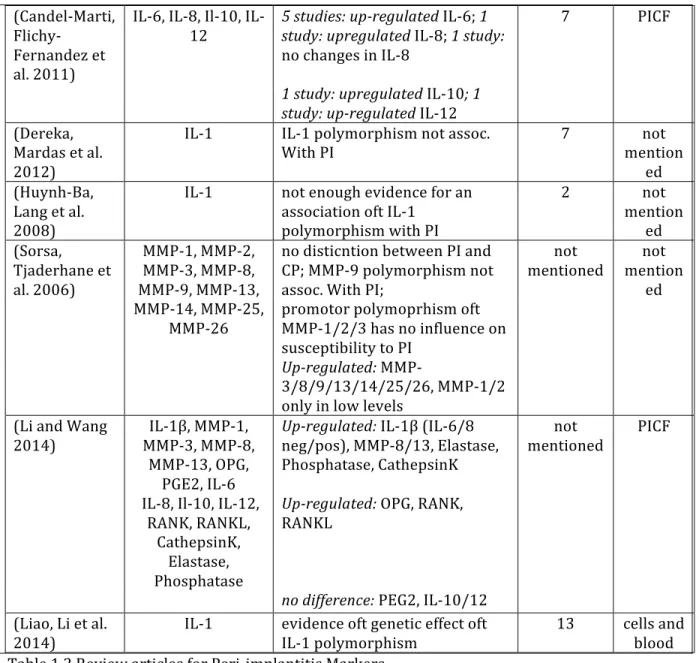
2011, Javed, Al-‐Hezaimi et al. 2011, Petkovic-‐Curcin, Matic et al. 2011, Li and Wang 2014). The relationship of levels of suspect proteases was considered in three reviews (Sorsa, Tjaderhane et al. 2006, Javed, Al-‐Hezaimi et al. 2011, Li and Wang 2014). The possible relationships of genetic polymorphisms influencing the levels of inflammatory mediators has also been summarized previously (Andreiotelli, Koutayas et al. 2008, Bormann, Stuhmer et al. 2010, Javed, Al-‐Hezaimi et al. 2011, Dereka, Mardas et al. 2012, Liao, Li et al. 2014). A general conclusion from these different summaries is that peri-‐implantitis involves upregulation of the general mediators of inflammation including TNF-‐α, IL1a, IL1b, IL6 and IL10. Fewer studies investigated more specific mediators of osteoclastogenesis, an essential aspect of peri-‐implantitis. The recognition of OPG, RANK and RANKL up-‐regulation (Sorsa, Tjaderhane et al. 2006), as well as elevated levels of more general mediators, TNF-‐α and IL6, affirm an existing appreciation that inflammation and osteoclastogenesis are central to the pathogenesis of peri-‐implantitis (Table 1.2).
Publications Investigated Findings Articles
included
Probes
(Javed, Al-‐ Hezaimi et al. 2011)
IL-‐1β, IL-‐6, IL-‐8, MMP-‐1, TNF-‐α, IL-‐
1
2 studies: upregulated IL-‐6 ; 4
studies: upreglated IL-‐1β; 15 PICF
1 study: up-‐regulated IL-‐6, IL-‐8,
MMP1, 6 studies: upredulated
TNF-‐α
2 studies: IL-‐1 polymorphism
assoc. With PI; 1 study: TNF-‐α not assoc. With PI
(Petkovic-‐ Curcin, Matic et al. 2011)
IL-‐1β,IL-‐6, IL-‐8,
MIP-‐1α, TNF-‐α Uup-‐regulated PI; 3x up-‐regulated IL-‐1β in early stage IL-‐1β; mentioned not PICF
Up-‐regulated IL-‐8, MIP-‐1α, TNF-‐
α
(Andreiotelli, Koutayas et al. 2008)
IL-‐1β IL-‐1β polymorphism not assoc. With PI, synergistic effect oft pos. IL-‐1β and smoking
8 PICF
(Bormann, Stuhmer et al. 2010)
IL-‐1 correlation between IL-‐1 polymorphism and PI with additional risk factors (eg. Smoking)
27 not
(Candel-‐Marti, Flichy-‐
Fernandez et al. 2011)
IL-‐6, IL-‐8, Il-‐10, IL-‐
12 5 studies: up-‐regulated study: upregulated IL-‐8; IL-‐6; 1 study: 1
no changes in IL-‐8
7 PICF
1 study: upregulated IL-‐10; 1
study: up-‐regulated IL-‐12
(Dereka, Mardas et al. 2012)
IL-‐1 IL-‐1 polymorphism not assoc.
With PI 7 mentionnot
ed (Huynh-‐Ba,
Lang et al. 2008)
IL-‐1 not enough evidence for an association oft IL-‐1
polymorphism with PI
2 not
mention ed (Sorsa,
Tjaderhane et al. 2006)
MMP-‐1, MMP-‐2, MMP-‐3, MMP-‐8, MMP-‐9, MMP-‐13,
no disticntion between PI and CP; MMP-‐9 polymorphism not assoc. With PI;
not
mentioned mentionnot ed
MMP-‐14, MMP-‐25,
MMP-‐26 promotor polymoprhism oft MMP-‐1/2/3 has no influence on susceptibility to PI
Up-‐regulated: MMP-‐
3/8/9/13/14/25/26, MMP-‐1/2 only in low levels
(Li and Wang
2014) MMP-‐3, MMP-‐8, IL-‐1β, MMP-‐1, MMP-‐13, OPG,
PGE2, IL-‐6
Up-‐regulated: IL-‐1β (IL-‐6/8
neg/pos), MMP-‐8/13, Elastase, Phosphatase, CathepsinK
not
mentioned PICF
IL-‐8, Il-‐10, IL-‐12, RANK, RANKL,
CathepsinK, Elastase, Phosphatase
Up-‐regulated: OPG, RANK,
RANKL
no difference: PEG2, IL-‐10/12
(Liao, Li et al. 2014)
IL-‐1 evidence oft genetic effect oft IL-‐1 polymorphism
13 cells and blood
Table 1.2 Review articles for Peri-‐implantitis Markers
et al. 2013, Rakic, Nikolic-‐Jakoba et al. 2013, Wohlfahrt, Aass et al. 2014). Some of the pro-‐
inflammatory cytokines were found to be unchanged or down-‐regulated, among them IL-‐10, MMP-‐8 (Severino, Napimoga et al. 2011, Casado, Villas-‐Boas et al. 2013, Irshad, Scheres et al. 2013).
In the present review, the knowledge regarding molecular basis of peri-‐implantitis were conveniently categorized according to the manner of investigation and are: a) peri-‐implant sulcus fluid (PISF) biomarkers (Table 1.3.), b) peri-‐implant tissue biomarkers (Table 1.4.), and c) genetic biomarkers (Table 1.5.). A rationale for this categorization is that the source of information available will reflect the manner of collecting patient-‐specific information (e.g., collection of PISF, Biopsy, DNA) and may further influence the data made available (e.g.; incipient disease screening vs. genetic risk assessment).
Peri-‐implant sulcular fluid molecular markers
Twenty-‐seven papers compared specific constituents of PISF among healthy and peri-‐ implantitis sites (Table 1.3.). Monitoring of molecular mediators in sulcular fluid in periodontitis has been adopted in assessment of PISF to study peri-‐implantitis (Periodontology 2013, Faot, Nascimento et al. 2015). Recent data suggests that there are differences in PISF and gingival
cervicular fluid (GCF) that may be important to consider and may imply a different pathogenesis for peri-‐implantitis and periodontitis (Recker, Avila-‐Ortiz et al. 2015).
Publications Investigated Findings PI
patients (Hall, Britse et
al. 2011) OC, IL-‐1β, TNF-‐α, RANKL, TRAP, DDK-‐1, OPG, CatK, ALP, GAPDH, PPIA, ACTB
no sign. Differences between
healthy and PI for all markers 7
YWHAZ, RRN18S, B2M,
UBC, RPLP, HPRT1 ev problem with strategy (Severino,
Napimoga et al. 2011)
IL-‐6, IL-‐8, IL-‐10, IL-‐17 Up-‐regulated: IL-‐17; no differences:
sign. Positive correlation bt. IL-‐6
and IL-‐8 in PI
(Rakic, Nikolic-‐ Jakoba et al. 2013)
RANK Up-‐regulated: 9x RANK 22
Rakic, 2013 #1}
sRANKL, RANK, OPG Up-‐regulated: SRANKL, RANK, OPG 23 Arikan, 2011
#52}
ICTP, sRANKL, OPG Up-‐regulated: ICTP, OPG ; up-‐
pregulated in healthy: OPG, sRANKL
12 Wohlfahrt,
2014 #28} MMP-‐8, TNF-‐α, OPN, OPG, OC, IL-‐6, PTH, Insulin Down-‐regulation Insulin, MMP-‐8 after Tx: IL-‐6, 12
no correlation bt. Change oft bone
and marker concentration
Irshad, 2013
#36} MMP-‐1, MMP-‐2, MMP-‐8, IL-‐1β IL-‐6, IL-‐8, MCP-‐1, TIMP-‐1, TGFβ-‐1
Up-‐regulated in non-‐challenged (P.
gingivalis) cells: IL-‐1β, IL-‐8, MCP-‐1,
MMP-‐8
7
Up-‐regulated in challenged cells: IL-‐
1β, IL-‐6/8, MCP-‐1, MMP-‐1
Down-‐regulated in challenged cells:
MMP-‐8
(Lachmann, Kimmerle-‐ Muller et al. 2007)
IL-‐1β, PAI-‐2, PGE2 no assoc. With genotypes 11
Casado, 2013 #40}
IL-‐1β, IL-‐10 Up-‐regulated: IL-‐1β; IL-‐10 in healthy; down-‐regulated: IL-‐10
10 Darabi, 2013
#35} TNF-‐α, IL-‐17 upregulated: TNF-‐α, IL-‐17 24
(Ozcakir-‐ Tomruk, Chiquet et al. 2012)
TNC, MMP-‐9 Up-‐regulated: MMP-‐9, small for TNC
18 total, PI not mentione
d Sarlati, 2010
#56}
sRANKL no sign. Difference in concentration 26 Slotte, 2012
#59} CatK, TNF-‐α, ALP, OC, IL-‐1β early loading, clinical complications with TNF-‐α, CatK, ALP immediate loading: 9
correlation with clinical
parameters and complications group: 9 test (Ramseier, Eick
et al. 2015) IL-‐1β, MMP-‐1, MMP-‐3, MMP-‐8, MMP-‐1/TIMP MMP-‐8 in 90% oft sites, IL-‐1β in 50% oft sites, in 30% oft sites MMP-‐1, MMP3, MMP-‐1/TIMP
504 implants (Hultin,
Gustafsson et al. 2002)
elastase, IL-‐1β,
Lactoferrin Up-‐regulated: elastase, Lactoferrin 17
no changes: IL-‐1β
(Paknejad, Emtiaz et al. 2006)
(Arakawa, Uehara et al. 2012)
MMP-‐1, MMP-‐8, MMP-‐13 Up-‐regulated: MMP-‐8 4
no changes: MMP-‐1-‐13
(Basegmez, Yalcin et al. 2012)
MMP-‐8, PGE2 MMP-‐8 might be early signal of
peri-‐implant inflammation implants 72 in 28 patients (Xu, Yu et al.
2008) MMP-‐8, collagenase-‐2 Up-‐regulated: MMP-‐8 highest activation collagenase-‐2 971%, 5 (Kivela-‐
Rajamaki, Maisi et al. 2003)
MMP-‐8, MMP-‐7 Up-‐regulated: MMP-‐7, MMP-‐8; correlated significantly with each other
13 total, PI not mentione
d (Tumer, Aksoy
et al. 2008) ICTP, OC Up-‐regulated: statistically significant)OC, ICTP (ICTP not 15
(Yaghobee, Khorsand et al. 2014)
IL-‐1β, IL-‐6 Up-‐regulated: IL-‐1β, IL-‐6 16
(Recker, Avila-‐ Ortiz et al. 2015)
IL-‐1α, IL-‐1β,IL-‐4, IL-‐6, IL-‐ 8, IL-‐10, IL-‐12, IL-‐17A, TNF-‐α, CRP, OPG, Leptin,
Adiponectin
u-‐pregulated: IL-‐17A, TNF-‐α 73, PI not
mentione d
no changes: IL-‐1α, IL-‐1β,IL-‐4, IL-‐6,
IL-‐8, IL-‐10, IL-‐12, CRP, OPG, Leptin, Adiponectin
(Fonseca, Moraes Junior et al. 2014)
GM-‐CSF, IL-‐1β, IL-‐2, IL-‐4, IL-‐5, IL-‐6, IL-‐7, IL-‐8, IL-‐
10, IL-‐12, IFN-‐γ, TNF-‐α
Up-‐regulated: IL-‐1β ,IL-‐8, IL-‐12 10
no changes: GM-‐CSF, IL-‐2, IL-‐4, IL-‐5,
IL-‐6, IL-‐7, IL-‐10, IFN-‐γ, TNF-‐α
(Xie, Deng et al. 2011)
HMGB1, HMGN2, IL-‐1β, IL-‐6, IL-‐8, TNF-‐α
Up-‐regulated: HMGB1, HMGN2, IL-‐
1β, IL-‐6, IL-‐8
15 (Rakic,
Struillou et al. 2014)
RANK, sRANKL, OPG,
CatK, sclerostin Up-‐regulated: sclerostin RANK, sRANKL, OPG, 52
(Casado, Canullo et al. 2013)
IL-‐1β, IL-‐10 Up-‐regulated: IL-‐1β, dow-‐
nregulated: IL-‐10 10
Table 1.3. Peri-‐implant Sulcular Fluid Molecular Markers
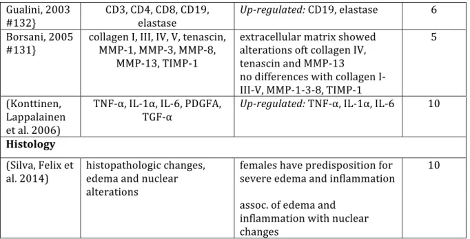
implicated specific cellular constituents by immunohistochemistry (Gualini and Berglundh 2003) the majority of papers focused on common mediators of inflammation and often those previously involved in periodontal disease (Yucel-‐Lindberg and Bage 2013).
Attemps to link peri-‐implantitis to specific genes has predominantly focused on general markers of inflammation (TNF-‐α, IL-‐1a and IL-‐1b) and inflammatory mediators (e.g. Interleukins). It is beyond the intent of this review to explore the knowledge regarding the fundamentals of tissue inflammation, however, IL1a, IL1b and TNF-‐α are known as central soluble mediators of PAMP mediated inflammation and have defined roles in peri-‐implantitis (Lindberg and Bage, 2013). Each of these are known to be produced by cells identified in the connective tissue inflammatory cell infiltrate that expands in response to plaque accumulation at implants.
The results for PISF measures of the inflammatory cytokines TNF-‐α and IL-‐1 are not
consistent. Hall et al. (Hall, Britse et al. 2011) observed no difference in TNF-‐α or other cytokines in PISF from healthy and PI sites. Wohlfart et al (Wohlfahrt, Aass et al. 2014) noted TNF-‐α levels were reduced following treatment of PI sites and other studies have observe increases in TNF-‐α in PISF associated with increased inflammation at implants. While studies of biomarkers in peri-‐implantits (Hultin, Gustafsson et al. 2002) and a cross sectional comparison of teeth vs. implants (Recker, Avila-‐Ortiz et al. 2015) found no up-‐regulation of IL-‐1β in peri-‐implantits, several other
In this review, IL-‐17 was prominently observed among studies involving PISF. Three studies reported that IL-‐17 levels are increased in PISF associated with peri-‐implantitis (Severino,
Napimoga et al. 2011, Darabi, Kadkhoda et al. 2013, Recker, Avila-‐Ortiz et al. 2015). IL-‐17 suggests an important regulatory role for TH17 t-‐cells. Secretion of IL-‐17 by these cells stimulates the production of TNF-‐α, IL-‐1β, IL-‐6 and IL-‐1β. IL-‐17 is speculated to play a role in bone resorption of rheumatoid arthritis as well as periodontitis (Kramer and Gaffen 2007).
Interleukin 6 is a pro-‐inflammatory cytokine tightly linked to osteoclastogenesis and involved in the pathogenesis of periodontitis. IL-‐6 is produced by numerous cell types in response to inflammatory mediators and is found in GCF and tissues of periodontitis. Three studies failed to demonstrate differences in the levels of IL-‐6 in PISF at healthy versus peri-‐implantitis sites
(Severino, Napimoga et al. 2011, Fonseca, Moraes Junior et al. 2014, Recker, Avila-‐Ortiz et al. 2015). However, in other comparisons, IL-‐6 levels were increased at peri-‐implantitis sites in a cross-‐ sectional studies that comparing PISF to GCF (Xie, Deng et al. 2011, Yaghobee, Khorsand et al. 2014) and in fibroblasts from peri-‐implant tissues compared to healthy sites (Irshad, Scheres et al. 2013). Further study is needed to determine if IL-‐6 levels are relatively elevated with inflammation at implant sites compared to tooth sites.
The matrix metalloproteinases in PISF were consistently elevated in peri-‐implantitis (Table 1.3.). MMPs are essential enzymes mediating inflammatory tissue destruction. Their tissue specificity and tightly regulated gene expression implies there may be pathology-‐specific roles important in peri-‐ implantitis. PISF MMP-‐8 levels, for example, may have a role in the early diagnosis of peri-‐
implantitis (Basegmez, Yalcin et al. 2012).
implantitis sites versus healthy sites. OPG levels were not consistently up regulated with peri-‐ implantitis, perhaps reflecting both the variability in disease severity as well as the limited number of subjects enrolled in these early studies. Similarly, variable outcomes were reported for sRANKL. ICTP levels in PISF were elevated at peri-‐implantitis sites and reflect high collagen turnover. Although general molecular markers of tissue turnover/destruction are observed in PISF, these studies of PISF did not demonstrate a consistent ability to identify specific molecular markers of increased osteoclast activity associated with peri-‐implantitis. Although the present data presents a general picture of increased inflammation leading to osteoclastogenesis (mediators of
osteoclastogenesis (OPG, IL-‐6 and RANKL, or mediators of tissue destruction (MMPs)), further study is required to identify PISF markers of increased osteoclast activity associated with this disease.
Peri-‐implant Immunohistology
Histological assessment of the peri-‐implant lesion has been provided by many investigators. The lesions are characterized predominantly by neutrophils, macrophages, T-‐ and B-‐cells.
Nevertheless, compared to periodontitis, peri-‐implantitis is marked by a more extensive
inflammatory infiltrate and innate immune response, a greater severity of tissue destruction and a faster progression rate (Belibasakis 2014). Different than in periodontitis, the lesion extends to the bony crest and it progresses spontaneous and continuously (Periodontology 2013).