R E S E A R C H
Open Access
Interfollicular epidermal stem-like cells for
the recreation of the hair follicle epithelial
compartment
Carla M. Abreu
1,2, Rogério P. Pirraco
1,2, Rui L. Reis
1,2, Mariana T. Cerqueira
1,2and Alexandra P. Marques
1,2*Abstract
Background:Hair follicle (HF) development and growth are dependent on epithelial-mesenchymal interactions (EMIs). Dermal papilla (DP) cells are recognized as the key inductive mesenchymal player, but the ideal source of receptive keratinocytes for human HF regeneration is yet to be defined. We herein investigated whether human interfollicular epidermal keratinocytes with stem-like features (EpSlKCs), characterized by aα6bri/CD71dimexpression, can replace human hair follicular keratinocytes (HHFKCs) for the recreation of the HF epithelium and respective EMIs.
Methods:Theα6bri/CD71dimcellular fraction was selected from the whole interfollicular keratinocyte population through fluorescence-activated cell sorting and directly compared with follicular keratinocytes in terms of their proliferative capacity and phenotype. The crosstalk with DP cells was studied in an indirect co-culture system, and EpSlKC hair forming capacity tested in a hair reconstitution assay when combined with DP cells.
Results:EpSlKCs exhibited a phenotypic profile similar to follicular keratinocytes and were capable of increasing DP cell proliferation and, for short co-culture times, the number of alkaline phosphatase-active cells, suggesting an improvement of their inductivity. Moreover, the recreation of immature HFs and sebaceous glands was observed after EpSlKC and DP cell co-grafting in nude mice.
Conclusions:Our results suggest that EpSlKCs are akin to follicular keratinocytes and can crosstalk with DP cells, contributing to HF morphogenesis in vivo, thus representing an attractive epithelial cell source for hair regeneration strategies.
Keywords:Epidermal keratinocytes with stem-like features, Dermal papilla cells, Epithelial-mesenchymal interactions, Hair follicle, Sebaceous gland
Background
The hair follicle (HF) singular capability to continually self-renew and undergo repeated cycles relies on the re-ciprocal interactions that occur between its mesenchy-mal and epithelial compartments [1, 2]. Therefore, successful strategies aiming to promote HF regeneration
rely on the combination of relevant dissociated cell pop-ulations to rescue epithelial-mesenchymal interactions (EMIs) [3, 4]. The HF mesenchyme is usually recreated with specialized dermal papilla (DP) cells, whereas the epithelial fraction is commonly reconstructed using ker-atinocytes (KCs) isolated from different follicular sources, including the bulge [5], the outer root sheath (ORS) [6–10], or the hair bulb [8,11]. Yet, the ideal epi-thelial source is not identified. Although representing a stem cell niche, the bulge can only be accessed from © The Author(s). 2021Open AccessThis article is licensed under a Creative Commons Attribution 4.0 International License, which permits use, sharing, adaptation, distribution and reproduction in any medium or format, as long as you give appropriate credit to the original author(s) and the source, provide a link to the Creative Commons licence, and indicate if changes were made. The images or other third party material in this article are included in the article's Creative Commons licence, unless indicated otherwise in a credit line to the material. If material is not included in the article's Creative Commons licence and your intended use is not permitted by statutory regulation or exceeds the permitted use, you will need to obtain permission directly from the copyright holder. To view a copy of this licence, visithttp://creativecommons.org/licenses/by/4.0/. The Creative Commons Public Domain Dedication waiver (http://creativecommons.org/publicdomain/zero/1.0/) applies to the data made available in this article, unless otherwise stated in a credit line to the data.
* Correspondence:apmarques@i3bs.uminho.pt
13B’s Research Group–Biomaterials, Biodegradables and Biomimetics,
Headquarters of the European Institute of Excellence on Tissue Engineering and Regenerative Medicine, University of Minho, Guimarães, Portugal
2
intact HF [12], which is hindered by fairly indistinguish-able anatomical boundaries [13]. Hence, KCs are fre-quently obtained from the hair bulb or ORS of plucked HFs, an easier access source. However, their isolation re-lies on the rough HF transection, followed by enzymatic digestion or cell outgrowth [12,14, 15]. This represents a low yield process [16] where population purity is re-duced due to the contamination with other cell fractions. Moreover, the isolated cells are at distinct differentiation stages, most of them being transit-amplifying (TA) cells, with shorter culture lifespan in comparison to stem cells [12].
Human interfollicular epidermal cells have also been used for HF regeneration, but successful strategies rely on their combination with murine DP cells [17] that bet-ter retain inductivity than human cells. Neonatal epithe-lial cell sources are potentially the most useful due to the immature state of the cells; however, accessibility and ethical constraints still feed the gap between de-mand and supply. Knowing that epidermal stem cells ex-press high levels of the α6-integrin (also known as CD49f) and can be distinguished from other basal cells based on their lower expression of the transferrin recep-tor (CD71) [18,19], we previously demonstrated that the
α6briCD71dim subpopulation of interfollicular epidermal stem-like cells can be isolated from adult skin [19]. Con-sidering their undifferentiated state and high clonogenic capacity [19], we hypothesized that these epidermal KCs with stem-like features (EpSlKCs) can replace follicular keratinocytes in the recreation of the HF epithelium and respective EMIs with DP cells, promoting HF formation. EpSlKCs were isolated by fluorescence-activated cell sorting (FACS) based on theirα6bri/CD71dimprofile, and their proliferative capacity and phenotype were directly compared with the commonly used human hair follicu-lar keratinocytes (HHFKCs). Next, EpSlKC capacity to affect and communicate with DP cells was tested in in-direct co-cultures focusing on cell proliferation, pheno-type, and release of the HF-related growth factors PDGF-A, VEGF, and BMP2. Finally, EpSlKC hair tricho-genic capacity was assessed in a hair inductive assay in combination with human DP cells.
Methods
Human skin was obtained from abdominoplasties, per-formed at Hospital da Prelada (Porto, Portugal), while human occipital scalp samples from hair transplantation surgeries at Sanare Unicapilar (Porto, Portugal). Samples were obtained under a collaboration protocol approved by the ethical committees of the involved institutions and after written informed consent from patients. Ani-mal experimentation was approved by the local AniAni-mal Welfare Body.
Cell isolation and culture
KCs were isolated as previously described [19] and cul-tured in keratinocyte serum-free medium (KSFM, Gibco) supplemented with 1% penicillin-streptomycin solution (PenStrep, Gibco) and with the ROCK pathway inhibitor Y-27632 (10μM, STEMCELL Technologies). HHFKCs were purchased from ScienCell (CA, USA) and cultured in the supplied keratinocyte medium. DPs were micro-dissected [20] from occipital scalp samples. The explants were cultured in Dulbecco’s modified Eagle’s medium (DMEM, Sigma-Aldrich) supplemented with 20% FBS and 1% antibiotic-antimycotic solution (Gibco). Cells growing out of the explant were further expanded in col-lagen (Sigma-Aldrich)-coated flasks and cultured in DMEM with 10% FBS and 1% antibiotic-antimycotic so-lution. Cell culture was conducted under standard con-ditions (37 °C, 5% CO2) in a humidified atmosphere and
with medium change every 2–3 days.
Feeder layer preparation
J2-3T3 mouse fibroblasts were cultured in DMEM with 10% bovine serum (Life Technologies). Cells were then inacti-vated with mitomycin C (4μg/ml, Sigma-Aldrich) for 2 h 30 min at 37 °C [21]. Feeder layers were prepared by seeding the inactivated 3T3 fibroblasts at a density of 2.4 × 10⁴cells/cm2.
Selection of theα6bri/CD71dimcellular subpopulation
A suspension of primary KCs with 5.0 × 10⁶cells/ml was incubated with CD71-PE (1:200, BioLegend) and CD49f-APC (1:85, eBioscience). The selection of α6bri/CD71dim cells was performed using a FACSAriaIII Cell Sorter and FACSDiva software (BD Biosciences). Sorted α6bri/ CD71dimcells were plated at 1.8 × 10³cells/cm2onto the feeder layer and cultured in FAD medium [1-part Ham’s F-12 (Sigma-Aldrich) and 3-parts DMEM supplemented with 10% non-inactivated FBS, 1.8 × 10⁴ M adenine, 0.5μg/ml hydrocortisone, 5μg/ml insulin, 10–10M cholera toxin (Sigma-Aldrich), 10 ng/ml epidermal growth factor (Peprotech), 1.8 mM CaCl2(Merck), and 1% PenStrep].
Flow cytometry analysis
For surface marker analysis, cell suspensions were dir-ectly incubated with fluorochrome-labeled antibodies (Supplemental Table S1). For intracellular staining, cells were firstly fixed with the FIX&PERM™ Cell Permeabilization Kit Reagent A (Life Technologies) for 15 min at room temperature (RT) and then perme-abilized with Reagent B and simultaneously incubated with the intracellular-labeled antibodies (20 min, RT). Cells were analyzed with FACSCalibur flow cytometer in CellQuest software.
Indirect co-culture
EpSlKCs were harvested from the feeder layer [21], re-suspended in KSFM with Y-27632, and seeded at a dens-ity of 500 cells/cm2. Co-cultures were established in the next day, using Transwell® inserts (0.4μm pore, Corn-ing). DP cells were plated in the insert at a density of 1000 cells/cm2. The co-culture medium consisted of an equal mixture of DMEM 10% FBS and KSFM with Y-27632 (DMEM:KSFM). Single cell-type cultures were prepared as controls, by culturing cells in their regular medium and in the co-culture medium, the latter to control possible medium effects on the cells.
DNA and alkaline phosphatase quantification
At each experimental time point, cells were washed with PBS and lysed with 0.01% SDS (NZYteck). DNA was quantified using the Picogreen™ dsDNA kit (Thermo Fisher Scientific), and active ALP was quantified using the ALP Fluorescent Detection Kit (Sigma-Aldrich), both following the manufacturer’s instructions.
Growth factor quantification
Growth factors present in the supernatants of the 48 h homotypic cultures or co-cultures were quantified using VEGF ELISA Development Kit (Peprotech), BMP2 ELISA Development Kit (Peprotech), and PDGF-A Duo-Set (R&D Systems), according to the manufacturer’s in-structions. DNA values were used to normalize results.
Hair reconstitution assay
Athymic male nude Balb/C mice (Charles River, 9 weeks) were randomly divided into three experimental groups: control, DP cells, and DP cells with EpSlKCs (n= 4, each group). The silicone chamber model was set and used as previously described [22]. Cell suspensions with 5.0 × 10⁶ DP cells and 2.5 × 10⁶ EpSlKCs (experi-mental group), 5.0 × 10⁶DP cells (DP cell group), or ve-hicle (FAD medium, control group) were injected in the chamber. The top of the chamber was cut-off after 1 week, and the chamber was fully removed after 2 weeks. Animals were euthanized 6 weeks after surgery to har-vest the wound area for histological analysis.
Immunofluorescence and histological analysis
Cells from the in vitro experiments were washed in PBS and fixed in 10% (v/v) formalin for 15 min (RT). Cell permeabilization was performed with 0.2% (v/v) Triton X-100 for 15 min (RT, intracellular staining) or 30 min (4 °C, nuclear staining), and blocking with a 3% (w/v) BSA solution for 30 min (RT). Samples were then incu-bated with primary antibodies (Supplemental Table S2) diluted in 1% BSA for 1 h (RT). After washing with PBS, Alexa Fluor (594/488)-conjugated secondary antibodies (1:500; Molecular Probes) were applied. Cellular
morphology was observed after staining F-actin with Phalloidin-TRITC (1:100, Sigma-Aldrich) for 1 h (RT) and nuclear counterstain performed using DAPI (0.02 mg/ml, Biotium) for 15 min (RT). Images were acquired with an Axio Imager Z1m microscope (Zeiss).
For histological analysis, 4-μm paraffin-embedded sec-tions were stained with hematoxylin and eosin (H&E), according to standard procedures, and analyzed with a DM750 microscope (Leica). For immunofluorescence studies, heat-mediated antigen retrieval was performed in the deparaffinized sections using the citrate buffer (pH 6.0), and the remaining procedure was performed as above described for the in vitro ones.
Statistical analysis
Statistical analysis was performed using GraphPad Prism 7.03. To test if data followed a Gaussian distribution, the D’Agostino and Pearson normality test was used. Non-parametric data were analyzed with a Mann-Whitney t test (two groups, unpaired); the Kruskal-Wallis (three groups, unpaired) or Friedman test (three groups, paired) was used coupled with Dunn’s post-test. Para-metric data were analyzed using a one-way ANOVA (two groups, paired) or RM two-way ANOVA (three groups, paired) in combination with Tukey’s post-test. Differences withp< 0.05 were considered significant. Results
EpSlKCs and HHFKCs are phenotypically similar
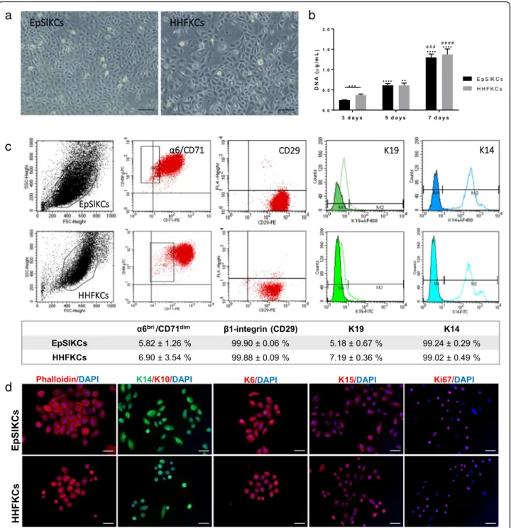
The isolated interfollicular KCs comprehended a low per-centage of epidermal stem cells (4.62 ± 1.47%; α6bri/ CD71dim fraction) and differentiated cells (3.72 ± 0.47%;
α6dim subpopulation), while TA cells (78.44 ± 3.26%;α6bri/ CD71bri subpopulation) represented the majority of the population (Supplemental Fig. S1a), as expected [18]. The selectedα6bri/CD71dimcells were cultured on feeders (Sup-plemental Fig. S1b,c), and the obtained cells—EpSlKCs—
were directly compared to HHFKCs. Most EpSlKCs and HHFKCs were small and bright cells displaying a cobble-stone morphology (Fig. 1a), characteristic of undifferenti-ated epithelial cells. However, cellular heterogeneity was higher for HHFKC cultures, with the presence of large size cells, representative of differentiated cells. Nevertheless, both cell types proliferated at similar rates (Fig. 1b), al-though at day 3 HHFKC numbers were higher than EpSlKCs. The percentage ofα6bri/CD71dimcells in both cell types was similar, as was the expression of the basal epider-mal markers integrin β1 (CD29) and keratin (K) 14 (Fig.
1c) . The expression of K19, typically considered a stem cell marker whose expression decreases with age [23], was also similar among cell types. Immunocytochemistry analysis confirmed their immature phenotype, with positive staining for the basal-specific markers K15, K6, and K14, and ab-sence of the differentiation marker K10 (Fig. 1d).
Additionally, most cells were positive for the proliferation-associated marker ki67. Together, these results demonstrate that EpSlKC and HHFKC proliferative capacity and pheno-type are equivalent.
EpSlKCs support DP cell growth and a partial restoration of their native phenotype
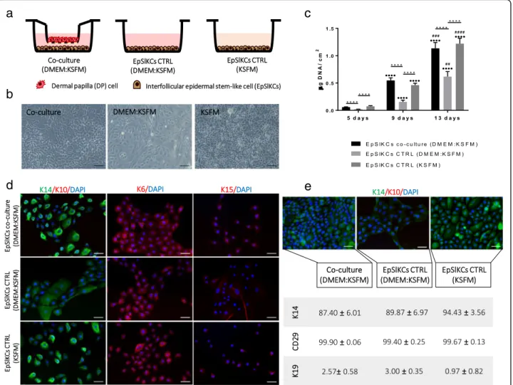
To study EpSlKC capacity to communicate with DP cells, an indirect co-culture was established (Fig. 2a). bri
a
c
d
b
Fig. 1Morphology, proliferation, and phenotype of EpSlKCs and HHFKCs.aRepresentative light microscopy images of human epidermal stem-like keratinocyte (EpSlKC) and human hair follicular keratinocyte (HHFKC) culture.bDNA quantification of the cells along the culture time (n= 5, EpSlKCs;n= 4, HHFKCs).cRepresentative flow cytometry histograms and respective quantification regarding the percentage ofα6bri/CD71dim cells, and those positive for integrinβ1 (CD29), keratin 19 (K19), and keratin 14 (K14) in EpSlKC (n= 4) and HHFKC (n= 3) cultures after 1 week.d
Immunofluorescence staining ofβ-actin filaments (phalloidin), keratin 14 (K14), keratin 10 (K10), keratin 6 (K6), keratin 15 (K15), and the proliferation-associated marker Ki67 in EpSlKCs and HHFKCs. DAPI was used as a nuclear counterstainer. Data shown are mean ± SEM. Scale bars are 100μm foraand 50μm ford. ***p< 0.001;··p< 0.01 and····p< 0.0001 vs. day 3;###p< 0.001 and####p< 0.0001 vs. day 5
After co-culture, EpSlKCs remained small and exhibited a cuboidal morphology (Fig. 2b). In the conventional culture medium (KSFM), EpSlKCs displayed a cobble-stone morphology and were slightly brighter and smaller than in co-culture. Despite this, co-cultured EpSlKCs were different from those kept in the co-culture medium (DMEM:KSFM), whose morphology, larger size, and lower brightness evidenced a more differentiated state. Co-culture with DP cells also prevented the differenti-ation of EpSlKCs, expected to be induced by DMEM components, such as calcium [24,25]. Furthermore, co-culture conditions were able to sustain EpSlKC prolifera-tion along culture, as demonstrated by the similar DNA amounts of the co-culture and KSFM, both significantly higher than in the medium control (Fig. 2c). Further evaluation of DP cell effects on EpSlKC differentiation showed that K14 expression was similar in the
co-cultures and in the monoculture established with KSFM. In contrast, K14-bright-positive cells were not found in the monocultures of EpSlKCs in the control medium (Fig.2d). Nevertheless, K6 and K15 staining were similar among conditions, with cells clearly stained for K6 but weakly expressing K15. After 13 days in co-culture, most cells were K14-positive, as confirmed by flow cytometry analysis, but K14-bright cells were only observed for EpSlKCs cultured in KSFM (Fig. 2e). Independently of the culture conditions, EpSlKCs retainedβ1-integrin ex-pression while K19 exex-pression was almost absent.
In addition to the effect of the co-culture over EpSlKCs, we looked to potential alterations on DP cells (Fig. 3a). In DMEM, most of the cells exhibited a flat-tened and enlarged morphology, typical of higher pas-sage cells, while in co-culture they were smaller, with a polygonal or spindle-shaped morphology (Fig. 3b),
a
c
e
b
d
Fig. 2Characterization of EpSlKCs co-cultured with DP cells.aSchematic representation andblight microscopy images of EpSlKCs in co-culture with dermal papilla (DP) cells and in the respective monoculture controls.cDNA quantification of EpSlKCs after 5, 9, and 13 days of culture (n= 5).dRepresentative images of immunofluorescence staining against the epithelial markers K14 and K10, K6, and K15 after 5 days in culture.e
Immunostaining against K14 staining in EpSlKCs after 13 days in culture and the respective flow cytometry quantitative data (n= 3). DAPI was used as nuclear counterstaining. Data shown are mean ± SEM. Scale bars are 100μm forband 50μm fordande. ****p< 0.0001;····p< 0.0001 vs. day 5;##p< 0.01,###p< 0.001, and####p< 0.0001 vs. day
evidencing a healthier state [26, 27]. When cultured in the control medium, DP cells had an intermediate morphology. Likewise, the cell number at each time point was successively higher from DMEM to the co-culture conditions revealing an increased proliferation capacity both due to the culture medium and the pres-ence of EpSlKCs (Fig.3c). The expression of the V1 iso-form of versican was uniiso-form among conditions, whereas the in vivo predominant V2 isoform [28] was only observed in co-cultured DP cells (Fig. 3d). More-over, the amount of active alkaline phosphatase (ALP) in co-culture was significantly higher than that in the con-trols after 5 days in culture, although this effect was not sustained over time (Fig. 3e). A decrease in α-SMA ex-pression was also observed in co-culture, although
C-C-X-C chemokine receptor type 4 (CD184) and low-density lipoprotein receptor-related protein 4 (LRP-4) expression was not restored (Fig.3f).
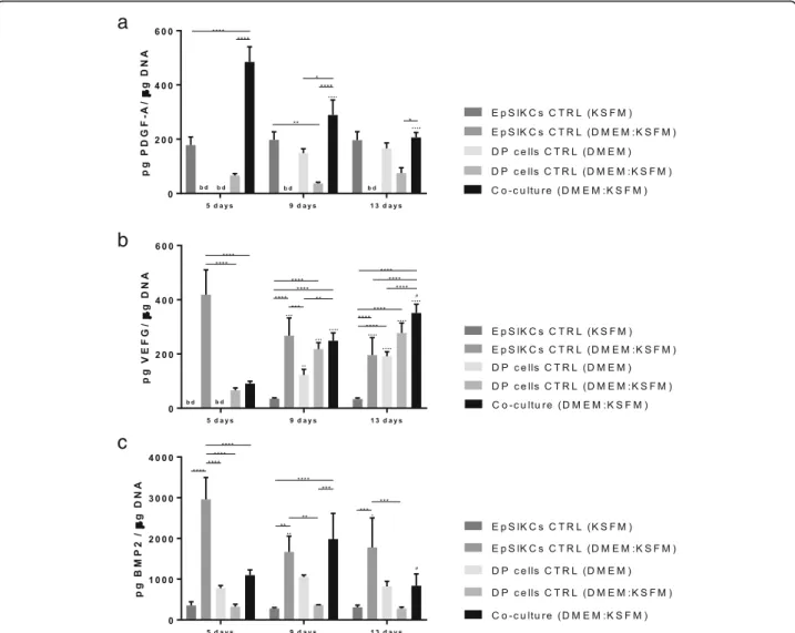
EpSlKC and DP cell co-culture impacts the release of EMI mediators
To study if DP cells and EpSlKCs could communicate through the release of mediators, as seen in the native HF EMIs, we studied the release of PDGF-A, VEGF, and BMP2, known to influence hair growth [29–31].
PDGF-A production was higher in the co-culture than in the other conditions although successively lower levels were detected along the culture (Fig.4a). EpSlKCs in KSFM were also able to secrete PDGF-A, which was at a constant level along the culture time. Secretion of
a
b
d
f
c
e
Fig. 3Characterization of DP cells co-cultured with EpSlKCs.aSchematic representation of the co-culture established with DP cells and EpSlKCs and the respective monoculture controls.bRepresentative images of DP cell morphology after staining against the mesenchymal cytoskeleton marker vimentin.cDNA quantification of DP cells along the culture time (n= 5).dExpression of the versican V1 and V2 isoforms by DP cells in the different culture conditions at day 5 of culture. The presence of versican V2-positive cells was observed when DP cells were co-cultured with EpSlKCs (zoomed-in image). Images were counterstained with DAPI.eThe inductivity of DP cells was assessed through the quantification of active alkaline phosphatase (ALP,n= 3).fQuantitative flow cytometry data (n= 3). *p< 0.05, **p< 0.01, ***p< 0.001, ****p< 0.0001;··p< 0.01 and
PDGF-A by DP cells was only detected at 9 and 13 days of culture in DMEM. However, in the monocultures established to control medium effects (DMEM:KSFM), PDGF-A values were below the detection level for EpSlKCs and lower than the standard culture for DP cells from day 9 onward. Considering this, it seems that the interaction between EpSIKCs and DP cells in co-culture promotes PDGF-A production, particularly at early time points.
VEGF levels in the co-culture increased with time, and both EpSlKCs (p< 0.05) and DP cells secreted more VEGF when cultured in the medium used for the co-culture (DMEM:KSFM) than in KSFM and DMEM, re-spectively (Fig. 4b). Moreover, the amount of VEGF se-creted by EpSlKCs decreased along the culture and increased for DP. Despite this, VEGF levels in the co-cultures and in the monoco-cultures of DP cells in the same
medium were similar independently of the time of cul-ture. Thus, except at day 5 at which the VEGF amount was significantly lower in the co-culture than in the EpSlKC cultures established in the co-culture medium, it seems that the co-culture does not affect VEGF secretion.
In what concerns BMP2 secretion, the levels detected in the co-culture peaked on day 9 (Fig.4c). The amount of BMP2 in the monocultures of EpSlKCs in KSFM was lower than that in the monoculture of DP cells in DMEM, and none varied with time. However, the medium used to establish the co-culture medium had contrary effects on each cell type, respectively promoting and diminishing BMP2 secretion by EpSlKCs and DP cells. Except for day 9, BMP2 levels in the co-cultures were always lower than those in the monocultures of EpSlKCs established with the same medium. This
a
b
c
Fig. 4Quantification of the amount ofaPDGF-A,bVEGF, andcBMP2 released by both co-cultured cells and monocultured EpSlKCs and DP cells by ELISA (n= 3). Data shown are mean ± SEM. *p< 0.05, **p< 0.01, ***p< 0.001, ****p< 0.0001;∙p< 0.05,··p< 0.01,···p< 0.001,····p< 0.0001 vs. day 5;#p< 0.05 vs. day 9. bd, bellow detection level; BMP2, bone morphogenetic protein 2; PDGF-A, platelet-derived growth factor A; VEGF, vascular endothelial growth factor
indicates that cells in co-culture are communicating, and suggests that DP cells may be responsible for the overall decreased BMP2 production in co-culture.
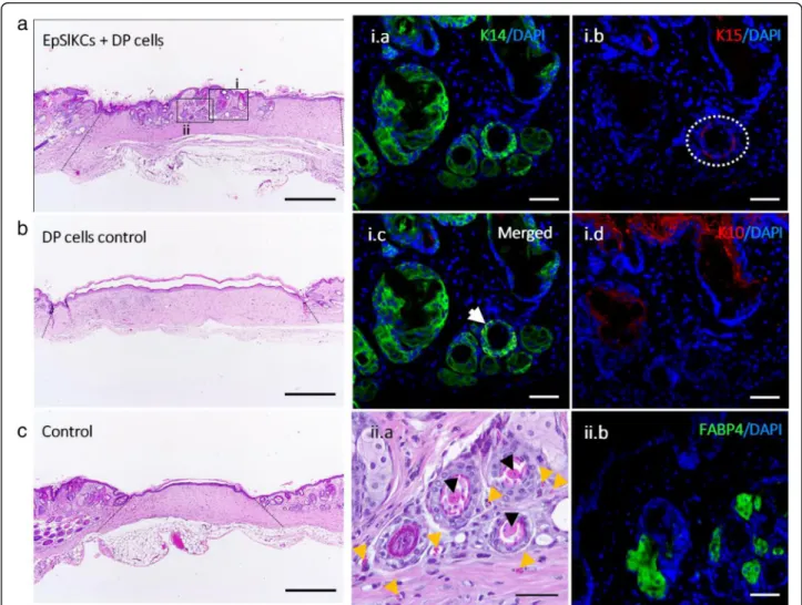
Generation of HF and sebaceous gland-like structure by co-grafted EpSlKCs and DP cells
Six weeks after EpSlKC and DP cell transplantation in mice, we observed the formation of structures that mor-phologically resembled HF and sebaceous glands (SG) in 3 out of 4 animals (Fig. 5a). These structures were not observed in the controls (Fig. 5b, c). The immunohisto-chemical analysis of the recreated structures showed the presence of epithelial layers positive for the basal markers K14 and K15 (Fig. 5i.a,b), in some cases co-expressed (Fig. 5i.c, white arrow), whereas K10 was ob-served in the skin outermost layer and in some of the
structures’core (Fig.5i.d). The presence of multiple con-centric epithelial layers, some of them with an appar-ently keratinized core (Fig. 5ii.a, black arrowhead), was also observed. This suggests that the differentiation process was initiated but remained incomplete, since features of mature HFs, including hair shaft formation, were not observed. Moreover, small vessels were ob-served around the recreated structures (Fig. 5ii.a, yellow arrowhead), while in the SG-like structures the expres-sion of the adipocyte marker FABP4 (Fig. 5ii.b) demon-strates sebocyte lineage differentiation. Labelling with a human-specific DNA probe failed to detect the presence of human cells within the recreated structures (Supple-mental Fig. S2). Nevertheless, the absence of structures in the controls suggests that the human EpSlKCs were critical for HF and SG induction process.
Fig. 5HF and SG induction in mice.aRepresentative hematoxylin and eosin (H&E) images of the area where EpSlKCs and DP cells,bDP cells alone, orcvehicle were transplanted (dashed lines indicating the wound area). Immunostaining of the recreated hair follicle (HF)- and sebaceous gland (SG)-like structures against the epithelial markersi.aK14,i.bK15 (dashed circle), ori.cboth (white arrow).i.dExpression analysis of the differentiation marker K10 within the wound area (ii.a). H&E higher magnification image evidencing an apparently differentiated core (black arrowheads) within the formed structures, and the presence of small vessels around them (yellow arrowheads).ii.bImmunodetection of the fatty acid-binding protein 4 (FABP4) within the structures resembling SGs. Scale bars are 500μm fora–cand 50μm fori.a–i.dandii.aandii.b
Discussion
Despite the advances in rodent HF regeneration, thought the combination of receptive-epithelial and inductive-mesenchymal cells, human HF regeneration remains elu-sive. Major hurdles reside in the lack of tissue sources capable of providing immature epithelial cells, and of suitable culture conditions capable of minimizing cell differentiation in vitro. Here, we propose the use of EpSlKCs as an alternative to HHFKCs for the recreation of the HF epithelial compartment. Compared to HHFK Cs, EpSlKCs represent a more homogeneous cell frac-tion and larger numbers of EpSlKCs can be obtained from surgically discarded skin, enabling hair regener-ation studies/therapies.
Despite their undifferentiated profile and high clono-genic capacity [19], using EpSlKCs for the recreation of the HF epithelial compartment depends on their cap-acity to crosstalk with DP cells. We showed that DP cells promoted an increase in EpSlKC numbers, which is in agreement with previous studies confirming that DP cells enhance the growth of co-cultured ORS cells [32,
33]. For longer culture times, EpSlKCs in co-culture in-creased in size and the K14-bright cells were lost, sug-gesting differentiation. Despite this, their proliferation was not affected, which seems to indicate that DP cells are tightly balancing EpSlKC division and differentiation. This is important considering that DP controls the num-ber of bulb KCs [34], whose continuous proliferation and differentiation are required for hair growth. Another indicator of this control was the release of VEGF and BMP2 by co-cultured cells. Epidermal VEGF is mainly expressed in the upper-to-middle epidermal layers, sup-porting a positive correlation between VEGF release and epithelial cell differentiation [35, 36]. This is also sup-ported by the higher amount of VEGF detected in EpSlKCs cultured in the co-culture medium, in which EpSIKCs have a more differentiated phenotype. Interest-ingly, VEGF levels in the co-culture increased with time, which may indicate that EpSlKC differentiation is occur-ring. However, their proliferative capacity remained
un-affected, once again indicating that EpSlKC
differentiation and proliferation is balanced by DP cells. Also, BMP expression is subtle among proliferating epi-thelial cells and higher in differentiating cells [37]. Herein we showed that BMP2 secretion in the co-cultures peaked at day 9 while decreasing in the EpSlKC monocultures established in the same medium, once again suggesting DP cell effect.
EpSlKCs also improved DP cell proliferation, an effect already reported for rodent KCs [38]. To this might have contributed the marked increase in PDGF-A levels ob-served in the co-culture, particularly up to 9 days of cul-ture. PDGF-A is a mitogenic factor typically produced by epidermal [39] and follicular keratinocytes [40],
capable of stimulating mesenchymal cell proliferation [41]. Most importantly, the PDGF-A receptor is acti-vated in the anagenic DP [42] and PDGF-A administra-tion has been demonstrated to induce anagen [42, 43]. This suggests that PDGF-A increase in co-culture may not only promote DP cell proliferation but also stimulate their hair growth capacity. In fact, co-culture conditions’ positive effect on DP cell phenotype was demonstrated by the loss of α-SMA expression. Although commonly referred as a DP cell marker [44], α-SMA is only expressed in 2D-cultured DP cells and not in situ [45]; therefore, its near absence in the co-cultures seems an indicator that EpSlKCs are positively influencing DP cells. ALP activity and versican expression, in turn, are well-established markers of DP cell inductivity [44]. The presence of the V2 isoform of versican at early culture times, which is predominant in the native DP [28], and the increased number of ALP-active DP cells also sug-gest an improvement in the inductivity. Nevertheless, ALP activity is known to vary along the HF cycle, and differences in DP spatial-temporal ALP activity might correlate with distinct functions along the cycle [44,46]. Furthermore, the release profile of BMP2 also suggests a hair inductive profile by co-cultured DP cells. BMP sig-naling is an anagen negative regulator [31, 47], which needs to be counteracted for HF induction to occur [31,
48,49]. Therefore, the lower BMP2 expression observed at day 5, associated with increased PDGF-A levels, sug-gest that EpSlKC-DP cell communication in vitro may be mimicking early anagen features. This is interesting considering previous reports where ORS cell co-culture with DP cells, precisely for 4–5 days, restored ORS cell trichogenic ability, with DP cells influencing their com-petence [50,51]. They also demonstrated that previously co-cultured ORS cells promoted HF formation, whereas monocultured ORS cells failed to do it. Remarkably, we observed the recreation of HF- and SG-like structures upon co-grafting of EpSlKCs and human DP cells, re-gardless of not having been co-cultured, which suggests that EpSlKCs can acquire follicle-specific competence in vivo upon interaction with DP cells.
The capacity of interfollicular epidermal cells to differ-entiate into follicular structures was already demon-strated in the chamber model, after their combination with murine DP cells [17]. However, to our knowledge, this is the first study to demonstrate the recreation of HF- and SG-like structures upon grafting adult human interfollicular KCs and DP cells. Additionally, the cap-acity of human epithelial cells to generate HF-like struc-tures is higher for neonatal cells than for adult or passaged cells [17], supporting the importance of EpSlKC immature state. We demonstrated that KSFM-cultured EpSlKCs, as performed before grafting, are kept in a high proliferative state. Moreover, these cells are
characterized by high production of PDGF-A, a factor involved in HF formation, and low secretion of the ana-gen inhibitor BMP2, which may have contributed to the observed response. The recreated structures displayed common features of the native HF, including the pres-ence of cell layers co-expressing K15 and K14, as ob-served in the bulge and outermost upper layer of the ORS, or only expressing K14 as in the ORS suprabasal layers and respective progeny [52]. Similarly, K10 ex-pression in the HF is limited to the differentiated follicu-lar portions adjacent to the epidermis [52]. This pattern was simulated in the recreated structures, as shown by the K10 expression in the innermost epithelial layer of the HF-like structures closer to the epidermis and its ab-sence in the ones located deeper in the dermis. We also observed the presence of blood vessels around the struc-tures, likely a consequence of EpSlKC and DP cell pro-duction of VEGF, essential to induce and maintain the microvasculature around the HF and in the dermal com-partment [30, 53]. Nevertheless, the follicle-like struc-tures failed to produce a hair shaft, demonstrating that hair formation remained incomplete. Furthermore, we did not detect human cells within the recreated struc-tures or in the grafting area, which has potentially lim-ited the required signaling for the complete hair formation. The absence of the transplanted cells is not completely unexpected considering that, unlike other models, the chamber assay represents a wound healing environment [4], with high cellular turnover. Moreover, although the chamber prevents the contribution of host cells for the initial regenerative process, dissociated hu-man cells are easily replaced by host KCs after wound closure [54]. Nonetheless, the lack of structures in the controls demonstrates that human EpSlKCs were critical for hair neogenesis.
Conclusions
Taken together, our results show that EpSlKCs are phenotypically similar to HHFKCs and can crosstalk and beneficially impact DP cells in vitro. When combined with cultured DP cells in vivo, EpSlKCs demonstrated their competence by promoting the recreation of struc-tures that resembled the HF and SG initial morpho-genetic events. Thus, EpSlKCs represent an alternative immature and available epithelial cell fraction for the re-creation of the HF epithelial compartment.
Supplementary Information
The online version contains supplementary material available athttps://doi. org/10.1186/s13287-020-02104-9.
Additional file 1: Fig. S1.Phenotype of the CD49fbri/CD71dim subpopulation. A) Representative image of fluorescent activated cell sorting (FACS) dotplot of the whole population of epidermal
keratinocytes and the gatting of the subpopulations of cells, based on the expression of the CD49f and CD71 markers, accompanied by the respective cellular percentages. CD49fbri/CD71dimcells represent epidermal stem-like cells, CD49fbri/CD71brisubpopulation are transit-amplifying (TA) cells, while differentiated (Diff.) cells are characterized by a CD49fdimphenotype. Epidermal stem-like cells derived colonies grow-ing in the inactivated feeders (B) 4 and (C) 6 days after seedgrow-ing. Data shown are mean ± SEM. Scale bars are 50μm.Fig. S2. Human cell detec-tion within skin explants from the hair reconstitudetec-tion assay. Staining with a human-specific probe demonstrates that, 6 weeks after cells injection, there were no human cells remaining in the wound area in the controls (A,B) and in the condition where interfollicular epidermal stem-like kerati-nocytes (EpSlKCs) and DP cells were co-grafted(C). Likewise, no staining was observed in the mice tissue (D), whereas nuclear orange-pink stain-ing was observed in human positive control specimens (E,F), demonstrat-ing the specificity of the staindemonstrat-ing. Scale bars are 200μm, for (A-D) and 50μm for (E,F).Table S1.List of antibodies used for flow cytometry stud-ies.Table S2.List of antibodies used for immunofluorescence studies.
Abbreviations
ALP:Alkaline phosphatase; BMP2: Bone morphogenetic protein 2; CD184: C-X-C chemokine receptor type 4; CD29: Integrinβ1; CD49f: Integrinα6; CD71: Transferrin receptor; DP: Dermal papilla; DMEM: Dulbecco’s modified eagle medium; EMIs: Epithelial-mesenchymal interactions; EpSlKCs: Epidermal keratinocytes with stem-like features; FABP4: Fatty acid-binding protein 4; FACS: Fluorescence-activated cell sorting; HF: Hair follicle; HHFKCs: Human hair follicular keratinocytes; K: Keratin; KSFM: Keratinocyte serum-free medium; KCs: Keratinocytes; LRP-4: Low-density lipoprotein receptor-related protein 4; ORS: Outer root sheath; PDGF-A: Platelet-derived growth factor A; SG: Sebaceous gland; SMA: Smooth muscle actin; TA: Transit-amplifying; VEGF: Vascular endothelial growth factor
Acknowledgements
We would like to acknowledge Prof Fiona Watt for her expert knowledge and Emanuel Rognoni and Simon Broad, also from King’s College London, for their input regarding the animal model used and the cell culture methodology. This collaboration was boosted by GENE2SKIN, a European Union’s Horizon 2020 project (Grant Agreement Number 692221). The support of Manuela Lago on the in vitro work is also acknowledged.
Authors’contributions
CA: collection and assembly of data, data analysis and interpretation, and manuscript writing
RPP: data analysis and interpretation and collection and assembly of data RLR: administrative and financial support
MTC: data analysis and interpretation and collection and assembly of data APM: administrative and financial support, conception and design, supervision, data analysis and interpretation, and final approval of the manuscript
The authors read and approved the final manuscript.
Funding
This study was supported by the FCT/MCTES (Fundação para a Ciência e a Tecnologia/Ministério da Ciência,Tecnologia,e Ensino Superior) through the PD/59/2013, PD/BD/113800/2015 (C.M. Abreu), CEECIND/00695/2017 (M.T. Cerqueira), IF/00347/2015 (R. P. Pirraco), and IF/00945/2014 (A.P. Marques) grants.
Availability of data and materials
The authors confirm that the data supporting the findings of this study are available within the article and its supplementary materials.
Ethics approval and consent to participate
All human tissue samples were collected after the patient consent and under a collaboration protocol approved by the ethic committees of the involved institutions. Experimental protocols involving animals were approved by the local animal welfare body and performed according to the applicable national regulations and international animal welfare rules.
Consent for publication
Not applicable
Competing interests
The authors declare that they have no competing interests.
Received: 30 September 2020 Accepted: 14 December 2020
References
1. Millar SE. Molecular mechanisms regulating hair follicle development. J Invest Dermatol. 2002;118:216–25.
2. Sennett R, Rendl M. Mesenchymal–epithelial interactions during hair follicle morphogenesis and cycling. Semin Cell Dev Biol. 2012;23:917–27. 3. Ohyama M, Veraitch O. Strategies to enhance epithelial–mesenchymal
interactions for human hair follicle bioengineering. J Dermatol Sci. 2013;70: 78–87.
4. Balañá ME. Epidermal stem cells and skin tissue engineering in hair follicle regeneration. World J Stem Cells. 2015;7:711–27.
5. Leirós GJ, Kusinsky AG, Drago H, et al. Dermal papilla cells improve the wound healing process and generate hair bud-like structures in grafted skin substitutes using hair follicle stem cells. Stem Cells Transl Med. 2014;3:1209–19. 6. Limat A, Breitkreutz D, Hunziker T, et al. Outer root sheath (ORS) cells
organize into epidermoid cyst-like spheroids when cultured inside Matrigel: a light-microscopic and immunohistological comparison between human ORS cells and interfollicular keratinocytes. Cell Tissue Res. 1994;275:169–76. 7. Havlickova B, Biro T, Mescalchin A, et al. Towards optimization of an
organotypic assay system that imitates human hair follicle-like epithelial-mesenchymal interactions. Br J Dermatol. 2004;151:753–65.
8. Wu J-J, Zhu T-Y, Lu Y-G, et al. Hair follicle reformation induced by dermal papilla cells from human scalp skin. Arch Dermatol Res. 2006;298:183–90. 9. Havlickova B, Bíró T, Mescalchin A, et al. A human folliculoid microsphere assay for exploring epithelial–mesenchymal interactions in the human hair follicle. J Invest Dermatol. 2009;129:972–83.
10. Lindner G, Horland R, Wagner I, et al. De novo formation and ultra-structural characterization of a fiber-producing human hair follicle equivalent in vitro. J Biotechnol. 2011;152:108–12.
11. Miao Y, Sun Y Bin, Liu BC, et al. Controllable production of transplantable adult human high-passage dermal papilla spheroids using 3D Matrigel culture. Tissue Eng Part A. 2014;20:2329–38.
12. Aasen T, Belmonte JCI. Isolation and cultivation of human keratinocytes from skin or plucked hair for the generation of induced pluripotent stem cells. Nat Protoc. 2010;5:371–82.
13. Ohyama M. Characterization and isolation of stem cell-enriched human hair follicle bulge cells. J Clin Invest. 2005;116:249–60.
14. Moll I. Proliferative potential of different keratinocytes of plucked human hair follicles. J Invest Dermatol. 1995;105:14–21.
15. Limat A, Noser FK. Serial cultivation of single keratinocytes from the outer root sheath of human scalp hair follicles. J Invest Dermatol. 1986;87:485–8. 16. Seo Y-K, Lee D-H, Shin Y-H, et al. Development of isolation and cultivation
method for outer root sheath cells from human hair follicle and construction of bioartificial skin. Biotechnol Bioprocess Eng. 2003;8:151–7. 17. Ehama R, Ishimatsu-Tsuji Y, Iriyama S, et al. Hair follicle regeneration using
grafted rodent and human cells. J Invest Dermatol. 2007;127:2106–15. 18. Li A, Simmons PJ, Kaur P. Identification and isolation of candidate human
keratinocyte stem cells based on cell surface phenotype. Proc Natl Acad Sci. 1998;95:3902–7.
19. Cerqueira MT, Frias AM, Reis RL, et al. Boosting and rescuing epidermal superior population from fresh keratinocyte cultures. Stem Cells Dev. 2014; 23:34–43.
20. Gledhill K, Gardner A, Jahoda CAB. Isolation and establishment of hair follicle dermal papilla cell cultures. Methods Mol Biol. 2013;989:285–92. 21. Watt F, Simon B, Prowse D. Cultivation and retroviral infection of human
epidermal keratinocytes. Cell Biol. 2006;1:133–8.
22. Jensen KB, Driskell RR, Watt FM. Assaying proliferation and differentiation capacity of stem cells using disaggregated adult mouse epidermis. Nat Protoc. 2010;5:898–911.
23. Michel M, Török N, Godbout MJ, et al. Keratin 19 as a biochemical marker of skin stem cells in vivo and in vitro: keratin 19 expressing cells are differentially localized in function of anatomic sites, and their number varies with donor age and culture stage. J Cell Sci. 1996;109:1017–28.
24. Bertolero F, Kaighn ME, Camalier RF, et al. Effects of serum and serum-derived factors on growth and differentiation of mouse keratinocytes. Vitr Cell Dev Biol. 1986;22:423–8.
25. Borowiec A-S, Delcourt P, Dewailly E, et al. Optimal differentiation of in vitro keratinocytes requires multifactorial external control. PLoS One. 2013;8: e77507.
26. Ohyama M, Kobayashi T, Sasaki T, et al. Restoration of the intrinsic properties of human dermal papilla in vitro. J Cell Sci. 2012;125:4114–25. 27. Upton JH, Hannen RF, Bahta AW, et al. Oxidative stress–associated
senescence in dermal papilla cells of men with androgenetic alopecia. J Invest Dermatol. 2015;135:1244–52.
28. Soma T, Tajima M, Kishimoto J. Hair cycle-specific expression of versican in human hair follicles. J Dermatol Sci. 2005;39:147–54.
29. Takakura N, Yoshida H, Kunisada T, et al. Involvement of platelet-derived growth factor receptor-αin hair canal formation. J Invest Dermatol. 1996; 107:770–7.
30. Yano K, Brown LF, Detmar M. Control of hair growth and follicle size by VEGF-mediated angiogenesis. J Clin Invest. 2001;107:409–17.
31. Botchkarev VA, Botchkareva NV, Roth W, et al. Noggin is a mesenchymally derived stimulator of hair-follicle induction. Nat Cell Biol. 1999;1:158–64. 32. Limat A, Hunziker T, Waelti ER, et al. Soluble factors from human hair papilla
cells and dermal fibroblasts dramatically increase the clonal growth of outer root sheath cells. Arch Dermatol Res. 1993;285:205–10.
33. Itami S, Kurata S, Sonoda T, et al. Interaction between dermal papilla cells and follicular epithelial cells in vitro: effect of androgen. Br J Dermatol. 1995; 132:527–32.
34. Li W, Man X-Y, Li C-M, et al. VEGF induces proliferation of human hair follicle dermal papilla cells through VEGFR-2-mediated activation of ERK. Exp Cell Res. 2012;318:1633–40.
35. Viac J, Palacio S, Schmitt D, et al. Expression of vascular endothelial growth factor in normal epidermis, epithelial tumors and cultured keratinocytes. Arch Dermatol Res. 1997;289:158–63.
36. Goldman CK, Tsai J-C, Soroceanu L, et al. Loss of vascular endothelial growth factor in human alopecia hair follicles. J Invest Dermatol. 1995;104:18–20. 37. Kulessa H. Inhibition of Bmp signaling affects growth and differentiation in
the anagen hair follicle. EMBO J. 2000;19:6664–74.
38. Inamatsu M, Matsuzaki T, Iwanari H, et al. Establishment of rat dermal papilla cell lines that sustain the potency to induce hair follicles from afollicular skin. J Invest Dermatol. 1998;111:767–75.
39. Ansel JC, Tiesman JP, Olerud JE, et al. Human keratinocytes are a major source of cutaneous platelet-derived growth factor. J Clin Invest. 1993;92:671–8. 40. Kamp H, Geilen CC, Sommer C, et al. Regulation of PDGF and PDGF
receptor in cultured dermal papilla cells and follicular keratinocytes of the human hair follicle. Exp Dermatol. 2003;12:662–72.
41. Karlsson L, Bondjers C, Betsholtz C. Roles for PDGF-A and sonic hedgehog in development of mesenchymal components of the hair follicle. Development. 1999;126:2611–21.
42. Festa E, Fretz J, Berry R, et al. Adipocyte lineage cells contribute to the skin stem cell niche to drive hair cycling. Cell. 2011;146:761–71.
43. Tomita Y, Akiyama M, Shimizu H. PDGF isoforms induce and maintain anagen phase of murine hair follicles. J Dermatol Sci. 2006;43:105–15. 44. Yang C-C, Cotsarelis G. Review of hair follicle dermal cells. J Dermatol Sci.
2010;57:2–11.
45. Jahoda CAB, Reynolds AJ, Chaponnier C, et al. Smooth muscle alpha-actin is a marker for hair follicle dermis in vivo and in vitro. J Cell Sci. 1991;99(Pt 3): 627–36.
46. Iida M, Ihara S, Matsuzaki T. Hair cycle-dependent changes of alkaline phosphatase activity in the mesenchyme and epithelium in mouse vibrissal follicles. Develop Growth Differ. 2007;49:185–95.
47. Botchkarev VA, Botchkareva NV, Nakamura M, et al. Noggin is required for induction of the hair follicle growth phase in postnatal skin. FASEB J. 2001;15: 2205–14.
48. Schmidt-Ullrich R, Paus R. Molecular principles of hair follicle induction and morphogenesis. BioEssays. 2005;27:247–61.
49. Fuchs E. Scratching the surface of skin development. Nature. 2007;445:834–42. 50. Chan C-C, Fan SM-Y, Wang W-H, et al. A two-stepped culture method for
efficient production of trichogenic keratinocytes. Tissue Eng Part C Methods. 2015;21:1070–9.
51. Bak SS, Kwack MH, Shin HS, et al. Restoration of hair-inductive activity of cultured human follicular keratinocytes by co-culturing with dermal papilla cells. Biochem Biophys Res Commun. 2018;505:360–4.
52. Ataç B, Kiss FM, Lam T, et al. The microfollicle: a model of the human hair follicle for in vitro studies. Vitr Cell Dev Biol - Anim. 2020;56:1–12. 53. Kozlowska U, Blume-Peytavi U, Kodelja V, et al. Expression of vascular
endothelial growth factor (VEGF) in various compartments of the human hair follicle. Arch Dermatol Res. 1998;290:661–8.
54. Ferraris C, Bernard BA, Dhouailly D. Adult epidermal keratinocytes are endowed with pilosebaceous forming abilities. Int J Dev Biol. 1997;41:491–8.
Publisher’s Note
Springer Nature remains neutral with regard to jurisdictional claims in published maps and institutional affiliations.