Breast Surgery
I
n previous articles, we thoroughly discussed the con-troversy related to the safety of silicone gel–filled breast implants that led to the widespread use of sub-muscular saline implant placement in the 1990s.1-4 Surgeons performing revisions of these surgeries confront thinned muscle tissues resulting from the placement of large implants in the subglandular or submuscular space. On the whole, revisionary breast surgery procedures are complex, challenging, and unpredictable because they often must address late complications of breast augmenta-tion, one of which is capsular contracture (CC). CC has plagued plastic surgeons as the most common complica-tion of aesthetic and reconstructive breast surgery for many years5,6; the majority of revisionary surgeries are performed because of CC.6,7 Many etiologies have been proposed for this process and it is clear that prevention of it in primary cases includes sound techniques, such as precise and atraumatic bloodless dissection, appropriate triple-antibiotic breast pocket irrigation, and the mini-mization of contamination points during the procedure.8,9Treatment of an established capsule can be challeng-ing and multiple treatment techniques have been used. As with any pathophysiologic process, understanding the cellular disease progression can lead to innovative solu-tions. In this case, it is perspicuous that, at the cellular level, CC is most likely caused by any process that will produce increased inflammation, leading to the forma-tion of deleterious cytokines. If this pathophysiological development is not controlled at an early stage, it can lead to increased thickness and contracture of the periprosthetic pocket. Consequently, in addition to all of the techniques for the treatment and prevention of CC described by many of our colleagues,1,5,8,10-16we believe that the acellular dermal matrix (ADM) is another modality in fighting the evolution of the capsule. ADM can counteract the inflammatory process adding greater availability of regenerative tissue to help control the interface of the implant pocket (Figure 1).
Over the course of many years, options for revision and improvement have included replacing saline implants with silicone gel–filled implants, using capsular flaps to gain additional stability and coverage, or performing a site change operation, none of which can achieve com-plete resolution of some of the implant issues.1,4,17
Volume 29 • Number 6 • November/December 2009 • 485
Aesthetic Surgery Journal
Background: Revisionary augmentation and revision of augmentation mastopexy are of considerable inter-est to plastic surgeons who perform breast surgery because of the procedures’ complexity. In these cases, sur-geons are faced with either thinned breast tissues resulting from large breast implants with tissue stretch or encapsulation caused by excessive scarring. To our knowledge, there are currently no large-series studies describing the use of acellular dermal matrices (ADM) in cosmetic breast surgery.
Objective: The authors describe the use of the ADM in revisionary breast surgery to establish the aesthetic breast form.
Methods: A retrospective chart review was conducted of 78 consecutive patients who underwent revisionary breast augmentation and augmentation mastopexies with ADM during a period of just over two years (October 2005 to January 2008). Data collected included patient characteristics, complications, outcomes, and reoperation rates.
Results: Seventy-eight procedures were performed with ADM during the two-year period, with a minimum of 12 months of follow-up. There were two complications requiring reoperations for a hematoma and implant malposition, respectively. There were no Baker III or IV capsular contractures at one year postprocedure. Conclusions: Revisionary augmentation and revision of augmentation mastopexy are commonly performed procedures and they have a significantly higher complication rate than primary procedures. This series shows that the ADM can be used both safely and effectively in revisionary cases, resulting in decreased rates of capsular contracture and implant cushioning/stabilization. (Aesthet Surg J; 29:485-493.)
Dr. Maxwell is Clinical Professor of Surgery and Dr. Gabriel is Director of Research in the Department of Plastic Surgery, Loma Linda University Medical Center, Loma Linda, CA.
Use of the Acellular Dermal Matrix in
Revisionary Aesthetic Breast Surgery
Capsular flaps are available, but some patients with implant malposition have extremely thin tissues and these flaps only allow for subtle improvements.
ADM products have been popularized lately in both breast and abdominal wall reconstructions.18-28 In cases of reconstruction, they have been used to either replace tissue, extend existing tissue, or act as a supplement. In
aesthetic cases, they are used to correct implant rippling and displacement, including symmastia.29-31 To our knowledge, no evaluations have been performed for the use of the ADM in CC. The authors believe that, with regard to CC, we are dealing with a phenomenon very similar to lamellar scarring in the eyelids. We hypothe-sized that by releasing or removing scar and replacing
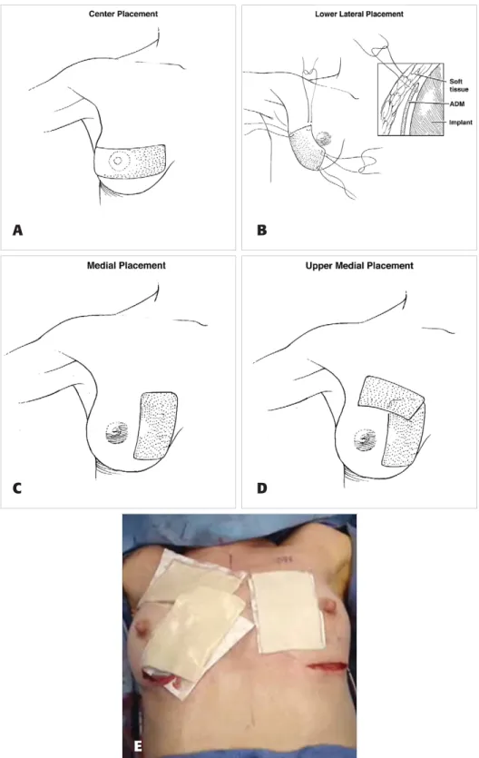
Figure 1. Placement of the acellular dermal matrix is shown in the center (A), lower lateral (B), medial (C), and upper medial (D) areas. In the intraoperative photo (E), medial placement can be seen on the patient’s left breast and upper medial placement is shown on the right breast.
A
B
C
E
Volume 29 • Number 6 • November/December 2009 • 487 Use of the Acellular Dermal Matrix in Revisionary Aesthetic Breast Surgery
the missing or scarred tissue with regenerative tissue, we would see a smaller incidence of CC in our patients. METHODS
A retrospective chart review was conducted of 78 consecu-tive patients who underwent revisionary breast augmenta-tion/mastopexies with ADM during a period of slightly more than two years (October 2005 to January 2008). Patients undergoing surgery for implant rupture, implant malposition, or other reasons without ADM were not included. Only charts with complete operative reports or those in which the use of the ADM could be definitively ascertained were included in the study. Data were collect-ed regarding the original augmentation date, the original implant location (subpectoral or subglandular), type of incision used, volume of the implant used, revision date, type of implant used for revision, incision used in revision, length of follow-up, and any complications that ensued.
All patients received perioperative antibiotics, with the majority receiving first-generation cephalosporin. All surgical pockets were irrigated with triple
antibi-otic solutions and the implants were bathed in the same solution before insertion into the new pocket. The implants were handled as little as necessary to minimize possible contamination.
For patients whose original implants were subglandu-lar, a site change to a subpectoral plane was performed, with lower pole coverage with ADM. For patients whose original implants were placed sub pectorally, a neopectoral pocket was dissected, with the addition of an ADM. For patients who did have adequate breast tissue, a subfascial pocket with an ADM sling was fashioned. In the subpec-toral procedures, the pecsubpec-toralis major muscle was released entirely across its inferior origin and the subpectoral pocket was released as far medially as necessary to achieve the desired pocket shape and medial breast border. Three to five half-mattress stabilizing marionette sutures were placed between the skin and the ADM to stabilize the tis-sue and hold it in the desired location (Figure 2).
Six different types of ADM products were used for this series (Table 1). The majority of patients received Alloderm or Strattice (LifeCell, Branchburg, NJ), FlexHD
Product Method of Year Time to Refrigeration
name Manufacturer Origin preservation introduced hydrate Shelf life required?
Alloderm LifeCell Human Lyophilized, 1994 20–40 minutes 2 years Yes
dermal patented with saline
collagen freeze-drying solution step
process prevents bath with
damaging ice agitation
crystals from forming
Neoform Regeneration Human Solvent dehydrated; 2007 A few minutes 5 years No
Technologies dermal gamma-irradiated with room
Tutogen Medical, collagen temperature
for Mentor saline
DermaMatrix Processed by Donated Aseptic processing 2005 None 18 months No
MTF for acellular method, lyophilized or 3 years
Synthes CMF human
dermis
FlexHD Processed by Donated Aseptic processing 2007 None 18 months No
the MTF for acellular method, lyophilized or 3 years
Ethicon human
dermis
SurgiMend TEI Biosciences Fetal Terminally stabilized 2007 60 seconds 3 years No
bovine with room
dermal temperature
collagen saline
Strattice LifeCell Porcine Patented 2008 Minumum of ? No
dermal freeze-drying 2 minutes in
collagen process that sterile saline
prevents damaging ice crystals from forming
or DermaMatrix (MTF, Edison, NJ) or SurgiMend (TEI Biosciences, Boston, MA). The majority of the products were listed as thick on the product label (if a product description was available). All dermal products were soaked in triple antibiotic solution before placement. RESULTS
A total of 78 patients who met all of the criteria for this review were identified. Fifty-six patients had their original implants in the subpectoral position and 22 had them in the
subglandular position. The average time from initial opera-tion to revision was seven years, nine months (originally subpectoral: seven years, 11 months; originally subglandu-lar: seven years, six months). The average time for follow-up after pocket conversion was at least 12 months for all patients (originally subpectoral: 11.9 months; originally sub-glandular, 12.1 months; Table 2). Eleven patients (20%) with previous subpectoral implants were noted to have implant rupture at the time of revision and the previous subglandular implants were ruptured in five patients (23%). Among the 56 patients with previously subpectoral implants, 11 patients were found to have ruptured implants. Five patients had silicone gel–filled implants, three patients had double-lumen implants in which only the saline component was found to be ruptured, and three patients had a double-lumen implant in which both components showed evidence of rupture. Among the 22 patients with original sub-glandular implants, five patients were found to have ruptured implants. All five patients had silicone gel–filled implants. None of the patients with ruptured implants had any preoperative complaints or physical findings suggestive of implant rupture.
Forty-five patients (60%) had replacement with silicone gel–filled implants (five smooth, 40 textured) and 33 (40%) had replacement with form-stable, highly cohesive gel implants (Table 3). Complications included two patients (2.5%) requiring reoperation, one for a hematoma and the other for an implant malposition (Table 4).
Of the 54 patients whose implants were initially sub-pectoral, 23 patients (43%) had silicone gel–filled implants, 15 patients (28%) had double-lumen implants, and 16 patients (30%) had saline implants. Of the 31 patients whose implants were originally sub -glandular, 20 patients (65%) had silicone gel–filled implants, three patients (10 %) had double-lumen implants, and eight patients (26%) had saline implants. Presenting clinical signs are listed in Table 5 and the types of operations performed are listed in Table 6. As expected, the majority of complaints were related
Figure 2. The acellular dermal matrix is secured with marionette sutures. This process is depicted in both the intraoperative photo (A) and the illustration (B).
Table 2.Time interval for conversion
Previous implant Total Subpectoral Subglandular
No. of patients 78 56 22
Time to revision, 93 95 90
months
Follow-up, months 12 11.9 12.1
Table 3.Types of implants used as replacements Total Silicone gel-filled Cohesive gel
Total 78 45 33
Textured 73 40 33
Smooth 5 5 —
Table 4.Complications
Complication No. of patients
Hematoma 1 Seroma 2 Implant malposition 1 Implant rupture 0 Infection 0 Total 4
A
B
Volume 29 • Number 6 • November/December 2009 • 489 Use of the Acellular Dermal Matrix in Revisionary Aesthetic Breast Surgery
to implant hardening. Of 78 patients, 77 (98.5%) were assessed as having soft implants with a Baker I level of CC at final follow-up; one patient (1.5%) had a Baker II CC. No patients had a Baker III or Baker IV classification postoperatively (Tables 7, 8, and 9). Between January 2008 and January 2009, an additional 41 cases have been performed for CC, increasing the total number of patients in the series to 119. Because we established a minimum follow-up period of one year, we have not included them in this series. However, it is worth noting that all of these newer
patients have maintained a Baker I classification at a mean follow-up of 6.2 months. Results from the study are shown in Figures 3 to 6.
DISCUSSION
The successful use of ADM products has been reported in a wide range of clinical settings, including abdominal wall repair, hernia repair, facial and eyelid surgery, cleft palate repair, soft tissue augmentation, tendon repair, ulcer repair, vaginal sling repair, and breast reconstruction.18-28 Immediate breast reconstruction using tissue expanders or implants has become one of the most commonly used sur-gical techniques, which has made visible rippling and con-tour deformity a more frequently encountered problem.31
Therefore, the applications of ADM to breast reconstruction have been of particular interest to plastic surgeons. The recent addition of allogenic tissue supplements avoids the problems of autologous tissue coverage and provides cam-ouflage, thereby decreasing rippling and increasing soft tis-sue padding.32 This rising demand, coupled with good
outcomes in breast reconstruction, has spurred tremendous interest about ADM use in aesthetic breast patients, partic-ularly in the management of CC.
In the past, revisionary surgeries for CC were generally performed with a total capsulectomy, removal of the implant from the subglandular plane, and the placement of a new implant in the subpectoral position.1,4,16This is a fairly simple procedure involving a change in implant placement from over the muscle to under the muscle. More recently, it
B
C
D
E
F
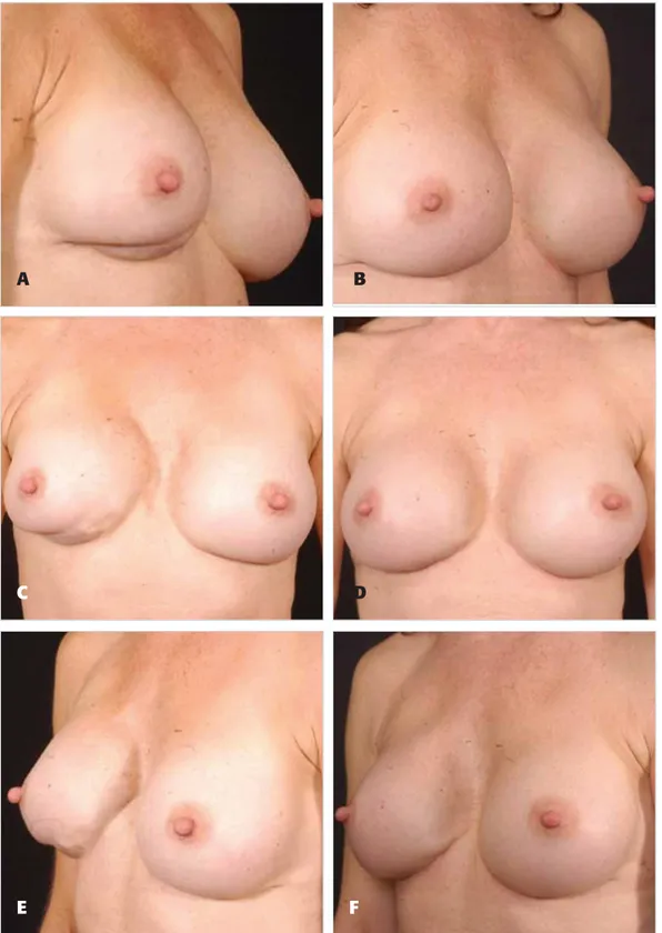
Figure 3. A, C, E, Preoperative views of a 42-year-old woman who had undergone multiple previous augmentation procedures. B, D, F, Sixteen months after revision augmentation mastopexy with circumvertical purse-string approach and an acellular dermal matrix. Her large implants were exchanged for higher-profile, lower-volume textured implants.
has become necessary to perform revisionary surgery on volume-depleted breasts as a result of large implants that were placed under the muscle or severe scarring in the breasts. We remain plagued with a paucity of tissue, leading to the use of allogenic materials such as ADM.
Our experience with 78 patients—all of whom uniformly had their implant-related complications successfully cor-rected by a site change and the use of an ADM—provides a number of important conclusions. First, the use of ADM enhanced soft tissue thickness or cushioning (Figure 5), decreasing and eliminating the visibility of implants. Second, the use of these products increased our ability to successfully manage implant displacements and their recur-rence. Third, lower pole thickness was enhanced in patients with dual plane or neopectoral pocket conversions, especially in those undergoing concurrent mastopexies, interposing regenerative tissue between skin closure and implant (lamellar interpositioning) (Figure 6). Lastly, it is of interest that 56 patients with Baker grade III and IV CC had their CC treated successfully.
The addition of the ADM to the management of breast augmentation intricacies gives us an exciting and, accord-ing to our data, reliable new surgical option. It is evident that by replacing the missing tissue at the implant interface, we are able to create an environment that is conducive to healing without excessive scarring (Figure 1). Unpublished results from our animal studies show decreased inflamma-tion within the implanted pocket at different time points with the addition of ADM.33Just as with any process that leads to excessive scarring, the key is to control the inflam-matory phase and allow the healing tissue to transition quickly and smoothly to the next (proliferative) phase. It is also important to keep in mind that the true etiology of CC remains multifactorial and, by controlling all of the factors, we are able to see the future in prevention of this compli-cated and prolonged issue.
ADM is not a miracle product that can eliminate CC, but it serves as an adjunct to the already well-described princi-ples in the management of CC, such as aseptic techniques with appropriate antibacterial pocket irrigations, postopera-tive drains, and the placement of textured implants.1,3,4,8,9,11,15,34 We postulate that in order to have a successful outcome with an ADM, the creation of a new pocket (raw surface) is essential to augment the interface (Figures 2 and 3). We do not believe that partial capsulecto-my or capsulotomies in conjunction with placement of an ADM are as effective as lining the ADM in a newly vascular-ized pocket, followed by placement of the implant (Figure 7). With our extensive use of all types of ADM, we have found no differences in terms of complications, as noted by our data. ADM products have their limita-tions and differences in terms of revascularization and cellular ingrowth and the acceptance between differ-ent thicknesses and sterility, but we noted no addi-tional drainage output and no infections related to the sterility of certain products. As previously noted, the majority of products were listed as thick on the prod-uct label (if available). It should be noted, however, Table 5.Clinical signs at presentation
Clinical signs No. of patients
Capsular contracture 56 Implant exposure 2 Rippling 7 Implant malposition 5 Bottoming out 4 Symmastia 4 Total 78
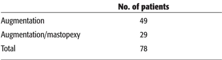
Table 6.Augmentation versus augmentation mastopexy No. of patients
Augmentation 49
Augmentation/mastopexy 29
Total 78
Table 7.Preoperative and postoperative Baker classifica-tion of patients with originally subpectoral implants
Baker Percent of patients
classification Preoperative Postoperative
I 5.4 98.5
II 21.1 1.5
III 67.9 0
IV 5.6 0
Table 8.Preoperative and postoperative Baker classifica-tion of patients with originally subglandular implants
Baker Percent of patients
classification Preoperative Postoperative
I 9.1 97.4
II 18.2 2.6
III 54.5 0
IV 18.2 0
Table 9.Preoperative and postoperative Baker classifica-tion in all patients
Baker Percent of patients
classification Preoperative Postoperative
I 6.4 97.4
II 20.5 2.6
III 64.1 0
Volume 29 • Number 6 • November/December 2009 • 491 Use of the Acellular Dermal Matrix in Revisionary Aesthetic Breast Surgery
that the different materials have different biomechani-cal properties, a discussion of which is beyond the scope of this article.
One obstacle that we will continue to face in aesthetic surgery is the cost–benefit ratio of these materials, as they are expensive. Further evaluation is needed to assess the
actual impact of these costs and their documented out-come improvements in our specialty.
CONCLUSIONS
ADM products show promise for applications in revision-ary breast surgery and specifically for the treatment of
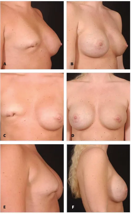
Figure 5. A, C, E, Preoperative views of a 40-year-old woman who had undergone multiple previous augmentation procedures. B, D, F, Fourteen months after mastopexy with an inframammary fold approach and acellular dermal matrix. Her implants were exchanged for textured silicone gel–filled implants.
C
E
F
complications of breast augmentation. This report describes different uses of the ADM that allow for the cor-rection of established CC, malposition, and other implant-related issues after previous subglandular or subpectoral breast augmentation. ◗
DISCLOSURES
Dr. Maxwell is a paid consultant to Allergan. Dr. Gabriel is on the speaker’s bureau of Lifecell.
Figure 6. A, C, E, Preoperative views of a 35-year-old woman with previous subpectoral augmentation with saline implants, with extrusion and lamellar scarring. B, D, F, Fourteen months after mastopexy with acellular dermal matrix. Her implants were exchanged for textured silicone gel–filled implants.
A
B
C
E
D
F
Volume 29 • Number 6 • November/December 2009 • 493 Use of the Acellular Dermal Matrix in Revisionary Aesthetic Breast Surgery
REFERENCES
1. Maxwell GP, Gabriel A. The neopectoral pocket in revisionary breast surgery. Aesthet Surg J2008;28:463–467.
2. Maxwell GP, Gabriel A. Possible future development of implants and breast augmentation. Clin Plast Surg2009;36:167–172.
3. Maxwell GP, Gabriel A. The evolution of breast implants. Clin Plast Surg2009;36:1–13.
4. Maxwell GP, Gabriel A. Efficacy of the neopectoral pocket in revi-sionary breast surgery. Aesthet Surg J2009;29:379–385.
5. Spear SL, Carter ME, Ganz JC. The correction of capsular contracture by conversion to “dual-plane” positioning: technique and outcomes.
Plast Reconstr Surg2006;118(7 suppl):103S–113S.
6. Spear SL, Murphy DK, Slicton A, Walker PS, Inamed Silicone Breast Implant U.S. Study Group. Inamed silicone breast implant core study results at 6 years. Plast Reconstr Surg2007;120(7 suppl 1):8S–16S. 7. Cunningham B. The Mentor Core Study on Silicone MemoryGel Breast
Implants. Plast Reconstr Surg2007;120(7 suppl 1):19S–29S. 8. Adams Jr WP, Rios JL, Smith SJ. Enhancing patient outcomes in
aesthetic and reconstructive breast surgery using triple antibiotic breast irrigation: six-year prospective clinical study. Plast Reconstr Surg2006;117:30–36.
9. Adams Jr WP. Capsular contracture: what is it? What causes it? How can it be prevented and managed? Clin Plast Surg2009;36:119–126. 10. Gancedo M, Ruiz-Corro L, Salazar-Montes A, Rincón AR,
Armendariz-Borunda J. Pirfenidone prevents capsular contracture after mammary implantation. Aesthetic Plast Surg2008;32:32–40.
11. Ma SL, Gao WC. Capsular contracture in breast augmentation with textured versus smooth mammary implants: a systematic review [in Chinese]. Zhonghua Zheng Xing Wai Ke Za Zhi2008;24:71–74. 12. Scuderi N, Mazzocchi M, Rubino C. Effects of zafirlukast on capsular
contracture: controlled study measuring the mammary compliance. Int J Immunopathol Pharmacol2007;20:577–584.
13. Weintraub JL, Kahn DM. The timing of implant exchange in the development of capsular contracture after breast reconstruction.
Eplasty2008;8:e31.
14. Wiener TC. Relationship of incision choice to capsular contracture.
Aesthetic Plast Surg2008;32:303–306.
glandular breast augmentation with textured versus smooth breast implants: a systematic review. Plast Reconstr Surg2006;118:1224–1236. 16. Zimman OA, Toblli J, Stella I, Ferder M, Ferder L, Inserra F. The effects
of angiotensin-converting enzyme inhibitors on the fibrous envelope around mammary implants. Plast Reconstr Surg2007;120:2025–2033. 17. Maxwell GP, Tebbetts JB, Hester TR. Site change in breast surgery.
Presented at the American Association of Plastic Surgeons annual meet-ing, St. Louis, MO, May 1994.
18. Bindingnavele V, Gaon M, Ota KS, Kulber DA, Lee DJ. Use of acellular cadaveric dermis and tissue expansion in postmastectomy breast recon-struction. J Plast Reconstr Aesthet Surg2007;60:1214–1218.
19. Breuing KH, Warren SM. Immediate bilateral breast reconstruction with implants and inferolateral AlloDerm slings. Ann Plast Surg
2005;55:232–239.
20. Breuing KH, Colwell AS. Inferolateral AlloDerm hammock for implant coverage in breast reconstruction. Ann Plast Surg2007;59:250–255. 21. Cothren CC, Gallego K, Anderson ED, Schmidt D. Chest wall recon-struction with acellular dermal matrix (AlloDerm) and a latissimus muscle flap. Plast Reconstr Surg2004;114:1015–1017.
22. Garramone CE, Lam B. Use of AlloDerm in primary nipple reconstruc-tion to improve long-term nipple projecreconstruc-tion. Plast Reconstr Surg
2007;119:1663–1668.
23. Glasberg SB, D’Amico RA. Use of regenerative human acellular tissue (AlloDerm) to reconstruct the abdominal wall following pedicle TRAM flap breast reconstruction surgery. Plast Reconstr Surg2006;118:8–15. 24. Kim H, Bruen K, Vargo D. Acellular dermal matrix in the management
of high-risk abdominal wall defects. Am J Surg2006;192:705–709. 25. Nahabedian MY. Secondary nipple reconstruction using local flaps and
AlloDerm. Plast Reconstr Surg2005;115:2056–2061.
26. Patton Jr JH, Berry S, Kralovich KA. Use of human acellular dermal matrix in complex and contaminated abdominal wall reconstructions.
Am J Surg2007;193:360–363.
27. Salzberg CA. Nonexpansive immediate breast reconstruction using human acellular tissue matrix graft (AlloDerm). Ann Plast Surg
2006;57:1–5.
28. Spear SL, Parikh PM, Reisin E, Menon NG. Acellular dermis-assisted breast reconstruction. Aesthetic Plast Surg2008;32:418–425. 29. Duncan DI. Correction of implant rippling using allograft dermis.
Aesthet Surg J2001;21:81–84.
30. Baxter RA. Intracapsular allogenic dermal grafts for breast implant-related problems. Plast Reconstr Surg 2003;112:1692–1696.
31. Colwell AS, Breuing KH. Improving shape and symmetry in mastopexy with autologous or cadaveric dermal slings. Ann Plast Surg
2008;61:138–142.
32. Gamboa-Bobadilla GM. Implant breast reconstruction using acellular dermal matrix. Ann Plast Surg2006;56:22–25.
33. Maxwell GP, Gabriel A, Perry LC. Role of acelluar dermal matrix in inflammation (unpublished) 2009.
34. Schreml S, Heine N, Eisenmann-Klein M, Prantl L. Bacterial coloniza-tion is of major relevance for high-grade capsular contracture after augmentation mammaplasty. Ann Plast Surg2007;59:126–130.
Accepted for publication February 16, 2009.
Presented at the 58th Annual Meeting of California Society of Plastic Surgeons, Dana Point, CA, June 2008, and the 24th Annual Meeting of Breast Surgery Symposium, Atlanta, GA, January 2008.
Reprint requests: Patrick Maxwell, MD, Department of Plastic Surgery, Loma Linda University Medical Center, 11234 Anderson St., Loma Linda, CA 92354. E-mail: gabrielallen@yahoo.com.
Copyright © 2009 by The American Society for Aesthetic Plastic Surgery, Inc. 1090-820X/$36.00
doi:10.1016/j.asj.2009.09.007
Figure 7. Interposing regenerative tissue between skin closure and implant (lamellar interpositioning).