The increasing use of foot-pressure measurement technology has given a new perspective to foot func-tion in a variety of clinical applicafunc-tions.1For exam-ple, in the diabetes literature, researchers2, 3 have identified high-pressure areas in the neuropathic foot as a precursor to ulceration. The insensate foot may be more susceptible to damage due to repetitive load-ing durload-ing gait, particularly where limited joint mo-tion or bony prominences occur.4, 5The new under-standing provided by foot-pressure measurement has enabled preventive care to be instituted early in the disease process. However, in rheumatology there are few articles demonstrating the clinical benefit of foot-pressure measurement in terms of instituting early foot care.
In the rheumatoid foot, typical clinical features commonly observed include a valgus deformity of the rearfoot, flattened medial longitudinal arch, sub-luxated and prominent metatarsal heads, hallux val-gus, and lesser-toe deformities, such as clawed or re-tracted toes.6In addition, numerous extra-articular features, including edema, bursitis, rheumatoid nod-ules, and plantar and digital calluses, are not uncom-mon.7It is well documented that the presence of foot deformities along with pain and inflammation may cause antalgic gait patterns.8, 9The gait alterations noted in rheumatoid patients include decreased ca-dence and prolonged double-limb support along with delayed heel rise.10-12Fortunately, owing to the pres-ence of protective sensation, rheumatoid patients rarely develop the plantar ulcers seen in the diabetic foot.4, 13Given the deformities commonly seen in the rheumatoid foot, most foot-pressure studies have predominantly focused on the spatial aspects of foot-pressure measurement, eg, the magnitude and
ana-Forefoot Plantar Pressures in
Rheumatoid Arthritis
Simon J. Otter, MSc, DPodM*
Catherine Jane Bowen, MSc, DPodM*
Adam K. Young, MD, FRCP†
We sought to investigate the magnitude and duration of peak forefoot plantar pressures in rheumatoid arthritis. The spatial and temporal char-acteristics of forefoot plantar pressures were measured in 25 patients with a positive diagnosis of rheumatoid arthritis of 5 to 10 years’ dura-tion (mean, 8 years) and a comparison group using a platform-based pressure-measurement system. There were no significant differences between groups in the magnitude of peak plantar pressure in the fore-foot region. Significant differences were, however, noted for temporal aspects of foot-pressure measurement. The duration of loading over sensors detecting peak plantar pressure was significantly longer in the rheumatoid arthritis group. In addition, the rheumatoid arthritis group demonstrated significantly greater force–time integrals. Significant in-creases in the temporal parameters of plantar pressure distribution, rather than those of amplitude, may be characteristic of the rheumatoid foot. (J Am Podiatr Med Assoc 94(3): 255-260, 2004)
*School of Health Professions, University of Brighton, Eastbourne, England.
†St Albans City Hospital, St Albans, England.
Corresponding author:Simon J. Otter, MSc, DPodM, School of Health Professions, Leaf Hospital, University of Brighton, St Annes Rd, Eastbourne BN21 2HW, England.
tomical location of plantar pressures.14-17However, in view of the changes seen in gait patterns in individu-als with rheumatoid arthritis, some authors18-20have suggested that the temporal parameters (eg, the dura-tion of plantar pressure during gait) may be more im-portant in the etiology of tissue pathology than the spatial parameters.
This study used foot-pressure measurements to in-vestigate temporal and spatial aspects of forefoot plantar pressures in individuals with a defined dura-tion of rheumatoid arthritis.
Methods
Patient SelectionThirty-seven consecutive patients were recruited from the rheumatology outpatient clinics of St Al-bans City Hospital in St AlAl-bans, England. All of the patients had a positive diagnosis of rheumatoid arthritis, as determined by the consultant rheumatol-ogist, and were taking part in the Early Rheumatoid Arthritis Study.21The inclusion criteria were a posi-tive diagnosis of rheumatoid arthritis of 5 to 10 years’ duration and an age of 18 to 70 years. The exclusion criteria were quite extensive. Individuals younger than 18 years were excluded because children with juve-nile idiopathic arthritis experience growth alterations. Furthermore, in juvenile idiopathic arthritis, the syn-ovitis rapidly leads to joint stiffness and fixation if not mobilized, and a fixed foot position can occur.22 Individuals older than 70 years were excluded owing to the increased incidence of osteoarthritis in this age group. Individuals with a concomitant systemic disease or impaired neurologic function were exclud-ed, as the results of a study by Chen et al23indicated that there may be a change in plantar pressure distri-bution with a changing sensory condition. Patients who had undergone a major surgical intervention, such as metatarsal head resection, were excluded. Patients who had undergone minor procedures, eg, single digital arthroplasty, were accepted. Other fac-tors that may affect plantar pressure distribution, such as congenital defects and recent abrupt trauma (ie, within 3 months), were also grounds for exclu-sion. The final exclusion group consisted of patients who could not comply with the study protocol, which included individuals who could not walk 4 m on five separate occasions owing to the severity of their dis-ability.
To discount variables due to age and gender, a comparison group was recruited from the staff of West Hertfordshire National Health Service Trust in St Albans and patients’ relatives; these subjects were
identified by the podiatry department’s patient data-base. The inclusion criteria for the comparison group were an age of 18 to 70 years and no positive diagno-sis of an inflammatory arthropathy. The exclusion criteria were the same as those for the rheumatoid arthritis group.
Twelve patients were subsequently excluded be-cause they either met one or more of the exclusion criteria or could not complete the study protocol. Thus plantar pressure measurements were recorded for 25 patients with rheumatoid arthritis and 25 indi-viduals in a comparison group.
Ethical Considerations
Ethical approval was granted for this project from West Hertfordshire Health Authority and St Albans and Hemel Hempstead National Health Service Trust local research ethics committees. Written informed consent was obtained from all participants.
Equipment
Plantar pressures were recorded barefoot using a midgait approach on a Musgrave Footprint system (Musgrave Systems Ltd, Llangollen, North Wales) in-terfaced with a laptop computer together with Win-foot data-acquisition software (version 1.0.15, 1998; Musgrave Systems Ltd). The Musgrave Footprint is a platform system incorporating 2,048 discrete sensors using force-sensitive resistor technology. Each sen-sor covered an area of 5 mm2 with a sampling fre-quency of 56 Hz. Pilot studies indicated that record-ing of consecutive footsteps usrecord-ing the Musgrave Footprint two-plate system adversely affected bal-ance, particularly in rheumatoid patients, because even with both plates side by side (though offset to allow for differences in step length), the active pres-sure-measuring area was wider than the natural base of gait of some individuals. Regard for the health and safety of participants necessitated that recordings be taken from the right foot only.
Data Collection
All participants were given an acclimatization period to ensure that they could step onto the pressure plate accurately without altering their natural stride pattern and while looking straight ahead. Each measurement was taken using a midgait (fourth step) approach. Participants were required to continue walking after stepping on the pressure plate for at least four steps, and they walked at a self-selected pace, with no at-tempt made to standardize walking speed. Five
foot-prints were taken from each participant, as results of pilot studies suggested that this method captured the maximum variation in magnitude and duration of plantar pressures from individuals. In pilot studies, recording more than five footprints did not reveal ad-ditional data and led to fatigue in some patients with rheumatoid arthritis. However, footprints produced when a participant was noted to have hesitated were discarded, and that trial was repeated.
Data Processing and Statistical Analysis
The system software generates the magnitude and duration of plantar pressure variables over the entire foot. A standardized division mask20was used for data interpretation. This mask divides the foot into six regions (distal medial and lateral, middle medial and lateral, and proximal medial and lateral) using the long plantar angle as a basis. The distal medial and lateral regions were used to define the forefoot in this study, and values found in these regions were exported into an Excel spreadsheet (Microsoft Corp, Redmond, Washington).
The peak pressure value is determined by the highest pressure measured by a single sensor. The duration of loading of this sensor was also measured. However, considering that these variables were mea-sured over a single sensor, it could be argued that this is not representative of the foot as a whole. There-fore, the force–time integral was selected to study the entire foot rather than the forefoot alone. This variable was selected because the Musgrave Foot-print system, like most other plantar pressure– measurement systems, measures vertical force and derives the pressure measurement using the follow-ing equation: pressure = force/area. In addition, ana-tomical landmarks cannot be accurately and repeat-edly located for a series of footsteps using plantar pressure–measurement patterns alone.
Baseline data were processed using a statistical software package (SPSS version 4; SPSS Science, Chicago, Illinois) and were shown to be normally dis-tributed using the Kolmogorov-Smirnov test. Differ-ences between peak plantar pressure and the dura-tion of pressure between rheumatoid patients and comparison subjects were determined using the un-paired two-tailed t-test (assuming unequal variance).
Results
The 25 rheumatoid patients (4 men and 21 women) had a mean disease duration of 8 years (range, 5 to 10 years) and a mean ± SD age of 55 ± 11.8 years (range, 28 to 70 years). The comparison group (3 men
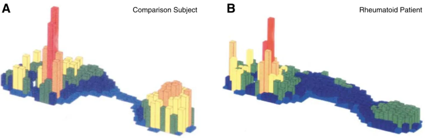
and 22 women) had a mean ± SD age of 49 ± 8.56 years (range, 28 to 66 years). No significant differ-ences were noted in the magnitude of the peak plan-tar pressure value in the forefoot between the two groups. The mean ± SD peak plantar pressure value for the rheumatoid group was 734 ± 147 kPa (range, 423 to 1,168 kPa) and for the comparison group was 780 ± 139 kPa (range, 337 to 1,616 kPa). Figure 1 shows typical three-dimensional peak plantar pres-sure diagrams for the rheumatoid and comparison groups.
Significant differences were noted in the temporal aspects of foot-pressure measurement. The duration of pressure over the sensor measuring the peak pres-sure value was significantly longer in the rheumatoid group (P< .001). For rheumatoid patients, the mean ± SD duration of pressure over the sensor measuring the peak pressure value was 734 ± 68 N.ms (range, 324 to 1,170 N.ms), whereas for the comparison group a mean ± SD of 624 ± 63 N.ms (range, 180 to 990 N.ms) was noted.
Rheumatoid patients also demonstrated signifi-cantly longer force–time integrals than comparison subjects (P < .001). The mean force–time integral seen in rheumatoid patients was 521 N.ms (range, 300 to 884 N.ms). In the comparison group, a lower mean force–time integral (413 N.ms) was found, to-gether with a much wider range of values for the force–time integral (131 to 1,017 N.ms). In typical force–time curves, comparison subjects generally demonstrate a double-peak curve, whereas a flat-tened, lengthened curve is typically seen in rheuma-toid patients (Fig. 2).
Discussion
The general goals of management of complications associated with rheumatoid arthritis are to reduce pain and joint inflammation and to alter the course of the disease by decreasing the progression of joint damage.24Cornerstone podiatric medical practices in the management of foot problems associated with rheumatoid arthritis include the removal of painful skin calluses and the use of foot orthoses and pre-scribed footwear. However, the efficacy of these in-terventions has been poorly addressed in the litera-ture. Indeed, Woodburn and Helliwell25highlighted the lack of robust evidence in the literature to sup-port these practices. Studies14, 26, 27that have attempted to provide measures for these cornerstone practices tended to focus on the use of peak plantar pressure readings. However, the results of this study indicate significant differences in the temporal aspects of foot-pressure measurement in individuals with
rheu-matoid arthritis. Therefore, spatial aspects of foot-pressure measurement alone may not be the most appropriate outcome measure for predicting tissue damage in the rheumatoid patient.
The results of this study suggest that temporal pa-rameters may be more appropriate outcome measures. It should be noted that original work focusing on gait changes in patients with rheumatoid arthritis recorded slower cadence and velocity, along with an increase in the duration of the double-support phase.9-12It has
been suggested that the changes seen in the gait of patients with rheumatoid arthritis are partly a pain-avoidance strategy.8, 9This study demonstrated in-creased temporal parameters of forefoot plantar pres-sures. It is possible that these changes could be explained by patients having a slower gait. However, the Musgrave Footprint system, being a pressure-plate system, does not measure the cadence or veloc-ity of the patient’s gait cycle. Measuring the con-tralateral limb using a second Musgrave Footprint plate was not possible because the patient’s balance was adversely affected. Furthermore, as patients walked at a self-selected speed, temporal parameters of gait would have to be calculated for each of the five trials. To date, there is little evidence of any re-search that has attempted to associate longer tempo-ral plantar pressures with the changes in gait seen in patients with rheumatoid arthritis. We recommend further study in this field.
If pain avoidance is a reason for the changes in the temporal elements of gait, the longer force–time integrals and increased loading time of the sensor measuring peak pressure would not seem to be con-sistent with reducing pain. Rather, given the patho-genesis of the rheumatoid foot, the possibility of fur-ther tissue damage may be enhanced. It may be more likely that changes in plantar pressure can be ex-plained by other factors. Keenan et al12noted sym-metrical weakness in the gastrocnemius and soleus muscles of patients with rheumatoid arthritis, reduc-ing the propulsion phase of gait. In addition, Woll-heim28reported that the ankle is involved in 30% to 50% of cases, and this may further impair the pa-tient’s mobility. These factors may help explain the significantly longer temporal parameters noted in this study.
Figure 2. Typical force–time curves for comparison (A) and rheumatoid (B) participants.
Force (N) 100 75 50 25 0 -400 800 1200 1600 Time (ms) Force (N) 100 75 50 25 0 -400 800 1200 1600 Time (ms)
A
Comparison Subject Rheumatoid PatientB
Figure 1.Typical peak pressure plots for comparison (A) and rheumatoid (B) participants.
The results of the present study indicate that peak forefoot plantar pressures in individuals with estab-lished rheumatoid arthritis are not significantly higher than those of a comparison group. However, the sig-nificantly increased duration of plantar pressures in this group of patients has the potential to cause tis-sue damage. In particular, this study investigated pa-tients with rheumatoid arthritis of 5 to 10 years’ dura-tion. As indicated previously in this article, this patient group often has joint deformities affecting the foot, which may have lost the protection provid-ed by subcutaneous tissue.29In addition, there is some evidence suggesting that the degree of erosion of the metatarsal heads together with the distal mi-gration of fibro-fatty padding seen in rheumatoid arthritis may be associated with high plantar pres-sure readings.11Studies of peak pressures under the forefoot have been inconclusive in finding associa-tions with lower plantar tissue thickness.30
In contrast to previous works,8, 14, 18, 31 this study did not demonstrate significant differences in peak forefoot plantar pressures in patients with estab-lished rheumatoid arthritis compared with healthy individuals. However, the results of this study indi-cate that there are significant increases in the tempo-ral characteristics of forefoot plantar pressure vari-ables in rheumatoid patients. It is possible, therefore, that temporal parameters may be better predictors of change (or more appropriate outcome measures for orthotic intervention) in the rheumatoid foot than peak plantar pressures. The argument for an altered approach to the choice of outcome measure in foot-pressure measurement is clouded, however. There are many different methodologic approaches to foot-pressure measurement. Early studies, for example, report plantar pressure in non-SI (Système Interna-tional) units (eg, percentage of body weight or pounds per square inch).31, 32Further difficulties are also encoun-tered in comparing different foot-pressure measure-ment systems. For example, Woodburn and Helliwell14 used an in-shoe system (F-Scan; Tekscan, Boston, Massachusetts) and reported higher plantar foot pres-sures in rheumatoid patients compared with healthy individuals. The present study used a force platform collecting barefoot data. This would seem to point to the need for the increased use of internationally rec-ognized standard protocols20to allow for better inter-study comparisons of foot-pressure measurement findings.
Conclusion
The results of this study indicate that significant in-creases in the temporal parameters of plantar
pres-sure distribution, rather than those of amplitude, are more characteristic of the rheumatoid foot. Thus temporal and spatial parameters should be incorpo-rated when using plantar pressure measurement in the evaluation of podiatric medical interventions in the rheumatoid foot.
Acknowledgment.Musgrave Systems Ltd, Llan-gollen, North Wales, for loaning the foot-pressure measurement equipment; Georgina Hollis, BSc(Hons), for her technical expertise; the patients and staff at St Albans and Hemel Hempstead National Health Service Trust and West Herts Community National Health Ser-vice Trust (St Albans); Elizabeth Davies, BSc(Hons), Worcester Health Authority, and Chris Foy, BSc(Hons), Gloucester Health Authority, for statistical advice; and Ivan Birch, MSc, and Ann Moore, PhD, University of Brighton, Eastbourne, England, for reviewing the manuscript.
References
1. ALEXANDERIM, CHAOEYS, JOHNSONKA: The assessment of dynamic foot to ground contact forces and plantar pressure distribution: a review of the evolution of cur-rent techniques and clinical applications. Foot Ankle 11:152, 1990.
2. BOULTONAJM, HARDISTYCA, BETTSRP: Dynamic foot pres-sure and other studies as diagnostic and management aids in diabetic neuropathy. Diabetes Care 6:26, 1983. 3. HSIWL, ULBRECHTJS, PERRYJC, ET AL: Plantar pressure
threshold for ulceration. Diabetes 42:103A, 1993. 4. FERNANDODJS, MASSONEA, VEVESA, ET AL: Relationship
of limited joint mobility to abnormal foot pressures and diabetic foot ulceration. Diabetes Care 14:8, 1991. 5. CAVANAGH PR, ULBRECHTJS: Clinical plantar pressure
measurement in diabetes: rationale and methodology. The Foot 4:123, 1994.
6. CRACCHIOLOA: The rheumatoid foot and ankle: pathol-ogy and treatment. The Foot 3:126, 1993.
7. O’BRIENTS, HARTTS, GOULDJ: Extraosseous manifes-tations of rheumatoid arthritis in the foot and ankle. Clin Orthop 340:26, 1997.
8. MINNSRJ, CRAXFORDAD: Pressure under the forefoot in rheumatoid arthritis: a comparison of static and dynamic methods of assessment. Clin Orthop 187:235, 1984. 9. GERBERLH, HUNTGC: Evaluation and management of
the rheumatoid foot. Bull N Y Acad Med 61:359, 1985. 10. DIAMONTEP, LIGHTH: Pathomechanics, gait deviations and treatment of the rheumatoid foot. Phys Ther 62: 1148, 1982.
11. ISACSONJ, BROSTROMLA: Gait deviations in rheumatoid arthritis: an electrogoniometric investigation. J Biomech 21:451, 1988.
12. KEENANMAE, PEABODYTD, GRONLEYJK, ET AL: Valgus deformities of the feet and characteristics of gait in pa-tients who have rheumatoid arthritis. J Bone Joint Surg Am 73:237, 1991.
13. DONAGHUE VM, VEVES A: Foot pressure measurement. Orthop Phys Ther Clin North Am 6:1, 1997.
14. WOODBURNJ, HELLIWELLPS: Relation between heel po-sition and the distribution of forefoot plantar pressures and skin callosities in rheumatoid arthritis. Ann Rheum Dis 55:806, 1996.
15. BITZANP, GIUREA A, WANIVENHAUS A: Plantar pressure distribution after resection of the metatarsal heads in rheumatoid arthritis. Foot Ankle Int 18:391, 1997. 16. BETTSRP, STOCKLEY I, GETTYJM, ET AL: Foot pressure
studies in the assessment of forefoot arthroplasty in the rheumatoid foot. Foot Ankle 8:315, 1988. 17. WOODBURNJ, STABLEFORDZ, HELLIWELLPS: Preliminary
investigation of debridement of plantar callosities in rheumatoid arthritis. Rheumatology 39:652, 2000. 18. LORDM, REYNOLDSDP, HUGHESJR: Foot pressure
mea-surement: a review of clinical findings. J Biomed Eng 8:283, 1986.
19. SOAMES RW: Foot pressure patterns during gait. J Biomed Eng 7:120, 1985.
20. BARNETTSJ: International protocol guidelines for plan-tar pressure measurement. The Diabetic Foot 1:137, 1998.
21. YOUNG A, DIXEYJ, COXN, ET AL: How does functional disability in early rheumatoid arthritis (RA) affect pa-tients and their lives? results of 5 years of follow-up in 732 patients from the Early RA Study (ERAS). Rheuma-tology 39:603, 2000.
22. FERRARIJ: A review of the foot deformities seen in ju-venile chronic arthritis. The Foot 8:193, 1998.
23. CHENH, NIGGBM, HULLIGERM, ET AL: Influence of sen-sory input on plantar pressure distribution. Clin Biomech 10:271, 1995.
24. SCOTTDL, SHIPLEYM, DAWSONA, ET AL: Strategies for
im-proving clinical effectiveness. Br J Rheumatol 37:546, 1998.
25. WOODBURNJ, HELLIWELLPS: Foot problems in rheuma-tology. Br J Rheumatol 36:932, 1996.
26. VEVESA, HEY EM, BOULTON AJM: The use of specially padded hosiery in the painful rheumatoid foot. The Foot 1:175, 1992.
27. POTTERJ, POTTERMJ: Effect of callus removal on peak plantar pressures. The Foot 10:23, 2000.
28. WOLLHEIMF: “Rheumatoid Arthritis: Clinical Picture,” in Oxford Textbook of Rheumatology, Vol 2, ed by PJ Maddison, DM Isenberg, P Woo, et al, p 639, Oxford University Press, Oxford, 1993.
29. TINLEY P: Appliances for the rheumatic foot. Baillieres Clin Rheumatol 1:383, 1987.
30. YOUNG M, COFFEY J, TAYLORPM, ET AL: Weight bearing ultrasound in diabetic and rheumatoid arthritis patients. The Foot 5:76, 1995.
31. SHARMAM, DHANENDRANM, HUTTONWC, ET AL: Changes
in load bearing in the rheumatoid foot. Ann Rheum Dis 38:549, 1979.
32. COLLISWJMF, JAYSONMIV: Measurement of pedal pres-sures. Ann Rheum Dis 31:215, 1972.