ISSN 2278- 4136
ZDB-Number: 2668735-5
IC Journal No: 8192
Volume 2 Issue 2
Online Available at www.phytojournal.com
Journal of Pharmacognosy and Phytochemistry
Vol. 2 No. 2 2013 www.phytojournal.com
Page | 140
In vitro and in vivo Methods for Anticancer Activity
Evaluation and Some Indian Medicinal Plants Possessing
Anticancer Properties: An Overview
Sumitra Chanda* and Krunal Nagani
1. Phytochemical, Pharmacological and Microbiological Laboratory,
2. Department of Biosciences, Saurashtra University, Rajkot 360005, Gujarat, India. [E-mail- svchanda@gmail.com]
Cancer is a major public health burden in both developed and developing countries. Anticancer activity is the effect of natural and synthetic or biological and chemical agents to reverse, suppress or prevent carcinogenic progression. Several synthetic agents are used to cure the disease but they have their toxicity and hence the research is going on to investigate the plant derived chemotherapeutic agents. Therefore an attempt has been made to review different in vitro and in vivo methods for estimating anticancer properties of natural products from medicinal plants. In this review, 50 anticancer medicinal plants of Indian origin belonging to 35 families are reported along with detailed information regarding part used, extract used, type of the model used, types of tested cancer cell lines, etc. These plants continue to be used against various types of tumours such as sarcoma, lymphoma, carcinoma and leukemia. All these plants are potential candidates for in vivo studies since they are showing good in vitro anticancer activity.
Keyword:Anticancer Medicinal Plants, Indian origin, Tumours, in vitro and in vivo Methods.
1. Introduction
Ayurveda, a traditional Indian medical practice
using plant drugs has been successful from very
early times in using these natural drugs and
preventing or suppressing various tumours with
different lines of treatment
[1]. In India, people of
different ethnic groups inhabiting various
terrains, possess their own distinct culture,
religious rites, food habit and a rich knowledge of
traditional medicine
[2]. They practice herbal
medicine to cure a variety of diseases. Natural
products, especially plants have been used in the
treatment of various diseases for thousands of
years. Terrestrial plants have been used as
medicines in Egypt, China, India and Greece
from ancient times and an impressive number of
modern drugs have been developed from them.
The first written records on the medicinal uses of
plants appeared about 2600 BC from the
Sumerians and Akkaidians
[3].
Cancer is a group of diseases caused by loss of
cell cycle control. Cancer is associated with
abnormal uncontrolled cell growth
[4]. Cancer is
caused by both external factors (tobacco,
chemicals, radiation and infectious organisms)
and
internal
factors
(inherited
mutations,
hormones, immune conditions, and mutations that
occur from metabolism). Cancer is a significant
worldwide health problem generally due to the
lack of widespread and comprehensive early
detection methods, the associated poor prognosis
of patients diagnosed in later stages of the disease
and its increasing incidence on a global scale.
Indeed, the struggle to combat cancer is one of
the greatest challenges of mankind
[5].
Vol. 2 No. 2 2013 www.phytojournal.com
Page | 141
The National Cancer Institute collected about
35,000 plant samples from 20 countries and has
screened around 114,000 extracts for anticancer
activity
[6]. Over 3000 species of plants with
antitumour properties have been reported
[7].
Cancer is one of the most prominent diseases in
humans and currently there is considerable
scientific and commercial interest in the
continuing discovery of new anticancer agents
from natural product sources
[8].
Chemoprevention is recognized as an important
approach to control malignancy and recent
studies have focused on the search for desirable
chemopreventive
agents.
Natural
products,
particularly dietary substances, have played an
important role in creating new chemopreventive
agents
[9]. Interesting patterns of differential
cytotoxicity have been associated with known
classes of compounds, such as cardenolides,
lignans or quassinoids
[10]. In any cancer drug
discovery program, a paradigm based on
ethnobotanical and ethnopharmacological data
would be more economical and beneficial in
identifying potential anticancer molecules than
mass screening of plant species
[11]. Natural
products have been regarded as important sources
of potential chemotherapeutic agents and many
anticancer drugs have originated from natural
sources
[12].
According to Cragg and Newman
[13]over 50 % of
the drugs in clinical trials for anticancer
properties were isolated from natural sources or
are related to them. Several natural products of
plant
origin
have
potential
value
as
chemotherapeutic agents. Some of the currently
used anticancer agents derived from plants are
podophyllotoxin,
taxol,
vincristine
and
camptothecin
[14]. The areas of cancer and
infectious diseases have a leading position in
utilization of medicinal plants as a source of drug
discovery. Among FDA approved anticancer and
anti-infectious drugs, drugs from natural origin
have a share of 60 % and 75 % respectively
[15].
A great number of
in vitro
and
in vivo
methods
have been developed to measure the efficiency of
natural anticancer compounds either as pure
compounds or as plant extracts.
In vitro
methods
like, Tryphan blue dye exclusion assay, LDH
(Lactic dehydrogenase) assay, MTT assay, XTT
assay and Sulforhodamine B assay are most
commonly used for estimating anticancer
properties of natural products from medicinal
plants. Among all
in vitro
methods MTT and
Sulforhodamine B assay most popular for
estimating anticancer activity.
2. Screening methods of anticancer activity:
2.1 In vitro methods
2.1.1 Tryphan blue
dye exclusion assay
The trypan blue dye exclusion assay is the most
commonly utilized test for cell viability. In this
assay, the cells are washed with HBSS (Hank's
Buffered Salt Solution) and centrifuged for 10 -
15 min at 10,000 rpm. The procedure is repeated
thrice. The cells are suspended in known quantity
of HBSS and the cell count is adjusted to 2 x 10
6cells /ml. The cell suspension is distributed into
Eppendorf tubes (0.1 ml containing 2 lakhs cells).
The cells are exposed to drug dilutions and
incubated at 37 °C for 3 h. After 3 h, dye
exclusion test, that is, equal quality of the drug
treated cells are mixed with tryphan blue (0.4 %)
and left for 1 min. It is then loaded in a
haemocytometer and viable and non-viable count
are recorded within 2 min. Viable cells do not
take up colour, whereas dead cells take up colour.
However, if kept longer, live cells also generate
and take up colour
[16]. The percentage of growth
inhibition is calculated using the following
formula:
(Total cells-Dead cells)
Growth inhibition (%) = 100 - ___________________________ X 100 Total cells
2.1.2 LDH (Lactic dehydrogenase) Assay
[17]Lactic
dehydrogenase
activity
is
spectrophotometrically measured in the culture
medium and in the cellular lysates at 340 nm by
analyzing NADH reduction during the
pyruvate-lactate transformation. Cells are lysed with 50
mM Tris-HCl buffer, pH 7.4 + 20 mM EDTA +
0.5 % Sodium Dodecyl Sulfate (SDS), further
disrupted by sonication and centrifuged at 13,000
X g for 15 min. The assay mixture (1ml final
volume) for the enzymatic analysis consists of 33
µl of sample in 48 mM PBS, pH 7.5 + 1 mM
pyruvate and 0.2 mM NADH. The percentage of
Vol. 2 No. 2 2013 www.phytojournal.com
Page | 142
LDH released is calculated as percentage of the
total amount, considered as the sum of the
enzymatic activity present in the cellular lysate
and that in the culture medium.
2.1.3 MTT assay
[18]The MTT assay, based on the conversion of the
yellow tetrazolium salt-MTT, to purple-formazan
crystals by metabolically active cells, provides a
quantitative determination of viable cells. Cells
are plated on to 96 well plates at a cell density of
2×10
5mL
-1per well in 100 µL of RPMI 1640 and
allowed to grow in CO
2incubator for 24 h (37
˚C, 5 % CO
2). The medium is then removed and
replaced by fresh medium containing different
concentrations of sample for 48 h. The cells are
incubated for 24-48 h (37 ˚C, 5 % CO
2). Then, 20
µL MTT ([3- (4, dimethylthiazol-yl)-2,
5-diphenyltetrazolium bromide]) stock solution (5
mg/mL in PBS) is added to each well and
incubated for 5 h. The medium is removed and
200 µL DMSO is added to each well to dissolve
the MTT metabolic product. Then the plate is
shaken at 150 rpm for 5 min and the optical
density is measured at 560nm. Untreated cells
(basal) are used as a control of viability (100 %)
and the results are expressed as % viability (log)
relative to the control.
2.1.4 XTT assay
[19]In order to measure the proliferation response, the
(2,3-bis[2-Methoxy-4-nitro-5-sulfophenyl]-
2H-tetrazolium-5-carboxyanilide inner salt (XTT)
assay is used. The tetrazolium salt, XTT, is
especially useful in quantifying viable cells. This
assay is designed for the spectrophotometric
quantification of cell growth and viability without
the use of radioactive isotopes and is based on the
cleavage of yellow tetrazolium salt, XTT, to form
an orange formazan dye by metabolically active
cells. XTT cleavages into an orange formazan
dye
by
the
mitochondrial
enzyme,
dehydrogenase, occurs exclusively in living cells.
Cells are grown in growth medium plus 10 %
FBS in 96-well plates until 70-80 % confluence.
They are then treated with the appropriate drug
sample for 24 h. An XTT assay is performed at
the end of incubation. Briefly, 50 mL of XTT
labeling mixture solution is add to each well, and
the cells are incubated at 37 °C for 4 h. The
formazan dye formed is soluble in aqueous
solutions and the optical density at 450 nm is
compared with that of control wells with a
screening multiwell spectrophotometer
enzyme-linked immunosorbent assay (ELISA) reader. The
reference wavelength is 650 nm.
2.1.5 Sulforhodamine B assay
Sulforhodamine B assay
is a bright pink
aminoxanthene dye that binds to basic amino
acids in mild acidic conditions and dissociates
under basic conditions. Cells are plated in 96-well
flat bottom plates at 5000-10000 cell/well. The
difference in cell numbers plated adjusts for
differences in the growth rates of the various cell
lines. Cells are allowed to adhere to the wells
overnight, then the samples are added to triplicate
wells in serial 3-fold dilutions. Water is added to
the control wells at a 1:10 dilution in medium.
These plates are incubated at 37 °C, 5 % CO
2for
3 days, then assayed for growth inhibition using a
sulforhodamine B (SRB) assay
[20]. The cells are
fixed by the addition of cold 50 % trichloroacetic
acid to a final concentration of 10 %. After 1 h
incubation at 4 °C, the cells are washed five times
with deionized water. The cells are then stained
with 0.4 % SRB (Sigma) dissolved in 1 % acetic
acid for 15-30 min and subsequently washed five
times with 1 % acetic acid to remove unbound
stain. After the plates are air dried at room
temperature, the bound dye is solubilized with 10
mm Tris base and the plates are analysed on a
microplate reader (Molecular Devices) at 595 nm.
2.2 In vivo model
2.2.1 Induction of Ehrlich ascites carcinoma
[21]Antitumor activity of the test compounds is
determined using Ehrlich ascites carcinoma
(EAC) tumor model in mice. The ascitic
carcinoma bearing mice (donor) are used for the
study, 15 days after tumor transplantation. The
animals are divided into groups of 12 animals
each. ((a) Normal mice (b) Tumor-bearing mice,
Vol. 2 No. 2 2013 www.phytojournal.com
Page | 143
(c) Tumor-bearing mice treated with standard
drug, (d) Tumor-bearing mice groups treated with
test drug) The ascitic fluid is drawn using an
18-gauge needle with sterile syringe. A small amount
is testing for microbial contamination. Tumor
viability is determine by Tryphan blue exclusion
test and cells are counted using haemocytometer.
The ascitic fluid is suitably diluted in normal
saline to get a concentration of 10
6cells/ml of
tumor
cell
suspension.
This
is
injected
intraperitoneally to obtain ascitic tumor. The
mice are weighed on the day of tumor inoculation
and then once in three days thereafter. Treatment
is started on the tenth day of tumor inoculation.
Standard (one dose) is injected on tenth day
intraperitoneally. The drug is administered from
tenth day for 5 days intraperitoneally. After the
administration of last dose followed by 18 h
fasting, six mice from each group are sacrifice for
the study of antitumor activity and hematological
parameters. The remaining animals in each of the
groups are kept to check the mean survival time
(MST) of the tumor-bearing hosts. Antitumor
effects of drug are assessed by observation of
following parameters.
i.
Percentage
increase
in
weight
as
compared to day-0 weight
ii.
Median survival time and increase in
lifespan [% ILS]
iii.
Hematological parameters
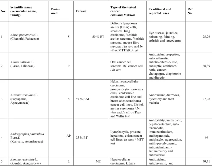
Table 1: List of Indian medicinal plants, their family, part used, solvents used for extraction and assay employed
for anticancer studies.
No. Scientific name (vernacular name, family) Part/s used Extract
Type of the tested cancer
cells and Method
Traditional and reported uses Ref. No. 1 Abrus precatorius L. (Chanothi, Fabaceae) S 50 % ET Dalton’s lymphoma ascites (DLA) cells, small cell lung carcinoma, Yoshida ascites sarcoma, Yoshida sarcoma, mouse fibro sarcoma / In vivo and In vitro /MTT,SRB test
Eye disease, jaundice, poisoning, fainting, arthritis and leucoderma
25,26
2
Allium sativum L.
(Lasan, Liliaceae) P
Oral cancer cell, sarcoma 180 cancer cell / In vivo Antioxidant properties, anti- asthmatic, anticholesterole- mic, antiseptic, antithrom- botic, cancer, cholagogue, diaphoretic and diuretic 38,39 3 Alstonia scholaris L. (Saptaparna, Apocynaceae) S 85 % EAL HeLa, hepatocellular carcinoma, promyelocytic leukemia cells, epidermoid carcinoma cell line and breast adenocarcinoma cancer cell lines, Ehrlich ascites carcinoma / In vivo and In vitro / Pratt and Willis test
Antioxidant, diarrhoea, dysentery and treat malaria 27,28 4 Andrographis paniculata Burn.f. (Kariyatu, Acanthaceae) AP 95 % ET Lymphocytic, prostate, hepatoma, colon cancer cell lines/ In vitro / MTT test Antifertility, antihepatic, hepatoprotective, anti- thrombotic, immunostimulant, antihepatotoxic, antiplatelet, aggregation, antihyper-glycaemic, antioxidant, anti- Inflammatory and antimalarial 69 5 Annona reticulate L. (Ramfal, Annonaceae) L ME Hepatocellular carcinoma, kidney Antioxidant, antidysentric, and 70,71
Vol. 2 No. 2 2013 www.phytojournal.com
Page | 144
carcinoma, colorectal
carcinoma cancer cell lines / In vitro / MTT test antihelminthic 6 Asparagus racemosus Willd.
(Shatavari, Liliaceae) R AQ Liver cancer / In vivo
Gastric ulcers, dyspepsia, inflammation, liver diseases and antioxidant
40,41
7 Azadirachta indica (Neem, Meliaceae)Juss. L 80 % ET Prostate cancer / In vivo
Immunomodulatory, anti- inflammatory, antiulcer, antimalarial, antifungal, antibacterial, antiviral, antioxidant, antimutagenic and anticarcinogenic properties 57,58 8 Bacopa monniera L.(Brahmi, Scrophulariaceae) WP 9 % ET
Mouse sarcoma Cell line/ In vitro / Trypan blue exclusion test
Mental disorders, tumors, ascites, antioxidant and inflammation 72 9 Bauhinia variegate L. (Kanchhanar, Caesalpiniaceae) S 95 % ET
Liver cancer cell, epithelial larynx cancer, human breast cancer / In vivo and In vitro line/MTT test
Bronchitis,leprosy, tumors ulcer, antibacterial, antifungal and antioxidant
45
10 Berberisvulgaris (Barberry, Berberidaceae)L. RB ME Breast cancer / SRB test In vitro /
Antioxidant, diarrhea, gallbladder, liver dysfunctions, leishmaniasis, malaria, stomach problems and urinary tract diseases
73
11 Beta vulgaris (Beet, Chenopodiaceae)L. J 95 % ET
Skin and lung cancer / In
vivo
Antioxidant, leukaemia, cancer such as breast, oesophagus, glands, head, intestines and leg
59,60
12 Bidens pilosa(Shemaro, Asteraceae) L. WP ME
Cervix carcinoma, nasopharyngeal epidermal carcinoma cancer cell lines / In vitro / MTT test
Antioxidant, wounds, colds, flu and acute or chronic hepatitis urinary tract infections 74,75 13 Calycopteris floribunda Lam. (Bukshi, Kokaranj Combretaceae) L DCM:ME (1:1)
Colon cancer cell line /
In vitro / MTT test
Colic, antihelminthic, astringent laxative, diarrhoea and malaria
89 14 Catharanthus roseus L. (Sadabahar, barmachi Apocynaceae) R,L EA Acute lymphocytic leukemia / In vivo, Colorectal Carcinoma cell line / In vitro / MTT test Anti cancer, menorrhagia and antioxidant 42-44
15 Cedrus deodara G. Don (Devdaar, Pinaceae) W - Acute lymphoblastic leukemia, promyelocytic leukemia, prostrate and lung cancer cell lines / In vitro / Trypan blue exclusion test
Astringent, antioxidant, antidiarrhoeal febrifuge, and antiseptic.
90,91
16 Citrullus colocynthis (Indrayan, Cucurbitaceae) L. L Glucosides Breast cancer cell line / In vitro / MTT test
Cytotoxic,
hepatoprotective, anti-inflammatory, cardiovascular, antioxidant and anti-diabetic effects
76,77
17 Crocus sativus L.
(Kesar, Iridaceae) dry stigmas 75 % ET
Cervical epithelioid carcinoma cancer cell line / In vitro / MTT test
Antioxidant properties 78,79 18 Curculigo orchioides Gaertn. (Kalimusli, Amaryllidaceae) R HE, CH, AN and ME
Breast cancer cell line /
In vitro / MTT test
Antioxidant, diarrhoea, jaundice, asthma and poultice for itch and skin diseases
80,81
19 Curcuma longa L.
(Haldi, Zingiberaceae) Rh -
Colon Cancer Cells / In vitro / Lactate
Antimutagenic,
Vol. 2 No. 2 2013 www.phytojournal.com
Page | 145
dehydrogenase test antigenotoxic,
anti-inflammatory and antioxidant properties 20 Cymbopogon flexuosus (Steud.) Wats. (Lemon grass, Poaceae)
G
Colon, cervix, oral, prostate, promyelocytic and leukemia cancer cell lines / In vitro and In vivo / SRB test Stress-related disorders, antifungal and antimicrobial properties 29 21 Emblica officinalis Gaertn. (Amla, Euphorbiaceae)
DFr ME Liver cancer/ In vivo
Liver protecting activity, antimutagenic, antioxidant and anticarcinogenic properties
61,62
22 Ephedra sinica Stapf
(Ephedra, Ephedraceae) AP ME
Murine melanoma cancer / In vivo
Colds, fever, flu, headaches, asthma, wheezing, and nasal congestion
63
23 Indigofera aspalathoides (Vahl, Papilionaceae) S 95% ET
Ehrlich’s ascites carcinoma cancer / In vivo
Antioxidant, various skin
disorders and cancer 45,46
24
Ipomoea aquatica Forskal. (Kalmisag,
Convolvulaceae)
L ME
Larynx epithelial carcinoma, small lung carcinoma cancer and normal African green monkey kidney cell line / In vitro / MTT and SRB test Antioxidant properties 82 25 Ipomoea squamosa
(Cairo Morning Glory, Convolvulaceae)
L - Ovarian cancer cell line / In vitro - 94
26
Jatropha curcas L. (Ratanjota, Huphorbiaceae)
S ME Skin cancer / In vivo
Skin diseases, antioxidant, ulcers, tumours
67,68
27 Lantana camara (Ghaneri, Verbenaceae)L. F,Fr.,L,R, S ME
Lung carcinoma cell line / In vitro / MTT and SRB test Antitumoral,antioxidant, antibacterial and antihypertensive 83
28 Mangifera indica (Keri, Anacardiaceae)L. Fr, B,L Lung cancer / In vivo
Antitumour, antioxidant, antiviral, antibacterial, analgesic, anti-inflammatory, antidiarrhoeal, antiamoebic, spasmolytic, immunostimulant and immunomodulatory properties 47 29 Melia azedarach L.
(White Cedar, Meliaceae) L 70 % ET
Lung cancer and glioma cancer cell line / In vitro
/ standard Cell Counting Kit (CCK)-8 test
Antiparasitic activity,
anthelmintic activity 95,96
30 Morinda citrifolia L.
(Noni, Rubiaceae) R, Fr. AQ
Colon cancer cell line /
In vitro / MTT test
Antidiabetic, antiviral, antibacterial, anticancer and antioxidant
97,98
31 Moringa oleifera (Saragavo, Moringacae) L. S ME, ET, EA and CH
Skin cancer/ In vivo and In vitro/ Natural red dye test
Antioxidant, antimicrobial, antigenotoxic and anti-inflammatory activities 36,37 32 Nigella sativa L. (Black seeds, Ranunculaceae)
S 90 % ET Colon Cancer / In vivo
Antioxidant, antidiabetic, antihistaminic, antiepileptogenic, antiinfective, antitumour and antiperoxidative 48,49
33 Ocimum gratissimum (Damro, Lamiaceae) L. S, L AQ Breast cancer / and In vitro / MTT test In vivo
Chemopreventive, anticarcinogenic, radioprotective and numerous others pharmacological uses 30
Vol. 2 No. 2 2013 www.phytojournal.com
Page | 146
34 Ocimum sanctum L.
(Tulsi, Lamiaceae) L ET Skin cancer / In vivo
Anti-stress, antioxidant, hepatoprotective, anti-inflammatory, antibacterial and radioprotective properties 64 35 Phellinus rimosus
(Berk, (Hymenochetaceae) sporocarps ME, AQE
Dalton’s lymphoma ascites, Ehrlich’s ascites carcinoma / In vivo and
In vitro / Trypan blue exclusion test
Antioxidant 31,32
36 Pinus resinosa Aiton
(Pinaceae) W
HE, DCM, ME and AQ
Colorectal
adenocarcinoma cell, lung carcinoma cell and normal skin
Fibroblast cell lines / In vitro / Resazurin reduction test Antioxidant, analgesic, antifungal and antibacterial 84,85 37 Polyalthia longifolia
Benth. & Hook. f. (Annonaceae)
L ET
Colon cell and leukemia HL-60 cancer cell line /
In vitro / SRB test
Antibacterial and antifungal activities
99
38 Psidium guajava L. (Jamphal, Myrtaceae) L AQ Prostate carcinoma cell/ In vitro / MTT test Antioxidant 100 39 Punica granatum L.
(Dadam, Lythraceae) J,P 70 % AC
Prostate carcinoma cell /
In vivo and In vitro / MTT test
Antioxidant and
anti-inflammatory 33
40 Tragia involucrata Linn.
(Euphorbiaceae) AP HE, EA Ehrlich’s ascites carcinoma/ in vivo Antimicrobial, antiinflammatory, antifertility activity 101 41 Rubia cordifolia L. (Manjistha, Rubiaceae) R 80 % ME Coloncarcinoma, breast carcinoma and liver carcinoma / In vitro / MTT test
Antitumor, antioxidant, anti inflammatory, urinary disorders, antistress, anti microbial, hepatoprotective, radio protective 86,87 42 Semecarpus anacardium L.
(Bhallika, Anacardiaceae) DFr 90 % ET and ME
Acute myeloblastic leukaemia,chronic myelogenic leukaemia, breast adenocarcinoma, cervical epithelial carcinoma and colon carcinoma cancer cell lines / In vitro / MTT test Antioxidant, immunomodu-latory, antiinflammatory, analgesic, antipyretic and ulcerogenic activities
88,11
43 Tephrosia purpurea (Sarapunkha, Fabaceae) Pers. R 95 % ET
Oral squamous cell carcinoma / In vivo Various inflammatory, liver, spleen and kidney disorders and antioxidant
50
44
Terminalia chebula Retz. (Karakkaya,
Combretaceae)
F ET COLO-205 cell line / vitro / MTT test In
Digestive, diabetes, colic pain, chronic cough, sore throat, asthma, oxidant, anti-inflammatory 102 45 Tiliacora racemosa Coleb. (Tiliacoru, Menispermaceae) R 90 % ET Acute myeloblastic leukaemia,chronic myelogenic leukaemia , breast adenocarcinoma and cervical epithelial cancer cell lines / In vitro / MTT test
- 88
46
Tinospora cordifolia
(Willd.) Hook. f. & Thom. (Guduchi, Menispermaceae) S PE, CH and DCM Ehrlich’s ascites carcinoma / In vivo General tonic, antioxidant, inflammatory, anti-arthritic, antiallergic, malarial, anti-diabetic and aphrodisiac properties 51,52 47 Viscum album L. (Vando,
L CO2 gas Ehrlich’s tumour cell /
In vivo
Nervine, hypotensive, cardiac depressant, antioxidant, vasodilator,
Vol. 2 No. 2 2013 www.phytojournal.com
Page | 147
Viscaceae) relaxant, diuretic and
stimulant 48 Withania somnifera L. (Ashwagandha, Solanaceae) R 70 % EAL Forestomach and skin carcinoma cancer / In vivo Antitumor, radiosensitizer, antioxidant, antistressor, immunomodulatory, anti-inflammatory and anti-bacterial 53,54 49 Woodfordia fruticosa Salisb. (Dhavdi, Lythraceae) F 70 % AC Sarcoma 180 cancer / In vivo Antipyretic, antioxidant, Anti- inflammatory, hepato-protective, antibacterial activity 55,56
50 Zingiber officinale Rosc.
(Adu, Zingiberaceae) Rh 50 % ET
Prostate cancer cellline /
In vitro and In vivo / MTT test Carminative, antioxidant, diaphoretic, antispasmodic, expectorant, peripheral circulatory stimulant, astringent, appetite stimulant, anti-inflammatory agent, diuretic and digestive
34,35
S: Stem, P: Peel, AP: Aerial parts, L: leaves, R: root, WP: whole plant, RB: rootbark, J: juice, W: wood, Rh: rhizomes, G: grass, DFr: dry fruits, F: flower, B: bark
ET: Ethanol, EAL: Ethyl alcohol, ME: Methanol, AQ: Aqueous, DCM: dichloromethane, EA: Ethyl acetate, HE: Hexane, CH: Chloroform, AN: Acetonitrile, AC: Acetone.
2.2.2 Anticancer Medicinal Plants of India
Anticancer
properties
of
many
natural
compounds isolated from different Indian plant
extracts have been reported. Research is being
carried out throughout the world to find a lead
compound which can block the development of
cancer in humans. Nature has always been a great
contributor towards this goal. Plant-derived
natural products such as flavonoids, terpenoids
and steroids have received considerable attention
due to their diverse pharmacological properties,
which include cytotoxic and chemopreventive
effects
[22]. The isolation of the vinca alkaloids,
vinblastine and vincristine from the Madagascar
periwinkle,
Catharanthus roseus
introduced a
new era in the use of plant material as anticancer
agents. They were the first agents to advance into
clinical use for the treatment of cancer
[23].
The medicinal plants contain many antioxidants
such as vitamins (A, C, E, K), carotenoids,
flavonoids (flavones, isoflavones, flavonones,
anthocyanins,
catenchins,
isocatechins),
polyphenols (ellagic acid, gallic acid, tannins),
saponins, enzymes and minerals (selenium,
copper, manganese, zinc, chromium, iodine,
etc)
[24].
In this review, 50 anticancer medicinal plants of
Indian origin belonging to 35 families are
reported
along
with
detailed
information
regarding part used, extract used, type of the
model used, types of tested cancer cell lines, etc.
(Table-1). These plants continue to be used
against various types of tumours such as sarcoma,
lymphoma, carcinoma and leukemia. Many of
these medicinal plants have been found to be very
effective in experimental as well as clinical cases
of tumours/cancers.
Some medicinal plants have been studied in
various
in vivo
and
in vitro
experimental models
of cancer and have shown significant inhibition
of cancer cell proliferation. For eg.
Abrus
precatorius
in Yoshida’s sarcoma, carcinoma
and Dalton’s lymphoma ascites cancer
[25,26];
Alstonia
scholaris
in
Ehrlich
ascites
carcinoma
[27,28];
Cymbopogon
flexuosus
in
Ehrlich ascites carcinoma, leukemia and sarcoma
180
[29];
Ocimum gratissimum
in breast cancer
[30];
Phellinus
rimosus
in
lymphoma
and
carcinoma
[31,32];
Punica granatum
in prostate
cancer
[33];
Zingiber officinale
in carcinoma
[34, 35];
Moringa oleifera
in skin cancer and Human
multiple myeloma cancer
[36,37];
Allium sativum
in
sarcoma 180
[38,39];
Asparagus racemosus
in liver
cancer
[40,41];
Catharanthus roseus
in P-1534
leukemia
[42-44];
Indigofera
aspalathoides
in
Ehrlich’s ascites carcinoma
[45,46];
Mangifera
indica
in lung cancer
[47];
Nigella sativaI
in colon
cancer
[48,49];
Tephrosia
purpurea
in
oral
Vol. 2 No. 2 2013 www.phytojournal.com
Page | 148
carcinoma
[50];
Tinospora cordifolia
in Ehrlich’s
ascites carcinoma
[51,52];
Withania
somnifera in
skin carcinoma
[53,54];
Woodfordia
fruticosa in
sarcoma 180
[55,56];
Azadirachta indica
in prostate
cancer
[57,58];
Beta vulgaris
in skin and lung
cancer
[59,60];
Emblica
officinalis
in
liver
cancer
[61,62];
Ephedra
sinica
in
Murine
melanoma
[63]; O
cimum sanctum
in skin cancer
[64];
Viscum album
in
Ehrlich’s carcinoma
[65,66];
Jatropha
curcas
in
skin
cancer
[67,68];
Andrographis paniculata
in lymphoma and
carcinoma
[69];
Annona reticulate
in kidney and
colorectal
carcinoma
cancer
[70,71];
Bacopa
monniera
in sarcoma
[72];
Berberis vulgaris
in
breast cancer
[73];
Bidens pilosa
in cervix
cancer
[74,75];
Citrullus colocynthis
in breast
cancer
[76,77];
Crocus sativus
in cervical
epithelioid carcinoma cancer
[78,79];
Curculigo
orchioides in breast cancer
[80,81];
Ipomoea
aquatica
in larynx epithelial carcinoma and small
lung carcinoma cancer
[82];
Lantana camara
in
lung carcinoma
[83];
Pinus resinosa
in Colorectal
adenocarcinoma cell, lung carcinoma cell and
normal skin Fibroblast
[84,85];
Rubia cordifolia
in
carcinoma
[86,87];
Tiliacora
racemosa
in
leukaemia
and
carcinoma
[88];
Calycopteris
floribunda
in colon cancer
[89];
Cedrus deodara
in
acute lymphoblastic leukemia, prostate and lung
cancer
[90,91];
Curcuma longa
in colon cancer
[92,93];
Ipomoea
squamosa
in ovarian cancer
[94];
Melia
azedarach
in lung cancer and glioma cancer
[95,96];
Morinda
citrifolia
in
colon
cancer
[97,98];
Polyalthia longifolia
in colon and leukemia
HL-60 cancer
[99];
Psidium gujava
in prostate
carcinoma cancer
[100];
Tragia involucrata
in
carcinoma cancer
[101];
Semecarpus anacardium
acute
myeloblastic
leukaemia,
chronic
myelogenic leukaemia, breast adenocarcinoma,
cervical
epithelial
carcinoma
and
colon
carcinoma cancer
[88,11];
Terminalia chebula
in
colon cancer
[102].
This review provides information on a number of
plants which show promising anticancer activity.
It lists various methods for evaluating anticancer
activity so it will be easy for the experimenter. It
emphasizes that
in vitro
anticancer assays have
been carried out for most of the plants, but
in vivo
remains to be done in majority of them.
3. Conclusion
In this review, some anticancer medicinal plants
of Indian origin have been presented. These
medicinal plants possess good antioxidant
properties, leading to anticancer activities. The
aim of this study was to give an overview on the
progress of anticancer medicinal plant research
around the continental India, focusing on the
most important findings of scientists in this field.
We have tried to explore the discovered plants
components with proved anticancer activity both
in vitro
and
in vivo
. India is one of the most
promising
regions
for
discovering
novel
biologically-active substances from its flora.
More efforts are needed to explore potent
anticancer plants from the mother earth and save
humans around the world from cancer.
4. References
1. Balachandran P, Govindarajan R. Cancer- an ayurvedic perspective. Pharmacol Res 2005; 51: 19-30.
2. Parinitha M, Srinivasa BH, Shivanna MB. Medicinal plant wealth of local communities in some villages in Shimoga distinct of Karnataka. India J Ethnopharmacol 2005; 98: 307-312. 3. Samuelsson G. Drugs of natural origin. A
textbook of pharmacognosy. 4th ed., Stockholm, Swedish Pharmaceutical Press. 1999
4. Krishnamurthi K. Screening of natural products for anticancer and antidiabetic properties. Health Administrator 2007; 1&2: 69-75. 5. Divisi D, Di TS, Salvemini S, et al., Diet and
cancer. Acta Biomed 2006; 77: 118-123.
6. Mohammad S. Anticancer agents from
medicinal plants. Bangladesh J Pharmacol 2006; 1: 35-41.
7. Hartwell JL. Plants used against cancer. A survey. Quarterman Publications, Lawrence; 1982.
8. Kinghorn AD, Farnsworth NR, Soejarto DD, et al.,Novel strategies for the discovery of plant-derived anticancer agents. Pharmaceutic Biol 2003; 41: 53-67.
9. Surh YJ. Cancer chemoprevention with dietary phytochemicals. Nature Rev Cancer 2003; 3: 768-780.
10. Cardellina JH, Gustafson KR, Beutler JA, et al., National cancer institute intramural research on human immunodeficiency virus inhibitory and antitumor plant natural products. Human
Vol. 2 No. 2 2013 www.phytojournal.com
Page | 149
Medicinal Agents from Plants; 1993; 15 pp. 218-227.
11. Nair PKR, Melnickb SJ, Wnukc SF, et al., Isolation and characterization of an anticancer
catechol compound from Semecarpus
anacardium. J Ethnopharmacol 2009; 122: 450-456.
12. Tan G, Gyllenhaal C, Soejarto DD. Biodiversity as a source of anticancer drugs. Curr Drug Targets 2006; 7: 265-277.
13. Cragg GM, Newman DJ. Antineoplastic agents from natural sources: achievements and future directions. Expet Opin Investig Drugs 2000; 9: 1-15.
14. Pezzuto JM. Plants derived anticancer agents. Biochem Pharmacol 1997; 53: 121-133. 15. Newman DJ, Cragg GM, Snader KM. Natural
products as sources of new drugs over the period 1981-2002. J Nat Prod 2003; 66: 1022-1037.
16. Unnikrishnan MC, Ramadasan K. Cytotoxicity of extracts of spices to cultured cells. Nutr Cancer 1998; 11: 251-257.
17. Russo A, Piovano M, Lombardo L, et al., Pannarin inhibits cell growth and induces cell death in human prostate carcinoma DU- 145 cells. Anti-Cancer Drugs 2006; 17: 1163-1169. 18. Mossman T. Rapid colorimetric assay for
cellular growth and survival: application to proliferation and cytotoxicity assays. J Immunol Methods 1983; 65: 55-63.
19. Economou MA, Andersson S, Vasilcanu D, et al.,. Oral picropodophyllin (PPP) is well tolerated in vivo and inhibits IGF-1R expression and growth of uveal melanoma. Acta Ophthalmologica 2008; 86: 35-41.
20. Skehan P, Storeng R, Scudiero D, et al., New colorimetric cytotoxicity assay for anticancer-drug screening. J Natl Cancer Inst 1990; 82: 1107-1112.
21. Devi PU, Rao BSS, Solomon FE. Effect of plumbagin on the radiation induced cytogenetic and cell cycle changes in mouse Ehrlich ascites carcinoma in vivo. Indian J Exp Biol 1998; 36: 891-895.
22. Abdullaev FI. Plant derived agents against cancer. In: Gupta, S. K., editor. Pharmacology and therapeutics in the new millennium. Narosa Publishing House: New Delhi, India, 2001; p. 345-354.
23. Cragg GM, Newman DJ. Plants as source of anticancer agents. J Ethnopharmacol 2005; 100: 72-79.
24. Gupta VK, Sharma SK. Plants as natural antioxidants. Nat Prod Rad 2006; 17: 326-334.
25. Sivakumar R, Alagesaboopathi C. Studies on cytotoxicity and antitumor screening of red and white forms of Abrus precatorius L. Afr J Biotechnol 2008; 7: 3984-3988.
26. Subba RVV, Sirsi M. Effect of Abrus precatorius L. on experimental tumors. Cancer Res 1969; 29: 1447-1451.
27. Jagetia GC, Baliga MS. Evaluation of anticancer activity of the alkaloid fraction of Alstonia scholaris in vitro and in vivo. Phytother Res 2006; 20: 103-109.
28. Kulkarni MP, Juvekar AR. Effect of Alstonia scholaris Linn. on stress and cognition in mice. Indian J Exp Biol 2008; 47: 47-52.
29. Sharma PR, Mondhe DM, Muthiah S, et al., Anticancer activity of an essential oil from Cymbopogon flexuosus. Chemico-Biol Interact 2009; 179: 160-168.
30. Makker PN, Tait L, Shekhar MPV, et al., Inhibition of breast tumor growth and angiogenesis by a medicinal herb Ocimum gratissimum. Int J Cancer 2007; 121: 884-894. 31. Ajith TA, Janardhanan KK. Antioxidant and
antihepatotoxic activities of Phellinus rimosus (Berk) Pilat. J Ethnopharmacol 2002; 81: 387-391.
32. Ajith TA, Janardhanan KK. Cytotoxic and antitumor activities of a polypore macrofungus, Phellinus rimosus Pilat. J Ethnopharmacol 2003; 84: 157-162.
33. Malik A, Afaq F, Sarfaraz S, et al., Pomegranate fruit juice for chemoprevention and chemotherapy of prostate cancer. Proc Natl Acad Sci 2005; 102: 14813-14818.
34. Shukla Y, Prasad S, Tripathi C, et al., In vitro and in vivo modulation of testosterone mediated alterations in apoptosis related proteins by [6]-gingerol. Mol Nutr Food Res 2007; 51: 1492-1402.
35. Stoilova I, Krastanov A, Stoyanova A, et al., Antioxidant activity of a ginger extract (Zingiber officinale). Food Chem 2007; 102: 764-770.
36. Guevara AP, Vargas C, Sakurai H, et al., An antitumor promoter from Moringa oleifera Lam. Mutation Res 1999; 440: 181-188.
37. Verma AR, Vijayakumar M, Mathela CS, et al., In vitro and in vivo antioxidant properties of different fractions of Moringa oleifera leaves. Food Chem Toxicol 2009; 47: 2196-2201. 38. Ejaz S, Woong LC, Ejaz A. Extract of garlic
(Allium sativum) in cancer chemoprevention. Exp Oncol 2003; 25: 93-97.
39. Balasenthila S, Ramachandranb CR, Naginia S. Prevention of 4-nitroquinoline 1-oxide-induced
Vol. 2 No. 2 2013 www.phytojournal.com
Page | 150
rat tongue carcinogenesis by garlic. Fitoterapia 2001; 72: 524-531.
40. Agrawal A, Sharma M, Rai S, et al., The effect of the aqueous extract of the roots of Asparagus racemosus on hepatocarcinogenesis initiated by diethyl nitrosamine. Phytother Res 2008; 22: 1175-1182.
41. Kamata JP, Boloora KK, Devasagayam TPA, et al., Antioxidant properties of Asparagus racemosus against damage induced by γ-radiation in rat liver mitochondria. J Ethnopharmacol 2000; 71: 425-435.
42. Sayed EA, Cordell GA. Catharanthus alkaloids atharanthamine, a new antitumor bisindole alkaloid from Catharanthus roseus. J Nat Prod 1981; 44: 289-293.
43. Johnson IS, Wright HF, Svoboda GH, et al., Antitumor principles derived from Vinca rosea L. Cancer Res 1960; 20: 1016-1022.
44. Jaleel C, Gopi AR, Manivannan P, et al., Antioxidant potential and indole alkaloid profile variations with water deficits along different parts of two varieties of Catharanthus roseus. Colloids Surf B Biointerfaces 2008; 62: 312-318.
45. Rajkapoor B, Jayakar B, Murugesh N. Antitumor activity of Indigofera aspalathoides on Ehrlich ascites carcinoma in mice. Indian J Pharmacol 2004; 36: 38-40.
46. Bakasso S, Meda LA, Lamien CE, et al., Polyphenol contents and antioxidant activities of five Indigofera species (Fabaceae) from Burkina Faso. Pakistan J Biol Sci 2008; 11: 1429-1435.
47. Rajendran P, Ekambaram G, Sakthisekaran D. Effect of mangiferin on benzo(a)pyrene induced lung carcinogenesis in experimental Swiss albino mice. Nat Prod Res., 2008; 22: 672-680. 48. Johar DA, Shinwari N, Arif J, et al., Role of
Nigella sativa and a number of its antioxidant constituents towards Azoxymethane-induced genotoxic effects and colon cancer in rats. Phytother Res 2008; 22:1311-1323.
49. Swamy SMK, Tan BKH. Cytotoxic and immunopotentiating effects of ethanolic extract of Nigella sativa L. seeds. J Ethnopharmacol 2000; 70: 1-7.
50. Kavitha K, Manoharan S. Anticarcinogenic and antilipidperoxidative effects of Tephrosia purpurea L. in 7,12-dimethylbenz(a)anthracene (DMBA) induced hamster buccal pouch carcinoma. Indian J Pharmacol 2006; 38: 185-189.
51. Rao SK, Rao PS, Rao BN. Preliminary investigation of the radiosensitizing activity of guduchi (Tinospora cordifolia) in tumor
bearing Mice. Phytother Res., 2008; 22: 1482-1489.
52. Prince PSM, Menon VP. Antioxidant activity of Tinospora cordifolia roots in experimental diabetes. J Ethnopharmacol 1999; 65: 277-281. 53. Padmavathi B, Rath PC, Rao AR, et al., Roots of Withania somnifera inhibit forestomach and skin carcinogenesis in mice. Adv Access Publi 2005; 2: 99-105.
54. Visavadiya NP, Narasimhacharya AVRL. Hypocholesteremic and antioxidant effects of
Withania somnifera (Dunal) in
hypercholesteremic rats. Phytomedicine 2007; 14: 136-142.
55. Yoshida T, Chou T, Nitta A, et al., Woodfordin C, a Micro-ring hydrolyzable tannin dimer with antitumor activity and accompanying dimmers from Woodfordia fruticosa flowers. Chem Pharm Bull 1990; 38: 1211-1217.
56. Kumaraswamy MV, Satish S. Free radical scavenging activity and lipoxygenase inhibition of Woodfordia fructicosa Kurz and Betula utilis Wall. Afr J Biotechnol 2008; 7: 2013-2016. 57. Gangar SC, Koul A. Azadirachta indica
modulates carcinogen biotransformation and reduced glutathione at peri-initiation phase of benzo(a)pyrene induced murine forestomach tumorigenesis. Phytother Res 2008; 22: 1229-1238.
58. Kumar S, Suresh PK, Vijayababu MR, et al., Anticancer effects of ethanolic neem leaf extract on prostate cancer cell line. J Ethnopharmacol 2006; 105: 246-250.
59. Kapadia GJ, Tokuda H, Konoshima T, et al., Chemoprevention of lung and skin cancer by Beta vulgaris root extract. Cancer Lett 1996; 100: 211-214.
60. Jiratanan T, Liu RH. Antioxidant activity of processed table beets (Beta vulgaris) and green beans (Phaseolus vulgaris). J Agric Food Chem 2004; 52: 2659-2670.
61. Sultana S, Ahmed S, Jahangir T. Emblica officinalis and hepatocarcinogenesis: A chemopreventive study in Wistar rats. J. Ethnopharmacol 2008; 118: 1-6.
62. Anila L, Vijayalakshmi NR. Antioxidant action of flavonoids from Mangifera indica and Emblica officinalis in hypercholesterolemic rats. Food Chem 2003; 83: 569-574.
63. Nam NH, Lee CW, Hong DH, et al., Antiinvasive, antiangiogenic and antitumour activity of Ephedra sinica extract. Phytother Res., 2003; 17: 70-76.
64. Rastogi S, Shukla Y, Paul BN, et al., Protective
effect of Ocimum sanctum on
7,12-Vol. 2 No. 2 2013 www.phytojournal.com
Page | 151
dimethylbenz(a)anthracene and aflatoxin B1 induced skin tumorigenesis in mice. Toxicol Appl Pharmacol., 2007; 224: 228-240.
65. Cebovic T, Spasic S, Popovic M. Cytotoxic effects of the Viscum album L. extract on Ehrlich tumour cells in vivo. Phytother Res 2008; 22: 1097-1103.
66. Ucar EO, Karagoz A, Arda N. Antioxidant activity of Viscum album ssp. album. Fitoterapia 2006; 77: 556-560.
67. Hirota M, Suttajit M, Suguri H, et al., A new tumor promoter from the seed oil of Jatropha curcas L., an intramolecular diester of 12-Deoxy-16-hydroxyphorbol. Cancer Res 1988; 48: 5800-5804.
68. Yan R, Gao S, Yang W, et al., Nickel toxicity induced antioxidant enzyme and phenylalanine ammonia-lyase activities in Jatropha curcas L. cotyledons. Plant Soil Environ 2008; 54: 294-300.
69. Geethangili M, Rao YK, Fang S, et al., Cytotoxic constituents from Andrographis paniculata induce cell cycle arrest in jurkat cells. Phytother Res 2008; 22: 1336-1341.
70. Mondal S, Mondal NB, Mazumder UK. In vitro cytotoxic and human recombinant caspase inhibitory effect of Annona reticulate leaves. Indian J Pharmacol 2007; 39: 253-254.
71. Baskar R, Rajeswari V, Kumar TS. In vitro antioxidant studies in leaves of Annona species. Indian J Exp Biol 2007; 45: 480-485.
72. Rohini G, Shyamala DCS. Bacopa monniera extract induces apoptosis in murine sarcoma cells. Phytother Res 2008; 22: 1595-1598. 73. Tomosaka H, Chin Y, Salim AA, et al.,
Antioxidant and cytoprotective compounds from Berberis vulgaris. Phytother Res 2008; 22: 979-981.
74. Sundararajan P, Dey A, Smith A, et al., Studies of anticancer and antipyretic activity of Bidens pilosa whole plant. Afr Health Sci 2006; 6: 27-30.
75. Abajo C, Boffill MA, Campo J, et al., In vitro study of the antioxidant and immunomodulatory activity of aqueous infusion of Bidens pilosa. J Ethnopharmacol 2004; 93: 319-323.
76. Tannin-Spitz T, Grossman S, Dovrat S, et al., Growth inhibitory activity of cucurbitacin glucosides isolated from Citrullus colocynthis on human breast cancer cells. Biochem Pharmacol 2007; 73: 56-67.
77. Spitz TT, Bergman M, Grossman S. Cucurbitacin glucosides: Antioxidant and free-radical scavenging activities. Biochem Biophys Res Commun 2007; 364:181-186.
78. Abdullaev FI. Cancer chemopreventive and tumoricidal properties of Crocus sativus L. Exp Biol Med 2002; 227: 20-25.
79. Escribano J, Alonso G, Coca-Prados M, et al., Crocin, safranal and picrocrocin from Crocus sativus L. inhibit the growth of human cancer cells in vitro. Cancer Lett 1996; 100: 23-30. 80. Singh R, Gupta AK. Antimicrobial and antitumor
activity of the fractionated extracts of kalimusli (Curculigo orchioides). Int J Green Pharm 2008; 2: 34-36.
81. Venukumar MR, Latha MS. Antioxidant activity of Curculigo orchioides in carbon tetrachloride induced hepatopathy in rats. Indian J Clin Biochem 2002; 17: 80-87.
82. Prasad KN, Ashok G, Raghu C, et al., In vitro cytotoxic properties of Ipomoea aquatica leaf. Indian J Pharmacol 2005; 37: 397-398.
83. Raghu C, Ashok G, Dhanaraj SA, et al., In vitro cytotoxic activity of Lantana camara Linn. Indian J Pharmacol 2004; 36: 93-95.
84. Simard F, Legault J, Lavoie S, et al., Isolation and identification of cytotoxic compounds from the wood of Pinus resinosa. Phytother Res 2008; 22: 919-922.
85. Pietarinen SP, Willfor SM, Ahotupa MO, et al., Knotwood and bark extracts: strong antioxidants from waste materials. J Wood Sci 2006; 52: 436-444.
86. Kaur P, Singh B, Kumar S, et al., In vitro evaluation of free radical scavenging activity of Rubia cordifolia L. J Chinese Clin Med 2008; 3: 278-284.
87. Son JK, Jung SJ, Jung JH, et al., Anticancer constituents from the roots of Rubia cordifolia L. Chem Pharm Bull 2008; 56: 213-216.
88. Chakraborty S, Roy M, Taraphdar AK, et al., Cytotoxic effect of root extract of Tiliacora racemosa and oil of Semecarpus anacardium nut in human tumour cells. Phytother Res 2004; 18: 595-600.
89. Ali H, Chowdhury AKA, Rahman AKM, et al., Pachypodol a flavonol from the leaves of Calycopteris floribunda, inhibits the growth of CaCo-2 colon cancer cell line in vitro. Phytother Res 2008; 22:1684-1687.
90. Sharma PR, Shanmugavel M, Saxena AK, et al., Induction of apoptosis by a synergistic lignan composition from Cedrus deodara in human cancer cells. Phytother Res 2008; 22:1587-1594.
91. Tiwari AK, Srinivas PV, Kumar SP, et al., Free radical scavenging active components from Cedrus deodara. J Agric Food Chem 2001; 49: 4642-4645.
Vol. 2 No. 2 2013 www.phytojournal.com
Page | 152
92. Lee YK, Lee WS, Hwang JT, et al., Curcumin exerts anti-differentiation effect through AMPKα-PPAR-γ in 3T3-L1 adipocytes and antiproliferatory effect through AMPKα-COX-2 in cancer cells. J Agric Food Chem., 2009; 57: 305-310.
93. Hsu Y, Weng H, Lin S, et al., Curcuminoids- Cellular uptake by human primary colon cancer cells as quantitated by a sensitive HPLC assay and its relation with the inhibition of proliferation and apoptosis. J Agric Food Chem 2007; 55: 8213-8222.
94. Cao S, Norris A, Wisse JH, et al., Ipomoeassin F, a new cytotoxic macrocyclic glycoresin from the leaves of Ipomoea squamosa from the Suriname rainforest. Nat Prod Res 2007; 21: 872-876. 95. Wu SB, Ji YP, Zhu JJ, et al., Steroids from the
leaves of Chinese Melia azedarach and their cytotoxic effects on human cancer cell lines. Steroids 2009; 74: 761-765.
96. Szewczuk VD, Mongelli ER, Pomilio AB. In vitro anthelmintic activity of Melia azedarach naturalized in Argentina. Phytother Res 2006; 20: 993-996.
97. Kamiya K, Hamabe W, Tokuyama S, et al.,. Inhibitory effect of anthraquinones isolated from the Morinda citrifolia root on animal A, B
and Y families of DNA polymerases and human cancer cell proliferation. Food Chem 2009; 118: 725-730.
98. Anekpankul T, Goto M, Sasaki M, et al., Extraction of anticancer damnacanthal from roots of Morinda citrifolia by subcritical water. Sep Purif Technol 2007; 55: 343-349.
99. Verma M, Singh SK, Bhushan S, et al., . In vitro cytotoxic potential of Polyalthia longifolia on human cancer cell lines and induction of apoptosis through mitochondrial dependent pathway in HL-60 cells. Chemico-Biol Interact 2008; 171: 45-56.
100. Chen KC, Hsieh CL, Peng CC, et al., Brain derived metastatic prostate cancer DU-145 cells are effectively inhibited in vitro by guava (Psidium gujava L.) leaf extracts. Nutr Cancer 2007; 58: 93-106.
101. Joshi CG, Gopal M, Kumari NS.
Antitumor activity of hexane and ethyl acetate extracts of Tragia involucrata. International Journal of Cancer Research 2011; 7: 267-277. 102. Reddy DB, Reddy TCM, Jyotsna G, et al.,
Chebulagic acid, a COX-LOX dual inhibitor isolated from the fruits of Terminalia chebula Retz., induces apoptosis in COLO-205 cell line. J Ethnopharmacol 2009; 124:506-512.