Review
Prostate cancer screening in Europe and Asia
Kai Zhang, Chris H. Bangma, Monique J. Roobol
*
Department of Urology, Erasmus University Medical Center, Rotterdam, The Netherlands
Received 30 June 2016; received in revised form 16 August 2016; accepted 16 August 2016
Available online 4 September 2016
KEYWORDS Prostate cancer; Early detection; Biomarkers; Imaging; Risk prediction; Europe; Asia
Abstract Prostate cancer (PCa) is the second most common cancer among men worldwide and even ranks first in Europe. Although Asia is known as the region with the lowest PCa inci-dence, it has been rising rapidly over the last 20 years mostly due to the introduction of prostate-specific antigen (PSA) testing. Randomized PCa screening studies in Europe show a mortality reduction in favor of PSA-based screening but coincide with high proportions of un-necessary biopsies, overdiagnosis and subsequent overtreatment. Conclusive data on the value of PSA-based screening and hence the balance between harms and benefits in Asia is still lack-ing. Because of known racial variations, Asian countries should not directly apply the European screening models. Like in the western world also in Asia, new predictive markers, tools and risk stratification strategies hold great potential to improve the early detection of PCa and to reduce the worldwide existing negative aspects of PSA-based PCa screening.
ª2017 Editorial Office of Asian Journal of Urology. Production and hosting by Elsevier B.V. This is an open access article under the CC BY-NC-ND license (http://creativecommons.org/ licenses/by-nc-nd/4.0/).
1. Incidence of prostate cancer in Europe and
Asia
Prostate cancer (PCa) is the second most common malig-nancy and the fifth leading cause of cancer death in men
worldwide[1]. However, the incidence differs by more than
25-fold among regions, with the highest in Australia/New Zealand and the lowest in South-Central Asia[1](Fig. 1).
This wide variation in incidence is strongly related to the use of the prostate-specific antigen (PSA) test as a
screening tool [2]. In most European countries such as
France, The Netherlands, and the Czech Republic, the PCa incidence increased significantly in the early 1990s, soon after the introduction of the PSA test, and is still increasing
[3,4]. The incidence in Asian countries like China and Japan, began to increase after 1995. Although later than in Europe because of the delayed use of the PSA test as a screening tool, the increase of PCa incidence in Asian
countries is more pronounced in a comparable period [3]
(Fig. 2).
As said, the worldwide variation in the use of the PSA test is probably the most important reason for the vari-ability in PCa incidence. It is interesting to note that in the early 1980s, when PSA was not yet used, a nearly 20-fold
PCa incidence difference already existed (USA 91.43 vs.
Japan 4.87, per 100,000) [5]. This could be explained by
factors like dietary differences (e.g., a high-fat diet in the
* Corresponding author. Department of Urology, Erasmus University Medical Center, PO Box 2040, Room Na1706, 3000 CA Rotterdam, The Netherlands.
E-mail address:m.roobol@erasmusmc.nl(M.J. Roobol).
Peer review under responsibility of Second Military Medical University.
http://dx.doi.org/10.1016/j.ajur.2016.08.010
2214-3882/ª2017 Editorial Office of Asian Journal of Urology. Production and hosting by Elsevier B.V. This is an open access article under the CC BY-NC-ND license (http://creativecommons.org/licenses/by-nc-nd/4.0/).
Available online atwww.sciencedirect.com
ScienceDirect
journal homepage:www.e lsevie r. com/l ocate/ ajur Asian Journal of Urology (2017)4, 86e95
western world), the prevalence of obesity, and genetic factors[3]. A striking example is the significantly higher PCa incidence in Japanese-American men than in native Japa-nese men, suggesting that westernization through a
high-fat diet is strongly related to the risk of having PCa [6].
Furthermore, the PCa incidence in African-American men is
2e3 times higher than in White and Asian-American men in
the US, which indicates that gene and race also contribute to PCa risk[7].
2. Screening trials in Europe and Asia
The European Randomized Study of Screening for Prostate Cancer (ERSPC) is the largest randomized trial for PCa screening and is still ongoing. It started in 1993 and includes
162,338 men, aged 55e69 years at time of randomization.
After 13 years of follow-up, the trial showed that PCa mortality was reduced by 21% in favor of the screening arm
[8]. This finding is contrary to that of a large American
randomized trial, the so-called prostate arm of the Pros-tate, Lung, Colorectal, and Ovarian Cancer Screening Trial (PLCO). This trial, also initiated in 1993 and in which 76,685 men were randomized showed no difference in PCa mor-tality between the screening and control arm with 13 years
of follow-up [9]. A recent publication on the basis of the
PLCO data showed a 90% PSA contamination rate in the control arm which seriously questions the value of the
re-ported outcomes[10].
The Goteborg Randomized Prostate Cancer Screening Trial is a European prospective, randomized trial that
started in 1995 and included 19,904 men, aged 50e64 years
at time of randomization. After 14 years of follow-up, it showed that PCa mortality decreased by 44% in the
screening group as compared to the control group[11].
It is important to note that all three PCa screening trials were based on Caucasian populations. Hence, their out-comes cannot be directly translated to an Asian population. The Japanese Prospective Cohort Study of Screening for Prostate Cancer (JPSPC) is the only known prospective controlled PCa screening study in Asia (Table 1). It started in 2002 and ended in 2014. The aim of the study is to compare the PCa mortality between the screening and control cohort. This study comprises of 200,000 men in the age range between 50 and 79 from Hokkaido, Gunma,
Hir-oshima and Nagasaki prefectures. A PSA3 ng/mL in men
aged 50e64 years, a PSA3.5 ng/mL in men aged 65e69
years and a PSA 4.0 ng/mL in men aged 70e79 years
triggered biopsy[12]. The compliance rate of PSA testing in
the Isesaki city screening cohort was about 75% over 5 years and the contamination (PCa screening) in the Kiryu city
control cohort was low at 8% between 1992 and 2006[13].
The contamination rate for the whole control cohort is expected to be considerably low, due to the absence of opportunistic PCa screening in Japan. The study outcome is eagerly awaited to show whether PSA-based screening has any potential in an Asian setting[14].
The Kanazawa population-based screening cohort study is another large PCa screening study in Japan. A total of
32,769 men aged 55e69 years participated in the program
from 2000 to 2006. Contrary to the JPSPC study, the indi-cation of biopsy varied among the different urologists participating in this study. From 2000 to 2002, all men with
a PSA > 2.1 ng/mL were recommended to undergo the
secondary screening (consisting of a digital rectal exami-nation (DRE) and transrectal ultrasonography (TRUS) ex-amination) and to consult a urologist who would decide whether or not to perform a systematic biopsy taking into
account the results of the DRE and TRUS[15]. From 2003
onwards, men with PSA values 2.1e10.0 ng/mL and a free
PSA/total PSA ratio (%fPSA) higher than 0.22, were not referred for further screening. 4766 men (14.9%) required secondary screening and 1041 men (3.2%) underwent pros-tate biopsy. A total of 249 men (0.76%) were diagnosed with PCa, of whom 231 (93.5%) were classified as clinically localized cancer. Comparing the outcomes of this screening study with those done among predominantly Caucasian men it again highlights the considerable difference in PCa 111.6 97.2 94.9 85 79.8 61.7 58.6 31.3 28 11.2 10.5 4.5 0 20 40 60 80 100 120 Australia/New Zealand Northern America West Europe Northern Europe Caribbean South Africa Southern Europe Central and Eastern Europe Western Asia South-Eastern Asia Eastern Asia South-Central Asia
Age-standardized rate per 100,000
Figure 1 Prostate cancer incidence worldwide[1]. Adapted with permission.
incidence; 0.76% and 8.33% in this Japanese screening study
and the ERSPC respectively)[8]. The percentage of
local-ized tumors (T1,T2) in Japan was, however, remarkably
higher than in the ERSPC (93.5%vs.78.2%)[16].
A South Korean screening study focused on the relation between the PSA value and PCa mortality. It included 118,665 men from 1994 to 2004, and followed these men up to 2011. The results showed a PCa death risk of 1.0%, 1.57%,
2.41%, 4.32% and 65.0% for baseline PSA values of <1.0,
1e2, 2e4, 4e10 and 10 ng/mL respectively after
adjusting for age, body mass index (BMI) and smoking status
[17]. By contrast, although having a longer follow-up, in a
subgroup of The Malmo Preventive Project which included 1167 men aged 60 years and who were followed up to age 85 years, the risk of dying of PCa at age 85 years was 0.9%, 2.7%, 11%, 17% and 30% for PSA levels 1.06, 1.50, 3.40, 5.17
and 14.8 ng/mL at age 60 respectively [18]. When
comparing similar PSA levels, European men seem to have a higher risk of dying of PCa than Asian men.
In Saudi Arabia in West Asia, a small PCa screening study
was conducted between January e December 2008 to
explore the prevalence of PCa in a healthy cohort of men and to assess the feasibility of a potential screening pro-gram. The study included 2100 healthy men among whom
223 men had elevated PSA values (4 ng/mL) and 132 men
underwent prostate biopsy. A total of 52 men were diag-nosed with PCa and nearly half of the cancers were already
locally advanced or metastatic[19].
Apart from the variance in incidence and mortality, differences in treatment outcome were also observed
[20e22]. In an American study, for instance, of the 294,160 patients diagnosed with clinically localized PCa, 42.1% un-derwent surgery, 34.5% unun-derwent radiotherapy and 23.3% underwent no treatment. The 10-year disease-specific survival rate was highest among Asian men (94.7%), as compared to White (93.5%), Hispanic (93.2%), and Black
men (91.8%) [20]. A Japanese study reported that
Japanese-American men had better outcomes following hormonal therapy in terms of overall and PCa-specific
sur-vival as compared to Caucasian men [21]. All these data
suggest that PCa characteristics vary between races and highlight the need for the development of a population-specific guideline.
It must be noted, however, that due to considerable differences in terms of the political system, the economic
climate and health policy in Asia, it will be difficult to organize a large randomised controlled PCa screening trial like the ERSPC study. It might therefore be an option to apply statistical modeling and combine results of the various Asian screening trials that are available. Whether PSA-based screening can reduce PCa mortality in Asia is currently still unknown.
3. Benefits and harms of PSA-based screening
Obviously, PCa screening can lead to the early detection of a tumor and subsequently in combination with adequate treatment can avoid cancer progression and even metas-tases. A British study calculated the lifetime risk of being diagnosed with and dying from PCa by different races in England between 2008 and 2010. It showed that the life-time risks of being diagnosed with and dying of PCa were 1 in 8 and 1 in 24 respectively in White men. Lifetime risks of being diagnosed with and dying of PCa in Asian men were 1
in 13 and 1 in 44 respectively[23]. It should be noted that
the risk of diagnosis in White men is 1.6 times higher than in Asian men, which is actually very close to the ratio of death risk between the two races, being 1.8. Hence, if the risk of diagnosis and death rise or decline equally, the harm-to-benefit ratio of screening will remain stable[24].
The Malmo Preventive Project study indicated that
starting PSA-based screening at age 45e49 years can avoid a
considerable number of men from suffering metastatic PCa
compared to starting screening at age 51e55 years[25]. On
the basis of this observation several guidelines currently suggest that men should start PCa screening at an early age. For instance, the European Association of Urology (EAU) recommended baseline PSA testing of men in their 40s to
predict the future risk of PCa [26]. The Memorial Sloan
Kettering Cancer Center (MSKCC) recommended to start
screening at age 45 years[27]. In addition, since young men
have a lower incidence of benign prostatic hyperplasia (BPH) that also influences the serum PSA value, an elevated PSA
level at young age better reflects the presence of PCa[28].
While these data show the potential of PSA-based risk stratification, it is crucial to acknowledge that a purely PSA-based screening algorithm also results in large numbers of unnecessary biopsies and the detection of potentially indolent PCa. This so-called overdiagnosis often leads to
Table 1 PCa screening trails in Asia.
Screening trial Country No. participants Age group (years)
Follow-up time (years)
Main conclusions
JPSPC[13] Japan 200,000 50e79 N/A N/A
Kanazawa[15] Japan 32,769 55e69 6 PCa incidence: 0.76%
Survival rate: 97.8% The Korean Heart Study[17] South Korea 118,665 20 Mean 11.6 PCa mortality: 0.047%
Changchun[84] China 12,027 50 3 PCa incidence: 0.34%
Riyadh[19] Saudi Arabia 2100 50 1 PCa incidence: 2.5%
Dharan[85] Nepal 1521 50 1 PCa incidence: 0.73%
Ho Chi Minh city[86] Vietnam 408 50 1.5 PCa incidence: 2.5%
overtreatment [29]. In the ERSPC study, up to 75.9% men
with an elevated PSA value (3 ng/mL) had a benign biopsy
result, and 72.2% of the PCa detected in the screening arm
had low-risk disease (Gleason score 6)[30]. In ERSPC
Rot-terdam, as many as 58.1% men with low-risk PCa underwent
aggressive treatment[31].
In another smaller Korean population screening study,
3943 men55 years of age were included. It showed that
among 719 men with PSA values 3 ng/mL 71.6% of the
biopsies showed a benign result and were unnecessary at that point in time. In addition, 53.9% of the PCa detected had a Gleason score of 2e6[32]. In a relatively small two-country (Japan, China) PCa screening study of 5778 men, a PSA value of 4.1 ng/mL was used as cut-off for prostate
biopsy, 73.4% of the biopsies had a benign result[33].
In 2012, having reviewed all the available data on prostate cancer screening, the United States Preventive Services Task Force (USPSTF) recommended against
PSA-based screening for PCa [34]. Recently the first studies
reported on the consequences of this recommendation. In
general there is a decreases in PSA testing rates[35e49].
However, while screening rates in men over 75 continued to decline, a decline was also noted in younger men for whom PSA screening has demonstrated benefit. In addition, men at high risk for developing PCa also received less screening
[38,46,50]. However, it is still unclear whether unnecessary
testing and over diagnosis has actually decreased[51].
In conclusion, it is unclear whether PSA based screening results in a PCa mortality reduction in Asian countries. Results of the JPSPC study are awaited and can hopefully give more insight. There is however reason to assume that the harm-to-benefit ratio of PSA-based screening in Asian men is comparable to that in the western world. This im-plies that also in Asia, optimization of PCa screening algo-rithms has to be based on introducing new biomarkers, risk prediction tools and individual risk based screening strategies.
4. Application of new markers and tools in
Europe and Asia
4.1. PSA subforms
PSA circulates in the serum in two forms, the complex form
(PSA binded to a1-antichymotrypsin) and the free form
known as free PSA (fPSA). proPSA is a molecular subform of fPSA and has three known forms: [-2], [-4], and [-5/-7] proPSA, in which [-2]proPSA (p2PSA) is the most stable one
[52,53].
The so-called Prostate Health Index (PHI) combines three PSA-based biomarkers using the following formula:
(p2PSA/fPSA) PSA1/2. It was developed by Beckman
Coulter, Inc in cooperation with the NCI Early Detection Research Network and then approved by U.S. Food and Drug
Administration (FDA) in 2012[54].
In a multicenter European study, a total of 883 men with elevated PSA values and/or suspicious DRE results under-went prostate biopsy. Total PSA (tPSA), fPSA, p2PSA levels were measured upfront and the %fPSA and PHI were calculated. On the basis of biopsy outcome (PCa detected yes or no), PHI showed the best discrimination of PCa with
areas under the curve (AUC) of 0.68 as compared to tPSA
(AUC 0.51) and %fPSA (AUC 0.64) [55]. In another
five-center European study of 646 biopsied men with PSA
levels 2e10 ng/mL, PHI also showed better performance
than tPSA, fPSA, %fPSA or p2PSA in predicting PCa with
Gleason 7 (AUC’s of 0.65 vs. 0.54, 0.56, 0.59, 0.54,
respectively)[56].
A study from Hong Kong included 230 biopsied men with
PSA levels 4e10 ng/mL and confirmed that PHI achieved the
highest predictive value for predicting PCa (AUC 0.781) as
compared to %fPSA (AUC 0.654) and PSA (AUC 0.547)[57]. In
another Chinese study of 636 biopsied men from Shanghai,
the AUC of PHI for predicting PCa was as high as 0.88[58].
These data, although being small in size suggest that PHI has more discriminatory capability (higher AUC) in Asian populations as compared to European populations. In addition, although the mean and median PSA values of men were similar in the Asian and European studies (mean PSA
6.29 ng/mL (range 4.0e9.5) vs. median PSA 6.39 ng/mL
(range 0.5e19.9)) [55,57], the cancer detection rate
differed remarkably (9.13% vs. 41.3%), suggesting that
these European and Asian studies might not be comparable. This difference in cancer detection is further confirmed
by the fact that when comparing the Chinese study[58]to
the European study [55], it was shown that the positive
biopsy rate was comparable: 43.1%vs.41.3% respectively.
However, the median PSA value of cancer cases was
significantly different (31.78vs.9.36 ng/mL).
The available data on PHI in Asian men suggest that PHI has a good, perhaps even better discriminative capability than PSA. It is, however, important to realize that the Asian and western study populations are not directly comparable and more data are needed to assess the value of PHI in predicting biopsy outcome in Asian men.
4.2. Genetic markers
Next to PSA subforms, gene-based tests have been devel-oped with the aim to improve the prediction biopsy outcome. Well-known tests are the prostate cancer gene 3
(PCA3) and the transmembrane protease, serine 2
(TMPRSS2):v-ets erythroblastosis virus E26 oncogene homo-log (ERG) tests[59]. Since these tests are urine-based, they are noninvasive and as such convenient in daily clinic
practice [60]. The clinical benefits of PCA3 and
TMPRSS2:ERG will have to be assessed further in Asian men as well as its cost-effectiveness. In the UK it has been shown that in an NHS-setting PCA3 was not cost-effective, but this may be different for an Asian population of change in the
future due to technological improvements[61].
PCA3 is a non-coding RNA that was found to be highly overexpressed in PCa cells as compared to normal prostate tissue. In 2012, PCA3 was approved by the FDA for PCa detection[62,63].
A multicenter study, with the aim to test the added value of PCA3 in pre-biopsy risk stratification was con-ducted in Europe and North America and included 809 men
[64]. The median PSA level of the study cohort was
6.3 ng/ml and PCa was detected in 319 men (39.1%). PCA3 showed a higher discriminative capability than PSA (AUC 0.679vs.0.527)[64].
To date, a Japanese multicenter study conducted be-tween 2009 and 2011 is the only large Asian study that assessed the value of PCA3. It comprised of 647 men with PSA values considered to be elevated (median PSA 7.6 ng/ml) and/or abnormal DRE. All men underwent prostate biopsy and 264 patients (41.7%) were diagnosed with PCa. PCA3 significantly outperformed total PSA in predicting biopsy outcome; with AUC’s of 0.748 and 0.583 respectively[65].
TMPRSS2:ERG is a fusion gene that is present in
approximately 50% of PCa cases[62]. In a European study
including 443 men, TMPRSS2:ERG (cut-off 10 copies
mRNA) showed a specificity of 93.2% and a sensitivity of 24.3% for clinically significant PCa defined as clinical stageT2, Gleason score7, PSA density>0.15, and>33% positive cores. In contrast, when using a PCA3 cut-off of
35, a specificity of 58.3% and a sensitivity of 68.4% were
found[66]. TMPRSS2:ERG showed lower AUC’s as compared
to PCA3 and PSA (AUC’s 0.59vs.0.72vs.0.67) due to its low
sensitivity[66].
In a Japanese study of 102 men, TMPRSS2:ERG (cut-off
20 copies mRNA) also showed a very high specificity of
93.5%, and a low sensitivity of 27.5% for PCa. The AUC of TMPRSS2:ERG, PCA3 and PSA were comparable with the European study mentioned above (0.604, 0.824 and 0.691, respectively). It should be noted that the cut-off of
TMPRSS2:ERG differed between the two studies [62].
Because of the low sensitivity of TMPRSS2:ERG, combining it with other markers, e.g., TMPRSS2:ERG with PSA or TMPRSS2:ERG with PSA and PCA3, is likely to improve test
performance [67]. The European study mentioned above
showed that TMPRSS2:ERG (cut-off10) plus PCA3 (cut-off
25) increased sensitivity from 24.3% to 88.1%, while a
decrease in specificity from 93.2% to 49.6% was seen[66].
Based on the existing data, PCA3 and TMPRSS2:ERG seem to have similar performance between European and Asian populations. However, and this applies for the European, but certainly also for the Asian setting, due to limited availability and unknown clinical effectiveness, PSA remains the most widely used marker for the detection of PCa.
4.3. Multiparametric MRI (mpMRI)
In Europe MRI has been used in the diagnosis of PCa since the 1980s. At first, only T1-weighted (T1W) and T2-weighted (T2W) pulse sequences were available, limiting the ability to distinguish benign prostate nodes from PCa
[68]. mpMRI is a new technique combining anatomic T2W
with functional and physiological assessments, including diffusion-weighted imaging (DWI) with apparent-diffusion coefficient (ADC) maps and dynamic contrast-enhanced (DCE) MRI. MpMRI not only improves detection of PCa, but in addition, as is shown in several studies can differentiate
between indolent and clinically significant PCa[68], which
is crucial in avoiding overdiagnosis of PCa.
A five-point scale system which is known as the Prostate Imaging-Reporting and Data System (PI-RADS) has been developed to classify suspicious lesions on prostate mpMRI with points ranging from 1 (no suspicion) to 5 (high
suspi-cion)[69]. In 2015, the PI-RADS system has been updated
(PI-RADS 2.0) and extensive information was added on how
to acquire, interpret and report mpMRI of the prostate[70].
In addition to having the availability of anatomical/struc-tural and functional imaging of the prostate, advances in technology have led to the development of the so-called MRI-targeted prostate biopsy. This technique comprises of MRI in-bore-guided biopsy, MRI visual estimation-guided
biopsy and MRI/TRUS fusion-guided biopsy[71,72].
In a European systematic review, 1926 men with a positive MRI from 16 studies were included with the aim to compare MRI-targeted biopsy (including in-bore, visual and fusion-guided biopsy) with transrectal ultrasound (TRUS)-targeted
biopsy in detecting PCa and significant PCa[73]. Analyses
showed that both modalities had similar performance in overall PCa detection (sensitivity: MRI-targeted biopsy 85%, TRUS-targeted biopsy 81%), but MRI-targeted biopsy had remarkably higher sensitivity in significant PCa detection
(91% vs. 76%) and lower sensitivity in insignificant PCa
detection (44% vs. 83%). In the subgroup of men with an
initial biopsy, the two approaches had similar results in overall PCa detection and a small difference in significant PCa detection. However, the differences were most obvious in men with a previous negative biopsy, either on overall PCa
detection (sensitivity: 88% vs. 54%) or significant PCa
detection (sensitivity: 87%vs.56%)[73]. On the basis of the available data, the EAU recommends mpMRI and
MRI-targeted biopsy in men with a previous negative biopsy[26].
Next to the European review, which mostly included European and US based studies (two Korean small-sample studies were also included), a Japanese study evaluated the value of mpMRI in the detection PCa and significant PCa
in 288 men[74]. In this study, all men underwent a mpMRI
scan and a 14-core systematic biopsy. In addition, 2 MRI-targeted biopsy cores were added for each suspicious or equivocal lesions seen on mpMRI. Although PI-RADS system was not yet available when the biopsies were taken, in retrospect a single experienced uroradiologist reviewed all MRI lesions according to the PI-RADS scoring system 2.0. It showed that the PI-RADS score was a strong predictor for the presence of PCa as compared to PSA and TRUS outcome
(AUC 0.835vs.0.622 and 0.543 respectively)[74].
In a Korean study of 76 men with a PSA level<10 ng/mL, MRI-targeted biopsy showed a remarkable higher significant
PCa detection rate than TRUS-targeted biopsy (74.1% vs.
35.1%). In addition, the positive prostate biopsy rate per biopsy core was 46.6% and 8.4% for MRI- and TRUS-targeted biopsy respectively. This finding translates into a poten-tially considerable reduction of unnecessary biopsies,
bi-opsy cores, as well as patients’ harm and costs[75].
Although the available Asian data on mpMRI and MRI-targeted biopsy are currently limited, both in Asia and Europe mpMRI holds great potential in improving the selective-detection of clinically significant PCa and as such reduce unnecessary biopsies and overdiagnosis. At present time MRI-targeted biopsy is only available in a few Asian centers and is not (yet) included in the guidelines of most Asian Urological Associations. It is likely that for MRI becoming more widespread available, visual (cognitive) MRI-targeted biopsy holds the most potential in Asia, due to costs and the relatively shorter learning curve.
4.4. Risk calculators
Crucial before deciding on a prostate biopsy, or even an mpMRI is a proper risk stratification with the main goal to avoid unnecessary interventions. In the three large PSA-based screening trials PLCO, ERSPC and the Goteborg screening trial, as well as in most Asian screening trials, every man within a certain age range and surpassing a pre-defined PSA cut-off value was biopsied resulting in large numbers of unnecessary biopsies and the detection and active treatment of many indolent tumors. An individual multivariate risk-based screening strategy is based on the concept of calculating a man’s personal risk using more relevant pre-biopsy information coming from multiple sources and subsequently apply a certain PCa risk value as a
threshold for prostate biopsy or subsequent screening[76].
There are a few well-known risk calculators based on western populations, such as the Prostataclass model, the Finne model, the Karakiewcz model, and the Prostate
Cancer Prevention Trial (PCPT) model[77e80].
Furthermore, an often-used, externally validated risk calculator with good performance is the so-called ERSPC
risk calculator. This risk calculator is developed on the Dutch data of the ERSPC trial, comprising of 19,970 men repeatedly screened and biopsied. The risk calculator consists of six different logistic regression models, each requiring different pre-biopsy information ranging from information available for lay men to information that re-quires a visit to the urologist. The calculator is easily
accessible through the internet (
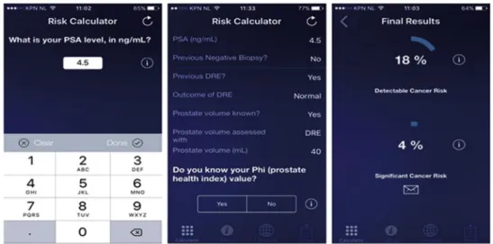
www.prostatecancer-riskcalculator.com) or through a smartphone-app (App:
Rotterdam Prostate Cancer Risk Calculator,Fig. 3). The risk
factors included in the various prediction models are age, family history, IPSS score, PSA, ultrasound-assessed pros-tate volume, DRE results, TRUS outcome, and previous
bi-opsy status[81]. The development study of risk calculator
step number 3 which predicts initial biopsy outcome reached an AUC of 0.77 as compared to 0.64 for PSA alone when predicting overall PCa[81](Fig. 4).
Retrospective analyses of 1850 men biopsied in ERSPC Rotterdam at initial screening, comparing a purely
PSA-based strategy (PSA3 ng/mL) with a risk-based strategy
combining the PSA3.0 ng/mL threshold with an
individ-ually calculated risk12.5%, showed that 33% of biopsies
Figure 3 Screenshots of the European Randomized Study of Screening for Prostate Cancer risk calculator (app).
could be avoided. Although 14% of PCa would potentially be missed with the risk-based strategy, 70% of these missed cancers were classified as potentially indolent. At repeat screening 4 years later, a similar strategy would result in 37% fewer biopsies while missing 16% of PCa diagnosis of which 81% could be classified as potentially indolent. As a comparison, applying a higher PSA threshold for biopsy
(4.0 ng/mL) could avoid similar numbers of unnecessary
biopsies, but would miss larger numbers of potentially
aggressive PCa’s (12% vs.10% at initial screening, 50%vs.
15% at repeat screening)[81].
In Asia, the Seoul National University Prostate Cancer Risk Calculator (SNUPC-RC) was developed. The risk calculator is based on data from 3482 Korean men who were all biopsied. In a validation cohort of 1112 Korean men, the SNUPC-RC showed a high predictive value with an AUC of 0.811, the AUC of the ERSPC risk calculator validated in the
same Korean population was 0.768[82]. When applying the
30%-threshold of the SNUPC-RC instead of a PSA level of
>4 ng/mL for biopsy, 21.5% of biopsies would be avoided
and only 1.4% of the PCa diagnoses would be missed[82].
When comparing the two risk calculators, it is notable that the ERSPC risk calculator was developed on 6-core
biopsy scheme while the SNUPC-RC was based on a
12-core biopsy scheme[82]. Furthermore, Korean men have a
different distribution of PSA levels than the European
population [83]. External validation studies confirm the
existence of racial variations and highlights that caution should be paid to using risk calculator models outside their region of origin, predominantly when proper validation studies have not been conducted.
It must however be noted that as compared to a purely PSA based strategy a risk based strategy is always better in avoiding unnecessary biopsies and reducing potential overdiagnosis, both in Europe and Asia.
5. The future of PCa screening
Since the incidence and tumor characteristics differ be-tween European and Asian populations, it is crucial to acknowledge that European-based screening outcomes are not directly transferrable to an Asian setting. Similar con-cerns, e.g., unnecessary testing and overdiagnosis exist and like in the western world PSA-based population screening is not recommended (yet).
Risk stratification has proven to be of aid in identifying men at low risk of harboring aggressive disease and can as such reduce unnecessary testing and overdiagnosis. Hence, Asian countries need to develop or adapt western-based risk stratification strategies using region-specific data.
mpMRI-guided biopsy holds great potential to replace conventional systematic TRUS-guided biopsy. It can provide direct visualization of suspicious lesions and improve diag-nostic accuracy for significant PCa, especially in men with a previous negative biopsy. As a new technique, however, performance and cost-effectiveness needs validation in Asian countries which is currently hampered by the avail-ability of data.
PCa screening in most Asian countries is still in its in-fancy, with the priority of assessing the effect on disease specific mortality. This is contrary to most European
countries, where the focus now lies in reducing over-diagnosis and overtreatment. Further research on PCa screening in Asia should however be based on the European experiences to avoid making the same mistakes. The con-ventional model “PSA screening-biopsy-active treatment” should not be duplicated. Instead, multivariate risk pre-diction, tailored to the Asian setting, followed by imaging in those considered at high risk for a potentially life threat-ening PC is advised, preferably in a controlled setting of a governmental regulated screening program.
Conflicts of interest
The authors declare no conflict of interest.
References
[1] Torre LA, Bray F, Siegel RL, Ferlay J, Lortet-Tieulent J, Jemal A. Global cancer statistics. CA Cancer J Clin 2012; 2015(65):87e108.
[2] Attard G, Parker C, Eeles RA, Schroder F, Tomlins SA, Tannock I, et al. Prostate cancer. Lancet 2016;387:70e82. [3] Center MM, Jemal A, Lortet-Tieulent J, Ward E, Ferlay J,
Brawley O, et al. International variation in prostate cancer incidence and mortality rates. Eur Urol 2012;61:1079e92. [4] Wong MC, Goggins WB, Wang HH, Fung FD, Leung C, Wong SY,
et al. Global incidence and mortality for prostate cancer: analysis of temporal patterns and trends in 36 countries. Eur Urol 2016;70:862e74.
[5] Curado MP, Edwards B, Shin HR, Storm H, Ferlay J, Heanue M, et al. Cancer incidence in five continents, volumes I to IX: IARC CancerBase No. 9. Lyon, France: International Agency for Research on Cancer. World Health Organization Web site.
http://ci5.iarc.fr.
[6] Watanabe M, Nakayama T, Shiraishi T, Stemmermann GN, Yatani R. Comparative studies of prostate cancer in Japan versus the United States. A review. Urol Oncol 2000;5:274e83. [7] Siegel R, Ward E, Brawley O, Jemal A. Cancer statistics, 2011: the impact of eliminating socioeconomic and racial disparities on premature cancer deaths. CA Cancer J Clin 2011;61:212e36. [8] Schroder FH, Hugosson J, Roobol MJ, Tammela TL, Zappa M, Nelen V, et al. Screening and prostate cancer mortality: re-sults of the European Randomised Study of Screening for Prostate Cancer (ERSPC) at 13 years of follow-up. Lancet 2014;384:2027e35.
[9] Andriole GL, Crawford ED, Grubb 3rd RL, Buys SS, Chia D, Church TR, et al. Prostate cancer screening in the randomized prostate, lung, colorectal, and ovarian cancer screening trial: mortality results after 13 years of follow-up. J Natl Cancer Inst 2012;104:125e32.
[10] Shoag JE, Mittal S, Hu JC. Reevaluating PSA testing rates in the PLCO trial. N Engl J Med 2016;374:1795e6.
[11] Hugosson J, Carlsson S, Aus G, Bergdahl S, Khatami A, Lodding P, et al. Mortality results from the Goteborg rando-mised population-based prostate-cancer screening trial. Lan-cet Oncol 2010;11:725e32.
[12] Kobayashi M, Takezawa Y, Takechi H, Ito K, Yamamoto T, Suzuki K, et al. MP-11.12: Japanese prospective cohort study of screening for prostate cancer (JPSPC): the study concept and the first analyses on compliance and contamination for the PSA test (gunma section). Urology 2007;70(Supplement):97. [13] Ito K, Kakehi Y, Naito S, Okuyama A, Japanese Urological
Association. Japanese Urological Association guidelines on prostate-specific antigen-based screening for prostate cancer
and the ongoing cluster cohort study in Japan. Int J Urol 2008; 15:763e8.
[14] Kitagawa Y, Namiki M. Prostate-specific antigen-based popu-lation screening for prostate cancer: current status in Japan and future perspective in Asia. Asian J Androl 2015;17:475e80. [15] Kitagawa Y, Mizokami A, Nakashima K, Koshida K, Shimamura M, Miyazaki K, et al. Clinical outcomes of prostate cancer patients detected by prostate-specific antigen-based population screening in Kanazawa City, Japan. Int J Urol 2011;18:592e6. [16] Schroder FH, Hugosson J, Roobol MJ, Tammela TL, Ciatto S,
Nelen V, et al. Prostate-cancer mortality at 11 years of follow-up. N Engl J Med 2012;366:981e90.
[17] Mok Y, Kimm H, Shin SY, Jee SH, Platz EA. Screening prostate-specific antigen concentration and prostate cancer mortality: the Korean heart study. Urology 2015;85:1111e6.
[18] Vickers AJ, Cronin AM, Bjork T, Manjer J, Nilsson PM, Dahlin A, et al. Prostate specific antigen concentration at age 60 and death or metastasis from prostate cancer: case-control study. BMJ 2010;341:c4521.
[19] Rabah DM, Arafa MA. Prostate cancer screening in a Saudi population: an explanatory trial study. Prostate Cancer Pros-tatic Dis 2010;13:191e4.
[20] Tyson 2nd MD, Castle EP. Racial disparities in survival for pa-tients with clinically localized prostate cancer adjusted for treatment effects. Mayo Clin Proc 2014;89:300e7.
[21] Fukagai T, Namiki TS, Carlile RG, Yoshida H, Namiki M. Com-parison of the clinical outcome after hormonal therapy for prostate cancer between Japanese and Caucasian men. BJU Int 2006;97:1190e3.
[22] Faisal FA, Sundi D, Cooper JL, Humphreys EB, Partin AW, Han M, et al. Racial disparities in oncologic outcomes after radical prostatectomy: long-term follow-up. Urology 2014;84: 1434e41.
[23] Lloyd T, Hounsome L, Mehay A, Mee S, Verne J, Cooper A. Lifetime risk of being diagnosed with, or dying from, prostate cancer by major ethnic group in England 2008e2010. BMC Med 2015;13:171.
[24] Bokhorst LP, Roobol MJ. Ethnicity and prostate cancer: the way to solve the screening problem? BMC Med 2015;13:179. [25] Vickers AJ, Ulmert D, Sjoberg DD, Bennette CJ, Bjork T,
Gerdtsson A, et al. Strategy for detection of prostate cancer based on relation between prostate specific antigen at age 40e55 and long term risk of metastasis: case-control study. BMJ 2013;346:f2023.
[26] Heidenreich A, Bastian PJ, Bellmunt J, Bolla M, Joniau S, van der Kwast T, et al. EAU guidelines on prostate cancer. Part 1: screening, diagnosis, and local treatment with curative intent-update 2013. Eur Urol 2014;65:124e37.
[27] Vickers AJ, Eastham JA, Scardino PT, Lilja H. The memorial sloan kettering cancer center recommendations for prostate cancer screening. Urology 2016;91:12e8.
[28] Catalona WJ. Baseline PSA testing for men in their 40s: currently available evidence strongly supports baseline PSA measurements in this age group. Oncol Willist Park 2014;28: 154e6.
[29] Loeb S, Bjurlin MA, Nicholson J, Tammela TL, Penson DF, Carter HB, et al. Overdiagnosis and overtreatment of prostate cancer. Eur Urol 2014;65:1046e55.
[30] Schroder FH, Hugosson J, Roobol MJ, Tammela TL, Ciatto S, Nelen V, et al. Screening and prostate-cancer mortality in a randomized European study. N Engl J Med 2009;360:1320e8. [31] Bokhorst LP, Venderbos LD, Schroder FH, Bangma CH,
Steyerberg EW, Roobol MJ. Do treatment differences between arms affect the main outcome of ERSPC Rotterdam? J Urol 2015;194:336e42.
[32] Song C, Ahn H, Lee MS, Park J, Kwon TG, Kim HJ, et al. Mass screening for prostate cancer in Korea: a population based study. J Urol 2008;180:1949e53.
[33] Kuwahara M, Tochigi T, Kawamura S, Ogata Y, Xu N, Wang H, et al. Mass screening for prostate cancer: a comparative study in Natori, Japan and Changchun, China. Urology 2003;61:137e41. [34] Moyer VA, Force USPST. Screening for prostate cancer: U.S. preventive services task force recommendation statement. Ann Intern Med 2012;157:120e34.
[35] Abdollah F, Dalela D, Sood A, Meyer CP, Hansen M, Han P, et al. The impact of 2011 United States preventive services task force panel update on PSA screening practice: a nation-wide, and state-by-state level analyses. In: American Urological Association Annual Meeting San Diego 2016; 2016. [36] Aslani A, Minnillo BJ, Johnson B, Cherullo EE, Ponsky LE,
Abouassaly R. The impact of recent screening recommenda-tions on prostate cancer screening in a large health care system. J Urol 2014;191:1737e42.
[37] Drazer MW, Huo D, Eggener SE. National prostate cancer screening rates after the 2012 US preventive-services task force recommendation discouraging prostate-specific antigen-based screening. J Clin Oncol 2015;33:2416e23.
[38] Frendl D, Epstein M, Fouyazi H, Krajenta R, Rybicki B, Sokoloff M. Impact of guidelines on prostate cancer screening in a population-based setting. In: 2000e2014: preliminary results from the first AUA data Grant American Urological Association Annual Meeting San Diego 2016; 2016.
[39] Jemal A, Fedewa SA, Ma J, Siegel R, Lin CC, Brawley O, et al. Prostate cancer incidence and PSA testing patterns in relation to USPSTF screening recommendations. JAMA 2015;314: 2054e61.
[40] Li J, Berkowitz Z, Hall IJ. Decrease in prostate cancer testing following the US preventive services task force (USPSTF) recommendations. J Am Board Fam Med 2015;28:491e3. [41] Miller C, Kabarriti A, Pulido J, Ziemba J, Guzzo T, Wein A,
et al. United States preventive services task force prostate cancer screening guidelines were associated with age and race dependent changes in primary care prostate specific antigen based screening at a tertiary care center. In: Amer-ican Urological Association Annual Meeting San Diego 2016; 2016.
[42] Rezaee ME, Ward CE, Odom BD, Pollock M. Prostate cancer screening practices and diagnoses in patients age 50 and older, Southeastern Michigan, pre/post 2012. Prev Med 2016; 82:73e6.
[43] Sammon JD, Abdollah F, Choueiri TK, Kantoff PW, Nguyen PL, Menon M, et al. Prostate-specific antigen screening after 2012 US preventive Services Task Force recommendations. JAMA 2015;314:2077e9.
[44] Sammon JD, Dalela D, Abdollah F, Sood A, Han P, Hansen M, et al. Age dependent variation in the effect of physician recommendations to undergo prostate specific antigen (PSA) screening following the United States preventive services task force 2012 statement against PSA screening. In: American Urological Association Annual Meeting San Diego 2016; 2016. [45] Shoag J, Halpern JA, Lee DJ, Mittal S, Ballman KV, Barbieri CE,
et al. Decline in prostate cancer screening by primary care physicians: an analysis of trends in the use of digital rectal examination and prostate specific antigen testing. J Urol 2016;196(4):1047e52.
[46] Turini GI, Gjelsvik A, Golijanin D, Pareek G, Renzulli JI. The role of patient race and ethnicity in predicting physician recommendation of prostate-specific antigen (PSA) testing. In: American Urological Association Annual Meeting San Diego 2016; 2016.
[47] Zavaski M, Meyer CP, Hanske J, Friedlander D, Cheng P, Menon M, et al. Differences in prostate specific antigen testing among urologists and primary care providers in the United States following the 2011 USPSTF recommendations. In: American Urological Association Annual Meeting San Diego 2016; 2016.
[48] Zeliadt SB, Hoffman RM, Etzioni R, Gore JL, Kessler LG, Lin DW. Influence of publication of US and European prostate cancer screening trials on PSA testing practices. J Natl Cancer Inst 2011;103:520e3.
[49] Cohn JA, Wang CE, Lakeman JC, Silverstein JC, Brendler CB, Novakovic KR, et al. Primary care physician PSA screening practices before and after the final U.S. preventive services task force recommendation. Urol Oncol 2014;32:e23e30. [50] Zargar H, van den Bergh R, Moon D, Lawrentschuk N,
Costello A, Murphy D. The impact of United States preventive Services Task Force (USPTSTF) recommendations against PSA testing on PSA testing in Australia. BJU Int 2016;119:110e5. [51] Fleshner KCS, Carlsson SV, Roobol MJ. A review of
prostate-specific antigen screening and prostate cancer incidence patterns after the 2011e2012 United States preventive ser-vices task force recommendation. Nat Rev Urol 2016. in press.
[52] Mikolajczyk SD, Grauer LS, Millar LS, Hill TM, Kumar A, Rittenhouse HG, et al. A precursor form of PSA (pPSA) is a component of the free PSA in prostate cancer serum. Urology 1997;50:710e4.
[53] Mikolajczyk SD, Millar LS, Wang TJ, Rittenhouse HG, Marks LS, Song W, et al. A precursor form of prostate-specific antigen is more highly elevated in prostate cancer compared with benign transition zone prostate tissue. Cancer Res 2000;60: 756e9.
[54] Sartori DA, Chan DW. Biomarkers in prostate cancer: what’s new? Curr Opin Oncol 2014;26:259e64.
[55] Lughezzani G, Lazzeri M, Haese A, McNicholas T, de la Taille A, Buffi NM, et al. Multicenter European external vali-dation of a prostate health index-based nomogram for pre-dicting prostate cancer at extended biopsy. Eur Urol 2014;66: 906e12.
[56] Lazzeri M, Haese A, de la Taille A, Palou Redorta J, McNicholas T, Lughezzani G, et al. Serum isoform [-2]proPSA derivatives significantly improve prediction of prostate cancer at initial biopsy in a total PSA range of 2e10 ng/mL: a mul-ticentric European study. Eur Urol 2013;63:986e94.
[57] Ng CF, Chiu PK, Lam NY, Lam HC, Lee KW, Hou SS. The pros-tate health index in predicting initial prospros-tate biopsy out-comes in Asian men with prostate-specific antigen levels of 4-10 ng/mL. Int Urol Nephrol 2014;46:711e7.
[58] Na R, Ye D, Liu F, Chen H, Qi J, Wu Y, et al. Performance of serum prostate-specific antigen isoform [-2]proPSA (p2PSA) and the prostate health index (PHI) in a Chinese hospital-based biopsy population. Prostate 2014;74:1569e75. [59] Leapman MS, Carroll PR. New genetic markers for prostate
Cancer. Urol Clin North Am 2016;43:7e15.
[60] Sanguedolce F, Cormio A, Brunelli M, D’Amuri A, Carrieri G, Bufo P, et al. Urine TMPRSS2: ERG fusion transcript as a biomarker for prostate cancer: literature review. Clin Geni-tourin Cancer 2016;14:117e21.
[61] Nicholson A, Mahon J, Boland A, Beale S, Dwan K, Fleeman N, et al. The clinical effectiveness and cost-effectiveness of the PROGENSA(R) prostate cancer antigen 3 assay and the prostate health index in the diagnosis of prostate cancer: a systematic review and economic evaluation. Health Technol Assess 2015;19(iexxxi):1e191.
[62] Okihara K, Ochiai A, Kamoi K, Fujizuka Y, Miki T, Ito K. Comprehensive assessment for novel prostate cancer markers in the prostate-specific antigen era: focusing on Asians and Asian countries. Int J Urol 2015;22:334e41.
[63] Bussemakers MJ, van Bokhoven A, Verhaegh GW, Smit FP, Karthaus HF, Schalken JA, et al. DD3: a new prostate-specific gene, highly overexpressed in prostate cancer. Cancer Res 1999;59:5975e9.
[64] Chun FK, de la Taille A, van Poppel H, Marberger M, Stenzl A, Mulders PF, et al. Prostate cancer gene 3 (PCA3): development
and internal validation of a novel biopsy nomogram. Eur Urol 2009;56:659e67.
[65] Ochiai A, Okihara K, Kamoi K, Oikawa T, Shimazui T, Murayama S, et al. Clinical utility of the prostate cancer gene 3 (PCA3) urine assay in Japanese men undergoing prostate biopsy. BJU Int 2013;111:928e33.
[66] Leyten GH, Hessels D, Jannink SA, Smit FP, de Jong H, Cornel EB, et al. Prospective multicentre evaluation of PCA3 and TMPRSS2-ERG gene fusions as diagnostic and prognostic urinary bio-markers for prostate cancer. Eur Urol 2014;65:534e42. [67] Tomlins SA, Day JR, Lonigro RJ, Hovelson DH, Siddiqui J,
Kunju LP, et al. Urine TMPRSS2:ERG plus PCA3 for individu-alized prostate cancer risk assessment. Eur Urol 2016;70: 45e53.
[68] Weinreb JC, Barentsz JO, Choyke PL, Cornud F, Haider MA, Macura KJ, et al. PI-RADS prostate imagingereporting and data system: 2015, version 2. Eur Urol 2016;69:16e40. [69] Barentsz JO, Richenberg J, Clements R, Choyke P, Verma S,
Villeirs G, et al. ESUR prostate MR guidelines 2012. Eur Radiol 2012;22:746e57.
[70] Turkbey B, Choyke PL. PIRADS 2.0: what is new? Diagn Interv Radiol 2015;21:382e4.
[71] Pinto PA, Chung PH, Rastinehad AR, Baccala Jr AA, Kruecker J, Benjamin CJ, et al. Magnetic resonance imaging/ultrasound fusion guided prostate biopsy improves cancer detection following transrectal ultrasound biopsy and correlates with multiparametric magnetic resonance imaging. J Urol 2011; 186:1281e5.
[72] Bjurlin MA, Mendhiratta N, Wysock JS, Taneja SS. Multi-parametric MRI and targeted prostate biopsy: improvements in cancer detection, localization, and risk assessment. Cent Eur J Urol 2016;69:9e18.
[73] Schoots IG, Roobol MJ, Nieboer D, Bangma CH, Steyerberg EW, Hunink MG. Magnetic resonance imaging-targeted biopsy may enhance the diagnostic accuracy of significant prostate cancer detection compared to standard transrectal ultrasound-guided biopsy: a systematic review and meta-analysis. Eur Urol 2015;68:438e50.
[74] Washino S, Okochi T, Saito K, Konishi T, Hirai M, Kobayashi Y, et al. Combination of PI-RADS score and PSA density predicts biopsy outcome in biopsy naive patients. BJU Int 2016;119: 225e33.
[75] Lee DH, Nam JK, Park SW, Lee SS, Han JY, Lee SD, et al. Visually estimated MRI targeted prostate biopsy could improve the detection of significant prostate cancer in patients with a PSA level<10 ng/mL. Yonsei Med J 2016;57:565e71. [76] Zhu X, Albertsen PC, Andriole GL, Roobol MJ, Schroder FH,
Vickers AJ. Risk-based prostate cancer screening. Eur Urol 2012;61:652e61.
[77] Stephan C, Cammann H, Semjonow A, Diamandis EP, Wymenga LF, Lein M, et al. Multicenter evaluation of an artificial neural network to increase the prostate cancer detection rate and reduce unnecessary biopsies. Clin Chem 2002;48:1279e87.
[78] Finne P, Finne R, Bangma C, Hugosson J, Hakama M, Auvinen A, et al. Algorithms based on prostate-specific anti-gen (PSA), free PSA, digital rectal examination and prostate volume reduce false-positive PSA results in prostate cancer screening. Int J Cancer 2004;111:310e5.
[79] Karakiewicz PI, Benayoun S, Kattan MW, Perrotte P, Valiquette L, Scardino PT, et al. Development and validation of a nomogram predicting the outcome of prostate biopsy based on patient age, digital rectal examination and serum prostate specific antigen. J Urol 2005;173:1930e4.
[80] Thompson IM, Ankerst DP, Chi C, Goodman PJ, Tangen CM, Lucia MS, et al. Assessing prostate cancer risk: results from the prostate Cancer prevention trial. J Natl Cancer Inst 2006; 98:529e34.
[81] Roobol MJ, Steyerberg EW, Kranse R, Wolters T, van den Bergh RC, Bangma CH, et al. A risk-based strategy improves prostate-specific antigen-driven detection of prostate cancer. Eur Urol 2010;57:79e85.
[82] Jeong CW, Lee S, Jung JW, Lee BK, Jeong SJ, Hong SK, et al. Mobile application-based Seoul national university prostate cancer risk calculator: development, validation, and comparative analysis with two Western risk calculators in Korean men. PLoS One 2014;9:e94441.
[83] Ku JH, Ahn JO, Lee CH, Lee NK, Park YH, Byun SS, et al. Dis-tribution of serum prostate-specific antigen in healthy Korean men: influence of ethnicity. Urology 2002;60:475e9.
[84] Gao HW, Li YL, Wu S, Wang YS, Zhang HF, Pan YZ, et al. Mass screening of prostate cancer in a Chinese population: the relationship between pathological features of prostate cancer and serum prostate specific antigen. Asian J Androl 2005;7: 159e63.
[85] Belbase NP, Agrawal CS, Pokharel PK, Agrawal S, Lamsal M, Shakya VC. Prostate cancer screening in a healthy population cohort in eastern Nepal: an explanatory trial study. Asian Pac J Cancer Prev 2013;14:2835e8.
[86] Vu Le C, Dao OQ, Khac Tran LN. Mass screening of prostate cancer in Vietnam: current status and our opinions. Urol Oncol 2010;28:673e6.
![Figure 2 Prostate cancer incidence trend in Europe and Asia [3]. Adapt with permission.](https://thumb-us.123doks.com/thumbv2/123dok_us/9533088.2829413/2.892.204.686.869.1069/figure-prostate-cancer-incidence-trend-europe-adapt-permission.webp)