Boston University
OpenBU http://open.bu.edu
Theses & Dissertations Boston University Theses & Dissertations
2013
Periostin signaling regulates breast
cancer stem cells
https://hdl.handle.net/2144/11111 Boston University
BOSTON UNIVERSITY SCHOOL OF MEDICINE
Dissertation
PERIOSTIN SIGNALING REGULATES BREAST CANCER STEM CELLS
by
ARTHUR W. LAMBERT
B.S., University of New Hampshire, 2006
Submitted in partial fulfillment of the requirements for the degree of
Doctor of Philosophy 2013
© Copyright by ARTHUR W. LAMBERT 2013
Approved by
First Reader
Sam Thiagalingam, Ph.D.
Associate Professor of Medicine, Genetics & Genomics, and Pathology & Laboratory Medicine
Second Reader
Douglas V. Faller, M.D., Ph.D.
Professor of Medicine, Pediatrics, Biochemistry, Microbiology, and Pathology
&
Laboratory MedicineDEDICATIONS
This work is dedicated to my wonderful parents, Robert and Sharon Lambert, who have always inspired me and worked hard to support my education.
ACKNOWLEDGEMENTS
I am deeply thankful to so many people who have graciously provided help and support to me during my graduate years, without which none of the work presented here would have been possible. First, I would like to thank my advisor, Dr. Sam Thiagalingam, for allowing me to join his laboratory in the first place. Since coming here, in 2008, he has always allowed me to work independently and encouraged me to explore projects that I found interesting. This, more than anything, gave me tremendous space in which to develop scientifically, often doing so at the expense of time and productivity, and I very much appreciate Dr. Thiagalingam's patience and trust during this period of my scientific training. I would also like to recognize the many contributions of my dissertation committee, which includes the chair, Dr. Herbert T. Cohen; the second reader, Dr. Douglas V. Faller; as well as Dr. Gerald V. Denis and Dr. Anurag Singh. In numerous committee meetings and in various other venues they generously offered their time, expertise and advice, which has been instrumental in the development of this work and my career.
I am also lucky to have had such wonderful colleagues in the laboratory. am especially grateful to Dr. Panos Papageorgis, who took the time to train me when I arrived here as a rotation student and throughout much of the following year. He made the lab an exciting place to be and I have always enjoyed discussing science with Panos. Often this has led to new ideas and experiments;
Abdolmaleky has been a constant calming presence throughout these years. His easy-going, relaxed demeanor has made it a pleasure to work with Hamid, and his wise advice is always appreciated. I would also like to thank Dr. Asami Takashima who, while not part of our lab, is a great friend from the Cancer Research Center, where we would find ourselves many Thursday mornings. Finally, I must thank Sait Ozturk, who has truly been like a brother in the Thiagalingam laboratory. We have been classmates since the beginning of graduate school and have followed essentially the same path. At each and every step Sait has been there, offering scientific advice, support, or even a hand to help with an experiment. Most important, during this time Sait has become a great friend. I find it difficult, as it were, to image graduate school without his companionship and look forward to many years ahead as friends.
I would be remiss not to thank the joint BUSM/MET undergraduate program in Biomedical Laboratory and Clinical Sciences, where I have been lucky enough to teach for most of my time here. The director, Connie Phillips; associate director, Gloria Vachino; and laboratory manager Dr. Stefan Doerre have been incredibly supportive of my teaching endeavors. Among them, the other instructors and the growing student body I have found myself in a wonderful community in which I feel so fortunate to have had the opportunity to work. Teaching has been an incredible educational experience while also being fun and rewarding, even during long weeknights and on early Saturday mornings, and I count it among my best times here at BUSM.
I owe nearly everything, though, to my family. My parents, Robert and Sharon Lambert, have always worked hard to support my education and consistently motivated me through their hard work, patience and love. My two younger brothers, Bobby and Danny, have never been far, always there for a laugh, good time, or round of golf. But more than anyone, I have to thank my incredible wife, Catherine Smart, who has been by my side through this entire process, from graduate school applications to the defense of this dissertation. All the while she has supported me with delicious food, incredible patience and love, even while growing her own business and career. She has given me much more than I deserve.
Arthur W. Lambert Boston, Massachusetts July 2013
PERIOSTIN SIGNALING REGULATES BREAST CANCER STEM CELLS (Order No.
ARTHUR W. LAMBERT
Boston University School of Medicine, 2013
Major Professor: Sam Thiagalingam, Ph.D., Associate Professor of Medicine, Genetics & Genomics, and Pathology & Laboratory Medicine
ABSTRACT
Each individual breast tumor contains a heterogeneous mix of cancer cells. Recent evidence suggests that the cellular heterogeneity inherent to breast cancer, which underlies metastasis, resistance to treatment and disease recurrence, can be driven by a distinct population of tumor cells referred to as cancer stem cells. However, the extracellular cues and intracellular signaling pathways that cancer stem cells rely on remain largely unknown. The aim of this dissertation was to investigate the functional relevance of tumor cell-derived periostin, a secreted extracellular matrix (ECM) molecule, to cancer stem cells and determine the pathways activated downstream of periostin through the integrin av~3 receptor. The rationale for this work was based on correlative evidence that connected periostin and integrin av~3 expression to numerous phenotypes associated with cancer stem cells including residence in a cell state associated with epithelial-mesenchymal transition (EMT), mammosphere formation and expression of the CD44+/CD24- surface phenotype. Multiple breast cancer cell lines with knockdown of either periostin or integrin ~3 showed
characteristics suggestive of a reduced cancer stem cell population such as impaired mammosphere formation and an inability to maintain a subpopulation of aldehyde dehydrogenase (ALDH)-positive cancer cells, implying that this autocrine/paracrine signaling axis is required to maintain breast cancer stem cells. Furthermore, this axis was found to be expressed in a subset of basal-like breast cancer cell lines. Additionally, high periostin expression was associated with a poor prognosis in patients with estrogen receptor (ER)-negative breast
cancer. Gene expression analysis revealed that breast cancer cells with
knockdown of periostin exhibited significant suppression of a distinct cytokine network, which includes IL-6 and IL-8, two cytokines that have been linked to the regulation of cancer stem cells. In a different cellular context, periostin could regulate the transcription of multiple WNT ligands. Thus, periostin serves as a critical regulator of the cancer stem cell state and acts through integrin signaling to control the production of other secreted factors that can also modulate cancer stem cells. These findings suggest that basal-like breast cancers can establish a microenvironmental niche supportive of cancer stem cells, which might be relevant as a biomarker or therapeutic target.
TABLE OF CONTENTS
Title page ... i
Copyright page ... ii
Approval page ... iii
Dedication page ... iv
Acknowledgements ... v
ABSTRACT ... viii
TABLE OF CONTENTS ... x
LIST OF TABLES ... xiii
LIST OF FIGURES ... xiv
LIST OF ABBREVIATIONS ... xvii
GENERAL BACKGROUND AND LITERATURE REVIEW ... 1
Breast cancer ... 1
Pathological classification of breast cancer ... 2
Molecular subtypes of breast cancer ... 5
lntratumor heterogeneity and the cancer stem cell hypothesis ... 8
The tumor microenvironment can influence cancer development.. ... 12
Periostin is a component of the extracellular matrix ... 15
Functional roles of periostin ... 18
Periostin and cancer ... 22
Periostin and breast cancer ... 25
CHAPTER 1: THE FUNCTIONAL RELEVANCE OF PERIOSTIN EXPRESSION TO BREAST CANCER STEM CELLS
BACKGROUND ... 28
Characteristics of breast cancer stem cells ... 28
EMT and breast cancer stem cells ... 31
Clinical relevance of breast cancer stem cells ... 32
Periostin and breast cancer stem cells ... 34
MATERIALS AND METHODS ... 36
RESULTS ... 42
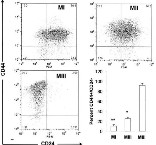
Characterization of cancer stem cell phenotypes in M cells ... 42
Periostin is highly expressed in cancer stem cell-enriched populations .... 46
A periostin-integrin signaling axis in basal-like breast cancer cells ... 51
Generation of breast cancer cells with knockdown of periostin ... 56
The effect of periostin knockdown on cancer stem cell phenotypes ... 59
lntegrin (33 and cancer stem cell phenotypes ... 63
Periostin is associated with a poor prognosis in ER-negative breast cancer ... 67
DISCUSSION ... 68
CHAPTER 2: THE PATHWAYS REGULATED BY PERIOSTIN IN BREAST CANCER STEM CELLS
lntegrins and breast cancer ... 77
lntegrins in normal and malignant stem cell biology ... 80
Signals that regulate breast cancer stem cells ... 82
MATERIALS AND METHODS ... 88
RESULTS ... 92
Genome-wide expression analysis of SUM159 cells with disruption of the periostin-integrin 133 signaling axis ... 92
Repression of a cytokine network in SUM159 shPN and shBeta3 cells ... 96
SUM159 shPN and shBeta3 cells exhibit reduced STAT3 signaling ... 1 01 Inhibition of the ERK pathway impairs cytokine transcription ... 1 03 Knockdown of periostin in Hs578T cells ... 1 08 Periostin regulates the transcription of Wnt ligands in Mill cells ... 110
Inhibition of the ERK pathway in Mill cells reduces the ALDH-positive population ... 114
DISCUSSION ... 116
FUTURE DIRECTIONS ... 126
GENERAL CONCLUSIONS ... 129
LIST OF ABBREVIATED JOURNAL TITLES ... 130
REFERENCES ... 136
LIST OF TABLES
Table 1. Characteristics of theM cell lines ... .43 Table 2. Limiting dilution analysis of SUM159 shGFP and shPN cells ...... 63 Table 3. Top genes repressed in SUM159 shPN that are coordinately regulated in shBeta3 cells ... 95 Table 4. Top pathways repressed in SUM159 shPN cells ...... 96 Table 5. Leading edge analysis of gene sets repressed in SUM159 shPN
LIST OF FIGURES
Figure 1. I ntertumor heterogeneity in breast cancer ...... 7
Figure 2. lntratumor heterogeneity ... 11
Figure 3. Periostin structure ....... 16
Figure 4. CD44/CD24 analysis of theM cell lines ....... .44
Figure 5. Mammosphere formation potential of M cells ...... .45
Figure 6. Periostin is highly expressed in Mill cells ....... .47
Figure 7. Periostin is a transcriptional target of TGF-13 signaling in M cells ... .48
Figure 8. Periostin is highly expressed in cancer stem cell-enriched populations ... 50
Figure 9. Expression of integrin
avl3
3
on the surface of M cells ... 52Figure 10. Periostin and integrin 133 are co-expressed in a subset of basal-like breast cancer cell lines ... 53
Figure 11. A subset of basal-like breast cancer cells produces periostin and exhibits high surface levels of integrin
avl33-
...
.
...
...
...
...
...
..
.
...
55Figure 12. Establishment of periostin knockdown cells ... 57
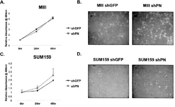
Figure 13. Proliferation and morphology of periostin knockdown cells ... 58
Figure 14. Periostin knockdown impairs mammosphere formation ... 59
Figure 15. Periostin knockdown does not alter the CD44/CD24 surface phenotype ... 60
Figure 16. Periostin is required for the maintenance of the ALDH-positive population in SUM159 cells ... 61
Figure 17. lntegrin avl3
3
expressing cells preferentially form mammospheres .... 64Figure 18. Knockdown of integrin 133 impairs mammosphere formation ..... 65
Figure 19. lntegrin 133 is required for the maintenance of the ALDH-positive
population in SUM159 cells ... 66
Figure 20. Periostin expression is associated with a poor prognosis in
ER-negative breast cancer ... 67
Figure 21. Periostin signaling pathways in cancer ...... 79
Figure 22. Genome-wide expression analysis of SUM159 shGFP, shPN and
shBeta3 cells ... 93
Figure 23. Expression of the cytokine gene set in SUM 159 shGFP and shPN
cells ... 97
Figure 24. Repression of IL6 and ILB expression in SUM159 shPN and shBeta3
cells ... 1 00
Figure 25. Impaired IL-8 secretion in SUM159 shPN and shBeta3 cells ... 1 01
Figure 26. Impaired STAT3 signaling in SUM159 shPN and shBeta3 cells ..... 102
Figure 27. Rescue of STAT3 signaling with conditioned media ...... 1 03
Figure 28. The NF-KB pathway is intact in SUM159 knockdown cells ..... 1 04
Figure 29. IPA identifies an ERK signaling defect in SUM159 shPN cells ....... 1 05
Figure 30. Knockdown of periostin or integrin 133 impairs ERK signaling ... 1 06
Figure 31. Inhibition of the ERK pathway reduces /L6 and ILB transcription .... 1 07
Figure 33. Knockdown of periostin in Mill cells impairs the transcription of WNT ligands and activation of Wnt signaling ... 111
Figure 34. Periostin expression is required for the induction of Wnt signaling
during mammosphere growth ... 112
Figure 35. Periostin knockdown impairs secondary mammosphere growth of Mill
cells ... 113
Figure 36. ERK inhibition reduces the ALDH-positive subpopulation in Mill
cells ... 114
Figure 37. Working model of periostin-integrin
avl3
3
signaling in breast cancer stem cells ... 125ACTB AKT ALDH ALDH1A1 AML AN OVA APC AV BAAA Bel Beta3 BMDC BMI1 BMP-1 BRCA1/2 BSA
c
C-terminal CAF CCRL1 LIST OF ABBREVIATIONS actin, betav-akt murine thymoma viral oncogene homolog aldehyde deydrogenase
aldehyde deydrogenase 1, family member A 1 acute myeloid leukemia
analysis of variance allophycocyan in atrioventricu Ia r BODIPY -aminoacetaldehyde B cell lymphoma integrin, beta 3
bone marrow derived cell
BM 11 polycomb ring finger oncogne bone morphogenetic protein 1 breast cancer 1/2, early onset bovine serum albumin
Celsius
carboxy terminal
cancer associated fibroblast
eDNA
C02
COX2 Cre cRNA CXCR1 Cy7 DEAB DHH Dl DKK DMEM DMSO DNA ECL ECM EDTA EGF EGFR ELISA EMT EpCAM complementary DNA carbon dioxie cyclooxygenase 2 Cre recombinase complementary RNAchemokine C-X-C motif receptor 1 cyanine dye 7
diethylaminobenzaldehyde desert hedgehog
aspartate-isoleucine dimer dickkopf-1
Dulbecco's Modified Eagle Medium dimethyl sulfoxide
deoxyribonucleic acid
enhanced chemiluminescence extracellular matrix
ethylenediaminetetraacetic acid epidermal growth factor
epidermal growth factor receptor enzyme-linked immunosorbent assay epithelial-mesenchymal transition epithelial cell adhesion molecule
ER ERBB2 ERK1 ERK2 ESA ESCC FAGS FAK FAS1 FBS FOR FGF FLK-1/KDR FOXC2 FOX03A G g GFP GLI1 GM-CSF estrogen receptor
v-erb-b2 erythoblastic leukemia viral oncogene homolog 2
mitogen-activated protein kinase 3 mitogen-activated protein kinase 1 epithelial specific antigen
esophageal squamous cell carcinoma fluorescence-activated cell sorting focal adhesion kinase
fasciclin domain fetal bovine serum false discovery rate fibroblast growth factor
kinase insert domain receptor forkhead box C2
forkhead box 03 glycine
gravity
green fluorescent protein GLI family zinc finger 1
GSEA H-RAS HCI HER-2 HNSCC hTERT I DC/NOS IHC IL IL 13RA2 IPA ITGB3 ITGB4 JAGGED JAK2 K kDa lacZ LAIR LB/amp LGR5
gene set enrichment analysis
v-Ha-ras Harvey rat sarcoma viral oncogene homolog hydrochloric acid
human epidermal growth factor receptor 2 head and neck squamous cell carcinoma telomerase reverse transcriptase
invasive ductal carcinoma, not otherwise specified immunohistochemistry
interleukin
interleukin 13 receptor, alpha 2 Ingenuity Pathway Analysis integrin, beta 3 integrin, beta 4 jagged 1 Janus kinase 2 thousand kilodalton beta-galactosidase
local acute inflammatory response lysogeny broth/ampicillin
leucine-rich repeat-containing G protein coupled receptor
LOX LRP MAPK MCP-1 MEK Ml miR ml MMP MMTV MPA mRNA MSCV-PGK MSigDB mTOR n N-terminal NES NF-kB ng nm lysyl oxidase
low density lipoprotein receptor-related protein mitogen activated protein kinase
monocyte chemoattractant protein 1 mitogen-activated protein kinase kinase myocardial infarction
micro RNA milliliter
matrix metalloproteinase mouse mammary tumor virus medroxyprogesterone acetate messenger RNA
murine stem cell virus/phosphoglycerate kinase Molecular Signatures Database
mammalian target of rapamycin sample size
amino terminal
normalized enrichment score nuclear factor-kappa-S nanogram
NOD/SCID NSCLC
oscc
OSF-2 p21 p53 p65 PBS PCA PCR PDACPE
pg PI PI3K PKB PN POSTN PR PROCR PTCH1non-obese diabetic/severe combined immunodeficiency
non-small cell lung cancer oral squamous cell carcinoma osteoblast-specific factor 2
cyclin-dependent kinase inhibitor 1 A tumor protein p53
v-rel reticuloendotheliosis viral oncogene homolog A phosphate buffered saline
Principal Component Analysis polymerase chain reaction
pancreatic ductal adenocarcinoma phycoerythrin picograms propidium idodide phosphatidylinositoi-3-0H kinase protein kinase B periostin periostin progesterone receptor
protein C receptor, endothelial patched recptor 1
PTGS2 PVDF PyMT qRT-PCR RANK RANKL RGD Rho A RIP A RMA RNA ROS RPM I SDS-PAGE SEM Ser SFRP1 SHH SHP2 shRNA prostaglandin-endoperoxide synthase 2 polyvinylidene fluoride
polyomavirus middle T-antigen
quantitative reverse transcription PCR receptor activator of nuclear factor-kappa-S
receptor activator of nuclear factor-kappa-S ligand a rg ig i ne-g lyci ne-aspartate motif
ras homolog family member A radioimmunoprecipitation assay robust multiarray average ribonucleic acid
reactive oxygen species
Roswell Park Memorial Institute
sodium dodecyl sulfate-polyacrylamide gel electrophoresis
standard error of the mean serine
secreted frizzled-related protein 1 sonic hedgehog
Src-homology 2 domain-containing phosphatase short hairpin RNA
SLUG SNAI1 Sost SPARC SRC
ss
STAB STAT TAM TBX3 TDLU TGF-~ TGFBI TIC TNBC TN F-a TSLP TWIST1 Tyrv
VEGF WNTsnail family zinc finger 2 snail family zinc finger 1 sclerostin
secreted protein rich in cysteine
v-src sarcoma viral oncogene homolog signal sequence
stabilin
signal transducer and activator of transcription tumor associated macrophage
transcription factor T-box protein
3
terminal ductal lobular end unit transforming growth factor beta
transforming growth factor, beta-induced tumor initiating cell
triple-negative breast cancer tumor necrosis factor, alpha thymic stromallymphopoietin
twist basic helix-loop-helix transcription factor 1 tyrosine
valine
vascular endothelial growth factor
XDH jJg !JM xanthine dehydrogenase microgram micromolar
GENERAL BACKGROUND AND LITERATURE REVIEW Breast cancer
Cancer is a staggering public health problem. The American Cancer Society expects over 1.6 million new cases of cancer to be diagnosed in 20131. In women, the majority of these cases -approximately 232,000 (29%) -will be classified as breast cancer1. Nearly 40,000 patients with breast cancer will die
this year, making it the second leading cause of cancer mortality in women, behind only lung cancer1. Fortunately, the number of newly-diagnosed cases has decreased steadily since 2000, largely as a result of a reduction in the number of post-menopausal women on hormone replacement therapy, which has been strongly linked to the development of breast cancer1·2. Death rates have also decreased in recent years and this has been attributed to improvements in screening and early detection with mammography as well as better treatment options 1. However, racial disparities in outcomes exist for breast cancer; African-American women have a lower incidence rate but an increased chance of death 1. While this likely reflects socioeconomic factors and access to health care, there also seem to be some biological differences since African-American women are more likely to be diagnosed with aggressive cancers3.4. Additionally, over 64,000
cases of carcinoma in situ are expected this year1. These are non-invasive
neoplasms and likely represent a pre-malignant stage in the progression towards invasive breast cancer5.
Numerous risk factors have been linked to the development of breast carcinomas. The most important factors that increase the risk of breast cancer are gender, age and family history, and modifiable factors such as obesity, physical inactivity and alcohol consumption are thought to confer risk as well1
.
There is also a strong relationship between exposure to reproductive hormones, such as estrogen and progesterone, and the development of breast cancer in postmenopausal women6, hence the connection between hormone replacement therapy and cancer incidence. There do seem to be inherited factors which predispose one to breast cancer because having a relative with a history of breast cancer increases risk. But this contribution is somewhat limited as most patients diagnosed do not have a close relative with breast cancer, and conversely, the majority of women that do have a relative with breast cancer will never develop the disease1. The exception here is individuals who inherent a mutant allele of BRCA 1 or BRCA2, two well-characterized breast and ovarian cancer-susceptibility genes. Women with these inherited mutations have up to an 80% chance of developing breast cancer, but these cases account for only 5%-10% of all breast cancer diagnoses7.
Pathological classification of breast cancer
Breast cancer arises from malignant transformation of epithelial cells in the mammary gland. The normal epithelium of the breast is organized as a series of ducts and lobules, designed to secrete milk and transport it to the
alveolar cells, are present in the lobules. Myoepithelial cells circumscribe these cells and form a basal layer of epithelium, which rests on the basement membrane separating the epithelial and connective tissue. The vast majority of carcinomas - upwards of 75% to 80% - arise in the ductal region and have the histological designation of invasive ductal carcinoma, not otherwise specified (IDC NOS)8-10. The remaining 20% to 25% of cases are referred to as "special types" and occupy one of 17 different histological subtypes, each with less than a 5% overall incidence9.
In addition to histological type, which categorizes tumors based on tissue morphology, the histological grade of the cancer reflects the degree of cellular differentiation as well as the mitotic rate. Histological grade is routinely used to classify cases of breast cancer and it serves as a good prognostic indicator, where poorly-differentiated and highly-proliferative cancers generally have worse clinical outcomes 11. Breast cancers are also subject to a staging process that evaluates the size of the primary tumor, the dissemination to regional lymph nodes and the presence or absence of distant metastases. The disease stage is an indicator of the extent to which a cancer has spread and it is the most important factor in determining the patient's prognosis and treatment options 12.
Finally, there are a number of well-characterized molecules that are also used to classify, prognosticate and guide treatment decisions. Foremost here is the estrogen receptor (ER), a nuclear hormone receptor that, upon binding estrogen, translocates to the nucleus and regulates the transcription of numerous
target genes, many of which are involved in cell proliferation 13. Roughly 70% of breast tumors are classified as ER-positive and the majority of these also express the progesterone receptor (PR), another member of the nuclear receptor superfamily that is commonly assessed by pathologists 14. Hormone receptor expression is less of a prognostic factor because it is correlated to histological grade15, however, because the growth of most of these cancers is hormone-dependent, ER/PR expression is strongly predictive of a response to endocrine therapies such as tamoxifen and aromatase inhibitors 16.
The human epidermal receptor-2 (HER-2, also known as ERBB2) is an oncogenic growth factor receptor that is now included in the standard pathological assessment of breast cancers. Up to 30% of cases harbor amplifications of this gene 17 that generally result in an increased abundance of the protein in the plasma membrane, where it functions to stimulate intracellular growth and division signaling pathways. Patients with amplified HER2 have an aggressive form of the disease and a substantially worse prognosis 17·18 but with the advent of targeted therapies, such as trastuzumab, which specifically targets the HER-2 protein, these patients have seen remarkable response rates and a corresponding benefit in terms of disease outcome 19. Today, the HER2
oncogene serves as a prime example of a patient-tailored molecular target, subject to standard pathologic detection and amenable to drug inhibition.
TNBC accounts for about 15% of all breast cancers, tends to occur in younger patients and is associated with poor short-term survival20. Importantly, these patients are not eligible for targeted therapies such as tamoxifen and trastuzumab because they do not express ER and HER-2. Despite intense study, no clear genetic lesion can be targeted clinically and TNBC remains a particularly aggressive malignancy.
Molecular subtypes of breast cancer
A major milestone in breast cancer research was reached in 2000 when it was reported that breast cancers could be classified into different subgroups based on their gene expression profiles21·22. This unprecedented molecular view
of breast tumors suggested that common transcriptional programs, shared between different tumors, could explain some of the biologic variability inherent to breast cancer that had been observed by pathologists and oncologists for years. Five subtypes were originally described: two luminal subtypes (luminal A and luminal B), which are hormone-receptor positive; a HER-2 positive subtype; a basal-like group that shares a gene expression profile similar to myoepithelial cells of the normal breast and generally lack expression of HER-2 and the hormone receptors; and a heterogeneous, "normal-like" collection of tumors. These subtypes have been repeatedly observed23'24 and, importantly, are associated with distinct clinical outcomes, where the HER-2 and basal-like subtypes have the worst prognosis22. There is a significant overlap between TNBC and the basal-like subtype as most cases of TNBC, but not all, are
classified as basal-like, and vice versa25. It is also worth noting that the majority of women with mutations in BRCA 1 develop basal-like cance~3. Recently, an additional claudin-low subtype that has reduced expression of tight junction and cell-cell adhesion genes has been observed as well26. These tumors exhibit an epithelial-to-mesenchymal (EMT) phenotype, are more resistant to chemotherapy and appear to include most cases of metaplastic breast cance~7.
The appearance of these intrinsic molecular subtypes may reflect a different cell of origin28, varying driver mutations29, differences in the microenvironment30 or some combination thereof (Figure 1). Regardless, these gene expression signatures help to capture and conceptualize the basic biological, pathological and clinical differences underlying the heterogeneous spectrum of breast cancers that arise in different patients.
INTERTUMOR HETEROGENEITY
•Cell of origin (epigenetic background)
•Mutational profile
•Transcriptional networks
•Microenvironment
Tumor A
Tumor B
TumorC
Differences could include: histological
type, molecular subtype, driver
mutations
,
metastatic
t
ropism and
response to chemotherapy.
Figure 1. lntertumor heterogeneity in breast cancer. lntertumor heterogeneity is used to describe the varying characteristics, such as those listed in the box below, that exist between tumors arising in different patients. Potential sources of this heterogeneity are listed above.
lntratumor heterogeneity and the cancer stem cell hypothesis
It is abundantly clear, as discussed above, that there are considerable differences among individual breast tumors - in their cellular origins, pathological and molecular classifications, genomic mutations and, importantly, in their propensity to metastasize and respond to chemotherapy. However, cancer cells within the same tumor are also heterogeneous and exhibit a range of phenotypes, an observation referred to as intratumor heterogeneitl1. This added level of complexity presents daunting challenges for both cancer research and clinical oncology, so recent efforts have been directed at understanding the basis for this heterogeneitl2-34.
One model put forth to explain this phenomenon, the clonal evolution model, is essentially Darwinian natural selection and posits that the cancer cells which acquire the most advantageous set of mutations will be selected for over time, and thus, drive disease progression35'36. This is perhaps best exemplified
in the classic "Vogelgram" that established a model to describe the sequential mutations acquired during colorectal tumorigenesis37. Recent genomic studies, which compare shared and unique mutations present in the tumor mass, have also suggested that a process of clonal evolution occurs in tumors, both spatially and temporally. In one example, sequencing of individual breast cancer cells from a TNBC revealed three discernable clonal populations which probably emerged early during tumorigenesis38. In another example, whole-genome
examine the cellular populations contributing to disease progression. In at least one basal-like breast cancer, the metastatic lesion was likely seeded from a small subpopulation of cells present in the primary tumor and exhibited relatively few new genetic alterations39. In a larger analysis of 21 breast cancers,
whole-genome sequencing revealed that most of these tumors harbor a dominant cellular population, although substantial subclonal differences evolved subsequently as most mutations were restricted to only a subset of all tumor cells40. Such genetic studies offer a powerful tool to examine intratumor heterogeneity and these early studies offer a fascinating view of the subclonal structure present in breast tumors. However, they do not causally link these evolutionary processes to tumor heterogeneity, they only describe the structure of the heterogeneity that is present.
The cancer stem cell hypothesis is another model that has been proposed to account for the heterogeneity found within tumors41-43. This idea was given new life in the mid-1990s when it was reported that acute myeloid leukemia (AML) is driven by a rare population of leukemia stem cells (marked as
CD34+/CD38-), apparently generated from mutation of a normal hematopoietic stem cell44·45. The salient implication from this work is that AML is organized as a hierarchy since only the leukemia stem cells had the ability to self-renew, differentiate into non-stem cancer cells, and initiate the disease when transplanted into immunocompromised mice45. While these studies firmly established the cancer stem cell paradigm, they also led to some confusion. The
cancer stem cell concept is meant to describe the source and organization of intratumor heterogeneity- that is, that the tumor is organized hierarchically- not the cell of origin46.47. Since then, cancer stem cells have been identified in other hematological malignancies such as chronic myeloid leukemia48, as well as in
solid tumors, most notably of the breast, brain and colon47·49, based on their
enhanced capacity to initiate tumorigenesis. Although this model might not be applicable to all cancers50'51, in tumors that are arranged hierarchically the
cancer stem cells assume a particularly important role, where they fuel tumor growth, give rise to other types of tumor cells and serve as the seeds of metastasis and recurrence52·53. In these cancers, it would seem that any efficacious therapeutic regimen would need to target the cancer stem cells54·55.
This notion is fundamentally different from that of the clonal evolution model, in which all cells of the tumor have a similar potential for tumorigenesis and disease progression is largely a stochastic process.
These two concepts (Figure 2) are not mutually exclusive, as even a population of cancer stem cells could be subject to the pressures of clonal evolution46·51·53. But it does seem logical that genetic differences between tumor
cells would play a prominent role in the clonal evolution model while the differences between cancer stem cells and non-cancer stem cells are more likely mediated by epigenetic changes47.
INTRATUMOR HETEROGENEITY
Clonal Evolution
--
... ->
...
-
..
->
Time
Cancer Stem Cell Hypothesis
r--->1
I I
I I
•---1
I+
Figure 2. lntratumor heterogeneity. lntratumor heterogeneity refers to differences between cancer cells within the same monoclonal tumor. In the clonal evolution model, cancer cells with a growth or survival advantage are selected for over time and due to the accumulation of new genetic mutations results in a heterogeneous cellular population. The cancer stem cell model predicts that tumors, similar to tissues, are organized as a hierarchy in which only the cancer stem cells are tumorigenic. Cancer stem cells can self-renew and
generate the various other cancer cell types. Note, that although these two models are depicted separately for conceptual purposes, they are not mutually exclusive.
The tumor microenvironment can influence cancer development
Oncogenesis, undoubtedly, is largely the result of cell-autonomous changes such as genetic mutations56·57 and epigenetic alterations58·59, which eventually lead to the acquisition of the six hallmarks of cancer: sustained proliferative signaling, evasion of anti-growth signals, resistance to apoptosis, cellular immortality, angiogenesis and invasion/metastasis60·61. However, the surrounding stromal cells, of what has been termed the tumor microenvironment, also exert a profound effect on the initiation and progression of cancers61-64. Various subtypes of infiltrating immune cells65, cancer-associated fibroblasts (CAFs)66, and vascular cells67 are either actively recruited by the tumor or arrive in response to the growing neoplasm. These diverse cell types act to module tumor development through heterotypic signaling with the cancer cells61. Early neoplastic cells are likely restrained by the surrounding normal stroma68, but as tumors progress they are able to manipulate this microenvironment to suit their own needs. One example of this involves tumor production of chemokines, such monocyte chemoattractant protein (MCP-1 ), which can stimulate the infiltration of tumor-associated macrophages (TAMs); TAMs then secrete vascular endothelial growth factor (VEGF) to promote angiogenesis69. Such interactions are
pervasive70 and likely influence most, if not all, of the hallmark capabilities shared by cancer cells64.
In addition to stromal cells, the tumor microenvironment also consists of extracellular matrix (ECM), which is secreted locally by resident cells in the tissue or tumor. Broadly, two types of ECM are recognized: the interstitial matrix which, occupies the space between intervening cells in the tissue and the basement membrane, made largely of collagen type IV, that forms the basal surface on which epithelial cells rest62. The ECM is comprised mainly of fibrous proteins and proteoglycans71·72, and can influence the surrounding cells in at least four ways. First, the ECM is well known to play a structural role, providing physical support and scaffolding for cell adhesion62. Second, the ECM can directly initiate downstream signaling through activation of heterodimeric transmembrane integrin receptors73. Third, ECM proteins are able to bind soluble growth factors, enhancing or sequestering their bioavailabilitl4. Finally, the ECM composition can alter the mechanical properties of the interstitial matrix, which can have surprisingly profound effects on various biological functions relevant to cancer such as proliferation, migration and differentiation74-77.
Dramatic changes to the ECM are characteristic during cancer progression and it can both restrain and promote neoplastic disease, depending on the specific context and components of the matrix. The basement membrane clearly counteracts the development of cancer since to progress an epithelial tumor must breach the basement membrane and invade the underlying stroma78.
The ECM is also a crucial regulator of normal tissue morphology and cellular polarity in the mammary gland79·80. But invasive cancers are often desmoplastic,
containing large numbers of CAFs, which results in tumor fibrosis with enhanced production of ECM proteins such as collagens, fibronectins and proteoglycans66·81. ECM proteins also play a central role in cancer cell migration
and invasion by activating intracellular signals downstream of integrin receptors, most notably focal adhesion kinase (FAK) and small GTPases such as RhoA82. Matrix metalloproteinases (MMPs), proteolytic enzymes that cleave various components of the ECM, are present in the tumor microenvironment and can modulate the structure and activity of the matrix to alter tumor progression83-85. For example, both MMP-2 and MMP-9 can process collagen in such a way that promotes angiogenesis86·87. And in recent years, as the process of metastatic
colonization has begun to be revealed88·89, ECM proteins have been shown to
function in this final stage of tumor cell dissemination. Interestingly, a premetastatic niche may be formed prior to the arrival of disseminated cancer cells90. In breast cancer this niche seems to be dependent on increased expression of fibronectin by resident fibroblasts91 and modulation of collagen to induce the recruitment of bone marrow-derived cells (BMDCs) and disseminated cancer cells92. Finally, it is important to note that the matrix is an essential aspect of the supportive niche for both normal and cancer stem cells74·76·93-97.
Periostin is a component of the extracellular matrix
The periostin gene, POSTN (also known as osteoblast-specific factor 2, OSF-2), is located on chromosome 13q13.3, contains 23 exons, and encodes a secreted ECM protein of up to 836 amino acids, with a molecular weight of approximately 90 kDa. The gene was cloned in 1993 from a murine osteoblastic cell line, MC3T3-E1, and was noted to share extensive homology to fasciclin I, an adhesion molecule important for axonal guidance in Drosophila melanogaster8
-100 In humans, the other three members of the fasciclin family include
transforming growth factor, beta-induced (TGFBI), stabilin I (STAB1) and stabilin 11 (STAB2)100•101. These proteins all have multiple FAS1 domains- named for their homology with fasciclin I - and each is comprised of around 150 amino acids, that appear to be important for cell adhesion and contain y-carboxylase recognition sites 100-102. Four periostin isoforms are currently validated and these represent alternatively-spliced transcripts containing different combinations of cassette exons (exons 17-21) in the C-terminal region 100·103-105. Although the precise function of these different splice variants is not known, an interesting cross-species comparison of the POSTN gene in vertebrates indicated that the C-terminal region evolved with considerably more plasticity than the rest of the gene, which suggests this region might harbor functions novel to periostin 103
, perhaps relating to interactions with other ECM proteins such as collagen 106,
fibronectin 107, heparin 108 and tenascin-C109. Other important characteristics of the protein include a typical N-terminal signal sequence common to secreted
proteins, a cysteine-rich EMI domain important for oligomerization of ECM proteins110·111, two aspartate-isoleucine (01) dimers similar in function to the arginine-glycine-aspartate (RGO) motif involved in binding integrins 112 and an N-glycosylation site 103. Consistent with the presence of the RGO-Iike 01 motif3,
periostin is known to act as a ligand for the following integrin receptors: av~3.
av~s. a
6
~4 and some ~1 containing complexes 100'105'113. The major biochemical domains of periostin are summarized in Figure 3.55 EMI FA51 FA511 FAS1 F.:-511 C-tenninal region
Figure 3. Periostin structure. Notable biochemical features of POSTN are depicted schematically. Full length POSTN is 836 amino acids long with an N-terminal signal sequence (SS) for secretion, a cysteine-rich oligomerization domain (EM I) and four fasciclin I adhesion domains (FAS1 ). The C-terminal region is subject to alternative splicing that defines the four different periostin
isoforms. The black bars in the second and fourth FAS1 domain specify
aspartate-isoleucine (01) integrin binding sites. The black triangle in the fourth FAS1 domain indicates an N-glycosylation site. This figure is adapted from reference 1 03.
Periostin is expressed widely in embryonic tissues including the periosteum, cardiac valves, placenta, peripheral nervous system and developing
teeth, where it seems to mark cells that have undergone an
epithelial-mesenchymal transition (EMT) 114-117. In adult tissues, periostin exhibits a more
restricted connective tissue expression pattern and remains expressed in the periosteum and periodontal ligaments 104 as well as in the valves of the heart114.
Recent work has also indicated that periostin may be expressed locally in various stem cell niches including the terminal ductal lobular end unit (TDLU) of the breast, the intestinal crypt and the area surrounding the bulge stem cells of skin follicles118. Furthermore, periostin expression can be induced in response to
cutaneous wound repair119-121, the Th2-type cytokines interleukin-4 (IL-4) and
interleukin-13 (IL-13)107'122, and in response to mechanical stimuli123·124. Based
in part on this expression pattern (along with its functional roles discussed below) periostin has been proposed to be a member of the matricellular family of ECM proteins 123·125. Matricellular proteins function as key modulators of cell-matrix interactions because they can bind to cells, through integrin receptors, as well as to soluble growth factors, cytokines and other structural ECM proteins 126·127. This class of molecules- including proteins such as osteopontin, secreted protein rich
in cysteine (SPARC), thrombospondin-1 and 2, and tenascin-C- are expressed
highly during development but in the adult their expression is generally induced in response to tissue injury128·129.
Functional roles of periostin
In attempts to understand the normal function of periostin, multiple mouse
models with knockout of the gene have been generated. The first such model, reported in 2005, was a periostin1acz knock-in mouse in which the /acZ cassette
was inserted in place of the first exon 117. Mice both heterozygous and
homozygous for the targeted allele were viable and grossly normal at birth and as neonates, consistent with the phenotypes observed for other matricellular proteins 128. However, in periostin-null animals there was a slight increase in postnatal lethality and by four months those animals surviving (86%) exhibited dwarfism and modest skeletal defects, which may be related to nutritional status since these mice have incisor enamel defects and periodontal-like disease that
may impair their ability to eat properli17. Another periostin knockout model (without the /acZ cassette) revealed that periostin was required for the proper eruption of incisors in the mouse, where it was found to localize to the shear zone and interact closely with collagen fibrils 130. Subsequent studies, following up on these initial observations, have added further details to the current understanding of how, when, and where periostin functions.
For instance, although knockout of periostin does not alter the development of cardiomyocytes, periostin has been found to be induced upon acute myocardial infarction (MI) and is required to recruit fibroblasts, properly form collagen fibrils and stiffen the ECM, which collectively acts to prevent
Ml131. Similarly, in a separate model, mice lacking periostin exhibited alterations
in cardiac hypertrophy, fibrosis and ventricular remodeling after myocardial infarction or long-term pressure overload132. Additional developmental studies of the atrioventricular (AV) values in the chick heart have shown that periostin is important for the differentiation of cushion mesenchymal cells to fibroblasts 133·134
and further examination of periostin-null mice revealed that these animals have leaflet defects and valve disease 135. Moreover, periostin appears to direct
progenitor cells away from the myocardial lineage in the developing heart125·136·137. Quite interestingly, in the adult heart, periostin has been reported
to stimulate the cycling of differentiated cardiomyocytes, which is dependent on activation of integrins av, 131, 133 and 135 as well as downstream activation of the
phosphatidylinositoi-3-0H kinase (P13K) pathwai38. Here too, periostin was
found to mitigate heart failure after Ml, which has prompted early pre-clinical studies to examine the prospect of using periostin, or periostin-derivatives, as therapeutic agents to treat myocardial damage 139-141.
Periostin also plays functional roles in osseous tissue142. In osteoblasts,
periostin promotes osteoblastic adhesion, proliferation, differentiation and
survival104·143·144. The expression of periostin is up-regulated in response to mechanical stimuli, such as moderate activity and pressure, which appears to suppress the expression of sclerostin (Sost), an inhibitor of the Wnt signaling pathwai45·146, to promote anabolic bone metabolism 124'147. Furthermore, the
the regulation of bone mineralization 142'148. Finally, periostin is suspected to play a role in the biological response to fractures as the expression of periostin is strongly induced in the days following a fracture 149.
As a prototypical matricellular protein, periostin is capable of regulating the organization of other ECM proteins, most notably collagen. Periostin is present in many collagen-rich connective tissues and co-localizes with collagen la1150. Interestingly, periostin was able to activate lysyl oxidase (LOX) via an interaction with bone morphogenetic protein-1 (BM P-1), which led to proteolytic activation of LOX and subsequent cross-linking of collagen fibrils 151. Periostin can directly interact with fibronectin and tenascin-C109 and this may generate a functionally important ECM architecture, especially in the context of bone formation, where most of these interactions have been studied152. Periostin can also alter the surrounding ECM, and perhaps collagen fibril formation as well153, by enhancing
the secretion of MMP-2, MMP-9 and MMP-13154·155. Furthermore, an association between periostin and laminin has been reported recently and this interaction was necessary for the proper proliferation of keratinocytes during wound healing 119. In addition to the specific interactions noted above, periostin also has been demonstrated to physically interact with Notch1, which might increase Bcl-xL levels to prevent cell death 156.
Apart from the well-studied function of periostin in the bone 142 and heart137, recent work has begun to reveal a role for periostin in chronic
accompanies such diseases 157. In asthmatic patients, periostin expression in the
airway epithelium was significantly increased relative to normal subjects 158 ;
subsequently, IL-13- a Th2-derived cytokine linked to bronchial asthma 159- was
found to induce periostin expression in human epithelial bronchial cells 160. In this
model system, overexpression of periostin resulted in increased transcription of TGF-13 ligands, which was dependent on the induction of MMP-2 and MMP-9160.
Through this signaling pathway periostin was able to drive enhanced secretion of type 1 collagen, as well as increased cross-linking of collagen fibrils, implying that periostin can promote airway fibrosis in asthma 160. Similar observations
have been made in a genetically-engineered mouse model of the allergic
inflammatory skin disease atopic dermatitis 161, although the mechanism of
periostin action was slightly different. Here, periostin expression was found to induce the proliferation of keratinocytes and skin inflammation downstream of
integrin av161. Periostin was also able to activate NF-KB signaling in
keratinocytes to stimulate the production of various Th2 cytokines including
thymic stromallymphopoietin (TSLP), and perhaps tumor necrosis factor-a
(TNF-a), granulocyte-macrophage colony-stimulating factor (GM-CSF) and interleukin-1 alpha (IL-interleukin-1 a), which all required the presence of periostin to fully induce their expression upon treatment with IL-13161. This places periostin as a central link in
a so-called "viscous circle in allergic skin inflammation 161." Finally, periostin
expression has been noted to be increased in dystrophic muscle tissue 162,
genetically-engineered model of muscular dystrophy, loss of periostin improved muscle function and regeneration, ostensibly by modifying cellular responses to TGF-13 signaling 163.
Periostin and cancer
Periostin function has been most extensively studied in the context of cancer. Periostin is overexpressed in numerous cancer types and, in general, an oncogenic role has been ascribed to this ECM protein 100. Initial studies were
mostly correlative and found that high periostin expression was associated with a
poor prognosis in non-small cell lung cancer164, thymoma 165 and
neuroblastoma 166. Early mechanistic studies revealed that epithelial ovarian
carcinoma cells secreted periostin, which led to the inclusion of integrins
a
v
l3
3 andavl3
5 in focal adhesion plaques and enhanced cell motility167 thus, for the firsttime, highlighting a potential functional role for periostin in the malignant progression of cancer. This notion was expanded on in a comprehensive study examining the molecular mechanisms of periostin action in colon cancer cells,
where periostin was highly expressed in 80% of human colon cancers and where high expression was associated with metastatic disease 168. Ectopic expression
of periostin in human colon cancer cells led to an increase in the number of
metastatic lesions formed in liver when these cells were injected
intrasplenically168. Interestingly, the data suggested that periostin can act on
induced metastases, and it could activate the AKT/protein kinase B (PKB) survival pathway downstream of integrin av~
3
in tumor cells 168. This study wasthe first to report a pro-survival function of periostin in the context of cancer and the first to examine, in any detail, the intracellular signaling pathways activated downstream of periostin in cancer cells, signals which appeared to be particularly beneficial to nascent metastatic cells colonizing a new tissue environment.
Additional studies have suggested that periostin can also act to facilitate tumor cell invasion in head and neck squamous cell carcinoma (HNSCC)169, oral
squamous cell carcinoma (OSCC)170, esophageal squamous cell carcinoma
(ESCC) 171, and pancreatic ductal adenocarcinoma (PDAC) 172. In ESCC,
periostin is represented in a gene expression signature of tumor invasion derived from transformed esophageal cells expressing telomerase reverse transcriptase (hTERT), epidermal growth factor receptor (EGFR) and mutant p53, and in these cells periostin expression depends on the presence of constitutively-active EGFR as well as mutated p53171. Gain and loss of function experiments both indicated
a role for periostin in cancer cell migration and invasion but the underlying
mechanism for this was not identified171. High periostin expression was
observed in human PDAC samples and cell line studies demonstrated that integrin a6~4 could act as a receptor for periostin, which activated P13K, FAK and AKT/PKB172. In line with earlier reports in other cancers, periostin-mediated
activation of this signaling pathway was found to be important for tumor cell invasion and survival under hypoxic conditions 172. A similar signaling pathway
involving integrin as~1 has been reported to be relevant for the invasion of cholangiocarcinoma cell invasion 173. Periostin can also stimulate activation of the MAPK pathway in pancreatic cancer cells 174.
Periostin is highly expressed in non-small cell lung cancer (NSCLC)175 and
it is correlated with lymphangiogenesis, angiogenesis and pathological markers of tumor progression, suggesting that periostin could serve as a useful prognostic biomarker176·177. In the A549 lung cancer cell line, periostin expression was induced in response to hypoxia and enhanced cell survival via activation of AKT/PKB178; in separate studies using this same cell line, periostin was noted to promote cell proliferation and migration 179. Tumors isolated from patients with
gastric180·181 and prostate182·183 cancer exhibit increased periostin expression and early studies suggest that it may function to promote cell survival and angiogenesis 181·184, although the relevant signaling pathways are not yet clear.
It should be noted that in bladder cancer, periostin was reported to be down-regulated185. In this context periostin has been found to counteract cell invasion 186, although these differences may be explained by isoform-specific
expression differences 187. However, in the vast majority of studies periostin has been found to play an oncogenic role, influencing nearly every hallmark of cancer100
Periostin and breast cancer
Periostin was first linked to breast cancer in 2003, when it was reported that patients with elevated serum levels of periostin were more likely to present with bone metastasis 177. Although periostin levels were also elevated in the
serum of patients with small cell lung cancer, this was not associated with metastasis to the bone suggesting that periostin could be a biomarker specific for
breast cancers with metastatic bone involvement177. Numerous other studies
have found that, relative to normal breast tissue, periostin is highly expressed in breast cancer188-193. Interestingly, high periostin expression has been reported to
be associated with triple-negative cancers 193, which is consistent with the
observation that a mutant BRCA 1 protein can lead to the up-regulation of periostin expression 194, since most BRCA 1 carriers develop triple-negative
disease23. In tumors, periostin is altered mainly at the level of gene expression,
and it can be derived from the carcinoma cells or the surrounding stromal cells 195. However, one study did uncover a single nucleotide polymorphism in
the periostin gene associated with high grade, ER-negative cancer196.
The known functional roles of periostin in breast cancer are somewhat limited but it does seem to impact tumor growth and progression in a variety of ways. First, periostin has been found to promote the growth of xenograft tumors,
ostensibly by enhancing angiogenesis 197. On the molecular level, periostin was
able to activate the integrin avr33-FAK pathway in cultured endothelial cells, which
growth factor197. Further support for the relevance of this mechanism derives from the fact that injection of monoclonal antibody (OC-20) that disrupts the interaction between periostin and integrins avf33 and avf3s was able to significantly reduce tumor growth 198. Periostin signaling through integrin avf35 has also been
linked to the induction of EMT105'199, a process known to play a key role in the
malignant progression of tumors200-202, implying that periostin might also play
important roles in the steps leading to the dissemination and metastatic outgrowth of breast cancer cells. Some preclinical data suggests this may true as inhibiting periostin with either a neutralizing antibody2°3 or DNA aptame~04 was able to impede the metastatic growth of the murine 4T1 breast cancer cell line. This may be related to the inhibition of periostin-stimulated FAK and SRC activation204. Recent evidence suggests that periostin is also a key component of a microvascular niche that supports the outgrowth of dormant micrometastatic breast cancer cells205. Finally, periostin appears to play a role in the regulation of highly-malignant breast cancer stem cells96'206, a topic addressed in more detail
Specific aims
It is within this context that the current project was conceived of and carried out. We hypothesized that periostin expression is required for the maintenance of a cancer stem cell phenotype, which occurs through activation of integrin signaling, and sought to test this under the following specific aims:
Aim 1: Define the functional relevance of periostin expression to breast cancer stem cells.
Aim 2: Identify the pathways regulated by periostin in breast cancer stem cells.
For the purpose of this dissertation each aim has been divided into a separate chapter.
CHAPTER 1: THE FUNCTIONAL RELEVANCE OF PERIOSTIN EXPRESSION TO BREAST CANCER STEM CELLS
BACKGROUND
Characteristics of breast cancer stem cells
The first evidence for the presence of a cancer stem cell subpopulation in breast cancer, indeed the first evidence for cancer stem cells in any solid tumor,
was reported in 2003207. To differentiate between highly tumorigenic cancer stem cells - also referred to as tumor-initiating cells (TICs) - and the non-tumorigenic bulk population, primary human cancers were engrafted into NOD/SCID mice and fractionated with flow cytometry based on the expression of multiple cell surface markers207. Tumorigenic breast cancer cells, capable of
initiating tumor growth at limiting dilution, with as few as 200 cells, were enriched in the population of cancer cells with the combination of CD44+/CD24-110w surface
phenotype; the percentage of tumor cells expressing this combination of markers ranged from 11-35%207. Perhaps most interesting was the observation that the tumors formed from injection of this subpopulation were not uniformly CD44+/CD24-110w, but rather recapitulated, to a large extent, the original
heterogeneity found to exist in the tumor with respect to these markers207. Taken
together with the fact that this subpopulation could serially propagate tumors, the implication was that cancer cells within this fraction could both self-renew and
differentiate into non-tumorigenic cells, suggesting a hierarchy for tumor growth and heterogeneity consistent with a cancer stem cell model49·207.
Since this seminal study, additional markers have been suggested to enrich for breast cancer stem cells, such as retention of the dye PKH-26208, efflux
of Hoechst 33342209, and expression of CD133 (also known as prominin-1 )210 or
PROCR (protein C receptor, endothelial)34. Foremost here has been the
detoxifying enzyme aldehyde dehydrogenase 1 (ALDH1 )211, which has been
found to be highly active in both normal mammary stem/progenitor cells as well as breast cancer stem cells212. ALDH1 was originally discovered to be a marker
of stem and progenitor cells in the hematopoietic system213-215, but it can also be
used to enrich for mammary cells with stem cell properties such as
mammosphere formation, in vitro self-renewal, multi-lineage differentiation
potential and in vivo outgrowth of ductal structures upon injection into cleared,
humanized fat pads of NOD/SCID mice212. Moreover, only ALDH-positive
primary human breast cancer cells, which represented 3-10% of the total cancer cell population, were found to be tumorigenic in a limiting dilution tumorigenesis analysis and the resulting tumors that were formed reflected the initial heterogeneity in ALDH activity, suggesting that these cells have cancer stem cell-like properties212. Interestingly, there was little overlap (1.2%) between
ALDH-positive cancer cells and those expressing the previously characterized CD44+/CD24-surface marker profile; however, this small population was highly tumorigenic and could initiate a tumor when as few as 20 cells were injected212.
High protein expression of ALDH1, as assessed by immunohistochemistry (IHC), was found to be an independent prognostic factor in breast cancer, associated with a higher relative risk of death212. In addition to identifying a new cancer stem cell marker, this study suggested that molecular similarities exist between normal mammary stem cells and their malignant counterparts.
Such similarities also extend to an in vitro method to culture and propagate human mammary stem/progenitor cells as what has been termed mammospheres216. When normal human mammary epithelial cells are grown
under these non-adherent conditions, most cells die but the small surviving fraction is highly enriched with stem/progenitor cells, which are able to self-renew to generate new mammospheres and are capable of differentiating into all three epithelial lineages in the mammary gland- luminal, myoepithelial and alveola~16. This is analogous to similar assays to enrich for neural stem cells that preceded the development of mammospheres217. Interestingly, such sphere-forming cells
have been identified in primary human breast tumors as well as breast cancer cells lines and the cancer cells capable of growth as spheres are predominantly CD44+/CD24- and were tumorigenic when injected at limiting dilution218,
suggesting that, similar to high ALDH activity, the ability to generate non-adherent mammospheres is a characteristic shared by both normal and cancerous stem-like cells.