Analysis and Detection of Retinal
Microvascular Network: A Comparative Study
Kanchan S. Bhagat
Dept. of Electronics and Telecommunication Engineering, J T Mahajan College of Engineering, Faizpur,
Tal, Yawal, Dist. Jalgaon, India
Dr. Pramod B Patil
Principal, Jhulelal Institute of Technology, Nagpur
Abstract— Among the various biometrics technologies, iris recognition is the most reliable and accurate biometric identification system available. Iris Recognition using the features of retinal microvascular network is the new area of research. In this paper we have provided a study of iris recognition using the retinal microvascular network. The proposed algorithm for recognition of iris consists of four stages; i.e. pre-processing, segmentation, feature extraction and finally the matching process. This paper provides the detailed review of related work in the iris recognition based on retinal microvascular network.
Index Terms— biometric system, Iris recognition, segmentation, feature extraction
I. INTRODUCTION
Various commonly used biometrics used in automated personal recognition and identification systems especially at high security regions are face, fingerprint, iris and hand [1]. All these systems are proved to be accurate but these biometrics can be easily forged Human retina contains vascular pattern which is unique in every individual and can be used in biometric system [2].
Retina: In the center of the retina is the optic nerve, a circular to oval white area measuring about 2 x 1.5 mm across. From the center of the optic nerve radiates the major blood vessels of the retina. Approximately 17 degrees (4.5-5 mm), or two and half disc diameters to the left of the disc, can be seen the slightly oval-shaped, blood vessel-free reddish spot, the fovea, which is at the center of the area known as the macula by ophthalmologists
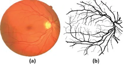
Unlike traditionally used biometric systems, vascular pattern of human retina is the most reliable and stable source for biometrics. It is not easy to forge it as it lies at back end of human eye and is not directly accessible [2]. There is always a confusion regarding iris and retina as they both belong to human eye but their functions and patterns are completely different. Iris is the colored region between the pupil and sclera whereas retina is located at back region of eye. The foundation of retinal recognition is the pattern of blood vessels present in human retina [2]. Figure 1 shows the human retinal and vascular pattern extracted from digital retinal image.
Figure 1 a. Digital Image of Human Retina b. Vascular Pattern
importance in recent years. Comparison of various biometric traits shows that iris is very attractive biometric because of its uniqueness, stability, and non-intrusiveness.
x Pattern of retina’s blood vessels rarely changes during people's lives.
x In addition, retina has not contact with environment unlike other biometrics such as fingerprint; therefore it is protected from external changes.
x Moreover people have not access to their retina and hence they could not deceive identification system. x Small size of the feature vector is another advantage of retina to the other biometrics; this property
leads to faster identification and authentication than other biometrics.
Here, we propose a comparative study of automated system for person identification based on microvascular network of human retina.
The proposed algorithm consists of four stages; i.e. pre-processing, segmentation, feature extraction and finally the matching process. In pre-processing, it removes the noise from image and improves the quality of the image, in segmentation, then it extracts /segments the vascular pattern from input retinal image. Third stage extracts all possible feature points and represents each feature point with a feature vector. The proposed system matches the template feature vectors and input image feature vector using any suitable distance measure. We will evaluate the performance of the proposed system for various segmentation algorithms for blood vessel segmentation from the color retina images and will propose a suitable segmentation method. Also various features of the segmented vascular pattern will be extracted and evaluated for performance.
The rest of the paper is organized as follows: In section II we have presented a study of various techniques for noise removal. Section III deals with the related work of segmentation of blood vessels from digital retina image. In section IV we have studied various methods for feature point extraction from segmented vascular pattern. Section V provides related work considering various methods for image matching.
II. RELATED WORK FOR NOISE REMOVAL
Image noise is the random variation of brightness or color information in images produced by the sensor and circuitry of a scanner or digital camera. Image noise can also originate in film grain and in the unavoidable shot noise of an ideal photon detector [3].Image noise is generally regarded as an undesirable by-product of image capture. Although these unwanted fluctuations became known as "noise" by analogy with unwanted sound they are inaudible and such as dithering. The types of Noise are Amplifier noise (Gaussian noise), Salt-and-pepper noise, Shot noise (Poisson noise), Speckle noise.
Image noising is very important task in image processing for the analysis of images. Ample image de-noising algorithms are available, but the best one should remove the noise completely from the image, while preserving the details. Broadly speaking, De-noising filters can be classified in the following categories:
A. Mean Filter
Mean filter is an averaging linear filter. Here the filter computes the average value of the corrupted image in a predefined area. Then the center pixel intensity value is replaced by that average value. This process is repeated for all pixel values in the image.
The algorithm proposed in [4] considers first order neighborhood pixels for detecting the noisy pixel and mean filter is considered. Color images are also de-noised by extracting the R, G and B pixels from noisy image and then they are de-noised separately and then merged together to again form the color image. The algorithm is compared with all other standard and well known algorithms and found to have good noise removal capabilities at high densities.
In order to improve the speed of mean filter algorithm and to reduce the redundancy operation, an efficient algorithm is presented in [5]. At first, an assistant array is built up to store the pixels' value summation of each column. Then, according to the relationship between adjacent windows when filter window is siding along the rows, a recursive formula for calculating the midpoint mean of a new window is schemed out. Meanwhile, the recursive formula for updating assistant array items is obtained based on the relationship between adjacent rows' pixels in the changing row process of midpoint of window. On the basis of these two recursion formulas, a high efficient mean filter algorithm is achieved.
1.In general the mean filter acts as a lowpass frequency filter and, therefore, reduces the spatial intensity derivatives present in the image.
2. Mean filtering is a simple, intuitive and easy to implement method ofsmoothingimages,i.e.reducing the amount of intensity variation between one pixel and the next. It is often used to reduce noise in images.
Limitations:
1. A single pixel with a very unrepresentative value can significantly affect the mean value of all the pixels in its neighborhood.
2. When the filter neighborhood straddles an edge, the filter will interpolate new values for pixels on the edge and so will blur that edge. This may be a problem if sharp edges are required in the output.
B. Median Filter
The Median filter is a nonlinear digital filtering technique, often used to remove noise. Such noise reduction is a typical preprocessing step to improve the results of later processing (for example, edge detection on an image). Median filtering is very widely used in digital image processing because under certain conditions, it preserves edges whilst removing noise. The main idea of the median filter is to run through the signal entry by entry, replacing each entry with the median of neighboring entries. Note that if the window has an odd number of entries, then the median is simple to define: it is just the middle value after all the entries in the window are sorted numerically. For an even number of entries, there is more than one possible median. The median filter is a robust filter. Median filters are widely used as smoothers for image processing, as well as in signal processing and time series processing. A major advantage of the median filter over linear filters is that the median filter can eliminate the effect of input noise values with extremely large magnitudes.
Authors in [6] proposes a novel decision-based median filter that replaces each corrupted pixel with the median value of their k-nearest noise-free pixels. Advantages of the median filter using k-nearest noise-free pixels instead of k-nearest pixels are two facets: first, it guarantees that pixels after being restored must be noise-free, because the median filter operator is executed on noise-free pixels; second, the median filter using k-nearest noise-free pixels adaptively adjusts its window size for each pixel such that the number of noise-free pixels locating in the window increases up to k. To realize it, the median filter using k-nearest noise-free pixels firstly detects noise-free pixels in an image, then replaces each corrupted pixel with the median value of their knearest noise-free pixels.
[7] Presents the removal of high density salt and pepper noise in gray scale Images using fuzzy based median filter (FBMF) algorithm. FBMF replaces the noisy pixel by median value when O's, 255's and other pixel values are present in the chosen window and when all pixel values are either O's or 255's, or combination of these, then the noise pixel is replaced by fuzzy membership value of a selected window.
Advantages:By calculating the median value of a neighborhood rather than the mean filter, the median filter has two main advantages over the mean filter:
1. The median is a more robust average than the mean and so a single very unrepresentative pixel in a neighborhood will not affect the median value significantly.
2. Since the median value must actually be the value of one of the pixels in the neighborhood, the median filter does not create new unrealistic pixel values when the filter straddles an edge. For this reason the median filter is much better at preserving sharp edges than the mean filter.
Limitations:
1. One of the major problems with the median filter is that it is relatively expensive and complex to compute.
2. To find the median it is necessary to sort all the values in the neighborhood into numerical order and this is relatively slow, even with fast sorting algorithms such asquicksort.
C. Wiener Filter
from a different angle. One is assumed to have knowledge of the spectral properties of the original signal and the noise, and one seeks the LTI filter whose output would come as close to the original signal as possible.
A new widely linear noise-reduction Wiener filter based on the variance and pseudo-variance of the short-time Fourier transform coefficients of speech signals is proposed in [8]. Authors show that this
new noise-reduction filter has many interesting properties, including but not limited to: 1) it causes less speech distortion as compared to the classical noise-reduction Wiener filter; 2) its minimum mean squared error (MSE) is smaller than that of the classical Wiener filter; 3) it can increase the subband signal-to-noise ratio (SNR), while the classical Wiener filter has no effect on the subband SNR for any given signal frame and subband.
An adaptive wiener filter design method is presented in [9] using quality based hybrid algorithms. The proposed method restore degraded image using Wiener filter with Gradient based smoothing and Median filter with an adaption of Bilateral filter for edge preserving and Guided filter for reducing staircase effect and gradient reversal. Using this method, clear images are obtained from degraded images without increasing noise, ringing artifacts and halo effect.
Advantages
1. Begins to exploit signal. 2. Controls output error. 3. Straightforward to design. Limitations
1. Results often too blurred. 2. Spatially invariant.
D. Wavelet Transform
In several applications, it might be essential to analyze a given signal. The structure and features of the given signal may be better understood by transforming the data into another domain. There are several transforms available like the Fourier transform, Hilbert transform, wavelet transform, etc. The Fourier transform is probably the most popular transform. However the Fourier transform gives only the frequency-amplitude representation of the raw signal. The time information is lost. So we cannot use the Fourier transform in applications which require both time as well as frequency information at the same time. The Short Time Fourier Transform (STFT) was developed to overcome this drawback.
According to the characteristic of river sediment image, the two-dimensional image data is transformed into one-dimensional array data from horizontal, vertical, + diagonal line, - the diagonal line four directions, by using different threshold value on the different wavelet decomposition high frequency coefficient and enhancing the processed coefficient, de-noise image is obtained after inversing wavelet transform and calculating on four two-dimensional pictures by unequal coefficient. The results indicate the proposed algorithm in [10] is effective both in reserving the edge and in removing noise.
Authors in [11] have proposed an improved wavelets shrinkage
method that uses different parameters across different wavelet scales to denoise the Besov images and it outperforms the existing á fixed algorithms. It is obvious that different parts of an image or different images have different structures and properties, so we assume that these differences could be described by means of their distinct smoothness properties. In experiments, authors used three simple mathematic functions to denote the relationship between the smoothness index and wavelet scale and found that the linear function achieved a better denoising effect.
Advantages:
Limitations: Wavelet analysis does not provide as accurate information about the measured surface as the Fourier transform.
E. Order Statistics Filter
Order-Statistics filters are non-linear filters whose response depends on the ordering of pixels encompassed by the filter area. When the center value of the pixel in the image area is replaced by 100th percentile, the filter is called max-filter. On the other hand, if the same pixel value is replaced by 0th percentile, the filter is termed as minimum filter.
Directional order statistics filtering approaches are performance limited owing to their implementation with larger window sizes (to a maximum size of 9x9) and with the need of iterative operation to filter the residual noise for highly corrupted images. Authors in [12] presents a methodology to improve the performance of directional order statistics filters for suppression of salt-and-pepper noise. The proposed filtering approach involves eight directions to identify boundary and no boundary pixels. If the non-boundary pixels are noise free, they are left unprocessed otherwise; updated by order statistics parameters of the local window.
A theoretical study and experimental validation on a binaural hearing aid setup of this standard SDW-MWF implementation is presented in [13], where the effect of estimation errors in the second-order statistics is analyzed. In this case, and for a single target speech source, the standard SDW-MWF implementation is found not to behave as predicted theoretically. Second, two recently introduced alternative filters, namely the rank-one SDW-MWF and the spatial prediction SDW-MWF, are also studied in the presence of estimation errors in the second-order statistics. These filters implicitly assume a single target speech source, but still only rely on the speech and noise correlation matrices. It is proven theoretically and illustrated through experiments that these alternative SDW-MWF implementations behave close to the theoretical optimum, and hence outperform the standard SDW-MWF implementation.
Advantages:
1. This filter is more robust because a single pixel in neighbourhood never affects the median value. 2. This filter is very effective in removing gaussian and uniform noise while preserving image detail.
3. It is useful in situations such as which multiple types of noises, for eg, combination of salt and pepper and Gaussian noise.
Limitations:
1. It is more expensive and complex to execute.
2.More time is spent calculating the median of each window.
3.This filter is unable to find out the black colored or dark colored pixels in an image. 4.The Min filter can't locate the light or white colored points in an image.
5.It works well only for randomly distributed noise. 6.Used only where noise density is high.
7.These are not used for low noise density images.
III. RELATED WORK OF SEGMENTATION
A complete review of existing methods for retinal blood vessel segmentation can be referenced at [14]. The state-of-the art methods for segmentation of vascular pattern from digital retina images can be classified as follows-
A. Tracking Based Segmentation
The first proposed methods [15-20] are based on tracking techniques to trace the vessels starting from some seed points (which are identified either manually or automatically) and following the vessel center line guided by local information. Advantage:
1. An advantage of tracking based methods is its efficiency since only pixels close to the initial positions are examined and evaluated.
Limitations:
1. Sophisticated methods have to be used to deal with a bifurcation or crossover point due to the complexity of the intensity profile at these regions.
2. Since vessel branching or crossover points are not well modeled, this approach often tends to terminate at these points and this leads to the incompletion in the detection result.
B. Segmentation Using Matched Filter
Another approach is the use of matched filter concept [21] for blood vessel enhancement. This approach is based on the assumption that the intensity profile along the cross-section of a vessel has the shape of a Gaussian. A set of 12 Gaussian shaped filters is used to match the vessel at different directions. For each pixel, the maximum response is retained. Different variants [22-25] have been proposed to improve the performance of the original matched filter response, ranging from the use of a threshold probing technique [22], ‘double-sided’ thresholding [24], or the first order derivative of Gaussian image [25] to provide a better thresholding method and reduce false vessel responses at non vessel structures.
Advantages:
1. Among the various retinal vessel extraction methods, the matched filter (MF) method is a representative one and it has advantages of simplicity and effectiveness.
Limitation:
1. A limitation of matched filter based methods is its naive assumption that the cross-sectional intensity profile of a vessel follows the shape of a Gaussian, which is not always the case (for example, in the presence of central reflex).
2. Moreover, the assumptions that the vessels are piece-wise linear and have constant width along a certain distance make it difficult to adapt to the variations in vessel width and orientations.
C. Segmentation Using Supervised Methods
Recently, there is an emerging trend of using supervised methods [26-31] to perform this segmentation problem. Methods belonging to this category often follow the same framework where each image pixel is represented by a feature vector, which is computed using local or global information of the image. A supervised classifier (ranging from the use of artificial neural networks [26], kNN [27], support vector machines [26], Bayesian classifier [7], etc.) is used to train the model and classify each image pixel as vessel or background.
Advantages: Supervised methods have been shown to provide higher accuracy than other unsupervised methods.
Limitations:
1. They require the ground truth segmentations for training the models which is not always available in real life applications.
2. Furthermore, these methods often require the ‘re-training’ process when performing on the new set of images to achieve optimal performance. In other words, the performance of these methods is highly dependent on the training dataset.
IV. FEATURE EXTRACTION RELATED WORK
of neighboring image segments for each pixel in a retinal image, a multi-scale vessel filter is defined which combines the responses of tubular structures and the responses of bifurcations and crossovers [32].
Azzopardi and Petco, proposed a method for automatic detection of vascular bifurcations in segmented retinal images using trainable COSFIRE filters. The vascular tree observed in a retinal fundus image can provide clues for cardiovascular diseases. Its analysis requires the identification of vessel bifurcations and crossovers. Authors use a set of trainable key point detectors that are called as Combination of Shifted Filter Responses or COSFIRE filters to automatically detect vascular bifurcations in segmented retinal images [33].
Bhuiyan et. al., introduced a method to detect and classify the vascular bifurcation, branch and crossover points (landmarks) based on the vessel geometrical features. They utilize the vessel's centerline and width information to detect and classify these landmarks, which can be used for image matching in medical diagnosis and biometric security applications. The geometrical properties of the blood vessels passing through the potential landmarks are obtained. Perceptual grouping and Support Vector Machine (SVM) are used to classify the landmarks into the vascular bifurcations, branches and crossovers [34].
Ardizzone et. al., presents an effective algorithm for automated extraction of the vascular tree in retinal images, including bifurcations, crossovers and endpoints detection. Correct identification of these features in the ocular fundus helps the diagnosis of important systematic diseases, such as diabetes and hypertension. The pre-processing consists in artifacts removal based on anisotropic diffusion filter. Then a matched filter is applied to enhance blood vessels. The filter uses a full adaptive kernel because each vessel has a proper orientation and thickness. The kernel of the filter needs to be rotated for all possible directions. As a consequence, a suitable kernel has been designed to match this requirement. The maximum filter response is retained for each pixel and the contrast is increased again to make easier the next step. A threshold operator is applied to obtain a binary image of the vascular tree [35].
VI. CONCLUSIONS
In the paper, we propose a review of methods for recognizing retinal images based on microvascular networks. The proposed method is divided into four steps such as noise removal, segmentation, feature extraction and classification. A detailed study of segmentation and noise removal along with the related work done in feature extraction such as bi-furcation angle detection is provided.
ACKNOWLEDGMENT
The authors would like to welcome the reviewers for their critical and constructive comments and suggestions. The authors would like to thank the Authorities of National Institute of Electronics and InformatioTechnology, Dr. Babasaheb Ambedkar Marathwada University, Aurangabad for providing the infrastructure to carry out the research.
REFERENCES
[1] S. Prabhakar, S. Pankanti, and A. K. Jain, “Biometric recognition: Security and privacy concerns”, IEEE Security and Privacy, vol. 1, No. 2, pp. 33-42, 2003.
[2] Ravi Das, “Retinal recognition Biometric technology in practice”, Keesing Journal of Documents & Identity, vol. 22, pp. 11-14, 2007. [3] Charles Boncelet (2005).”Image Noise Models”. In Alan C. Bovik. Handbook of Image and Video Processing.
[4] Priyanka Shrivastava, Uday Pratap Singh, “Noise Removal using First Order Neighborhood Mean Filter”, 978-1-4799-3064-7/14/$31. 00, 2014 IEEE.
[5] Fu Bin, Xiong XinyanI, Sun Guozhen, “An efficient Mean Filter Algorithm”,, Proceedings of the 2011 IEEEIICME International Conference on Complex Medical Engineering May 22 - 25, Harbin, China.
[6] Yi Hong, Sam Kwong, Hanli Wang, “Decision-Based Median Filter Using K-Nearest Noise-Free Pixels”, ICASSP 2009, 978-1-4244-2354-5/09/$25.00 ,2009 IEEE.
[7] B. Sravani, M. V. N ageswara Rao, “Removing of high density salt and pepper noise using Fuzzy Median Filter”, 978-1-4799-5958-7114/$31.00 , 2014 IEEE.
[8] Jacob Benesty, Jingdong Chen, and Yiteng (Arden) Huang, “Study of the Widely Linear Wiener Filter for Noise Reduction”, ICASSP 2010, 978-1-4244-4296-6/10/$25.00, 2010 IEEE.
[9] SmitTrambadia, Paresh Dholakia, “Design and Analysis of an Image Restoration Using Wiener Filter with a Quality Based Hybrid Algorithms”, IEEE Sponsored 2nd International Conference on Electronics and Communication System (ICECS 2015), 978-1- 4788-7225 -8/15/$31.00,2015 IEEE.
[10] Peng Xuange,Xiao Ying, Zhu Bing, Liu Yong, “River Sediment image denoising algorithm based on multidirectional 1D wavelet Transform”, 2009 International Conference on Information Management, Innovation Management and Industrial Engineering, 978-0-7695-3876-1/09 $25.00, 2009 IEEE DOI 10.1109/ICIII.2009.215
[11] Hong Yang,Yiding Wang, “An Improved Method of Wavelets Basis Image Denoising Using Besov Norm Regularization”, Fourth International Conference on Image and Graphics, 0-7695-2929-1/07 $25.00, 2007 IEEE
[13] Awanish Kumar Shukla,R. L. Verma, Mohd. Sanawer Alam, Vikrant Bhateja, “Directional Order Statistics Filtering for Suppression of Salt and Pepper Noise”, 2014 International Conference on Signal Processing and Integrated Networks (SPIN). 978-1-4799-2866-8/14/$31.00, 2014 IEEE.
[14] Bram Cornelis, Marc Moonen, and Jan Wouters, “Performance Analysis of Multichannel Wiener Filter-Based Noise Reduction in Hearing Aids Under Second Order Statistics Estimation Errors”, IEEE Transactions On Audio, Speech, And Language Processing, VOL. 19, NO. 5, JULY 2011
[15] 14.Fraz,P.Remagnino,A.Hoppe,B.Uyyanonvara,A.Rudnicka,C.Owen,Barman, “Blood vessel segmentation methodologies in retinal images—a survey” , Computer Methods and Programs in Biomedicine.
[16] Liu, Y.Sun, “Recursive tracking of vascular networks in angiograms based on the detection-deletion scheme”, IEEE Transactions on Medical Imaging12 (2) (1993) 334–341.
[17] L.Zhou, M.S.Rzeszotarski, L.J.Singerman, J.M.Chokreff, “The detection and quantification of retinopathy using digital angiograms”, IEEE Transactions on Medical Imaging 13(4)(1994)619–626.
[18] O.Chutatape,L.Zheng,S.Krishnan, “Retinal blood vessel detection and tracking by matched Gaussian and kalman filters”, Engineering in Medicine and Biology Society, 1998., Proceedings of the 20th Annual International Conference of the IEEE,vol.6,
IEEE,1998,pp.3144–3149.
[19] A.Tolias, S.M.Panas, “A fuzzy vessel tracking algorithm for retinal images based on fuzzy clustering”, IEEE Transactions on MedicalImaging17 (2) (1998) 263–273.
[20] A.Can, H.Shen, J.N.Turner, H.L.Tanenbaum, B.Roysam, “Rapid automated tracing and feature extraction from retinal fundus images using direct exploratory algorithms”, IEEE Transactions on Information Technology in Biomedicine 3(2) (1999)125–138.
[21] Y. Yin, M. Adel, S. Bourennane, "Retinal vessel segmentation using a probabilistic tracking method", Pattern Recognition 45 (2012) 1235–1244.
[22] S. Chaudhuri, S. Chatterjee, N. Katz, M. Nelson, M. Goldbaum," Detection of blood vessels in retinal images using two-dimensional matched filters", IEEE Transactions on Medical Imaging 8 (3) (1989) 263–269.
[23] A. Hoover, V. Kouznetsova, M. Goldbaum," Locating blood vessels in retinal images by piecewise threshold probing of a matched filter response", IEEE Transactions on Medical Imaging 19 (3) (2000) 203–210.
[24] M. Al-Rawi, M. Qutaishat, M. Arrar," An improved matched filter for blood vessel detection of digital retinal images", Computers in Biology and Medicine 37 (2) (2007) 262–267.
[25] L. Zhang, Q. Li, J. You, D. Zhang," A modified matched filter with double-sided thresholding for screening proliferative diabetic retinopathy", IEEE Transac- tions on Information Technology in Biomedicine 13 (4) (2009) 528–534.
[26] B. Zhang, L. Zhang, F. Karray, "Retinal vessel extraction by matched filter with first-order derivative of gaussian", Computers in Biology and Medicine 40 (4) (2010) 438–445.
[27] C. Sinthanayothin, J.F. Boyce, H.L. Cook, T.H. Williamson, "Automated localisation of the optic disc, fovea, and retinal blood vessels from digital colour fundus images", British Journal of Ophthalmology 83 (8) (1999) 902.
[28] 27.Niemeijer,J.Staal,B.vanGinneken,M.Loog,M.D.Abramoff,"Comparative study of retinal vessel segmentation methods on a new publicly available database", in: Proceedings of SPIE,vol.5370,2004,pp.648–656.
[29] 28. M.Sofka,C.V.Stewart, "Retinal vessel center line extraction using multiscale matched filters, confidence and edge measures" ,IEEE Transactions on Medical Imaging 25(12)(2006)1531–1546.
[30] 29.C.A.Lupascu, D.Tegolo,E.Trucco, "Fabc:retinal vessel segmentation using adaboost", IEEE Transactions on Information Technology in Biomedicine 14(5) (2010) 1267–1274.
[31] 30.D.Marin,A.Aquino,M.E.Arias, J.M.Bravo,"A new supervised method for blood vessel segmentation in retinal images by using gray-level and moment invariants-based features", IEEE Transactions on Medical Imaging 30(1)(2011)146–158.
[32] 31. X.You,Q.Peng,Y.Yuan,Y.Cheung,J.Lei, "Segmentation of retinal blood vessels using the radial projection and semi-supervised approach", Pattern Recognition 44(2011)2314–2324.
[33] Li Guodong, Xiang Dehui, Yang Fei, Wan Xiaonan, Yang Xin, Tian Jie, "Automatic detection of retinal vascular bifurcations and crossovers based on isotropy and anisotropy ", Proc. SPIE 8669, Medical Imaging 2013: Image Processing, 86691T (March 13, 2013); Doi:10.1117/12.2006710;
[34] 33.Azzopardi George, and Petkov Nicolai, "Detection of Retinal Vascular Bifurcations by Trainable V4-Like Filters", A. Berciano et al. (Eds.): CAIP 2011, LNCS 6854, pp. 451–459, 2011, Springer-Verlag Berlin Heidelberg 2011.
[35] 34. Bhuiyan A., Nath B., Chua J., Ramamohanarao K., "Automatic Detection of Vascular Bifurcations and Crossovers from Color Retinal Fundus Images" SignalImage Technologies and Internet-Based System, 2007. SITIS '07. Third International IEEE Conference on Signal-Image Technologies and Internet-Based System, pp: 711 – 718, 2007.