RESEARCH
The kinase LRRK2 is differently expressed
in chronic rhinosinusitis with and without nasal
polyps
Yue Ma
1,2, Chunquan Zheng
1*and Le Shi
1,2Abstract
Background: Chronic rhinosinusitis (CRS), commonly divided into CRS with nasal polyps (CRSwNP) and without nasal polyps (CRSsNP) is an inflammatory disease which mechanism remain unclear. Leucine-rich repeat kinase 2 (LRRK2) has been proved to be a negative regulator of inflammation response while its role in pathogenesis of CRS has yet to be revealed. This research study was designed to investigate the relationship between the expression level and biologic role of LRRK2 in CRS.
Methods: Expression of LRRK2 mRNA and noncoding repressor of NFAT (NRON) were examined by qRT-PCR. Protein levels of LRRK2 were performed by western blot and immunohistochemistry. Nuclear factor of activated T cells (NFAT) nuclear translocation was analyzed by immunohistochemistry. Additionally, LRRK2 mRNA and NRON expression in response to specific inflammatory stimulation was measured in human nasal epithelia cells (HNECs).
Results: The expression of LRRK2 was increased in CRSsNP patients (p < 0.05) and positively correlated with the expression levels of CD3 and Charot-Leyden crystal. Meanwhile, the NRON expression level is much lower in CRSsNP patients compared to both the control group and CRSwNP group (p < 0.05). Marked enhanced NFAT nuclear localiza-tion was observed in CRSwNP groups compared with the CRSsNP and control group (p < 0.0001). And the over-expression of LRRK2 was significantly regulated by lipopolysaccharide (LPS) in HNECs (p < 0.05). Moreover, IL-17A can increase LRRK2 expression and suppress NRON expression in vitro and dexamethasone can rescue the NRON inhibition.
Conclusion: LRRK2 and NRON may play different role in CRSsNP and CRSwNP. The molecular mechanisms identified here may aid in the design of novel therapeutic strategies to improve clinical outcomes.
Keywords: LRRK2, NRON, Long non-coding RNA, Chronic rhinosinusitis, Pro-inflammatory cytokine
© The Author(s) 2018. This article is distributed under the terms of the Creative Commons Attribution 4.0 International License (http://creat iveco mmons .org/licen ses/by/4.0/), which permits unrestricted use, distribution, and reproduction in any medium, provided you give appropriate credit to the original author(s) and the source, provide a link to the Creative Commons license, and indicate if changes were made. The Creative Commons Public Domain Dedication waiver (http://creat iveco mmons .org/ publi cdoma in/zero/1.0/) applies to the data made available in this article, unless otherwise stated.
Background
In general, chronic rhinosinusitis (CRS) is an inflam-matory disease that is composed of CRS with nasal pol-yps (CRSwNP) and without nasal polpol-yps (CRSsNP). Although their pathogenesis is not yet clear, it is com-monly acknowledged that the two types of CRS possess distinct inflammation and remodeling patterns [1, 2]. CRSsNP is characterized by Th1-biased inflammation
and an elevated expression of transforming growth fac-tor (TGF-β1), while CRSwNP is characterized by a Th2-biased inflammation [3]. Meanwhile, the dysregulation of innate immunity has been regarded as the key point for the initiation and perpetuation of inflammatory responses in CRS patients [4]. The toll-like receptor sign-aling pathway induced by lipopolysaccharide (LPS)has been reported as one of critical factors in the pathogen-esis of CRS, but the mechanism remains unclear. Thus, there is an urgent need for a new biomarker that can fur-ther explain this mechanism.
Leucine-rich repeat kinase 2 (LRRK2), also known as Dardarin, is a large complex protein that contains several
Open Access
*Correspondence: zheng_ent96@163.com
1 Department of Otolaryngology-Head and Neck Surgery, Eye Ear Nose and Throat Hospital, Fudan University, 83 Fenyang Road, Xuhui District, Shanghai 200031, People’s Republic of China
domains [5–7]. Accumulating evidence has revealed that LRRK2 is involved in regulating inflammatory processes [8–10]. Participation in the signaling of IFN-γ [11, 12], enhancement of NF-kB-dependent transcription, and interference in reactive oxygen species (ROS) produc-tion all indicate that LRRK2 may play a pivotal role in the anti-microbial process [11].
It has already been identified that LRRK2 negatively regulates NFAT by interacting with NRON, a known repressor of NFAT. Long non-coding RNA (lncRNA) NRON and 11 additional proteins, including LRRK2, constitute a protein-RNA complex which could directly inhibit the translocation of NFAT to the nucleus [13, 14] such that cell activation and the proceeding immune reactivity are suppressed. For example, enhanced nuclear location of NFAT was associated with more severe colitis in LRRK2-deficient mice [14].
Therefore, LRRK2 is a negative regulator of this inflam-mation response. These findings implied that the changes in LRRK2 expression levels resulting from extrinsic sig-nals may play a vital role in the regulation of immune responses. However, the role of LRRK2 in the patho-genesis of CRS has yet to be revealed. Our present study was designed to investigate the relationship between the expression level and biologic role of LRRK2 in CRS.
Methods
Subjects
The subjects (n = 74) were all recruited from the Depart-ment of Otolaryngology-Head and Neck Surgery, Eye, Ear, Nose, and Throat Hospital, Fudan University. All were CRS patients who had not taken oral and/or topi-cal corticosteroids or any other sinonasal medications for at least 1 month prior to the study and were diagnosed
based on previously published criteria [15]. NPs were obtained from CRSwNP patients (n = 34), and nasal mucosa of middle turbinates were from CRSsNP patients (n = 23). At the same time, the nasal mucosa of inferior turbinates were collected from those patients (n = 17) who had no clinical symptoms or radiographic evi-dence of CRS and who underwent a septoturbinoplasty. Patients responding positively to a skin-prick test were diagnosed with an allergy. All of the patients’ clinical data are presented in Table 1. The exclusion criteria for the study group ran as follow: age < 18 or > 80 years, a diag-nosis of cystic fibrosis, Churg-Strauss syndrome, immu-nodeficiency, or autoimmune disease.
This study obtained permission from the local ethical committee of the Otolaryngology-Head and Neck Sur-gery, Eye, Ear, Nose, and Throat Hospital, Fudan Univer-sity, and informed consent was signed by every subject.
RNA extraction and real‑time polymerase chain reaction
Total RNA was extracted using TRIzol reagent (Invit-rogen) according to the manufacturer’s instructions. Then, PrimeScript RT master mix (Takara) was used to synthesize complementary DNA (cDNA). An ABI 7900 Sequence Detection System (ABI) was used to perform qRT-PCR with SYBR Green chemistry. Table 2 showed the primers (Sangon Biotech). The expression of each gene was calculated using the comparative threshold cycle (2−ΔΔCT) method.
Immunohistochemistry (IHC) staining
Immunohistochemistry was performed following the protocol for the streptavidin-biotin complex (SABC) kit (Weiao Biological Technology). The sections were incubated with primary antibody (polyclonal rabbit
Table 1 Characteristics of included subjects
Control subjects Patients with CRSsNP Patients with CRSwNP
No. of patients 17 23 34
Sex, male/female 12/5 12/11 20/14
Age (years), mean (SD) 36 (8) 52 (17) 37 (13)
Atopy, no. 0 4 6
Asthma, no. 0 0 2
Aspirin intolerance, no. 0 0 0
Smoking, no. 0 10 16
Operation history, no. 0 0 2
Methodologies used
Tissue IHC 10 16 25
Tissue mRNA 15 21 30
anti-human LRRK2; Abcam; 1:200 dilution; polyclonal goat anti-human NFAT1; Abcam; 1:100 dilution) at 4 °C overnight. 3′3-Diaminobenzidine (DAB) was used for the final visualization. To analyze LRRK2 expression, two pathologists scored the results independently according to the immunostaining intensity scale, which ran as fol-lows: 0 = absent; 1 = mild; 2 = moderate; and 3 = marked. The numbers of immuno-positive cells within the sam-ples were also counted in at least five random areas at a 400× magnification, and at the same time, the percent-age of immuno-positive cells was scored according to the standard scale as follows: 0 (0–9%); 1 (10–25%); 2 (26–50%); 3 (51–75%); and 4 (> 76%). Multiplication of the two abovementioned scores provided the final score for each sample. The highest final score was 12 while the lowest was 0. To analyze NFAT nuclear translocation, the percentage of cells with NFAT1 nuclear staining in all immuno-positive cells were also counted in five random areas at a 400x magnification.
Western blots
Protein samples (30 μg) were separated by electrophore-sis using 10% sodium dodecyl sulfate polyacrylamide gels and then transferred to polyvinylidene difluoride (PVDF) membranes for incubation with anti-LRRK2 (Abcam; 1:1000) antibody. Image J (NIH) analysis and processing software was used to quantify data, which was expressed as densitometry units (DU). The expression of β-actin (Abcam; 1:1000) was regarded as an internal reference.
Cell culture and stimulation
Following a previously established protocol, HNECs (Human Nasal Epithelia Cells) isolated from the middle turbinates of CRSsNP patients were cultured [16]. Briefly, nasal specimens were immersed in DMEM/F12 media (Hyclone) containing 1.4 mg/ml protease K and 0.1 mg/ ml DNase for a 1.5 h incubation at 37 °C. Next, all cells were collected and immersed in DMEM/F12 (Hyclone)
containing 1% ITS for 2 h at 37 °C before being cultured in BEGM medium (Lonza).
When 80–90% confluence was reached, fresh media without hydrocortisone was added in the presence of the following stimulators or control PBS for 12 h: the recombinant cytokines human IFN-γ (100 ng/mL), IL-4 (100 ng/mL), IL-13 (100 ng/mL), IL-17A (100 ng/mL), TGF-β (10 ng/mL), and IL-1α (100 ng/mL; all purchased from Peprotech); the TLR agonists LPS (500 ng/mL, from Escherichia coli serotype 0111: B4; purchased from Sigma) and the glucocorticoid dexamethasone (Sigma; 10 μg/mL). After stimulation, HNECs were collected for qRT-PCR analysis.
Statistical analysis
Statistical analyses were performed by SPSS v22.0 soft-ware (IBM Corporation). The data were presented as medians and interquartile ranges. Tests for Gaussian dis-tribution were performed by Kolmogorov–Smirnov test. Differences between groups were evaluated by one-way analysis of variance and either by the two independent sample t test or the Mann–Whitney U test. The corre-lation analysis was performed to assess the correcorre-lation between two groups by Spearman’s rank correlation.
p < 0.05 was regarded as statistically significant.
Results
NRON and LRRK2 mRNA levels in nasal tissues
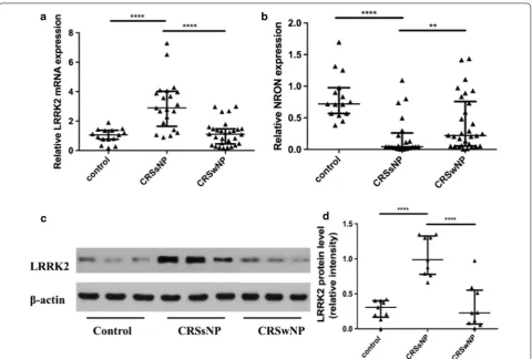
As showed in Fig. 1a, significant upregulation of LRRK2 mRNA levels was found in CRSsNP groups but not in CRSwNP groups compared with the control group (p < 0.0001), and NRON levels were significantly higher in the inferior turbinate than in the middle turbinate of CRSsNP groups (p < 0.0001) and in NPs (p < 0.01) (Fig. 1b).
LRRK2 protein levels in nasal tissues
To verify the results at the protein level, western blots were performed. Strong bands for LRRK2 were observed in CRSsNP groups, whereas weak bands were found in the CRSwNP group and in the control group (Fig. 1c). The LRRK2 protein level was significantly higher in the CRSsNP group than in the CRSwNP group and the con-trol group (Fig. 1d; p < 0.0001).
LRRK2 and NFAT immunoreactivity in nasal tissues
Immunohistochemistry was performed to further con-firm the results pertaining to the protein levels of LRRK2 in the various samples. As depicted by immunohisto-chemistry staining (Fig. 2a–c), LRRK2 was significantly overexpressed in the CRSsNP group compared with the control group and CRSwNP group. The cytoplasmic or nuclear staining of LRRK2 was mainly located at the
Table 2 Primers
Name Sense Antisense
GAPDH CAA GGT CAT CCA TGA CAA CTT
TG GTC CAC CAC CCT GTT GCT GTAG LRRK2 GGA TGT TGG TGA TGG AGT T GGC TGA GTG GAG GTA TCT NRON AAC AAC CCA GCA AGG GAA GT
nasal epithelium and in submucosal inflammatory cells. In comparison with control groups and NPs, quantita-tive analysis of LRRK2 revealed an obvious elevation in immuno-labeling of LRRK2 in the CRSsNP group (Fig. 2d; p < 0.01). NFAT nuclear translocation was also detected by immunohistochemistry (Fig. 2e–g). Marked enhanced NFAT nuclear localization was observed in CRSwNP groups compared with the CRSsNP group and control group (Fig. 2h; p < 0.0001).
Detection of LRRK2 producing cells in nasal mucosa
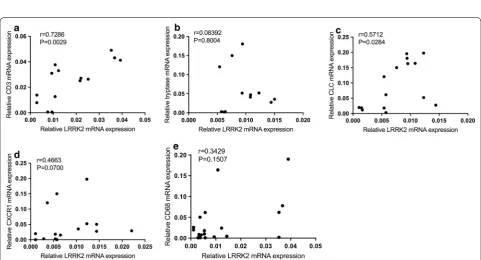
These results revealed that LRRK2+ cells were highly accumulated in the submucosal region of CRSsNP tis-sues and that the mRNA and protein expression levels of LRRK2 were also significantly higher in CRSsNP tissues (Fig. 2). Next, the expression of LRRK2 and the markers of inflammatory cells in CRSsNP were assessed by qRT-PCR and the relationship between these markers were analyzed by Spearman’s rank correlation. The results demonstrated that the mRNA expression levels of LRRK2 in CRSsNP tissue were significantly and positively related to the expression of CD3 (r = 0.7286; p= 0.0029),
Charot–Leyden crystal (CLC; r = 0.5712; p= 0.0284), CD68 (r = 0.146; p < 0.05), but not CXCR1, tryptase and CD68, which indicated that the expression of LRRK2 may derive from T cells, eosinophils (Fig. 3).
LPS and pro‑inflammatory cytokines differentially regulate LRRK2 and NRON expression in human nasal epithelial cells (HNECs) in vitro
To further expose the role and mechanism of the LRRK2 signaling pathway in the nasal mucosa of CRS, the effects of TLR activation and pro-inflammatory cytokine stimu-lation were examined in cultured human nasal epithelial cells. As presented in Fig. 4a, mRNA expression levels of LRRK2 were significantly increased after stimulating the HNECs with LPS, the TLR agonists (p < 0.001). Mean-while, stimulation with IL-17A increased very low LRRK2 mRNA expression (p < 0.01) compared with stimulation by the other pro-inflammatory cytokines, including IFN-γ, IL-4, IL-13, IL-1α, and TGF-β which also increased LRRK2 expression (p < 0.01). In contrast, NRON levels were significantly inhibited by stimulation with IL-17A (p < 0.001; Fig. 4b).
Fig. 1 The expression of LRRK2 and NRON in nasal tissues. a LRRK2 mRNA levels was upregulation in CRSsNP groups compared with CRSwNP groups and the control group; b NRON expression was significantly reduced in CRSsNP patients; c representative western blot results of LRRK2; d
Glucocorticoid treatment increases LRRK2 expression in vitro
Because glucocorticoid treatment is recommended as one of the primary treatment of choice for patients with
CRS, the effect of glucocorticoids on LRRK2 mRNA and NRON expression in HNECs was examined. After incu-bating cells with dexamethasone, LRRK2 mRNA and NRON expression was more upregulated in the presence
Fig. 2 LRRK2 inhibits NFAT1 nuclear translocation. (a–c; ×400 magnification) LRRK2 expression and distribution in control, CRSsNP and CRSwNP groups; d LRRK2 was significantly overexpressed in the CRSsNP group compared with the control group and CRSwNP group; (e–g; ×400 magnification) NFAT1 localization in control, CRSsNP and CRSwNP groups; blue arrows indicate cells with cytosolic NFAT1 staining, red arrows indicate cells with nuclear NFAT1 staining; the top right corner is an enlargement of immuno-positive cells; h the percentage of cells with NFAT1 nuclear staining was much higher in CRSwNP group compared with the control group and CRSsNP group. **p < 0.01, ****p < 0.0001
of pro-inflammatory cytokines (IL-17A, IL-4, IL-13, IL-1α; p < 0.05) than without dexamethasone stimula-tion (Fig. 4c, d). In contrast, NRON expression was lower when the cells were stimulated with IFN-γ plus dexa-methasone compared to only IFN-γ stimulation.
Discussion
This study demonstrated that the expression of LRRK2 was increased in patients suffering from CRSsNP (Figs. 1,
2), and the over-expression of LRRK2 was regulated by LPS in HNECs (Fig. 4). Moreover, LRRK2 expression in CRSsNP tissues was correlated with the expression levels of CD3, CLC, and CD68, which suggested that T cells, eosinophils, and macrophages may be the main LRRK2-producing cells in nasal mucosa (Fig. 3). Mean-while, NRON expression level is much lower in CRSsNP patients compared to both the control group and CRSwNP group (Fig. 1). Its expression can be strongly suppressed by stimulation with IL-17A in vitro, and dex-amethasone can rescue this phenomenon.
CRS, commonly divided into CRSwNP and CRSsNP, is characterized by chronic inflammation of the paranasal sinuses. Due to the poor awareness of the initiation and progression of CRS, the development of effective treat-ments for this disease remains stagnant [17]. LRRK2 can
be physically associated with NRON and this protein-RNA complex has been proven to be a NFAT suppres-sor in many inflammatory diseases such as inflammatory bowel disease [18], and LRRK2 deficiency can lead to a more serious inflammation response. In the results pre-sented herein, considerable data indicates that the up-regulation of LRRK2 expression in CRSsNP is obviously higher than in the CRSwNP group, and the percentage of cells with NFAT nuclear staining in CRSsNP is much lower in CRSwNP group (Fig. 2), which suggests that a lower expression of LRRK2 exhibits less of an inhibitory effect on NFAT. Moreover, this data supports that the idea that the defection of innate immunity may lead to a robust adaptive immune response, which triggers the generation of pro-inflammatory cytokines and the devel-opment of chronic inflammation [19].
NRON is a cytoplasmic lncRNA which has previously been reported to be highly expressed in human immune cells including macrophages, dendritic cells, and neutro-phils along with its vital role in T cell cytokine produc-tion. NRON acts as a scaffold to maintain the structure of a ribonucleoprotein complex which can retain the transcription factor NFAT in the cytoplasm [13], mean-ing that NRON plays a significant role in the immune response. However, in this study, the expression level of
Fig. 4 LRRK2 mRNA and NRON expression in cultured HNECs in response to pro-inflammatory cytokines, LPS and dexamethasone. a LRRK2 mRNA,
NRON is lower in CRSsNP group, which indicates that in the RNA-protein complex, LRRK2 may play a much more important role in the generation and the develop-ment of CRS than NRON.
Within the immune system, the expression of LRRK2 has been reported in lymphocytes, dendritic cells, and macrophages [11]. In this study, LRRK2-producing cells were identified in CRSsNP nasal mucosa. Considerable data had already shown that T cells and eosinophils may be the main LRRK2-producing cells in CRSsNP nasal mucosa, which was in line with the published reports.
It has also been reported that the induction of LRRK2 expression could be triggered in mouse bone marrow-derived macrophages under the stimulation of toll-like receptor 4 [20], whereas opposite findings were issued in another study [14]. Hence, to further explore the mechanism of LRRK2 and NRON expression in CRS, HNECs were incubated with LPS and pro-inflammatory cytokines (IFN-γ, TGF-β, IL-1α, IL-4, IL-13, IL-17A). The results showed that IL-17A can increase LRRK2 and sup-press NRON exsup-pression which were coincidence with the expression of LRRK2 and NRON in nasal mucosa of CRSsNP patients, while other cytokines can induce both LRRK2 and NRON. The results indicate that IL-17A may play a significant role in the LRRK2 signaling pathway in CRSsNP patients, while both Th1 and Th2 cytokines par-ticipate in the inflammation activities of CRSsNP nasal mucosa. Further studies are urgently needed to confirm this theory.
Notably, this study found that dexamethasone univer-sally enhanced the induced role of pro-inflammatory cytokines (IL-13, IL-1α, IL-4) in LRRK2 mRNA and NRON expression. What’s more, it can rescue IL-17A cytokine-induced NRON suppression. Although the underlying molecular mechanisms require further char-acterization, this finding demonstrates that glucocorti-coid treatment can make more LRRK2-NRON complex so that it can play a more significant role in the inhibition of NFAT translocation. Then, the inflammation response will be inhibited.
Conclusions
In summary, it has been revealed here that LRRK2 is elevated in patients with CRSsNP, while NRON is lower in this group. LRRK2 can strongly inhibit the nuclear function of NFAT. IL-17A may play a significant role in the LRRK2 signaling pathway in CRSsNP patients. Fur-ther investigation will concentrate on the interaction of LRRK2 and NRON in CRS to reveal their effects on inflammation and the immune system. The molecular mechanisms identified here will help clarify the patho-genic processes involved in these two CRS subsets, as
well as aid in the design of novel therapeutic strategies to improve clinical outcomes.
Abbreviations
CRS: chronic rhinosinusitis; LPS: lipopolysaccharide; LRRK2: leucine-rich repeat kinase 2; NRON: noncoding repressor of NFAT; NFAT: nuclear factor of activated T cells; ROS: reactive oxygen species; lncRNA: long non-coding RNA; IHC: immunohistochemistry; SABC: streptavidin-biotin complex; PVDF: polyvi-nylidene difluoride; DU: densitometry units; HNECs: human nasal epithelia cells; CLC: Charot–Leyden crystal.
Authors’ contributions
Yue Ma performed most of the experiment and was a major contributor in writing the manuscript. Chunquan Zheng designed the experiment and analyzed the results. Le Shi collected all the specimens and participant in the experiment. All authors read and approved the final manuscript.
Author details
1 Department of Otolaryngology-Head and Neck Surgery, Eye Ear Nose and Throat Hospital, Fudan University, 83 Fenyang Road, Xuhui District, Shang-hai 200031, People’s Republic of China. 2 Shanghai Key Clinical Disciplines of Otorhinolaryngology, Shanghai, People’s Republic of China.
Acknowledgements Not applicable.
Competing interests
The authors declare that they have no competing interests.
Availability of data and materials
All data generated or analysed during this study are included in this published article.
Consent for publication Not applicable.
Ethics approval and consent to participate
All procedures performed in studies involving human participants were in accordance with the ethical standards of the institutional and/or national research committee and with the 1964 Helsinki declaration and its later amendments or comparable ethical standards. Informed consent was obtained from all individual participants included in the study This study obtained permission from the local ethical committee of the Otolaryngology-Head and Neck Surgery, Eye, Ear, Nose, and Throat Hospital, Fudan University.
Funding
This work was supported by grants from the National Natural Science Founda-tion of China (Nos.81470672).
Publisher’s Note
Springer Nature remains neutral with regard to jurisdictional claims in pub-lished maps and institutional affiliations.
Received: 31 October 2017 Accepted: 28 February 2018
References
1. Stevens WW, Lee RJ, Schleimer RP, Cohen NA. Chronic rhinosinusitis pathogenesis. J Allergy Clin Immunol. 2015;136:1442–53. https ://doi. org/10.1016/j.jaci.2015.10.009.
2. Van Zele T, Claeys S, Gevaert P, Van Maele G, Holtappels G, Van Cauwen-berge P, Bachert C. Differentiation of chronic sinus diseases by measure-ment of inflammatory mediators. Allergy. 2006;61:1280–9. https ://doi.org /10.1111/j.1398-9995.2006.01225 .x.
• We accept pre-submission inquiries
• Our selector tool helps you to find the most relevant journal
• We provide round the clock customer support
• Convenient online submission
• Thorough peer review
• Inclusion in PubMed and all major indexing services
• Maximum visibility for your research
Submit your manuscript at www.biomedcentral.com/submit
Submit your next manuscript to BioMed Central
and we will help you at every step:
and collagen deposition in chronic rhinosinusitis. J Allergy Clin Immunol. 2009;124:253–9. https ://doi.org/10.1016/j.jaci.2009.04.013.
4. Kern RC, Conley DB, Walsh W, Chandra R, Kato A, Tripathi-Peters A, Gram-mer LC, SchleiGram-mer RP. Perspectives on the etiology of chronic rhinosinusi-tis: an immune barrier hypothesis. Am J Rhinol. 2008;22:549–59. https :// doi.org/10.2500/ajr.2008.22.3228.
5. Paisan-Ruiz C, Jain S, Evans EW, Gilks WP, Simon J, van der Brug M, Lopez de Munain A, Aparicio S, Gil AM, Khan N, Johnson J, Martinez JR, Nicholl D, et al. Cloning of the gene containing mutations that cause PARK8-linked Parkinson’s disease. Neuron. 2004;44:595–600. https ://doi. org/10.1016/j.neuro n.2004.10.023.
6. Zimprich A, Biskup S, Leitner P, Lichtner P, Farrer M, Lincoln S, Kachergus J, Hulihan M, Uitti RJ, Calne DB, Stoessl AJ, Pfeiffer RF, Patenge N, et al. Muta-tions in LRRK2 cause autosomal-dominant parkinsonism with pleomor-phic pathology. Neuron. 2004;44:601–7. https ://doi.org/10.1016/j.neuro n.2004.11.005.
7. Meylan E, Tschopp J. The RIP kinases: crucial integrators of cellular stress. Trends Biochem Sci. 2005;30:151–9. https ://doi.org/10.1016/j. tibs.2005.01.003.
8. Russo I, Bubacco L, Greggio E. LRRK2 and neuroinflammation: Partners in crime in Parkinson’s disease? J Neuroinflamm. 2014;11:52. https ://doi. org/10.1186/1742-2094-11-52.
9. Dzamko N, Halliday GM. An emerging role for LRRK2 in the immune sys-tem. Biochem Soc Trans. 2012;40:1134–9. https ://doi.org/10.1042/bst20 12011 9.
10. Mamais A, Cookson MR. LRRK2: dropping (kinase) inhibitions and seek-ing an (immune) response. J Neurochem. 2014;129:895–7. https ://doi. org/10.1111/jnc.12691 .
11. Gardet A, Benita Y, Li C, Sands BE, Ballester I, Stevens C, Korzenik JR, Rioux JD, Daly MJ, Xavier RJ, Podolsky DK. LRRK2 is involved in the IFN-gamma response and host response to pathogens. J Immunol. 2010;185:5577–85.
https ://doi.org/10.4049/jimmu nol.10005 48.
12. Kuss M, Adamopoulou E, Kahle PJ. Interferon-gamma induces leucine-rich repeat kinase LRRK2 via extracellular signal-regulated kinase ERK5 in macrophages. J Neurochem. 2014;129:980–7. https ://doi.org/10.1111/ jnc.12668 .
13. Willingham AT, Orth AP, Batalov S, Peters EC, Wen BG, Aza-Blanc P, Hogen-esch JB, Schultz PG. A strategy for probing the function of noncoding RNAs finds a repressor of NFAT. Science. 2005;309:1570–3. https ://doi. org/10.1126/scien ce.11159 01.
14. Liu Z, Lee J, Krummey S, Lu W, Cai H, Lenardo MJ. The kinase LRRK2 is a regulator of the transcription factor NFAT that modulates the severity of inflammatory bowel disease. Nat Immunol. 2011;12:1063–70. https ://doi. org/10.1038/ni.2113.
15. Rosenfeld RM, Piccirillo JF, Chandrasekhar SS, Brook I, Kumar KA, Kramper M, Orlandi RR, Palmer JN, Patel ZM, Peters A, Walsh SA, Corrigan MD. Clini-cal practice guideline (update): adult sinusitis executive summary. Otolar-yngol Head Neck Surg. 2015;152:598–609. https ://doi.org/10.1177/01945 99815 57424 7.
16. Wang H, Bai J, Ding M, Liu W, Xu R, Zhang J, Shi J, Li H. Interleukin-17A contributes to the expression of serum amyloid A in chronic rhinosinusi-tis with nasal polyps. Eur Arch Otorhinolaryngol. 2013;270:1867–72. https ://doi.org/10.1007/s0040 5-012-2295-x.
17. Fokkens WJ, Lund VJ, Mullol J, Bachert C, Alobid I, Baroody F, Cohen N, Cervin A, Douglas R, Gevaert P, Georgalas C, Goossens H, Harvey R, et al. EPOS 2012: European position paper on rhinosinusitis and nasal polyps 2012. A summary for otorhinolaryngologists. Rhinology. 2012;50:1–12.
https ://doi.org/10.4193/Rhino 50E2.
18. Liu Z, Lenardo MJ. The role of LRRK2 in inflammatory bowel disease. Cell Res. 2012;22:1092–4. https ://doi.org/10.1038/cr.2012.42.
19. Tieu DD, Kern RC, Schleimer RP. Alterations in epithelial barrier function and host defense responses in chronic rhinosinusitis. J Allergy Clin Immu-nol. 2009;124:37–42. https ://doi.org/10.1016/j.jaci.2009.04.045. 20. Hakimi M, Selvanantham T, Swinton E, Padmore RF, Tong Y, Kabbach G,