Open Access
Research article
Risk of prostate cancer after detection of isolated high-grade
prostatic intraepithelial neoplasia (HGPIN) on extended core
needle biopsy: a UK hospital experience
Paras B Singh*
1,2, Caroline M Nicholson
1, Narasimhan Ragavan
2,
Rosemary A Blades
1, Francis L Martin
2and Shyam S Matanhelia
1Address: 1Lancashire Teaching Hospitals NHS Foundation Trust, Fulwood, Preston, UK and 2Centre for Biophotonics, Lancaster Environment
Centre, Lancaster University, Bailrigg, Lancaster, UK
Email: Paras B Singh* - [email protected]; Caroline M Nicholson - [email protected];
Narasimhan Ragavan - [email protected]; Rosemary A Blades - [email protected]; Francis L Martin - [email protected]; Shyam S Matanhelia - [email protected]
* Corresponding author
Abstract
Background: High-grade prostatic intraepithelial neoplasia (HGPIN) is a precursor lesion to prostate cancer (CaP). UK-based studies examining the occurrence of isolated HGPIN and subsequent risk of CaP are lacking. Our aim was to assess the occurrence of HGPIN in a regional UK population and to determine whether in a retrievable cohort of such patients that had repeat extended core biopsies, there was an elevated risk of CaP.
Methods: A retrospective analysis of the pathology database was conducted at our institution (Lancashire Teaching Hospitals NHS Foundation Trust) for prostate biopsies recorded between January 2001 and December 2005 (all extended core biopsies). Those patients with isolated HGPIN on 1st set of biopsies were identified and, their clinical characteristics and pathological findings from subsequent biopsies (if any) were determined. The risk of CaP on subsequent biopsies based on presenting baseline PSA was stratified.
Results: Of 2,192 biopsied patients, there were 88 cases of isolated HGPIN of which 67 patients underwent one or more repeat biopsies. In this repeat-biopsy group, 28 CaP diagnoses were made. Age at first biopsy (P < 0.001), higher mean baseline prostate-specific antigen (PSA) (P < 0.005) and higher mean change in PSA (P < 0.05) were predictive of CaP detection on repeat biopsies. PSA ranges and their associated predictive values for cancer were: 0 to 5 ng/ml – 11%; 5 to 10 ng/ml – 34%; 10 to 20 ng/ml – 50%; and > 20 ng/ml – 87.5%.
Conclusion: Based on our results, we recommend delaying the 1st repeat biopsy at low PSA range but to have a shorter interval to repeat biopsies at intermediate and higher PSA ranges.
Background
High-grade prostatic intraepithelial neoplasia (HGPIN) is a precursor lesion to prostate cancer (CaP). Like CaP, the
incidence of HGPIN increases with age and has a higher prevalence in African-American men [1]. HGPIN is more frequently found in prostates with cancer than those
with-Published: 27 May 2009
BMC Urology 2009, 9:3 doi:10.1186/1471-2490-9-3
Received: 1 December 2008 Accepted: 27 May 2009
This article is available from: http://www.biomedcentral.com/1471-2490/9/3
© 2009 Singh et al; licensee BioMed Central Ltd.
out [2] and, tends to be multifocal and located in the peripheral zone [3]. Similar genetic and molecular changes in HGPIN and CaP have been described [4]. Stud-ies prior to the mid-1990s that examined cancer risk on subsequent prostate biopsies after diagnosis of isolated HGPIN showed a 27% to 100% elevated risk. Since the widespread adoption of extended core biopsy techniques, it has been suggested that there is a lower risk of CaP detection on repeat biopsies [5,6]. In fact, some studies now cast doubt on whether isolated HGPIN is associated with a high risk of CaP on repeat biopsies [5,7,8]. Reduced rates of CaP detection on repeat biopsies have been attrib-uted to widespread prostate-specific antigen (PSA) screen-ing resultscreen-ing in early, small-volume cancer at diagnosis, and adoption of extended core biopsy techniques that sample more extensively the lateral regions of the prostate [5].
Almost all the recent studies that examine the risk of CaP detection following diagnosis of isolated HGPIN are based on US populations where PSA screening is wide-spread [6,9]. The corresponding UK-based experience examining the occurrence of isolated HGPIN and subse-quent risk of CaP detection on repeat biopsies remains to be determined. The aim of this retrospective study was to exploit the pathology database in a UK hospital in order to evaluate the occurrence of isolated HGPIN in a specific regional population and, to determine whether in a retrievable cohort of such patients who had repeat biop-sies, there was a subsequent elevated risk of CaP.
Methods
With appropriate approval from the institutional audit department, a retrospective analysis of the pathology database was conducted at our institution (Lancashire Teaching Hospitals NHS Foundation Trust) for prostate biopsies recorded between January 2001 and December 2005. Those patients during this period with isolated
HGPIN on 1st set of prostate biopsies were identified. In
these men with isolated HGPIN, the clinical characteris-tics and pathological findings from subsequent biopsies (if any) were also determined. Patient characteristics between those that went on to develop CaP and those that were found to have benign histology on subsequent
biop-sies were compared (unpaired Student t-test). All P-values
given are two-tailed.
A further sub-group analysis looking at the subsequent risk of CaP based on the level of presenting PSA was per-formed to determine whether the risk was purely due to HGPIN or possibly due to concurrent CaP that was not
biopsied at the time of 1st biopsy. A Kaplan-Meier type
estimator plot was generated using GraphPad Prism soft-ware (Version 4) based on this PSA sub-group data;
diag-nosis of CaP (on repeat biopsies) was assessed as the endpoint of interest.
Results
During this period, extended core needle prostate biopsies were performed in a cohort of 2,087 men, of which 88 men (4.2%) had isolated HGPIN and 972 (46.6%) were diagnosed with CaP. In the 88 patients with isolated
HGPIN on 1st set of prostate biopsies, the median age was
68 y (range 53 y to 84 y). Their mean PSA before the 1st set
of prostate biopsies was 11.45 ng/ml (median 8.5; range 0.6–68.2) and they had a median of 12 core prostate biop-sies (range 8 to 14).
Of the 88 patients with isolated HGPIN, 67 patients underwent one or more repeat biopsies at our institution. In those who had repeat biopsies, CaP was subsequently diagnosed in 28 patients (41.8% of 67 patients).
Twenty-four (32.8%) were found to have CaP on 1st set of repeat
prostate biopsies at a median interval of 13.5 months. The number cores taken on the initial prostate biopsy and the
number of cores with HGPIN on the 1st biopsy were not
associated with subsequent CaP detection (data not shown). The isolated HGPIN was originally reported as unifocal in 20 patients (71%) who were subsequently diagnosed with CaP on repeat biopsies.
Patient characteristics between those that went on to develop CaP and those that were found to have benign histology on subsequent repeat biopsies were compared (Table 1). Those who were subsequently diagnosed with
CaP were significantly older (y; P < 0.001) and had a
higher mean PSA (P < 0.005) at 1st set of prostate biopsies.
Patients that remained CaP-free had on average 1.4 repeat biopsies (range 1 to 3) at a median interval of 13.6 months (range 1 to 57 months) while, those that devel-oped CaP had on average 1.25 (range 1 to 3) biopsies at a median interval of 14.5 months (range 1 to 42 months).
There was a significantly higher change in mean PSA (P <
0.05) in those that were found to have CaP on repeat biopsies. Gleason 7 or higher tumours were detected in 35.7% of these patients who were subsequently diag-nosed with CaP (Table 1).
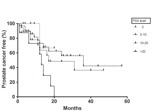
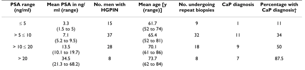
We performed a sub-group analysis at different PSA ranges
(Table 2; Figure 1). At PSA range ≤ 5 ng/ml there were 15
Table 1: Comparison between patients with benign and malignant histology on repeat prostate biopsies
Patient characteristics Remained benign (n = 39) Developed CaP (n = 28) P-value
HGPIN at 1st set of biopsies
Mean age (y) 63.5 69.8 < 0.001
Mean PSA (ng/ml) 8.4 17.5 < 0.005
Mean number of biopsy cores 11 10.54 0.3
Unifocal HGPIN 27 20
At repeat biopsies
Mean number of repeat biopsies (range) 1.4 (1–3) 1.3 (1–3)
Mean interval (months) to 1st repeat biopsies 7.38 15.11 < 0.0005
Mean change in PSA (ng/ml) 0.5 4.8 < 0.05
Gleason score
< 7 na 18
7 na 7
> 7 na 3
Prostate cancer-free interval from the time of 1st biopsy, according to PSA levels at presentation
Figure 1
mean change in PSA was only 1.3 ng/ml in this cohort of men. The cancer detection rate was markedly higher in
men with PSA ≥ 10 ng/ml. For PSA range > 10 ≤ 20 ng/ml,
it was 50%; in those with presenting PSA > 20 ng/ml, it was 87.5%.
Discussion
Recent reviews on HGPIN have found that it has a median incidence of 5.2% (mean of 7.7%) on needle biopsies. From data derived from recent studies, in men who are
identified to have isolated HGPIN on a 1st set of biopsies,
there is a median elevated risk of 24% (mean 31.5%) of subsequent CaP on repeat needle biopsies [5,6]. Two-thirds of the 22 contemporary studies (since 2000) reviewed reported the median elevated risk of subsequent CaP to be below 24% [6]. In fact, with the widespread
implementation of extended core (≥ 8) biopsies that
result in a more rigorous examination of the lateral regions of the prostate, it has been suggested that identifi-cation of isolated HGPIN may no longer be associated with higher CaP risk on repeat biopsies. Thus, the
CaP-detection rate post-HGPIN on 1st set of biopsies may not
be markedly dissimilar to that on repeat biopsies follow-ing an original benign diagnosis [5,10].
There appears to be no common consensus regarding the optimal biopsy technique and schedule for repeat
biop-sies after isolated HGPIN diagnosis on 1st set of biopsies.
Three to six monthly repeat biopsies for 2 y followed by yearly biopsies for life has been recommended [11]. Some studies have shown a low detection rate when biopsies are repeated within 12 months after isolated HGPIN on
extended core 1st biopsies [6,10]. Lefkowitz et al. (2002)
looked at follow-up biopsies at 3 years that showed a CaP-detection rate of 25.8%; based on these results they rec-ommended delaying the repeat biopsies for 3 years [12]. There has also been a suggestion for site-directed repeat
biopsies after isolated HGPIN on extended core 1st
biop-sies [13]. Men with multifocal isolated HGPIN have been implicated to be at greater risk of developing CaP in some studies [13,14].
Our current UK-based study showed a 41.8% risk of sub-sequent diagnosis CaP in men with an original diagnosis
of isolated HGPIN on 1st set of biopsies, which is higher
than most contemporary studies examining extended core repeat biopsy techniques [6]. However, this rate is compa-rable to earlier U.S.-based study from the mid-1990s when there was lower level of PSA screening and fewer cores were sampled on prostate biopsies [15].
The sub-analysis based on PSA ranges showed that the ele-vated risk of CaP detection was only observed at higher PSA ranges. This would suggest two important findings. Firstly, as purely isolated HGPIN should not raise the PSA so patients with low age specific PSA (especially in the PSA
range ≤ 5 ng/ml) may be appropriate candidates for repeat
biopsies at longer intervals as suggested by Lefkowitz et al.
(2002) [12]. Secondly, the negative 1st biopsy in the
pres-ence of a high PSA may be a reflection of a missed concur-rent focus of CaP and in these patients a repeat biopsy strategy at a shorter interval may be more appropriate. A similar strategy in the intermediate PSA range (between 5 to 10 ng/ml) may be justified as the risk of subsequent diagnosis of CaP on repeat biopsies seems to be still high (Table 2; Figure 1). We present part of the data in the Kap-lan-Meier type plot (Figure 1); it is important to realise that as the Kaplan-Meier method uses a finite number of observations to provide information for estimation pur-poses, one should use caution in deducing important con-clusions based solely from the curve.
There are a number of limitations to the current study. Either due to patient choice or due to their co-morbidities or for some unknown reasons, 21 patients with diagnosis of HGPIN on first set of biopsies did not undergo repeat biopsies at our institution. Though these patients were rel-atively older with a mean age of 73 y, no significant differ-ence in the mean PSA level or mean number of biopsy
cores at 1st set of biopsies was observed when compared to
those that had repeat biopsies (data not shown). In 18 of these 21 patients the clinic follow-up data was available and none of these men have shown rapid rise in PSA or Table 2: Detection rates of prostate cancer in men with HGPIN based on the baseline PSA level at first biopsy
PSA range (ng/ml)
Mean PSA in ng/ ml (range)
No. men with HGPIN
Mean age [y (range)]
No. undergoing repeat biopsies
CaP diagnosis Percentage with CaP diagnosis†
≤5 3.3
(1.5 to 5)
15 61.7
(52 to 74)
9 1 11
> 5 ≤ 10 7.1 (5.2 to 9.5)
37 65.4
(52 to 81)
32 11 34
> 10 ≤ 20 13.5 (10.1 to 19.7)
28 70.1
(61 to 86)
18 9 50
> 20 34.5
(21.3 to 68.2)
8 73.7
(62 to 84)
8 7 87.5
Publish with BioMed Central and every scientist can read your work free of charge "BioMed Central will be the most significant development for disseminating the results of biomedical researc h in our lifetime."
Sir Paul Nurse, Cancer Research UK
Your research papers will be:
available free of charge to the entire biomedical community
peer reviewed and published immediately upon acceptance
cited in PubMed and archived on PubMed Central
yours — you keep the copyright
Submit your manuscript here:
http://www.biomedcentral.com/info/publishing_adv.asp
BioMedcentral clinical progression to CaP. Also, there does not appear to
have been a uniform protocol for repeat biopsies. Further-more, this is a retrospective study looking at a regional population and, thus may not be representative of the sit-uation in the UK as a whole. Prospective or retrospective studies looking at multiple regions may be needed to examine this more fully.
Conclusion
In this UK-based study a presentation of isolated HGPIN
at 1st set of extended core needle prostate biopsies was
associated with a high risk (41.8%) of a subsequent diag-nosis of CaP on repeat biopsies. Patient age, higher PSA level at isolated HGPIN diagnosis and higher mean changes in serum PSA levels were predictive of CaP detec-tion on repeat biopsies. Based on the results of our study, we recommend delaying the first repeat biopsy at low PSA range and repeat biopsies at shorter interval at intermedi-ate and higher PSA ranges.
Competing interests
The authors declare that they have no competing interests.
Authors' contributions
PBS, NR and FLM conceived the idea of the study. PBS and CMN collected data. PBS drafted the manuscript. PBS and NR did data analysis. CMN, RAB, FLM and SSM helped with manuscript drafting and critical analysis. All authors read and approved the final manuscript.
Acknowledgements
This work is funded by Rosemere Cancer Foundation.
References
1. Sakr WA, Grignon DJ, Haas GP: Pathology of premalignant lesions and carcinoma of the prostate in African-American men. Semin Urol Oncol 1998, 16:214-20.
2. McNeal JE, Bostwick DG: Intraductal dysplasia: a premalignant lesion of the prostate. Hum Pathol 1986, 17:64-71.
3. Haggman MJ, Macoska JA, Wojno KJ, Oesterling JE: The relation-ship between prostatic intraepithelial neoplasia and prostate cancer: critical issues. J Urol 1997, 158:12-22.
4. Sakr WA, Partin AW: Histological markers of risk and the role of high-grade prostatic intraepithelial neoplasia. Urology 2001, 57:115-20.
5. Schlesinger C, Bostwick DG, Iczkowski KA: High-grade prostatic intraepithelial neoplasia and atypical small acinar prolifera-tion: predictive value for cancer in current practice. Am J Surg Pathol 2005, 29:1201-7.
6. Epstein JI, Herawi M: Prostate needle biopsies containing pros-tatic intraepithelial neoplasia or atypical foci suspicious for carcinoma: implications for patient care. J Urol 2006, 175:820-34.
7. Lefkowitz GK, Sidhu GS, Torre P, Lepor H, Taneja SS: Is repeat prostate biopsy for high-grade prostatic intraepithelial neo-plasia necessary after routine 12-core sampling? Urology 2001, 58:999-1003.
8. Moore CK, Karikehalli S, Nazeer T, Fisher HA, Kaufman RP Jr, Mian BM: Prognostic significance of high grade prostatic intraepi-thelial neoplasia and atypical small acinar proliferation in the contemporary era. J Urol 2005, 173:70-2.
9. Sirovich BE, Schwartz LM, Woloshin S: Screening men for pros-tate and colorectal cancer in the United Spros-tates: does prac-tice reflect the evidence? JAMA 2003, 289:1414-20.
10. Herawi M, Kahane H, Cavallo C, Epstein JI: Risk of prostate cancer on first re-biopsy within 1 year following a diagnosis of high grade prostatic intraepithelial neoplasia is related to the number of cores sampled. J Urol 2006, 175:121-4.
11. Bostwick DG, Liu L, Brawer MK, Qian J: High-grade prostatic intraepithelial neoplasia. Rev Urol 2004, 6:171-9.
12. Lefkowitz GK, Taneja SS, Brown J, Melamed J, Lepor H: Follow up interval prostate biopsy 3 years after diagnosis of high grade prostatic intraepithelial neoplasia is associated with high likelihood of prostate cancer, independent of change in pros-tate specific antigen levels. J Urol 2002, 168:1415-8.
13. Akhavan A, Keith JD, Bastacky SI, Cai C, Wang Y, Nelson JB: The proportion of cores with high-grade prostatic intraepithelial neoplasia on extended-pattern needle biopsy is significantly associated with prostate cancer on site-directed repeat biopsy. BJU Int 2007, 99:765-9.
14. Schoenfield L, Jones JS, Zippe CD, Reuther AM, Klein E, Zhou M, Magi-Galluzzi C: The incidence of high-grade prostatic intraep-ithelial neoplasia and atypical glands suspicious for carci-noma on first-time saturation needle biopsy, and the subsequent risk of cancer. BJU Int 2007, 99:770-4.
15. Davidson D, Bostwick DG, Qian J, Wollan PC, Oesterling JE, Rudders RA, Siroky M, Stilmant M: Prostatic intraepithelial neoplasia is a risk factor for adenocarcinoma: predictive accuracy in nee-dle biopsies. J Urol 1995, 154:1295-9.
Pre-publication history
The pre-publication history for this paper can be accessed here: