Original Research Article.
Long Term Consumption of High Dose Sucralose Induces
Hepatic Necrosis and Fibrosis in Male Albino Rats
Manar M. Bayoumi
1, Ghada M. El Gallad
2, Sherin S. Ghaleb
3, Hala M. El-Hanbuli
4*1Msc. Department of Forensic Medicine and Clinical Toxicology, Fayoum University, Egypt. 2MD, Department of Forensic Medicine and Clinical Toxicology, Fayoum University, Egypt. 3MD, Department of Forensic Medicine and Clinical Toxicology, Cairo University, Egypt. 4*MD, Pathology Department, Faculty of Medicine, Fayoum University, Egypt.
ABSTRACT
Background: Sucralose is one of the most popular non-nutritive sweeteners in the market. Whoever its toxicological effect are still need further investigation
Materials and Methods: Three groups of 50 rats formed of a control group and two sucralose groups that received sucralose dissolved in water for 12 weeks formed of a control group and two sucralose groups. For all, liver function tests and hepatic histopathologic examination were done.
Results: There was a statistically significant difference (P <0.05) among rats’ liver function tests in different study groups,
with high mean of liver enzymes (ALT, AST and ALP) levels in sucralose groups (low dose group & high dose group). Also, a statistically significant difference (P <0.0001) in rats' liver necrosis in different study groups with higher effect on liver tissues among the high dose group. There was a statistically significance difference (P <0.0001) in rats' liver fibrosis in different study groups; about 55% liver tissues of the high dose group shows Grade 2 hepatic fibrosis, 30% shows Grade 1 and
about 15% no fibrosis. There was no hepatic fibrosis among control and the low dose group.
Conclusion: Long term use of sucralose in male albino rats produces hepatotoxicity which is more in large doses.
Key words: Sucralose, Hepatotoxic, Necrosis, Fibrosis.
*Correspondence to:
Hala M. El-hanbuli, Pathology Department,
Faculty of Medicine, Fayoum University, Egypt. Article History:
Received: 07-07-2017, Revised: 05-08-2017, Accepted: 16-09-2017 Access this article online
Website: www.ijmrp.com
Quick Response code
DOI:
10.21276/ijmrp.2017.3.5.047
INTRODUCTION
Nutritive and non-nutritive are the two known types of sweeteners. Nutritive sweeteners are naturally occur as sucrose and fructose, while non-nutritive sweeteners are synthetically made as aspartame, stevia and sucralose.1
Sucralose is one of the most popular non-nutritive sweeteners in the market as it is highly similar to sucrose and it is very similar to carbohydrate in structure.2 It is produced by chlorination of
sucrose3, although sucralose is made from sugar, the human body
does not recognize it as a sugar so it does not metabolize and as a result it does not provide the body with calories as all artificial sweeteners.4
At the beginning of 1998, Food and Drug Administration (FDA) approved sucralose as a food additive in some varieties of foods and beverages after evaluating many studies in animals and humans.5 In spite that the safety of sucralose as an additive in
human food products has been well established by several safety studies3, some case studies reported that sucralose consumption
has a harmful effects on the body.6 Because some studies has
associated artificial sweeteners with health conditions such as cancers, hepatotoxicity and migraines. So the aim of this study is to assess the potential toxicological effects of long term use of sucralose on the liver of male albino rats.
MATERIAL AND METHODS Material
Sucralose was purchased from Eva Pharma Company in the form of powder 12.5 mg.
Experimental Animals
The study was conducted on 50 albino rats weighing between (150-230 g) which were used as experimental animals. Albino rats were obtained from animals' house of Research Institute of Ophthalmology, El-Giza, Egypt. The rats were acclimatized for 7days before the onset of the experiment.
The chosen animals were individually housed in plastic cages with good aerated covers at normal atmospheric temperature (25 ± 5ºC) as well as under good ventilation and received water and standard balanced diet.
Experimental Design
Rats were divided into 3 groups. Two groups received sucralose dissolved in water by oral route daily for 12 weeks.
Group (A): Control group (10 rats): Animals untreated and served as negative control, received distilled water.
Group (B) (20 rats): Sucralose treated group at a dose of 5 mg/kg/day via oral route for 12 weeks.7 This dose is the
Group (C) (20 rats): Sucralose treated group at a dose of 125 mg/kg/day via oral route for 12 weeks.7 Doses higher than this
dose were not palatable for rats.
The mean body weight was measured at the beginning and at the end of the study in all different groups. The entire experimental protocol that involved the use of animals was approved by the Ethics Committee for Animal Research and was conducted in accordance with the institutional and National Institutes of Health guidelines for the care and use of animals.
Blood Collection and Biochemical Analysis
At the end of the experimental period animals were anesthetized using diethyl ether, blood samples were collected from the orbital sinus. The blood samples were centrifuged at 3000 round per minute (r.p.m.) for 20 minutes to obtain serum. The supernatant sera were separated and frozen at -80ºC for biochemical analysis:
Liver Function Tests
1. ALT (Alanine aminotransferase): Normal 14-18 U/L 2. AST (Aspartate aminotransferase): Normal 21-23U/L 3. Alkaline phosphatase: Normal 50-160 U/L
4. Albumin: Normal 3.5-5.5 g/dL Tissue Harvesting Procedures
At the end of experiment, the animals sacrificed by cervical decapitation and laparotomy were carried out to remove the livers that were stored in a formalin solution.
Sample of 0.5cmз of the livers were removed and fixed in 10%
neutral formalin for 24 hours followed by washing, dehydration in ascending grades of alcohol, cleared in xylene and embedding in hard paraffin. Samples were then serially sectioned at thickness of 5-6µ, mounted on slides and left for 24 hours at 37°C to dry and to avoid detachment of sections during subsequent steps of staining. The tissue sections were stained by Hematoxylin and Eosin stain and then examined under the light microscope.
The histopathological examination of liver was carried out to determine any associated changes and compare between groups. The liver tissue sections were also stained by special stain (Masson's trichrome stain) to clearly show the fibrosis of liver
tissue. A numerical scoring system for histologically assessing the extent of fibrosis was adapted from the formula of Scheuer8, with
minor modifications.9 Briefly, fibrosis was graded as:
0: No fibrosis.
Grade 1: Enlarged, fibrous portal tracts.
Grade 2: Periportal or portal- portal septa, but intact architecture. Grade 3: Fibrosis with architectural distortion;
Grade 4: Probable or definite cirrhosis.
Additionally, hepatocyte necrosis or degeneration severity was also graded as:
0: No hepatocyte necrosis or degeneration.
Grade 1: Focal necrosis or degeneration of hepatocytes (mild
lesion no. ≤ 3).
Grade 2: Multifocal necrosis or degeneration of hepatocytes (moderate lesion no.> 3).
Grade 3: Locally extensive or diffuse necrosis or degeneration of hepatocytes (severe).
Hepatocyte degeneration is mainly associated with cytoplasmic vacuolation and swelling, with the nuclear contour generally intact, whereas hepatocyte necrosis is associated with karyopyknosis (nuclear shrinkage) and karyorrhexis (nuclear rupture), in addition to degenerative changes.10
Statistical Analysis
The collected data was organized, tabulated and statistically analyzed using SPSS software statistical computer package version 18 (SPSS Inc, USA). For quantitative data, the mean and standard deviation were calculated. ANOVA (Analysis of variance) was used to test the difference about mean values of measured parameters among groups, multiple comparison between pairs of groups were performed using LSD (Post hoc range test). Paired t test was used in comparison between the difference of body weight in before and after intervention. For qualitative data the
number and percent distribution was calculated, chi square (χ2)
was used as a test of significance. For interpretation of results of
tests of significance, significance was adopted at P ≤ 0.05.
Table 1: Comparisons of body weights between study groups before and after interventions
Mean ± SD P-value
Initial weight (gm.) Control 183.00 ± 31.38 0.849*
Dose 5 180.75 ± 33.06 0.938**
Dose 125 180.00 ± 26.66 0.799***
Last weight (gm.) Control 247.50 ± 24.86 0.086*
Dose 5 262.00 ± 26.92 0.001**
Dose 125 286.00 ± 10.46 <0.0001***
*Between dose 5 and control; **Between dose 5 and 125; ***Between dose 125 and control
RESULTS
Rat survival was not affected by sucralose administration as survival in the sucralose treatment groups was similar to that of the controls. The percentages of rats that survived for 12 weeks were 100% of all study groups. The body weight of rats was determined in the beginning of the study and after finishing it (after 12 week) (Table 1). There was no statistically significant differences in the initial bodyweight between all studied groups,
Table 2: Comparison of liver function tests in different study groups
Mean ± SD P-value
ALT Control 26.60 ± 6.50 <0.0001*
Dose 5 60.65 ± 8.38 <0.0001**
Dose 125 71.70 ± 9.57 <0.0001***
AST Control 29.80 ± 6.19 <0.0001*
Dose 5 46.80 ± 6.80 0.912**
Dose 125 46.50 ± 10.67 <0.0001***
ALP Control 131.40 ± 13.54 <0.0001*
Dose 5 193.80 ± 56.86 <0.0001**
Dose 125 246.65 ± 29.54 <0.0001***
Albumin Control 3.81 ± 0.34 0.331*
Dose 5 3.65 ± 0.35 0.334**
Dose 125 3.78 ± 0.51 0.855***
*Between dose 5 and control; **Between dose 5 and 125; ***Between dose 125 and control; SD= Standard deviation
Figure 1: Liver of rat from control group showing no histopathological changes (H& E X 400).
There was a statistically significant difference (P <0.05) among
rats’ liver function tests in different study groups, with high mean
of liver enzymes (ALT, AST and ALP) level in sucralose groups (low dose group & high dose group) with exception for AST level which showed non-significant increase (P=0.912) in low dose group when compared with the high dose one. Also, insignificant reduction in albumin level was observed in different study groups. Microscopically, Livers of rats of the control group revealed normal histological structure of hepatic lobule (Figure 1).
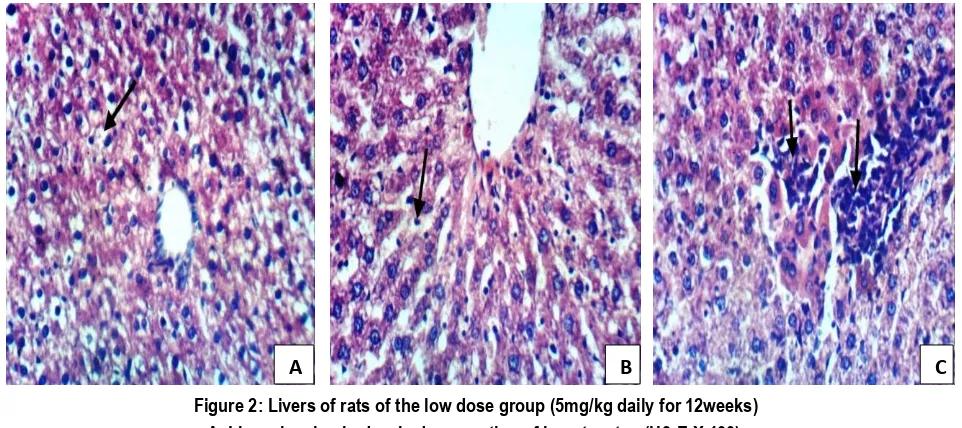
Examination of livers of rats of the low dose group (Figure 2) revealed: Most (n=12) of examined livers of rats of the low dose group revealed hydropic degeneration of hepatocytes (A), Kupffer cell activation (B), focal hepatic necrosis (Grade 1 hepatic necrosis) with inflammatory cells infiltration (C), while some (n=8) livers of rats of the same group showed normal histological examination.
Figure 2: Livers of rats of the low dose group (5mg/kg daily for 12weeks) A. Liver showing hydropic degeneration of hepatocytes (H& E X 400).
B. Liver showing Kupffer cells activation (H& E X 400).
C. Liver showing focal necrosis of hepatocytes associated with inflammatory cells infiltration (H& E X 400).
Figure 3: Liver of rat from high dose group(125mg/kg daily for 12weeks)
A. Liver showing slight hydropic degeneration of hepatocytes (arrow) and sinusoidal congestion (H& E X 400).
B. Liver showing portal infiltration with inflammatory cells (H& E X 400).
Figure 4: Liver of rat from high dose group showing fibroplasia in the portal triad (A) (H & E X 400), confirmed by Masson Trichrome (B) (X400)
Figure 5: The variation in hepatic necrosis according to study groups of the rats. 0
10 20 30 40 50 60 70 80 90 100
Control Dose 5 Dose 125
100
40
0 0
60
30
0 0
70
0 Grade 1 Grade 2
Hepatic necrosis
A
B
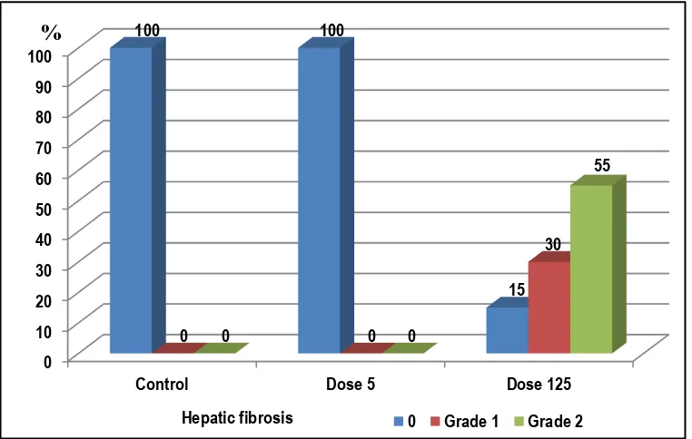
Figure 6: The variation in hepatic fibrosis according to study groups of the rats.
Livers of rats of the high dose group (Figure 3): Most (n=14) of examined livers of rats of the high dose group) revealed hydropic degeneration of hepatocytes, congestion of hepatic sinusoids (A), multifocal necrosis of hepatocytes (Grade 2 hepatic necrosis), Kupffer cell activation, portal infiltration with inflammatory cells (B), while the rest of the same group (n=6) showed milder liver affection with Grade 1 hepatic necrosis. In addition, most of the livers of rats of the high dose group showed fibroplasia in portal triad (Figure 4) (of Grade 1 and Grade 2) and only 3 of them show no hepatic fibrosis.
There was a statistically significant difference (P <0.0001) in rats' liver necrosis in different study groups with higher effect on liver tissues among the high dose group (Figure 5); about 70% liver tissues of the high dose group showed Grade 2 hepatic necrosis and 30% showed Grade 1) but about 60% liver tissues of the low dose group showed Grade 1 hepatic necrosis and 40% showed no hepatocyte necrosis or degeneration. Also, there was a statistically significance difference (P <0.0001) in rats' liver fibrosis in different study groups (Figure 6); about 55% liver tissues of the high dose group showed Grade 2 hepatic fibrosis, 30% showed Grade 1 and about 15% showed no fibrosis. There was no hepatic fibrosis among control and the low dose group.
DISCUSSION
There is increasing movement toward the consumption of artificial sweeteners due to increased incidence of diseases like obesity, diabetes, and metabolic syndrome. These artificial sweeteners, also called nonnutritive sweeteners provide more intense sweetness sensation with no or minimal calories per gram and are used in beverages, dietary products, drugs, and even mouthwashes.11
Sucralose (1, 6-dichloro-1, 6-dideoxy-β-D-fructofuranosyl- chloro-4-deoxy-α-D-galactopyranoside) is one of the most commonly used artificial sweeteners. It is characterized by its sweetness, immunity to metabolic degradation, non-hygroscopic nature, and relative heat resistance. It has a taste which is approximately 600 times as sweet as sucrose without any aftertaste.12
Despite the approved safety by FDA, sucralose appeared to have some harmful effects in some case report. In comparison with other commonly used artificial sweeteners, some studies reported that both aspartame and saccharin may induce a dose-dependent hepatotoxicity.13 So there was a need to substantiate whether long
term oral consumption of sucralose induces hepatic toxicity, so the present study highlights the effect of long-term consumption of two different doses of sucralose on liver.
ALT and AST are normally highly concentrated in the liver however an increase in ALT serum levels is, consequently, more specific for liver damage. While an increased activity of ALP is an indication of liver damage. In addition determining serum albumin
levels is considered “test of liver function” and this is mainly
because hepatic synthesis of albumin tends to diminish in end-stage liver disease.14
The study revealed significantly elevated levels of plasma ALT, AST and ALP were recorded in all treated groups of the present study compared to those of the control. On the other side albumin levels are within normal ranges.
In a similar study conducted on albino rats, sucralose was dissolved in saline and administered once daily subcutaneously at 0.625 mg/kg and 1.875mg/kg (low doses) and 5.625 mg/kg (high dose) for 2 weeks and reported that sucralose group showed slightly non-significant increase in AST, ALT and ALP level when compared to the control group in low doses but significant increase in high dose.15
While these results disagree with a recent study concluded that sucralose had no adverse effects one liver enzymes activity when used at dose of 15 mg/kg/day for one month.16
On microscopic examination, there were no histological abnormalities in the control groups, while sucralose groups displayed obvious histological changes in the liver tissue in most of them in addition to hepatic fibrosis which appeared only in the high dose group.
The observed hepatic histopathological changes agree with Mann et al.17 who reported that sucralose administration to
groups formed of 52 male and 52 female mice in their diet at 0
10 20 30 40 50 60 70 80 90 100
Control Dose 5 Dose 125
100 100
15
0 0
30
0 0
55
0 Grade 1 Grade 2
concentrations of 0.3%, 1% or 3% continuously, for 104 weeks revealed chronic inflammation of the liver in the high-dose group males.
In disagreement with other studies15,18 which noted that the livers
of rats administered sucralose showed normal histological structure of hepatic lobule from central vein and hepatocytes. It is known that Kupffer cells activation leads to release of toxic secretory products that have the potential to kill the hepatocytes19,
and so Kupffer cells are included in the pathogenesis of liver injury mediated by chemical substances, toxins and pharmacological agents. The cytokine and chemokine produced by activated Kupffer cells are also involved in processes of liver fibrosis through the production of cytokines and growth factors that induce Ito cell (hepatic stellate cells) myofibroblastic transformation, and regulate the production of metalloproteinase and their inhibitors.20
The significant hepatotoxic effects of long term use of sucralose especially in high dose reported in this work needs to be confirmed in further studies using larger study groups.
CONCLUSION
Long term use of sucralose in male albino rats produces hepatotoxicity which is more in large doses.
REFERENCES
1. Villareal LMA, Cruz RAM, Ples MB and Vitor RJS. Neurotropic effects of aspartame, stevia and sucralose on memory retention and on the histology of the hippocampus of the ICR mice (Mus musculus). Asian Pacific Journal of Tropical Biomedicine. 2016;6(2):114-118.
2. Brown AW, Bohan Brown MM, Onken KL and Beitz DC. Short-term consumption of sucralose, a nonnutritive sweetener, is similar to water with regard to select markers of hunger signaling and short-term glucose homeostasis in women. Nutrition Research Journal. 2011;31(12):882-888.
3. Stoddard KI and Huggett DB. Early Life Stage (ELS) Toxicity of Sucralose to Fathead Minnows, Pimephales promelas. Bulletin of Environmental Contamination and Toxicology Journal.2014; 93(4):383-387.
4. Kroger M, Meister K and Kava R. Low-calorie sweeteners and other sugar substitutes: A review of the safety issues. Comprehensive Reviews in Food Science and Food Safety Journal.2006; 5(2):35-47.
5. Rodero AB, Rodero LDS and Azoubel R. Toxicity of Sucralose in Humans: A Review. International Journal of Morphology.2009; 27(1):239-244.
6. Bigal, M. E., & Krymchantowski, A. V. Migraine triggered by sucralose: A case report. Headache. 2006; 46(3), 515-517. 7. Baird IM, Shephard NW, Merritt RJ and Hildick-Smith G. Repeated dose study of sucralose tolerance in human subjects. Food and Chemical Toxicology Journal.2000; 38:123-129. 8. Scheuer PJ. Classification of chronic viral hepatitis: a need for reassessment. Journal of Hepatology.1991; 13(3): 372-37. 9. Hsu YC, Lin YL, Chiu YT, Shiao MS, Lee CY, Huang YT: Antifibrotic effects of Salvia miltiorrhiza on dimethylnitrosamine-intoxicated rats. J Biomed Sci 2005, 12:185-195.
10. Weng T, Shen C, Chiu Y, Lin Y, Kuo C and Huang Y. Inhibitory effects of armepavine against hepatic fibrosis in rats. Journal of Biomedical Science. 2009; 16(78):1-13.
11. Sharma A, Amarnath S, Thulasimani M and Ramaswamy S. Artificial sweeteners as a sugar substitute: Are they really safe?. Indian Journal of Pharmacology. 2016; 48(3):237-240.
12. Idris M, Rao VJ, Middha D, Shukla SK and Rao Baggi TR ( 2013). Determination of sucralose by controlled UV photodegradation followed by UV spectrophotometry. Journal of AOAC International;96(3):603-606.
13. Alkafafy ME, Zein SI, Mohamed MA, Samir AE. Impact of aspartame and saccharin on the rat liver: biochemical, molecular, and histological approach. Int J Immunopathol Pharmacol. 2015, pp. 1-9
14. Giannini EG, Testa R and Savarino V. Liver enzyme alteration: A guide for clinicians.Canadian Medical Association Journal. 2005;172(3):367–379.
15. Abd El Fatah HH, El Gallad GM, Tawfik HM and Saddeik WF. Effect of Aspartame on Different Organs in Albino Rats and Comparing It With Some Other of Sugar Substitutes(Thesis). Department of Forensic Medicine and Clinical Toxicology, Fayoum University.2015.
16. Pałkowska-goździk E, Bigos A, Rosołowska-Huszcz D. Type of sweet flavour carrier affects thyroid axis activity in male rats. European Journal of Nutrition. 2016; pp 1–10
17. Mann SW, Yuschak MM and Amyes SJG. A Carcinogenicity Study of Sucralose in the CD-1 Mouse. Food and Chemical Toxicology Journal. 2000; 38(2):91-97.
18. Grice H and Goldsmith L. Sucralose—an overview of the toxicity data. Food and Chemical Toxicology Journal. 2000;38(2):1-6.
19. Muriel P and Escobar Y. Kupffer Cells are Responsible for Liver Cirrhosis Induced by Carbon Tetrachloride. Journal of Applied Toxicology. 2003;23(2) :103–108.
20. Kolios G, Valatas V, Kouroumalis E, Kolios G, Valatas V and Kouroumalis E. Role of Kupffer cells in the pathogenesis of liver disease. World Journal of Gastroenterology.2006;12(46):7413-7420.
[
Source of Support: Nil.
Conflict of Interest: None Declared.
Copyright: © the author(s) and publisher. IJMRP is an official publication of Ibn Sina Academy of Medieval Medicine & Sciences, registered in 2001 under Indian Trusts Act, 1882. This is an open access article distributed under the terms of the Creative Commons Attribution Non-commercial License, which permits unrestricted non-commercial use, distribution, and reproduction in any medium, provided the original work is properly cited.