R E V I E W
Open Access
Oligomeric A
β
-induced synaptic dysfunction in
Alzheimer
’
s disease
Shichun Tu
*, Shu-ichi Okamoto, Stuart A Lipton and Huaxi Xu
*Abstract
Alzheimer’s disease (AD) is a devastating disease characterized by synaptic and neuronal loss in the elderly. Compelling evidence suggests that soluble amyloid-βpeptide (Aβ) oligomers induce synaptic loss in AD. Aβ-induced synaptic dysfunction is dependent on overstimulation ofN-methyl-D-aspartate receptors (NMDARs) resulting in aberrant activation of redox-mediated events as well as elevation of cytoplasmic Ca2+, which in turn triggers downstream pathways involving phospho-tau (p-tau), caspases, Cdk5/dynamin-related protein 1 (Drp1), calcineurin/PP2B, PP2A, Gsk-3β, Fyn, cofilin, and CaMKII and causes endocytosis of AMPA receptors (AMPARs) as well as NMDARs. Dysfunction in these pathways leads to mitochondrial dysfunction, bioenergetic compromise and consequent synaptic dysfunction and loss, impaired long-term potentiation (LTP), and cognitive decline. Evidence also suggests that Aβmay, at least in part, mediate these events by causing an aberrant rise in extrasynaptic glutamate levels by inhibiting glutamate uptake or triggering glutamate release from glial cells. Consequent extrasynaptic NMDAR (eNMDAR) overstimulation then results in synaptic dysfunction via the aforementioned pathways. Consistent with this model of Aβ-induced synaptic loss, Aβsynaptic toxicity can be partially ameliorated by the NMDAR antagonists (such as memantine and NitroMemantine). PSD-95, an important scaffolding protein that regulates synaptic distribution and activity of both NMDA and AMPA receptors, is also functionally disrupted by Aβ. PSD-95 dysregulation is likely an important intermediate step in the pathological cascade of events caused by Aβ. In summary, Aβ-induced synaptic dysfunction is a complicated process involving multiple pathways, components and biological events, and their underlying mechanisms, albeit as yet incompletely understood, may offer hope for new therapeutic avenues.
Keywords:Alzheimer’s disease, Synaptic loss, Aβoligomers, Cognitive decline, Calcium, NMDA receptors, PSD-95, Mitochondrial dysfunction, Tau hyperphosphorylation, Aberrant neuronal network activity
Introduction
Alzheimer’s disease (AD) is the most common form of dementia among the elderly. It is clinically characterized by progressive memory loss and cognitive dysfunction, with the eventual inability to perform activities of daily living (ADLs). According to the Alzheimer’s Association (http://www.alz.org), 5.4 million Americans are currently living with Alzheimer’s disease, and one in eight older Americans will develop AD. While drugs are available to temporarily improve memory and cognitive function or delay the progress of dementia, AD remains a devastating neurodegenerative disorder without any effective cure or disease-modifying therapy [1,2]. Following diagnosis, AD patients generally survive from several years to 20 years,
depending on age and other health conditions.
Although AD is pathologically characterized by the presence of extracellular deposition of plaques com-prised of Aβpeptide and neurofibrillary tangles (NFTs) comprised of hyperphosphorylated-tau protein, accu-mulating evidence suggests that these abnormal protein deposits are unlikely the causative events in AD as Aβ plaque or NFT volume poorly correlate with the severity of dementia. Instead, the degree of dementia in premor-tem patients correlates more closely with the level of soluble oligomers of Aβspecies in postmortem brains, especially in hippocampal and cortex regions associated with learning and memory function [3-5]. Aβ peptides are proteolytic products of the amyloid precursor pro-tein (APP) and are sequentially cleaved by β- and γ -secretases [6]. Although Aβ peptides of varying length are produced, Aβ1-42is considered to be comparatively
* Correspondence:[email protected];[email protected]
Neuroscience and Aging Research Center, Sanford-Burnham Medical Research Institute, 10901 North Torrey Pines Road, La Jolla, CA 92037, USA
more amyloidogenic and readily assembles into soluble oligomers and consequent fibril deposits. Aβoligomers, also termed as Aβ-derived diffusible ligands (ADDLs), are thought to induce synaptic loss and progressive cog-nitive decline in AD, whereas monomers and fibrillary ag-gregates may be more inconsequential to pathogenesis [7]. In addition, synaptic protein depletion [8-10] and synaptic loss [11,12] are also found in the same regions in human AD or mild cognitive impairment (MCI) postmortem brains, and the level of synaptic reduction closely parallels the degree of premortem cognitive impairment. These studies suggest that oligomeric Aβelevation and synaptic loss, rather than Aβ plaque load, may represent the best indicators of the severity of dementia or cognitive impair-ment in AD. Moreover, these findings imply that rescue of synapses could prove to be disease modifying in AD.
Using transgenic animal models of AD, additional stud-ies suggest that synaptic loss is induced by pathological Aβ elevation [13]. Although the molecular mechanism is still not fully understood, it is generally believed that Aβ oligomers at pathological concentrations trigger (most likely via an indirect pathway) overstimulation of extrasy-naptic NMDA receptors (eNMDARs), leading to aberrant redox events and Ca2+ upregulation. Subsequent activa-tion of downstream signal transducactiva-tion pathways trigger a cascade of pathological events leading to synaptic dis-ruption and neuronal loss. These include increased oxi-dative/nitrosative stress and mitochondrial dysfunction with consequent bioenergetic compromise, leading to dys-regulation of synaptic neurotransmission and abnormal neuronal network activity [7,14]. A number of synaptic pro-teins have been proposed as potential Aβ-binding partners under pathological conditions and their interactions are be-lieved to mediate Aβ-induced synaptic dysfunction. These proteins include, but are not limited to,α7-nicotinic acetyl-choline receptors (α7nAChRs) [15], NMDARs [16,17], mGluR5 [18], neurotrophin receptor p75NTR [19], cellular prion protein (PrPC) [20], PSD-95 [21], glutamate trans-porter [22], ephrin type-B receptor 2 (EphB2) [23], and ephrin type-A receptor 4 (EphA4) [24,25]. The involvement of some of these proteins in Aβ-mediated neurotoxicity will be discussed in the following sections.
Oligomeric Aβinduces synaptic dysfunction in AD mice
AD or amnestic MCI patients usually have trouble with spatial orientation in their daily routine and perform poorly in clinically-designed, hippocampus-dependent memory and navigation tests [26-28]. Studies using animal models of AD have suggested that soluble oligomeric Aβspecies are critical in initiating a pathogenic cascade leading to synaptic dysfunction, neuronal loss, and AD-like cognitive impairment [7]. The association between amyloidogenic Aβ species and age-dependent memory loss was first described in Tg2576 mice expressing a human APP695
transgene containing the Swedish mutation (K670N/ M671L) [29]. These mice show elevated brain Aβ1-42/43 levels by ELISA and perform poorly in Morris water maze tests of spatial memory [29,30]. The association between Aβ and cognitive impairment has also been documented in other transgenic (Tg) animal models of AD including 3XTg-AD mice, which express three human mutant AD gene variants (PS1M146V, APPSwe, and tauP301L) and develop progressive plaques and tangles [31,32] and the human amyloid precursor protein-overexpressing (hAPP) J20 mouse model [33]. Synaptic dysfunction was also ob-served in these AD transgenic mice with aberrantly ele-vated levels of oligomeric Aβand deficits in learning and memory (see selected reviews: [34-36]). Moreover, these studies showed that synaptic and cognitive impairments are associated with the elevation of soluble oligomeric Aβ species and usually evident prior to the appearance of pla-ques and tangles in the relevant brain regions.
NMDAR-dependent long-term potentiation (LTP) in hippocampus has attracted broad attention as an elec-trophysiological measurement of synaptic strength and plasticity in various AD models. LTP induction requires activation of NMDARs, which triggers a signaling cas-cade that induces the recruitment of AMPARs into the postsynaptic membrane [37]. On the other hand, NMDAR-dependent long-term depression (LTD) is an activity-dependent reduction in the efficacy of neuronal synapses, which is mediated, at least in part, by AMPAR endocytosis [37]. Multiple studies have shown that LTP is impaired in AD or Aβ-exposed wild-type hippocampus, and this im-pairment is dependent on NMDARs and downstream path-ways [38-44]. In contrast to LTP, Aβapplication enhances LTD [22,45,46], consistent with the notion that Aβcauses synaptic depression.
AMPARs [45]. In support of this, surface AMPARs are downregulated through endocytosis in wild-type neurons rapidly upon Aβ-application [51] and in AD transgenic mice [52]. Concurrently, AMPAR-mediated synaptic currents are also downregulated in AD double knock-in (mutant APP and PS1) transgenic mice [53]. Aβ-induced AMPAR endocytosis or surface removal is dependent on the activation of calcineurin/PP2B [54] and requires downregulation of Ca2+/calmodulin-dependent protein kinase II (CaMKII) [55]. Similar to AMPARs, Aβcan in-duce surface removal or endocytosis of NMDARs, which is mediated by dephosphorylation of NR2B (GluN2B), an NMDAR subunit, at p-Tyr1472 by the tyrosine phosphat-ase STEP [56,57]. Interestingly, the apoptotic effector component caspase-3 has also been suggested to play a role in synaptic plasticity and its activation is required in AMPAR removal and consequent LTD induction [58]. Ac-tivation of caspase-3 has been further shown to trigger early synaptic dysfunction in AD transgenic mice [59]. This is consistent with the observation that caspase-3 is enriched in postsynaptic densities [60]. In addition to ionic glutamate receptor NMDARs and AMPARs, metabotropic
glutamate receptors (mGluRs) at extrasynaptic or perisy-naptic sites have also been shown to play an important role in Aβ-induced synaptic dysfunction [46], likely acti-vated by pathologically eleacti-vated glutamate at extrasynaptic sites [22].
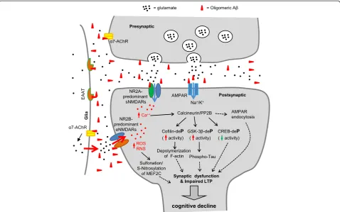
hyperactivation of neuronal nitric oxide synthase (nNOS) and Ca2+-overload of mitochondria, generating exces-sive reactive nitrogen and oxygen species (RNS/ROS). This excess results in aberrant oxidation reactions, such as S-nitrosylation and sulfonation, on various proteins, thereby disrupting their normal activity. One example is illustrated by excessive S-nitrosylation or sulfonation of the transcription factor MEF2C, thereby adversely af-fecting downstream effectors of neuronal survival and adult neurogenesis [68,70]. Importantly, memantine, an FDA-approved, uncompetitive NMDAR open-channel blocker, at proper concentration, preferentially blocks pathologically-stimulated eNMDARs over physiologically-activated synaptic NMDARs (sNMDARs) [71]. Indeed, application of memantine, and to a greater degree its im-proved derivative NitroMemantine [72], has been shown to mitigate Aβ-induced synaptic dysfunction and cognitive deficits [63,73]. Taken together, these studies depict an al-ternative mechanism underlying Aβ-induced synaptic dys-function based on glutamate-mediated hyperexcitability and synaptic excitotoxicity: Aβ-induced accumulation of excessive glutamate in the extrasynaptic space can occur through disruption of the glutamate uptake system or by triggering astrocytic glutamate release, which in turn aber-rantly activates eNMDARs and induces synaptic dysfunc-tion (Figure 1).
NMDAR and AMPAR function at postsynaptic sites are influenced by interaction with PSD-95, an important postsynaptic scaffolding protein that plays a critical role in protein assembly, synaptic development and neural plasticity [74,75]. Under pathological conditions how-ever, synaptic PSD-95 levels are decreased in AD post-mortem brains, and the degree of reduction correlates with both the level of Aβoligomers and the severity of dementia [21,76-78]. Similarly, PSD-95 is also reduced in AD transgenic mouse neurons or neurons exposed to Aβoligomers, concomitant with dendritic spine loss and surface AMPAR removal [51,52,79]. These results sug-gest that synaptic disruption of PSD-95 may play a role in the pathogenesis of AD. Consistent with this idea, it has been shown by co-immunoprecipitation that Aβ in-teracts with PSD-95 and is co-localized with PSD-95 specifically at excitatory synapses in human postmortem AD brain as well as in cultured murine neurons exposed to Aβ oligomers [21,80]. Therefore, it is possible that Aβ may interact with PSD-95 directly to cause synaptic damage under pathological conditions. Interestingly, over-expression ofα1-takusan, a PSD-95-binding and synaptic-stabilizing protein [81], decreases Aβ-induced synaptic damage, protecting from dendritic spine loss, de-creased synaptic expression of PSD-95, and downregu-lated AMPAR-mediated synaptic currents in cultured neurons [82]. Further evidence has shown that increased PSD-95 expression in hippocampus after treatment with
the Hsp90 inhibitor 17-AAG can improve cognitive func-tion in an animal model of AD, apparently via synaptic en-hancement [83]. Therefore, molecules that modulate the integrity of PSD-95 and improve synaptic functions may have therapeutic potential for reducing Aβ-induced synap-tic injury and cognitive impairment in AD.
A number of other synaptic proteins have been pro-posed to be putative receptors for Aβ oligomers, and play important roles in oligomeric Aβ-induced synaptic dysfunction and cognitive impairment. For example, sol-uble Aβ oligomers can bind with nanomolar affinity to PrPC, and PrP knockout or anti-PrP antibodies can rescue oligomeric Aβ-induced synaptic dysfunction and spatial memory. These results suggest that prion proteins play an important role in AD pathogenesis [20]. The α7nAChR modulates calcium homeostasis and release of the neuro-transmitter glutamate, two important parameters involved in learning and memory. Endogenous Aβ species have been reported to bind toα7nAChRs with nanomolar affin-ity in co-immunoprecipitation experiments on human AD postmortem brain extracts [15]. This interaction conse-quently triggersα7nAChR-dependent NMDAR endocyto-sis leading to synaptic and cognitive dysfunction [15,56]. The ephrin family of receptor tyrosine kinases has also been found to be a potential receptor that interacts with Aβ oligomers under pathological conditions. It has been shown that Aβoligomer binding to EphB2 induces its deg-radation, leading to impairments in NMDAR-mediated synaptic activity and cognitive function [23]. Conversely, EphB2 overexpression reverses deficits in NMDAR-dependent LTP and cognitive impairments in AD Tg mice [23]. Recently, ephrin A4 (EphA4), another ephrin receptor family member, was also identified as a putative Aβ recep-tor [24,25]. In contrast to EphB2 degradation, interaction with Aβ activates EphA4, leading to suppression of LTP and spine loss in AD transgenic mice. Application of either EphA4 shRNA or EphA4 inhibitors/antagonists has been reported to rescue these deficits, suggesting that EphA4 ac-tivation plays a critical role in Aβ-induced synaptic dys-function [24,25].
Alternative pathways contribute to Aβ-induced synaptic and neuronal loss in AD
higher in human postmortem AD brains compared to age-matched controls [85]. Interestingly, APP cleavage is differ-entially regulated by the neurotrophin high-affinity receptor TrkA and the low-affinity receptor p75NTR; p75NTR pro-motes whereas TrkA decreases APPβ-cleavage [86]. There-fore, aberrantly upregulated p75NTR together with TrkA downregulation in aged brains results in increased Aβ gen-eration [87]. In addition, p75NTRmay also enhance Aβ pro-duction via its ability to stabilize BACE1 or β-secretase through the activation of sphingomyelinase and consequent ceramide production [87,88].
In addition to neurotrophin receptors, insulin and insulin-like growth factor receptors and their cognate sig-naling pathways play a critical role in synaptic plasticity and cognitive function by affecting both excitatory and in-hibitory synaptic activity [89,90]. Growing evidence sug-gests that AD may represent a metabolic disease of the brain associated with brain insulin and insulin-like growth factor-I (IGF-I) resistance and deficiency. Impaired insulin signaling may contribute to dysregulation of downstream pro-survival pathways, including decreased signaling me-diated by PI3K, Akt, and Wnt/β-catenin; moreover, dis-rupted insulin-related signaling may enhance pathogenic pathways such as GSK-3βto trigger tau hyperphosphory-lation [91,92]. Therefore, disrupted components of brain insulin signaling pathways may represent potential thera-peutic targets in AD [91,92]. It has been suggested that both extracellular and intracellular Aβoligomers contrib-ute to neuronal dysfunction. Intracellular Aβinhibits insu-lin receptor signainsu-ling by interfering with the interaction between phosphoinositide-dependent kinase (PDK) and Akt, thus inhibiting Akt activation and abolishing insulin-mediated neuroprotection [93]. Extracellular Aβoligomers or ADDLs bound to synaptic sites can induce removal of surface insulin receptors and contribute to synaptic loss, which can be rescued by insulin treatment [94]. Encour-aging results obtained from intranasal insulin therapy in aged adults [95] as well as in AD and MCI patients [96] support the idea that insulin signaling is disrupted during normal aging and in clinical cases of dementia.
Furthermore, Wnt family members promote synaptic formation and regulate synaptic function by binding to receptors of the Frizzled (Fz) and low-density lipoprotein-related protein (LRP) families on the cell surface to acti-vate eitherβ-catenin-dependent canonical signaling (Wnt/
β-catenin) orβ-catenin-independent non-canonical signal-ing pathways; the latter pathways include the Wnt/PCP and Wnt/Ca2+cascades [97]. Multiple Wnt signaling com-ponents are dysregulated in AD and such impairments are likely to contribute to synaptic dysfunction and cognitive decline in AD. Wnt co-receptor LRP6 variants suppress Wnt signaling activity and are associated with late-onset AD [98]. An endogenous Wnt inhibitor, Dickkopf-1 (Dkk1), which disrupts Wnt-induced Fz/LRP complex
formation [99], is increased in human postmortem AD brains [100] and mouse model AD brains [101], where Dkk1 has been found to co-localize with active GSK-3 and phospho-tau. Additional studies suggest that Dkk1 levels increase after Aβoligomer exposure [102]. Dkk1 upregula-tion appears to be required for Aβ-induced synaptic loss since synaptic damage as well as tau phosphorylation are abolished by either Dkk1 knockdown [100] or Dkk1 block-ing antibodies [102]. Interestblock-ingly, it has been shown that Aβmay directly interact with Fz receptors of Wnt ligands [103] although the significance of this interaction remains to be determined. Taken together, these studies suggest that dysregulated Wnt signaling contributes to Aβ-induced syn-aptic loss in AD, which raises the possibility that Wnt sig-naling components may represent potential therapeutic targets in AD.
Aberrant neuronal network activity and seizures in AD
Epileptic seizures were once considered to be rare or an epiphenomenon in AD. However, accumulating evidence suggests that increased seizure activity may be the con-sequence of a disrupted neuronal network, contributing to cognitive decline and the onset of dementia [104]. It has been estimated that 10 to 22% of AD patients ex-perience at least one episode of an unprovoked seizure [105]. However, it is likely that these numbers are under-estimates for the following reasons: (1) AD patients with dementia may not recall having seizures if they were un-witnessed, and (2) some types of seizure activity, includ-ing complex partial seizures, manifest symptoms such as confusion or delirium, which are similar to those nor-mally seen in AD patients, and therefore may go un-detected. Prior observational studies have shown that the occurrence of a first unprovoked seizure in patients 55 years or older is significantly greater in AD and other dementias compared to the general population [106]. The increased incidence can be as high as 87-fold in AD patients with early-onset FAD [107]. Consistent with this result, a more recent study found that FAD mutations in
APP, PSEN1, and PSEN2 are all associated with higher
risk for a first unprovoked seizure [108]. These results suggest a tight association between epileptic seizures and genetic mutations that cause aberrant expression of Aβoligomers and induce early-onset AD.
video-electroencephalography (EEG) monitoring in AD transgenic mice expressing FAD mutations [112-114]. Acute application of levetiracetam (LEV or Keppra®), an antiepileptic drug, can effectively suppress abnormal EEG spike activity, and chronic treatment with LEV can even reverse AD-like phenotypes including synaptic dysfunc-tion, hippocampal remodeling, and learning and memory deficits in human APP transgenic mice [115]. Therefore, antiepileptic drugs, such as LEV, that suppress the aber-rant electrical activity that contributes to seizures may provide an alternative approach to the treatment of AD.
Aβ-induced tau hyperphosphorylation and its role in synaptic loss
The presence of hyperphosphorylated tau-enriched neuro-fibrillary tangles is one of the classical pathological hall-marks of AD. Tau is a microtubule-associated protein (MAP) that was originally identified as an important protein for microtubule (MT) assembly [116] and for stabilization of the MT network [117]. Under pathological conditions, tau becomes hyperphosphorylated and disas-sociated from microtubules, subsequently forming soluble aggregates, insoluble filaments, and eventually neurofibril-lary tangles (NFTs) in affected brain regions. This path-ology occurs not only in AD but also in several other neurological disorders, which are collectively termed tauo-pathies [118,119]. Phosphorylated tau (p-tau) colocalizes with Aβin synaptic terminals from both postmortem AD brain [120,121] and transgenic mouse AD brain [122]. These prior studies have shown that expression of p-tau in synaptic terminals correlates with Aβlevels, and increased p-tau expression also correlates with a reduction in total synapse number. A causal association between oligomeric Aβexposure and p-tau formation has been demonstrated in several studies. For example, Talantova et al. [63] re-ported that oligomeric Aβcaused astrocytic glutamate re-lease, which in turn activated extrasynaptic NMDARs, resulting in increased p-tau levels. Additional studies have shown that Aβ-induced synaptic loss is tau-dependent since tau deletion or reduction can rescue Aβ-induced synaptic loss and cognitive impairment in AD transgenic mice [123-126]. Moreover, a recent study characterized in-teractions between oligomeric Aβand p-tau in both hu-man and animal AD brains by co-immunoprecipitation and immunohistology, and this interaction progressively increased with disease progression [127]. It is thus possible that pathological interactions between oligomeric Aβand p-tau are important intermediate steps in Aβ-induced syn-aptic loss and neuronal damage. Although previously known as an axonal protein, tau is also expressed in den-drites and in the postsynaptic density, albeit at much lower levels [124]. Depletion of dendritic or postsynaptic tau prevents abnormal postsynaptic targeting of the tyro-sine kinase Fyn and rescues impaired learning and
memory function in AD transgenic mice, suggesting a vital role of dendritic and postsynaptic tau in Aβ-induced syn-aptic loss [124]. In cultured neurons, Aβ-induced tau hyperphosphorylation and dendritic disruption can be at-tenuated by anti-Aβ antibodies or tau reduction using RNAi-targeting strategies [128-130]. Therefore, it is possible that Aβ-induced tau hyperphosphorylation is an important intermediate event that leads to synaptic dysfunction. Indeed, only pseudohyperphosphorylated tau, which mimics hyperphosphorylated tau, but not phosphorylation-deficient tau, which mimics regular tau, is mislocalized and accumulated in dendritic spines [131]. Consequently, hyperphosphorylated tau that is mislocalized to dendritic spines has been reported to in-duce synaptic dysfunction by impairing AMPAR surface expression and synaptic transmission [131]. Consistent with this finding, tau deletion or inhibition of tau hyper-phosphorylation using a glycogen synthase kinase 3β (GSK-3β) inhibitor can prevent Aβ-induced impairment of LTP [132]. GSK-3β-mediated tau phosphorylation and Aβ production can also be reduced by RPS23R1 protein via activation of the adenylate cyclase/cAMP/ PKA pathway. In turn, this pathway leads to synaptic enhancement and improved AD pathology [133]. Tau-dependent synaptic dysfunction may also involve the tyrosine kinase Fyn [124]. Pseudohyperphosphorylated tau binds Fyn more tightly than wild-type tau, thus in-creasing Fyn activity and leading to synaptic damage [134]. Intriguingly, a study using human AD induced pluripotent stem cell (iPSC)-derived neurons has suggested that increased tau phosphorylation at Thr231 is mediated byβ-secretase activity [135]. This finding raises the possi-bility that Aβ-induced tau pathology and synaptic damage can be mediated by an alternative, non-Aβ-mediated path-way. Thus, future in-depth studies will determine the role of dendritic/postsynaptic tau and its hyperphosphorylation in Aβ-induced synaptic loss.
Aβ-induced mitochondrial dysfunction and synaptic loss in AD
[144-147]. Therefore, it is possible that Aβat pathological concentrations can trigger mitochondrial impairment, which in turn leads to bioenergetics compromise, synaptic starvation and damage in AD. Consistent with this hypoth-esis, mitochondrial impairment caused by oligomeric Aβ or toxic factors generated downstream of Aβ, such as RNS/NO, occurs prior to synaptic and neurite injury [148] and precedes AD pathology [149]. It has been suggested that abnormal interactions between Aβand the mitochon-drial fission protein dynamin-related protein 1 (Drp1) play an important role in mitochondrial dysfunction and synap-tic damage in AD [150]. Additional evidence suggests that Aβ-induced mitochondrial impairment is likely mediated by abnormal interaction between oligomeric Aβand mito-chondrial matrix protein ABAD [151] or cyclophilin D (CypD) [152]. It has also been suggested that appopto-sin, a glycine/5-amino-levulinic acid transporter medi-ating heme synthesis in mitochondria, is involved in Aβ-induced neurodegeneration by inducing ROS re-lease and resultant apoptosis under pathological condi-tions; these pathogenic processes can be prevented by appoptosin downregulation [133].
Synaptic mitochondria, or mitochondria residing in synapses, are usually older, and thus much more vulnerable to persistent insults such as Aβ compared to nonsynaptic
mitochondria. Aβ species reportedly also accumulate at much higher levels in synaptic mitochondria compared to nonsynaptic mitochondria [153,154]. Accordingly, synaptic mitochondria show greater susceptibility to damage com-pared to nonsynaptic mitochondria in AD transgenic mice [153,154]. Mitochondrial function such as respiratory rate, ROS production, membrane potential, and cytochrome c oxidase activity are compromised more severely in synaptic mitochondria than in nonsynaptic mitochondria [153,154]. The degree of mitochondrial impairment is also region-specific. Greater damage is found in areas related to learning and memory such as the hippocampus and cor-tex, while only moderate damage is found in other brain regions [154].
Tau pathology strongly correlates with mitochondrial impairment [125,130,155-158], suggesting that tau may play a role in Aβ-induced mitochondrial dysfunction. It has been shown using immunoprecipitation and im-munofluorescence that hyperphosphorylated tau abnor-mally interacts and colocalizes with the mitochondrial fission protein Drp1 in postmortem AD brains [158]. In the same brain specimens, elevated levels of Drp1 and mitochondrial fragmentation have also been identified [150]. These studies suggest that hyperphosphorylated tau is associated with Aβ-induced mitochondrial dysfunction,
likely through its abnormal interaction with Drp1 in AD neurons. Consistent with this notion, Aβ application in cultured neurons results in elevated expression of mito-chondrial fission proteins Drp1 and Fis1 and reduced expression of fusion proteins Mfn1, Mfn2, and Opa1, consistent with mitochondrial fragmentation phenotypes observed in AD [146]. Additional recent evidence also sup-ports a role for hyperphosphorylated tau in mitochondrial impairment. For example, overexpression of the postsyn-aptic protein α1-takusan inhibits Aβ-induced tau hyper-phosphorylation and prevents Aβ-induced mitochondrial fragmentation in cultured neurons [82].
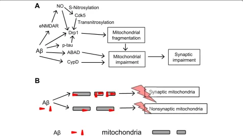
Moreover, pathological redox reactions of mitochondrial proteins triggered by oligomeric Aβ, in part mediated by aberrant eNMDAR stimulation as discussed above, have been described. These reactions include S-nitrosylation of Drp1 (to form SNO-Drp1), resulting in excessive mito-chondrial fission with consequent bioenergetic comprom-ise and hence synaptic damage [64]. Interestingly, Cdk5 has been shown to act as a transnitrosylase, transferring NO from Cdk5 to Drp1 (as opposed to the classical role of Cdk5 as a kinase). Transnitrosylation hyperactivates Drp1 under these disease conditions, and, importantly, S-nitrosylation of Cdk5 is triggered by oligomeric Aβpeptide [67]. Thus, the initial nitrosylation of Cdk5 may represent in inciting event in mitochondrial fragmentation, with resulting bioenergetic failure and synaptic loss. Figure 2 summarizes our current understanding of Aβ-induced mitochondrial impairment as discussed above.
Conclusions
Accumulating evidence suggests that oligomeric Aβplays a central role in synaptic dysfunction and cognitive decline in AD. Studies using animal models have revealed that Aβ -induced synaptic dysfunction involves multiple pathological events and various integral signaling systems, including glutamate receptors and their downstream pathways, ab-normal elevation of extrasynaptic glutamate levels and subsequent eNMDAR-mediated excitotoxicity, tau hyper-phosphorylation, and impaired mitochondria. However, the relevance of these studies to AD in human remains de-batable. Furthermore, the detailed molecular mechanisms underlying these events are still not fully understood. Sev-eral fundamental questions remain, such as the role of tau phosphorylation in Aβ-induced synaptic dysfunction, the physiological relevance of Aβ-binding partners or recep-tors in neuronal degeneration, and even the direct role of Aβitself in AD. Since a large variety proteins and distinct-ive pathways may be involved in the pathogenesis of AD, there may be no definitive treatment that can ubiquitously treat all AD patients. Indeed, the complexity of AD is ex-emplified by a diverse set of genetic mutations associated with AD. If this holds true, personalized drug candidates may need to be developed to cater to various genetic
profiles, severity in cognitive decline, and other envir-onmental factors. These challenges remain daunting. However, development of new technologies to treat dys-regulated molecular pathways downstream of Aβ and phospho-tau may enable us to utilize our emerging knowledge of these pathways in order to develop novel strategies in the treatment and prevention of AD.
Abbreviations
ADDLs:Aβ-derived diffusible ligands; ADLs: Activities of daily living; α7nAChRs:α7-nicotinic acetylcholine receptors; AD: Alzheimer’s disease; AMPARs: AMPA receptors; Aβ: Amyloid-βpeptide; APP: Amyloid Precursor Protein; CaMKII: Ca2+/calmodulin-dependent protein kinase II; Cdk5: Cyclin-dependent kinase 5; CypD: Cyclophilin D; Dkk1: Dickkopf-1; Drp1: Dynamin-related protein 1; EEG: Electroencephalography; eNMDARs: Extrasynaptic NMDARs; EAAT1: Excitatory Amino Acid Transporter 1; EAAT2: Excitatory Amino Acid Transporter 2; GSK-3β: Glycogen synthase kinase 3β; hAPP: Human amyloid precursor protein-overexpressing; iPSC: Induced pluripotent stem cell; LTD: Long-term depression; LilrB2: Leukocyte immunoglobulin-like receptor B2; IGF-I: Insulin-like growth factor-I; LTP: Long-term potentiation; LRP: Low-density lipoprotein-related protein; mGluRs: Metabotropic glutamate receptors; MT: Microtubule; MCI: Mild cognitive impairment; NFTs: Neurofibrillary tangles; nNOS: Neuronal nitric oxide synthase; NMDARs:N-methyl-D-aspartate receptors; PirB: Paired immunoglobulin-like receptor B; p-tau: Phospho-tau; PDK: Phosphoinositide-dependent kinase; PrPC: Prion protein; PKA: Protein kinase A; PP2A: Protein phosphatase 2A; PP2B: Protein phosphatase 2B; RNS: Reactive nitrogen species; ROS: Reactive oxygen species; sNMDARs: Synaptic NMDARs; Tg: Transgenic.
Competing interests
The authors declare that S.A.L. is the inventor on world-wide patents for the use of memantine and NitroMemantine for neurodegenerative disorders. Per Harvard University guidelines, S.A.L. participates in a royalty-sharing agreement with his former institution Boston Children’s Hospital/Harvard Medical School, which licensed the drug memantine (Namenda®) to Forest Laboratories, Inc. The other authors declare no financial conflicts of interest.
Authors’contributions
ST and HX wrote the manuscript. SO and SAL revised the manuscript for intellectual content. All authors read and approved the final manuscript.
Acknowledgments
This work was supported in part by grants from the National Institutes of Health (R01AG038710, R01AG021173, R01AG044420 and R01NS046673), the Alzheimer’s Association (to HX), NIH grants (P01 HD29587, R01 NS086890, R21 NS080799 and P30 NS076411), Department of Defense (Army) grant W81XWH-13-0053, the Brain & Behavior Research Foundation (to S.A.L.), Shiley-Marcos Alzheimer’s Disease Research Center (UCSD) Pilot Award, and NIH grant R21 MH102672 (to S.-I.O.).
Received: 30 July 2014 Accepted: 8 October 2014 Published: 14 November 2014
References
1. Patel L, Grossberg GT:Combination therapy for Alzheimer’s disease.
Drugs Aging2011,28:539–546.
2. Delrieu J, Piau A, Caillaud C, Voisin T, Vellas B:Managing cognitive dysfunction through the continuum of Alzheimer’s disease: role of pharmacotherapy.CNS Drugs2011,25:213–226.
3. Terry RD, Masliah E, Salmon DP, Butters N, DeTeresa R, Hill R, Hansen LA, Katzman R:Physical basis of cognitive alterations in alzheimer’s disease: synapse loss is the major correlate of cognitive impairment.Ann Neurol 1991,30:572–580.
4. Lue LF, Kuo YM, Roher AE, Brachova L, Shen Y, Sue L, Beach T, Kurth JH, Rydel RE, Rogers J:Soluble amyloidβpeptide concentration as a predictor of synaptic change in Alzheimer’s disease.Am J Pathol1999,155:853–862. 5. McLean CA, Cherny RA, Fraser FW, Fuller SJ, Smith MJ, Vbeyreuther K, Bush AI,
6. Kang J, Lemaire HG, Unterbeck A, Salbaum JM, Masters CL, Grzeschik KH, Multhaup G, Beyreuther K, Muller-Hill B:The precursor of Alzheimer’s disease amyloid A4 protein resembles a cell-surface receptor.
Nature1987,325:733–736.
7. Selkoe DJ:Soluble oligomers of the amyloidβ-protein impair synaptic plasticity and behavior.Behav Brain Res2008,192:106–113.
8. Ingelsson M, Fukumoto H, Newell KL, Growdon JH, Hedley-Whyte ET, Frosch MP, Albert MS, Hyman BT, Irizarry MC:Early Aβaccumulation and progressive synaptic loss, gliosis, and tangle formation in AD brain.
Neurology2004,62:925–931.
9. Reddy PH, Geethalakshmi M, Byung SP, Joline J, Geoffrey M, William W Jr, Jeffrey K, Maria M:Differential loss of synaptic proteins in Alzheimer’s disease: implications for synaptic dysfunction.J Alzheimers Dis2005,
7:103–117.
10. Masliah E, Mallory M, Alford M, DeTeresa R, Hansen LA, McKeel DW Jr, Morris JC:Altered expression of synaptic proteins occurs early during progression of Alzheimer’s disease.Neurology2001,56:127–129. 11. Scheff SW, Price DA, Schmitt FA, Mufson EJ:Hippocampal synaptic loss in
early Alzheimer’s disease and mild cognitive impairment.Neurobiol Aging 2006,27:1372–1384.
12. Scheff SW, Price DA, Schmitt FA, DeKosky ST, Mufson EJ:Synaptic alterations in CA1 in mild Alzheimer disease and mild cognitive impairment.Neurology2007,68:1501–1508.
13. Selkoe DJ:Alzheimer’s disease is a synaptic failure.Science2002,
298:789–791.
14. Palop JJ, Mucke L:Amyloid-β-induced neuronal dysfunction in Alzheimer’s disease: from synapses toward neural networks.Nat Neurosci 2010,13:812–818.
15. Wang HY, Lee DHS, D’Andrea MR, Peterson PA, Shank RP, Reitz AB:
β-Amyloid1–42binds toα7 nicotinic acetylcholine receptor with high
affinity: implications for Alzheimer’s disease pathology.J Biol Chem2000,
275:5626–5632.
16. De Felice FG, Velasco PT, Lambert MP, Viola K, Fernandez SJ, Ferreira ST, Klein WL:Aβoligomers induce neuronal oxidative stress through an N-methyl-D-aspartate receptor-dependent mechanism that is blocked by the Alzheimer drug memantine.J Biol Chem2007,282:11590–11601. 17. Decker H, Jürgensen S, Adrover MF, Brito-Moreira J, Bomfim TR, Klein WL,
Epstein AL, De Felice FG, Jerusalinsky D, Ferreira ST:N-Methyl-d-aspartate receptors are required for synaptic targeting of Alzheimer’s toxic amyloid-βpeptide oligomers.J Neurochem2010,115:1520–1529. 18. Renner M, Lacor PN, Velasco PT, Xu J, Contractor A, Klein WL, Triller A:
Deleterious effects of amyloidβoligomers acting as an extracellular scaffold for mGluR5.Neuron2010,66:739–754.
19. Yaar M, Zhai S, Pilch PF, Doyle SM, Eisenhauer PB, Fine RE, Gilchrest BA:
Binding ofβ-amyloid to the p75 neurotrophin receptor induces apoptosis: a possible mechanism for Alzheimer’s disease.J Clin Invest 1997,100:2333–2340.
20. Lauren J, Gimbel DA, Nygaard HB, Gilbert JW, Strittmatter SM:Cellular prion protein mediates impairment of synaptic plasticity by amyloid-β oligomers.Nature2009,457:1128–1132.
21. Pham E, Crews L, Ubhi K, Hansen L, Adame A, Cartier A, Salmon D, Galasko D, Michael S, Savas JN, Yates JR, Glabe C, Masliah E:Progressive accumulation of amyloid-βoligomers in Alzheimer’s disease and in amyloid precursor protein transgenic mice is accompanied by selective alterations in synaptic scaffold proteins.FASEB J2010,277:3051–3067. 22. Li S, Hong S, Shepardson NE, Walsh DM, Shankar GM, Selkoe D:Soluble
oligomers of amyloidβprotein facilitate hippocampal long-term depression by disrupting neuronal glutamate uptake.Neuron2009,62:788–801. 23. Cisse M, Halabisky B, Harris J, Devidze N, Dubal DB, Sun B, Orr A, Lotz G, Kim
DH, Hamto P, Ho K, Yu G-Q, Mucke L:Reversing EphB2 depletion rescues cognitive functions in Alzheimer model.Nature2011,469:47–52. 24. Vargas LM, Leal N, Estrada LD, Gonzalez A, Serrano F, Araya K, Gysling K,
Inestrosa NC, Pasquale EB, Alvarez AR:EphA4 activation of c-Abl mediates synaptic loss and LTP blockade caused by amyloid-βoligomers.
PLoS One2014,9:e92309.
25. Fu AK, Hung KW, Huang H, Gu S, Shen Y, Cheng EY, Ip FC, Huang X, Fu WY, Ip NY:Blockade of EphA4 signaling ameliorates hippocampal synaptic dysfunctions in mouse models of Alzheimer’s disease.Proc Natl Acad Sci U S A2014,111:9959–9964.
26. Pai MC, Jacobs WJ:Topographical disorientation in community-residing patients with Alzheimer’s disease.Int J Geriatr Psychiatry2004,19:250–255.
27. Hort J, Laczo J, Vyhnalek M, Bojar M, Bures J, Vlcek K:Spatial navigation deficit in amnestic mild cognitive impairment.Proc Natl Acad Sci U S A 2007,104:4042–4047.
28. Monacelli AM, Cushman LA, Kavcic V, Duffy CJ:Spatial disorientation in Alzheimer’s disease: the remembrance of things passed.Neurology2003,
61:1491–1497.
29. Hsiao K, Chapman P, Nilsen S, Eckman C, Harigaya Y, Younkin S, Yang F, Cole G:Correlative memory deficits, Aβelevation, and amyloid plaques in transgenic mice.Science1996,274:99–103.
30. Westerman MA, Cooper-Blacketer D, Mariash A, Kotilinek L, Kawarabayashi T, Younkin LH, Carlson GA, Younkin SG, Ashe KH:The relationship between Aβand memory in the Tg2576 mouse model of Alzheimer’s disease.
J Neurosci2002,22:1858–1867.
31. Oddo S, Caccamo A, Shepherd JD, Murphy MP, Golde TE, Kayed R, Metherate R, Mattson MP, Akbari Y, LaFerla FM:TTriple-transgenic model of Alzheimer’s disease with plaques and tangles: intracellular Aβand synaptic dysfunction.Neuron2003,39:409–421.
32. Billings LM, Oddo S, Green KN, McGaugh JL, LaFerla FM:Intraneuronal Aβ causes the onset of early Alzheimer’s disease-related cognitive deficits in transgenic mice.Neuron2005,45:675–688.
33. Mucke L, Masliah E, Yu GQ, Mallory M, Rockenstein EM, Tatsuno G, Hu K, Kholodenko D, Johnson-Wood K, McConlogue L:High-level neuronal expression of Aβ1-42in wild-type human amyloid protein precursor
transgenic mice: synaptotoxicity without plaque formation.J Neurosci 2000,20:4050–4058.
34. McGowan E, Eriksen J, Hutton M:A decade of modeling Alzheimer’s disease in transgenic mice.Trends Genet2006,22:281–289. 35. Haass C, Selkoe DJ:Soluble protein oligomers in neurodegeneration:
lessons from the Alzheimer’s amyloidβ-peptide.Nat Rev Mol Cell Biol 2007,8:101–112.
36. Yamin G:NMDA receptor-dependent signaling pathways that underlie amyloidβ-protein disruption of LTP in the hippocampus.J Neurosci Res 2009,87:1729–1736.
37. Malenka RC, Bear MF:LTP and LTD: an embarrassment of riches.
Neuron2004,44:5–21.
38. Cullen WK, Suh Y-H, Anwyl R, Rowan MJ:Block of LTP in rat hippocampus in vivo by amyloid precursor protein fragments.Neuroreport1997,
8:3213–3217.
39. Chen QS, Kagan BL, Hirakura Y, Xie CW:Impairment of hippocampal long-term potentiation by Alzheimer amyloidβ-peptides.J Neurosci Res 2000,60:65–72.
40. Chen Q-S, Wei W-Z, Shimahara T, Xie C-W:Alzheimer amyloidβ-peptide inhibits the late phase of long-term potentiation through calcineurin-dependent mechanisms in the hippocampal dentate gyrus.
Neurobiol Learn Mem2002,77:354–371.
41. Stephan A, Laroche S, Davis S:Generation of aggregatedβ-amyloid in the rat hippocampus impairs synaptic transmission and plasticity and causes memory deficits.J Neurosci2001,21:5703–5714.
42. Walsh DM, Klyubin I, Fadeeva JV, Cullen WK, Anwyl R, Wolfe MS, Rowan MJ, Selkoe DJ:Naturally secreted oligomers of amyloidβprotein potently inhibit hippocampal long-term potentiation in vivo.Nature2002,
416:535–539.
43. Knobloch M, Farinelli M, Konietzko U, Nitsch RM, Mansuy IM:Aβ oligomer-mediated long-term potentiation impairment involves protein phosphatase 1-dependent mechanisms.J Neurosci2007,27:7648–7653.
44. Zhao D, Watson JB, Xie CW:Amyloidβprevents activation of calcium/ calmodulin-dependent protein kinase II and AMPA receptor phosphorylation during hippocampal long-term potentiation.
J Neurophysiol2004,92:2853–2858.
45. Hsieh H, Boehm J, Sato C, Iwatsubo T, Tomita T, Sisodia S, Malinow R:
AMPAR removal underlies Aβ-Induced synaptic depression and dendritic spine loss.Neuron2006,52:831–843.
46. Shankar GM, Li S, Mehta TH, Garcia-Munoz A, Shepardson NE, Smith I, Brett FM, Farrell MA, Rowan MJ, Lemere CA, Regan CM, Walsh DM, Sabatini BL, Selkoe DJ:Amyloid-βprotein dimers isolated directly from Alzheimer’s brains impair synaptic plasticity and memory.Nat Med2008,14:837–842. 47. Kamenetz F, Tomita T, Hsieh H, Seabrook G, Borchelt D, Iwatsubo T, Sisodia
S, Malinow R:APP processing and synaptic function.Neuron2003,
37:925–937.
48. Shankar GM, Bloodgood BL, Townsend M, Walsh DM, Selkoe DJ, Sabatini BL:
synapse loss by modulating an NMDA-type glutamate receptor-dependent signaling pathway.J Neurosci2007,27:2866–2875. 49. Wang X, Takata T, Bai X, Ou F, Yokono K, Sakurai T:Pyruvate prevents the
inhibition of the long-term potentiation induced by amyloid-βthrough protein phosphatase 2A inactivation.J Alzheimers Dis2012,30:665–673. 50. Kim T, Vidal GS, Djurisic M, William CM, Birnbaum ME, Garcia KC, Hyman BT,
Shatz CJ:Human LilrB2 Is aβ-amyloid receptor and its murine homolog PirB regulates synaptic plasticity in an Alzheimer’s model.Science2013,
341:1399–1404.
51. Roselli F, Tirard M, Lu J, Hutzler P, Lamberti P, Livrea P, Morabito M, Almeida OFX:Solubleβ-amyloid1-40induces NMDA-dependent
degradation of postsynaptic density-95 at glutamatergic synapses.
J Neurosci2005,25:11061–11070.
52. Almeida CG, Tampellini D, Takahashi RH, Greengard P, Lin MT, Snyder EM, Gouras GK:β-amyloid accumulation in APP mutant neurons reduces PSD-95 and GluR1 in synapses.Neurobiol Dis2005,20:187–198. 53. Chang EH, Savage MJ, Flood DG, Thomas JM, Levy RB, Mahadomrongkul V,
Shirao T, Aoki C, Huerta PT:AMPA receptor downscaling at the onset of Alzheimer’s disease pathology in double knockin mice.Proc Natl Acad Sci U S A2006,103:3410–3415.
54. Zhao W-Q, Santini F, Breese R, Ross D, Zhang XD, Stone DJ, Ferrer M, Townsend M, Wolfe AL, Seager MA, Kinney GG, Shughrue PJ, Ray WJ:Inhibition of calcineurin-mediated endocytosis and andα -amino-3-hydroxy-5-methyl-4-isoxazolepropionic acid (AMPA) receptors prevents amyloidβ
oligomer-induced synaptic disruption.J Biol Chem2010,285:7619–7632. 55. Gu Z, Liu W, Yan Z:β-Amyloid impairs AMPA receptor trafficking and
function by reducing Ca2+/calmodulin-dependent protein kinase II
synaptic distribution.J Biol Chem2009,284:10639–10649.
56. Snyder EM, Nong Y, Almeida CG, Paul S, Moran T, Choi EY, Nairn AC, Salter MW, Lombroso PJ, Gouras GK, Greengard P:Regulation of NMDA receptor trafficking by amyloid-β.Nat Neurosci2005,8:1051–1058.
57. Kurup P, Zhang Y, Xu J, Venkitaramani DV, Haroutunian V, Greengard P, Nairn AC, Lombroso PJ:Aβ-mediated NMDA receptor endocytosis in Alzheimer’s disease involves ubiquitination of the tyrosine phosphatase STEP61.J Neurosci2010,30:5948–5957.
58. Li Z, Jo J, Jia J-M, Lo S-C, Whitcomb DJ, Jiao S, Cho K, Sheng M:Caspase-3 activation via mitochondria is required for long-term depression and AMPA receptor internalization.Cell2010,141:859–871.
59. D’Amelio M, Cavallucci V, Middei S, Marchetti C, Pacioni S, Ferri A, Diamantini A, De Zio D, Carrara P, Battistini L, Moreno S, Bacci A, Ammassari-Teule M, Marie H, Cecconi F:Caspase-3 triggers early synaptic dysfunction in a mouse model of Alzheimer’s disease.Nat Neurosci 2011,14:69–76.
60. Louneva N, Cohen JW, Han LY, Talbot K, Wilson RS, Bennett DA, Trojanowski JQ, Arnold SE:Caspase-3 is enriched in postsynaptic densities and increased in Alzheimer’s disease.Am J Pathol2008,173:1488–1495. 61. Li S, Jin M, Koeglsperger T, Shepardson NE, Shankar GM, Selkoe DJ:Soluble
Aβoligomers inhibit long-term potentiation through a mechanism involving excessive activation of extrasynaptic NR2B-containing NMDA receptors.J Neurosci2011,31:6627–6638.
62. Jacob CP, Koutsilieri E, Bartl J, Neuen-Jacob E, Arzberger T, Zander N, Ravid R, Roggendorf W, Riederer P, Grunblatt E:Alterations in expression of glutamatergic transporters and receptors in sporadic Alzheimer’s disease.
J Alzheimers Dis2007,11:97–116.
63. Talantova M, Sanz-Blasco S, Zhang X, Xia P, Akhtar MW, Okamoto S, Dziewczapolski G, Nakamura T, Cao G, Pratt AE, Kang YJ, Tu S, Molokanova E, McKercher SR, Hires SA, Sason H, Stouffer DG, Buczynski MW, Solomon JP, Michael S, Powers ET, Kelly JW, Roberts A, Tong G, Fang-Newmeyer T, Parker J, Holland EA, Zhang D, Nakanishi N, Chen HS:Aβinduces astrocytic glutamate release, extrasynaptic NMDA receptor activation, and synaptic loss.Proc Natl Acad Sci U S A2013,110:E2518–E2527.
64. Cho DH, Nakamura T, Fang J, Cieplak P, Godzik A, Gu Z, Lipton SA:
S-nitrosylation of Drp1 mediatesβ-amyloid-related mitochondrial fission and neuronal injury.Science2009,324:102–105.
65. Nakamura T, Wang L, Wong CC, Scott FL, Eckelman BP, Han X, Tzitzilonis C, Meng F, Gu Z, Holland EA, Clemente AT, Okamoto S, Salvesen GS, Riek R, Yates JR III, Lipton SA:Transnitrosylation of XIAP regulates caspase-dependent neuronal cell death.Mol Cell2010,39:184–195. 66. Nakamura T, Tu S, Akhtar MW, Sunico CR, Okamoto S, Lipton SA:
Aberrant protein s-nitrosylation in neurodegenerative diseases.
Neuron2013,78:596–614.
67. Qu J, Nakamura T, Cao G, Holland EA, McKercher SR, Lipton SA:
S-Nitrosylation activates Cdk5 and contributes to synaptic spine loss induced byβ-amyloid peptide.Proc Natl Acad Sci U S A2011,108:14330–14335. 68. Okamoto S-I, Nakamura T, Cieplak P, Chan Shing F, Kalashnikova E, Liao L,
Saleem S, Han X, Clemente A, Nutter A, Sances S, Brechtel C, Haus D, Haun F, Sanz-Blasco S, Huang X, Li H, Zaremba JD, Cui J, Gu Z, Nikzad R, Harrop A, McKercher SR, Godzik A, Yates JR III, Lipton SA:S-Nitrosylation-mediated redox transcriptional switch modulates neurogenesis and neuronal cell death.Cell Rep2014,8:1–12.
69. Molokanova E, Akhtar MW, Sanz-Blasco S, Tu S, Piña-Crespo JC, McKercher SR, Lipton SA:Differential effects of synaptic and extrasynaptic NMDA receptors on Aβ-induced nitric oxide production in cerebrocortical neurons.J Neurosci2014,34:5023–5028.
70. Ryan SD, Dolatabadi N, Chan SF, Zhang X, Akhtar MW, Parker J, Soldner F, Sunico CR, Nagar S, Talantova M, Lee B, Lopez K, Nutter A, Shan B, Molokanova E, Zhang Y, Han X, Nakamura T, Masliah E, Yates JR III, Nakanishi N, Andreyev AY, Okamoto S, Jaenisch R, Ambasudhan R, Lipton SA:Isogenic human iPSC Parkinson’s model shows nitrosative stress-induced dysfunction in MEF2-PGC1αtranscription.Cell2013,155:1351–1364.
71. Xia P, Chen HS, Zhang D, Lipton SA:Memantine preferentially blocks extrasynaptic over synaptic NMDA receptor currents in hippocampal autapses.J Neurosci2010,30:11246–11250.
72. Wang Y, Eu J, Washburn M, Gong T, Chen HS, James WL, Lipton SA, Stamler JS, Went GT, Porter S:The pharmacology of aminoadamantane nitrates.
Curr Alzheimer Res2006,3:201–204.
73. Figueiredo CP, Clarke JR, Ledo JH, Ribeiro FC, Costa CV, Melo HM, Mota-Sales AP, Saraiva LM, Klein WL, Sebollela A, De Felice FG, Ferreira ST:
Memantine rescues transient cognitive impairment caused by high-molecular-weight Aβoligomers but not the persistent impairment induced by low-molecular-weight oligomers.J Neurosci2013,33:9626–9634. 74. Sheng M, Hoogenraad CC:The postsynaptic architecture of excitatory
synapses: a more quantitative view.Annu Rev Biochem2007,76:823–847. 75. Kim E, Sheng M:PDZ domain proteins of synapses.Nat Rev Neurosci2004,
5:771–781.
76. Gylys KH, Fein JA, Yang F, Wiley DJ, Miller CA, Cole GM:Synaptic changes in Alzheimer’s disease: increased amyloid-βand gliosis in surviving terminals is accompanied by decreased PSD-95 fluorescence.Am J Pathol 2004,165:1809–1817.
77. Proctor DT, Coulson EJ, Dodd PR:Reduction in post-synaptic scaffolding PSD-95 and SAP-102 protein levels in the Alzheimer inferior temporal cortex is correlated with disease pathology.J Alzheimers Dis2010,21:795–811. 78. Sultana R, Banks WA, Butterfield DA:Decreased levels of PSD95 and two
associated proteins and increased levels of BCl2 and caspase 3 in hippocampus from subjects with amnestic mild cognitive impairment: insights into their potential roles for loss of synapses and memory, accumulation of Aβ, and neurodegeneration in a prodromal stage of Alzheimer’s disease.J Neurosci Res2010,88:469–477.
79. Spires TL, Meyer-Luehmann M, Stern EA, McLean PJ, Skoch J, Nguyen PT, Bacskai BJ, Hyman BT:Dendritic spine abnormalities in amyloid precursor protein transgenic mice demonstrated by gene transfer and intravital multiphoton microscopy.J Neurosci2005,25:7278–7287.
80. Lacor PN, Buniel MC, Chang L, Fernandez SJ, Gong Y, Viola KL, Lambert MP, Velasco PT, Bigio EH, Finch CE, Krafft GA, Klein WL:Synaptic targeting by Alzheimer’s-related amyloidβoligomers.J Neurosci2004,24:10191–10200. 81. Tu S, Shin Y, Zago WM, States BA, Eroshkin A, Lipton SA, Tong GG, Nakanishi
N:Takusan: a large gene family that regulates synaptic activity.
Neuron2007,55:69–85.
82. Nakanishi N, Ryan SD, Zhang X, Khan A, Holland T, Cho E-G, Huang X, Liao F-F, Xu H, Lipton SA, Tu S:Synaptic proteinα1-takusan mitigates amyloid-β-induced synaptic loss via interaction with tau and postsynaptic density-95 at postsynaptic sites.J Neurosci2013,33:14170–14183. 83. Chen Y, Wang B, Liu D, Li JJ, Xue Y, Sakata K, Zhu LQ, Heldt SA, Xu H, Liao
FF:Hsp90 chaperone inhibitor 17-AAG attenuates Aβ-induced synaptic toxicity and memory impairment.J Neurosci2014,34:2464–2470. 84. Chakravarthy B, Gaudet C, Menard M, Atkinson T, Brown L, Laferla FM,
Armato U, Whitfield J:Amyloid-βpeptides stimulate the expression of the p75(NTR) neurotrophin receptor in SHSY5Y human neuroblastoma cells and AD transgenic mice.J Alzheimers Dis2010,19:915–925.
85. Chakravarthy B, Menard M, Ito S, Gaudet C, Dal Pra I, Armato U, Whitfield J:
86. Costantini C, Weindruch R, Della Valle G, Puglielli L:A TrkA-to-p75NTR molecular switch activates amyloidβ-peptide generation during aging.
Biochem J2005,391:59–67.
87. Costantini C, Scrable H, Puglielli L:An aging pathway controls the TrkA to p75NTR receptor switch and amyloidβpeptide generation.EMBO J2006,
25:1997–2006.
88. Puglielli L, Ellis BC, Saunders AJ, Kovacs DM:Ceramide stabilizesβ-site amyloid precursor protein-cleaving enzyme 1 and promotes amyloid β-peptide biogenesis.J Biol Chem2003,278:19777–19783.
89. Zhao W-Q, Alkon DL:Role of insulin and insulin receptor in learning and memory.Mol Cell Endocrinol2001,177:125–134.
90. Zhao W-Q, Chen H, Quon MJ, Alkon DL:Insulin and the insulin receptor in experimental models of learning and memory.Eur J Pharmacol2004,
490:71–81.
91. Liao FF, Xu H:Insulin signaling in sporadic Alzheimer’s disease.Sci Signal 2009,2:pe36.
92. de la Monte SM:Brain insulin resistance and deficiency as therapeutic targets in Alzheimer’s disease.Curr Alzheimer Res2012,9:35–66.
93. Lee H-K, Kumar P, Fu Q, Rosen KM, Querfurth HW:The insulin/Akt signaling pathway is targeted by intracellularβ-amyloid.Mol Biol Cell2009,
20:1533–1544.
94. De Felice FG, Vieira MN, Bomfim TR, Decker H, Velasco PT, Lambert MP, Viola KL, Zhao WQ, Ferreira ST, Klein WL:Protection of synapses against Alzheimer’s-linked toxins: insulin signaling prevents the pathogenic binding of Aβoligomers.Proc Natl Acad Sci U S A2009,106:1971–1976. 95. Reger MA, Watson GS, Green PS, Baker LD, Cholerton B, Fishel MA, Plymate
SR, Cherrier MM, Schellenberg GD, Frey Ii WH, Craft S:Intranasal insulin administration dose-dependently modulates verbal memory and plasma amyloid-βin memory-impaired older adults.J Alzheimers Dis2008,
13:323–331.
96. Craft S, Baker LD, Montine TJ, Minoshima S, Watson GS, Claxton A, Arbuckle M, Callaghan M, Tsai E, Plymate SR, Green PS, Leverenz J, Cross D, Gerton B:
Intranasal insulin therapy for Alzheimer disease and amnestic mild cognitive impairment: a pilot clinical trial.Arch Neurol2012,69:29–38. 97. Inestrosa NC, Arenas E:Emerging roles of Wnts in the adult nervous
system.Nat Rev Neurosci2010,11:77–86.
98. De Ferrari GV, Papassotiropoulos A, Biechele T, Wavrant De-Vrieze F, Avila ME, Major MB, Myers A, Saez K, Henriquez JP, Zhao A, Wollmer MA, Nitsch RM, Hock C, Morris CM, Hardy J, Moon RT:Common genetic variation within the low-density lipoprotein receptor-related protein 6 and late-onset Alzheimer’s disease.Proc Natl Acad Sci U S A2007,104:9434–9439. 99. Semënov MV, Zhang X, He X:DKK1 antagonizes Wnt signaling without
promotion of LRP6 internalization and degradation.J Biol Chem2008,
283:21427–21432.
100. Caricasole A, Copani A, Caraci F, Aronica E, Rozemuller AJ, Caruso A, Storto M, Gaviraghi G, Terstappen GC, Nicoletti F:Induction of Dickkopf-1, a negative modulator of the Wnt pathway, is associated with neuronal degeneration in Alzheimer’s brain.J Neurosci2004,24:6021–6027.
101. Rosi MC, Luccarini I, Grossi C, Fiorentini A, Spillantini MG, Prisco A, Scali C, Gianfriddo M, Caricasole A, Terstappen GC, Casamenti F:Increased Dickkopf-1 expression in transgenic mouse models of neurodegenerative disease.
J Neurochem2010,112:1539–1551.
102. Purro SA, Dickins EM, Salinas PC:The secreted Wnt antagonist Dickkopf-1 is required for Amyloidβ-mediated synaptic loss.J Neurosci2012,
32:3492–3498.
103. Magdesian MH, Carvalho MMVF, Mendes FA, Saraiva LM, Juliano MA, Juliano L, Garcia-Abreu J, Ferreira ST:Amyloid-βbinds to the extracellular cysteine-rich domain of Frizzled and inhibits Wnt/β-Catenin signaling.
J Biol Chem2008,283:9359–9368.
104. Larner AJ:Epileptic seizures in AD patients.Neuromolecular Med2010,
12:71–77.
105. Mendez M, Lim G:Seizures in elderly patients with dementia: epidemiology and management.Drugs Aging2003,20:791–803. 106. Hesdorffer DC, Hauser WA, Annegers JF, Kokmen E, Rocca WA:Dementia
and adult-onset unprovoked seizures.Neurology1996,46:727–730. 107. Amatniek JC, Hauser WA, DelCastillo-Castaneda C, Jacobs DM, Marder K, Bell
K, Albert M, Brandt J, Stern Y:Incidence and predictors of seizures in patients with Alzheimer’s disease.Epilepsia2006,47:867–872. 108. Noebels J:A perfect storm: converging paths of epilepsy and
Alzheimer’s dementia intersect in the hippocampal formation.
Epilepsia2011,52:39–46.
109. LaFerla FM, Tinkle BT, Bieberich CJ, Haudenschild CC, Jay G:The Alzheimer’s Aβpeptide induces neurodegeneration and apoptotic cell death in transgenic mice.Nat Genet1995,9:21–30.
110. Moechars D, Lorent K, De Strooper B, Dewachter I, Van Leuven F:
Expression in brain of amyloid precursor protein mutated in the α-secretase site causes disturbed behavior, neuronal degeneration and premature death in transgenic mice.EMBO J1996,15:1265–1274. 111. Kumar-Singh S, Dewachter I, Moechars D, Lübke U, De Jonghe C, Ceuterick
C, Checler F, Naidu A, Cordell B, Cras P, Van Broeckhoven C, Van Leuven F:
Behavioral disturbances without amyloid deposits in mice
overexpressing human amyloid precursor protein with Flemish (A692G) or Dutch (E693Q) mutation.Neurobiol Dis2000,7:9–22.
112. Palop JJ, Chin J, Roberson ED, Wang J, Thwin MT, Bien-Ly N, Yoo J, Ho KO, Yu G-Q, Kreitzer A, Finkbeiner S, Noebels JL, Mucke L:Aberrant excitatory neuronal activity and compensatory remodeling of inhibitory hippocampal circuits in mouse models of Alzheimer’s disease.Neuron2007,55:697–711. 113. Minkeviciene R, Rheims S, Dobszay MB, Zilberter M, Hartikainen J, Fülöp L,
Penke B, Zilberter Y, Harkany T, Pitkänen A, Tanila H:Amyloidβ-induced neuronal hyperexcitability triggers progressive epilepsy.J Neurosci2009,
29:3453–3462.
114. Harris JA, Devidze N, Verret L, Ho K, Halabisky B, Thwin MT, Kim D, Hamto P, Lo I, Yu G-Q, Palop JJ, Masliah E, Mucke L:Transsynaptic progression of amyloid-β-induced neuronal dysfunction within the entorhinal-hippocampal network.Neuron2010,68:428–441.
115. Sanchez PE, Zhu L, Verret L, Vossel KA, Orr AG, Cirrito JR, Devidze N, Ho K, Yu G-Q, Palop JJ, Mucke L:Levetiracetam suppresses neuronal network dysfunction and reverses synaptic and cognitive deficits in an Alzheimer’s disease model.Proc Natl Acad Sci U S A2012,109:E2895–E2903. 116. Weingarten MD, Lockwood AH, Hwo SY, Kirschner MW:A protein factor
essential for microtubule assembly.Proc Natl Acad Sci U S A1975,
72:1858–1862.
117. Lee G, Rook SL:Expression of tau protein in non-neuronal cells: microtubule binding and stabilization.J Cell Sci1992,102:227–237. 118. Ballatore C, Lee VM, Trojanowski JQ:Tau-mediated neurodegeneration in
Alzheimer’s disease and related disorders.Nat Rev Neurosci2007,8:663–672. 119. Mazanetz MP, Fischer PM:Untangling tau hyperphosphorylation in drug
design for neurodegenerative diseases.Nat Rev Drug Discov2007,6:464–479. 120. Fein JA, Sokolow S, Miller CA, Vinters HV, Yang F, Cole GM, Gylys KH:
Co-localization of amyloidβand tau pathology in Alzheimer’s disease synaptosomes.Am J Pathol2008,172:1683–1692.
121. Sokolow S, Henkins KM, Bilousova T, Miller CA, Vinters HV, Poon W, Cole GM, Gylys KH:AD synapses contain abundant Aβmonomer and multiple soluble oligomers, including a 56-kDa assembly.Neurobiol Aging2012,
33:1545–1555.
122. Takahashi RH, Capetillo-Zarate E, Lin MT, Milner TA, Gouras GK:Co-occurrence of Alzheimer’s diseaseβ-amyloid and tau pathologies at synapses.
Neurobiol Aging2010,31:1145–1152.
123. Roberson ED, Scearce-Levie K, Palop JJ, Yan F, Cheng IH, Wu T, Gerstein H, Yu G-Q, Mucke L:Reducing endogenous tau ameliorates amyloid β-induced deficits in an Alzheimer’s disease mouse model.Science2007,
316:750–754.
124. Ittner LM, Ke YD, Delerue F, Bi M, Gladbach A, van Eersel J, Wölfing H, Chieng BC, Christie MJ, Napier IA, Eckert A, Staufenbiel M, Hardeman E, Götz J:Dendritic function of tau mediates amyloid-βtoxicity in Alzheimer’s disease mouse models.Cell2010,142:387–397.
125. Vossel KA, Zhang K, Brodbeck J, Daub AC, Sharma P, Finkbeiner S, Cui B, Mucke L:Tau reduction prevents Aβ-induced defects in axonal transport.
Science2010,330:198.
126. Roberson ED, Halabisky B, Yoo JW, Yao J, Chin J, Yan F, Wu T, Hamto P, Devidze N, Yu G-Q, Palop JJ, Noebels JL, Mucke L:Amyloid-β/Fyn–induced synaptic, network, and cognitive impairments depend on tau levels in multiple mouse models of Alzheimer’s disease.J Neurosci2011,31:700–711. 127. Manczak M, Reddy PH:Abnormal interaction of oligomeric amyloid-βwith
phosphorylated tau: implications to synaptic dysfunction and neuronal damage.J Alzheimers Dis2013,36:285–295.
128. De Felice FG, Wu D, Lambert MP, Fernandez SJ, Velasco PT, Lacor PN, Bigio EH, Jerecic J, Acton PJ, Shughrue PJ, Chen-Dodson E, Kinney GG, Klein WL:
Alzheimer’s disease-type neuronal tau hyperphosphorylation induced by Aβoligomers.Neurobiol Aging2008,29:1334–1347.
hyperphosphorylation and neuritic degeneration.Proc Natl Acad Sci U S A 2011,108:5819–5824.
130. Zempel H, Thies E, Mandelkow E, Mandelkow EM:Aβoligomers cause localized Ca(2+) elevation, missorting of endogenous Tau into dendrites, Tau phosphorylation, and destruction of microtubules and spines.
J Neurosci2010,30:11938–11950.
131. Hoover BR, Reed MN, Su J, Penrod RD, Kotilinek LA, Grant MK, Pitstick R, Carlson GA, Lanier LM, Yuan L-L, Ashe KH, Liao D:Tau mislocalization to dendritic spines mediates synaptic dysfunction independently of neurodegeneration.Neuron2010,68:1067–1081.
132. Shipton OA, Leitz JR, Dworzak J, Acton CEJ, Tunbridge EM, Denk F, Dawson HN, Vitek MP, Wade-Martins R, Paulsen O, Vargas-Caballero M:Tau protein is required for amyloidβ-induced impairment of hippocampal long-term potentiation.J Neurosci2011,31:1688–1692.
133. Zhang H, Zhang Y-W, Chen Y, Huang X, Zhou F, Wang W, Xian B, Zhang X, Masliah E, Chen Q, Han J-DJ, Bu G, Reed JC, Liao F-F, Chen Y-G, Xu H:
Appoptosin is a novel pro-apoptotic protein and mediates cell death in neurodegeneration.J Neurosci2012,32:15565–15576.
134. Bhaskar K, Yen S-H, Lee G:Disease-related modifications in tau affect the interaction between Fyn and Tau.J Biol Chem2005,280:35119–35125. 135. Israel MA, Yuan SH, Bardy C, Reyna SM, Mu YL, Herrera C, Hefferan MP, Van
Gorp S, Nazor KL, Boscolo FS, Carson CT, Laurent LC, Marsala M, Gage FH, Remes AM, Koo EH, Goldstein LSB:Probing sporadic and familial Alzheimer’s disease using induced pluripotent stem cells.Nature2012,
482:216–220.
136. Mattson MP, Gleichmann M, Cheng A:Mitochondria in neuroplasticity and neurological disorders.Neuron2008,60:748–766.
137. Wang X, Su B, Zheng L, Perry G, Smith MA, Zhu X:The role of abnormal mitochondrial dynamics in the pathogenesis of Alzheimer’s disease.
J Neurochem2009,109:153–159.
138. Knott AB, Perkins G, Schwarzenbacher R, Bossy-Wetzel E:Mitochondrial fragmentation in neurodegeneration.Nat Rev Neurosci2008,9:505–518. 139. Reddy PH, Beal MF:Amyloidβ, mitochondrial dysfunction and synaptic damage: implications for cognitive decline in aging and Alzheimer’s disease.Trends Mol Med2008,14:45–53.
140. Hirai K, Aliev G, Nunomura A, Fujioka H, Russell RL, Atwood CS, Johnson AB, Kress Y, Vinters HV, Tabaton M, Shimohama S, Cash AD, Siedlak SL, Harris PL, Jones PK, Petersen RB, Perry G, Smith MA:Mitochondrial abnormalities in Alzheimer’s disease.J Neurosci2001,21:3017–3023.
141. Maurer I, Zierz S, Möller HJ:A selective defect of cytochrome c oxidase is present in brain of Alzheimer disease patients.Neurobiol Aging2000,
21:455–462.
142. Caspersen C, Wang N, Yao J, Sosunov A, Chen X, Lustbader JW, Xu HW, Stern D, McKhann G, Yan SD:Mitochondrial Aβ: a potential focal point for neuronal metabolic dysfunction in Alzheimer’s disease.FASEB J2005,
19:2040–2041.
143. Manczak M, Anekonda TS, Henson E, Park BS, Quinn J, Reddy PH:
Mitochondria are a direct site of Aβaccumulation in Alzheimer’s disease neurons: implications for free radical generation and oxidative damage in disease progression.Hum Mol Genet2006,15:1437–1449.
144. Wang X, Su B, Siedlak SL, Moreira PI, Fujioka H, Wang Y, Casadesus G, Zhu X:
Amyloid-βoverproduction causes abnormal mitochondrial dynamics via differential modulation of mitochondrial fission/fusion proteins.Proc Natl Acad Sci U S A2008,105:19318–19323.
145. Wang X, Su B, Lee H-G, Li X, Perry G, Smith MA, Zhu X:Impaired balance of mitochondrial fission and fusion in Alzheimer’s disease.J Neurosci2009,
29:9090–9103.
146. Manczak M, Mao P, Calkins MJ, Cornea A, Reddy AP, Murphy MP, Szeto HH, Park B, Reddy PH:Mitochondria-targeted antioxidants protect against amyloid-βtoxicity in Alzheimer’s disease neurons.J Alzheimers Dis2010,
20:609–631.
147. Calkins MJ, Manczak M, Mao P, Shirendeb U, Reddy PH:Impaired mitochondrial biogenesis, defective axonal transport of mitochondria, abnormal mitochondrial dynamics and synaptic degeneration in a mouse model of Alzheimer’s disease.Hum Mol Genet2011,20:4515–4529. 148. Barsoum MJ, Yuan H, Gerencser AA, Liot G, Kushnareva Y, Graber S, Kovacs I,
Lee WD, Waggoner J, Cui J, White AD, Bossy B, Martinou J-C, Youle RJ, Lipton SA, Ellisman MH, Perkins GA, Bossy-Wetzel E:Nitric oxide-induced mitochondrial fission is regulated by dynamin-related GTPases in neurons.EMBO J2006,25:3900–3911.
149. Yao J, Irwin RW, Zhao L, Nilsen J, Hamilton RT, Brinton RD:Mitochondrial bioenergetic deficit precedes Alzheimer’s pathology in female mouse model of Alzheimer’s disease.Proc Natl Acad Sci U S A2009,106:14670–14675. 150. Manczak M, Calkins MJ, Reddy PH:Impaired mitochondrial dynamics and
abnormal interaction of amyloidβwith mitochondrial protein Drp1 in neurons from patients with Alzheimer’s disease: implications for neuronal damage.Hum Mol Genet2011,20:2495–2509.
151. Lustbader JW, Cirilli M, Lin C, Xu HW, Takuma K, Wang N, Caspersen C, Chen X, Pollak S, Chaney M, Trinchese F, Liu S, Gunn-Moore F, Lue L-F, Walker DG, Kuppusamy P, Zewier ZL, Arancio O, Stern D, Yan SS, Wu H:ABAD directly links Aβto mitochondrial toxicity in Alzheimer’s disease.Science2004,
304:448–452.
152. Du H, Guo L, Fang F, Chen D, Sosunov AA, McKhann GM, Yan Y, Wang C, Zhang H, Molkentin JD, Gunn-Moore FJ, Vonsattel JP, Arancio O, Chen JX, Yan SD:Cyclophilin D deficiency attenuates mitochondrial and neuronal perturbation and ameliorates learning and memory in Alzheimer’s disease.Nat Med2008,14:1097–1105.
153. Du H, Guo L, Yan S, Sosunov AA, McKhann GM, ShiDu Yan S:Early deficits in synaptic mitochondria in an Alzheimer’s disease mouse model.
Proc Natl Acad Sci U S A2010,107:18670–18675.
154. Dragicevic N, Mamcarz M, Zhu Y, Buzzeo R, Tan J, Arendash GW, Bradshaw PC:Mitochondrial amyloid-βlevels are associated with the extent of mitochondrial dysfunction in different brain regions and the degree of cognitive impairment in Alzheimer’s transgenic mice.J Alzheimers Dis 2010,20:535–550.
155. Mandelkow EM, Stamer K, Vogel R, Thies E, Mandelkow E:Clogging of axons by tau, inhibition of axonal traffic and starvation of synapses.
Neurobiol Aging2003,24:1079–1085.
156. Shahpasand K, Uemura I, Saito T, Asano T, Hata K, Shibata K, Toyoshima Y, Hasegawa M, Hisanaga S-I:Regulation of mitochondrial transport and inter-microtubule spacing by tau phosphorylation at the sites hyperphosphorylated in Alzheimer’s disease.J Neurosci2012,
32:2430–2441.
157. Kopeikina KJ, Carlson GA, Pitstick R, Ludvigson AE, Peters A, Luebke JI, Koffie RM, Frosch MP, Hyman BT, Spires-Jones TL:Tau accumulation causes mitochondrial distribution deficits in neurons in a mouse model of tauopathy and in human Alzheimer’s disease brain.Am J Pathol2011,179:2071–2082.
158. Manczak M, Reddy PH:Abnormal interaction between the mitochondrial fission protein Drp1 and hyperphosphorylated tau in Alzheimer’s disease neurons: implications for mitochondrial dysfunction and neuronal damage.Hum Mol Genet2012,21:2538–2547.
doi:10.1186/1750-1326-9-48
Cite this article as:Tuet al.:Oligomeric Aβ-induced synaptic dysfunction
in Alzheimer’s disease.Molecular Neurodegeneration20149:48.
Submit your next manuscript to BioMed Central and take full advantage of:
• Convenient online submission
• Thorough peer review
• No space constraints or color figure charges
• Immediate publication on acceptance
• Inclusion in PubMed, CAS, Scopus and Google Scholar
• Research which is freely available for redistribution