INTRODUCTION
More than half century ago, Nobelists Huggins and Hodges provided clinical evidence that hormones can influence the development of prostate tumors, suggest-ing that androgens promote tumor growth and estrogens inhibit it (1). Since this innovative work, medical or surgical castration with antiestrogens remains the basic treatment for advanced prostate cancer (PCa). However, castrate-resistant prostate cancer cells (CRPC) can drive further disease progression (2). In this context, estrogen’s ability to decrease hy-pothalamic pituitary stimulation of luteinizing hormone (LH) and follicle-stimulating hormone (FSH) production and consequently reduce androgen syn-thesis made them suitable to be used as a PCa therapy. Unfortunately, estrogen ther-apy had numerous cardiovascular and thrombotic side effects hindering its clini-cal use as an alternative to castration (3).
The direct estrogens actions are medi-ated by estrogen receptors. Estrogen
re-ceptors α(ERα) and β(ERβ) are mem-bers of the nuclear receptor superfamily. ERβis suggested to have a growth in-hibitory role in prostate tissue and it was proposed as a new therapeutic target for prostate cancer (4,5). However, biological significance of ERβsignaling remains unclear (6).
THE ROLE OF ERβIN PROSTATE GLAND
Expression and Mechanism of Action
ERβis encoded by chromosome locus 14q22-24 (7) and it is expressed in both stromal and luminal epithelial cells of the human prostate (5). ERαis expressed mainly in prostate stroma (8,9).
As a member of the nuclear receptor family, ERβacts individually, forming homodimers (ERβ/β) or heterodimers (ΕRβ/α). Ligand-induced dimerization leads to translocation of dimer to the nu-cleus, binding with coregulatory proteins and interaction with responsive elements (binding sites) in the promoter regions
including nuclear factor-κB (NF-κB) and activator protein 1 (AP-1). ERβbinds in-directly to these alternative binding sites through the recruitment of cofactors to the receptor. However, less is known about the interactions between ERβand transcriptional cofactors (10). ERβ/βand ERα/βhomo- and hetero dimers, respec-tively, exhibit antiproliferative effects as they activate different target genes (11). Interestingly, ERα/βheterodimer is more stable than the ERβ/βhomodimer (12). Overall, ER dimerization is a crucial step in defining ER signaling (11).
ERβIsoforms in Prostate Gland
ERβmay play a significant role in human PCa affecting progression as in-dicated by the distinct expression of its spliced variants during the phases of progression (13). In humans, there are at least five identified isoforms of ERβ. ERβ1, ERβ2, ΕRβ4, and ΕRβ5 isoforms can be found in various cell types in the normal prostate and are differentially expressed during the prostate cell cycle (14,15). Recent studies suggested that ERβ1 is the only fully functional iso-form of the ERβfamily. Other ERβ iso-forms have no intrinsic activity, since they neither form homodimers or re-cruit coregulator proteins and are char-acterized as variable dimer partners of the ERβcomplex altering its activity (16). Thus, ERβactivity may depend on ERβ1expression and the ERβisoforms ratio.
Paraskevi Christoforou, Panagiotis F Christopoulos, and Michael Koutsilieris
Department of Experimental Physiology, Medical School, National and Kapodistrian University of Athens, Athens, Greece
Although androgen receptor (AR) signaling is the main molecular tool regulating growth and function of the prostate gland, estrogen receptor β(ERβ) is involved in the differentiation of prostatic epithelial cells and numerous antiproliferative actions on prostate cancer cells. However, ERβsplice variants have been associated with prostate cancer initiation and progression mech-anisms. ERβis promising as an anticancer therapy and in the prevention of prostate cancer. Herein, we review the recent experi-mental findings of ERβsignaling in the prostate.
Online address: http://www.molmed.org doi: 10.2119/molmed.2014.00105
Address correspondence toMichael Koutsilieris, Department of Experimental Physiology, Medical School, National & Kapodistrian University of Athens, 75 Micras Asias Street, 11527 Goudi-Athens, Greece. Phone: +30-210-7462597; Fax: +30-210-7462571; E-mail: [email protected].
Two clinically important isoforms of ERβare the ERβ2 (also known as ERβcx) and ERβ5 (17). ERβ2 and ERβ5 mRNA variants share identical sequences with ERβ1 from exon 1 to 7, but miss the se-quences of exon 8 (18,19). However, they contain extra sequences which are distinct from each other, followed by se-quences that are then identical (20). Consequently, ERβ2 and ERβ5 proteins have truncated c-terminal regions, re-sulting in loss of activation function 2 (AF-2) domains and have differences in ligand binding domains (LBDs) (17,20) (Figure 1). ERβ2 and ERβ5 isoforms can-not homodimerize, but they can form heterodimers with ERβ1 under the stim-ulation of estrogens (but not phytoestro-gens) (16). Overall, much knowledge about the function and signaling of the ERβisoforms family came from studies on ERβ1, while the distinct functions of other ERβisoforms in the prostate re-main unknown.
Steroidogenic Capacity and ERβ
One level of regulation of ERβfunction is on the local steroidogenesis of prostate cells. Various enzymes are essential for the transformation of steroid hormone precursors into ligands of the ERβin prostate cancer (21). One of them, aro-matase, catalyzes the estrogen
biosynthe-sis from androgens (22). The aberrant ex-pression of aromatase has an important role in the development of prostate ma-lignancy (23). Also, highly expressed en-zyme 5α-reductase converts testosterone (T) into dihydrotestosterone (DHT). Fur-thermore, DHT can be converted to 3β -adiol, directly by AKR1C1 or via 3α-adiol and 17βhydroxysteroid dehydrogenase (17βHSD6) (24). 3β-adiol is further me-tabolized to triols by CYP7B1 (25) (Fig-ure 2). Remarkably, AKR1C1 and AKR1C3 enzymes can serve as drug de-velopment targets in PCa (26).
Indeed, 3β-adiol is considered as the physiological ligand for ERβ(27). This hypothesis is further supported by the fact that ERβ, activated by 3β-adiol, is involved in regulating the prostate AR content in wild-type mice, and in re-straining epithelial growth (28). In addi-tion, 3β-adiol is considered a powerful DHT metabolite since its intraprostatic protein level is 100-fold higher than that of estradiol (E2) (29). Notably, 3β-adiol has antiproliferative actions which are not reproduced by 17β-estradiol (30). Activation of ERβby 3β-adiol induces apoptosis by upregulating the proapop-totic factor p53and upregulating modu-lator of apoptosis (PUMA), an effect that implicates transcription factor FOXO3a (31).
ERβin the Developing Prostate and Normal Function
Interestingly, prostate morphogenesis occurs under the control of androgens and is modulated by estrogens (32). However, ERβis not required in early stages of prostate development, as it ap-pears to be expressed in the prostate after 2 wks in the life of newborn mice following ERαexpression (33). More-over, in the developing rodent prostate gland, ERα-induced excessive estro-genic exposure leads to permanent al-ternation of the gland including squa-mous metaplasia, inflammation and epithelial dysplasia as reported by an in uterostudy (34). Notably, the develop-mental pattern for ERβin the human prostate is different from the rodent. ERβexpression starts early in fetal wk 7 throughout the urogenital sinus epithe-lium and stroma, and this high expres-sion is maintained during ductal mor-phogenesis. Apparently, ERβis the only detectable ER in the developing human fetal prostate. However, by year 11 post-natally, expression of ERβis restricted to the basal epithelial cells and prostate stromal compartments, similar to adult human prostate. (35). Thus, in the devel-oping human prostate, ERβis the pre-dominant ER in both stromal and ep-ithelial cells (36).
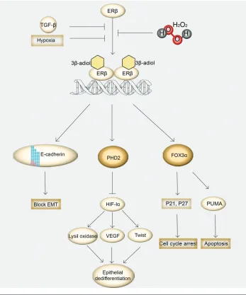
In the adult human prostate, ERβis characterized as an important mediator of epithelial differentiation (37). The mechanisms through which ERβ main-tain differentiation involve the degrada-tion of hypoxia-inducible factor 1α (HIF-1α) (38). ERβenhances transcrip-tion of prolyl hydroxylase containing protein 2 (PHD2) that hy-droxylates HIF-1αand marks HIF for destruction by the von Hippel-Lindau tumor suppressor (VHL) (39). Addition-ally, ERβappears to have antiprolifera-tive actions which are independent from the alternations of systemic androgen concentration and the activation of ERα, as documented in aromatase-knockout mice treated with ERβ-specific agonists (40). There, ERβseemed to have a sup-pressive role in the proliferation
cess, stimulating the differentiation of adult prostate epithelial cells.
THE ROLE OF ERβIN PROSTATE CARCINOGENESIS AND DISEASE PROGRESSION
ERβand Isoforms Expression
The role of ERβin the PCa initiation has been supported by studies using the ER-knockout (ERKO) mice. ERα -knock-out mice do not develop prostate cancer after testosterone and/or estrogen treat-ment, whereas mice lacking ERβreceptor develop prostate cancer after the addi-tion of sex hormones, similarly to wild-type mice (41). This antiproliferative role of ERβalso concurs with immunohisto-chemical findings in human PCa tissue samples, suggesting that ERβexpression is lost in high-grade tumors (42,43,44). Therefore, the loss of ERβmay be consid-ered as a prognostic factor of prostate cancer.
ERβisoforms in normal and cancerous prostate are differentially expressed. Transcriptional and posttranscriptional regulatory mechanisms may correlate with this phenomenon. In the transcrip-tional level, alternative promoter usage results in various amounts of ΕRβ tran-scripts. It has been proposed that pro-moters 0K and 0N upstream of exon1 (further described in [45]), are the regula-tory promoters of ERβexpression in the prostate and that ERβ1and ERβ2are transcribed from both 0N and 0K moters. On the contrary, only the 0K pro-moter is used for ERβ5transcription. The control of 0N promoter methylation of CpG islands located in the 5′-flanking se-quence of the 0N promoter results in loss of expression of ERβduring the develop-ment of PCa. AP-2 regulates the tran-scription of ERβby acting through a methylation hotspot of the 0N promoter in prostate cancer cells. Loss of protein AP-2 allows methylation at the critical AP-2 binding domain in ERβpromoter (46). Interestingly, very recently, a regula-tion between ERβ2 and ΕRβ1 has been addressed; ERβ2 is proposed to repress ERβ1transcription, thereby affecting its
Figure 2.
Meta
bolism of estr
ogen receptor
β
ph
ysiological lig
and,
3
β
-adiol.
Ar
om
atase con
v
er
ts estr
adiol into testoster
one,
w
hich is con
v
er
ted do
wnstream to dih
y-dr
otestoster
one (DHT) b
y 5
α
-reductase.
DHT is meta
bolized to 3
β
-adiol in tw
o w
ays; 3-k
eto reduction of DHT to 3
β
-adiol b
y enzyme
AKR1C1 (or
AKR1C3) or alter
na-tiv
ely 3-k
eto reduction of DHT to 3
α
-adiol b
y
AKR1C2 f
ollo
w
ed b
y 3
α
- to 3
β
-h
ydr
o
xyster
oid epimer
ization b
y B17b h
ydr
o
xyster
oid deh
ydr
ogenase (17bHSD6).
No m
at-ter the or
igin,
3
β
-adiol is fur
ther meta
bolized to tr
iols b
protective and favorable cellular re-sponses (47).
The combinations of 5′untranslated exons, known as exons 0Xs, are closely correlated with promoter 0K and are present in 5′untranslated regions of ERβ2and ERβ5 but not ERβ1. In the posttranscriptional level, upstream open reading frames (uORFs) can inhibit translation of transcripts composed of exons 0K and 0X. In PCa cells, there is a lower proportion of ERβ2 and ERβ5 tran-scripts containing exon 0Xs than in nor-mal cells, suggesting that elevated pro-tein expression in cancer cells promotes invasion and metastasis (48).
ERβand Oxidative Stress
An important factor favoring PCa initi-ation is oxidative stress (OS), which is as-sociated with inflammation, a possible precursor in neoplastic transformation of the prostate (49). Interestingly, oxidative stress is associated with aggressive phe-notypes of prostate cancer (50) while an-tioxidants, meanwhile, have a positive role in prostate cancer chemoprevention (51). Moreover, cell lines with high amounts of ERβand a low ratio of ERα/ERβhave a high expression of anti-oxidant enzymes and uncoupling pro-teins, resulting in lower oxidative stress (52). Origins of oxidative stress in prostate cancer include the mitochondr-ial hydrogen peroxide (H2O2) production through cytochrome c oxidation and the extramitochondrial origin of H2O2via nicotinamide adenine dinucleotide phos-phate-oxidase (NADPH) oxidases (53). PCa cells can produce increased levels of reactive oxygen species (ROS) and change the redox status of their microen-vironment in response to locally pro-duced transforming growth factor β1 (TGFβ1) prooxidant signals as revealed in DU145 PCa cells (54). Interestingly, ox-idative stress inactivates ERβby inhibit-ing the receptor’s dimerization by alter-ing the second zinc-falter-inger motif in the ERβstructure and therefore destabilizing its DNA-binding capacity (55). As a re-sult, ERβloses its ability to regulate vari-ous genes.
Switching Roles Theory
Recently, a theory of switching roles of ER during prostate carcinogenesis has been proposed, based on observations that elevated ERβprotein levels are de-tected in castration-resistant prostate cancer cells (CRPC), whereas these levels are related to lower survival in hormone-naive prostate cancer cells (HNPC). The theory suggests that in the early phases of PCa progression, ERβpresents a tumor-suppressing role and then is al-tered toward a tumor-promoting agent. It also proposes that ERβsignaling path-way in HNPC is mediated by Serin210-phosphorylated androgen receptor [pAR(S210)], but this observation disap-pears with the approach of CRPC, when AR gene stimulation takes over, perhaps in relevance to the lower levels of andro-gen in the body. The exact cause of this switch is not fully understood and the pathway that triggers the connection be-tween pAR(S210) and ERβin HNPC, if any exists, is currently unknown (56,57).
ERβIsoforms during Prostate Cancer Progression
Multiple variants of ERβexist, having possibly significant roles in PCa patho-physiology. Recent evidence suggests that apart from enhancing proliferation, ERβ2 promotes cancer cell migration and invasion, inducing the expression of fac-tors involved in bone metastasis (58). In addition to elevated levels of ERβ2 asso-ciated with epithelial-to-mesenchymal transition (EMT), ERβ2 may have the ability to suppress the expression of ERβ1 (47,59), leading to EMT (59). After immunolocalization of gastrin-releasing peptide receptor (GRPR) in human PCa, the association of ERβ2 with PCa was first determined by Nagasaki et al., indi-cating a correlation between immunore-activity of GRPR, Gleason score and ERβ2, supporting the hypothesis that ERβ2 contributes to prostate carcinogen-esis through GRPRexpression in PCa cells (60). Apparently, ERβ2 and ΕRβ5 can act as cancer-enhancing molecules involving cell migration and invasion under specific circumstances (17).
ERαand Epithelial-to-Mesenchymal Transition (EMT)
It is well documented that high grade PCa cells lose their epithelial characteris-tics and exhibit mesenchymal features, including increased hypoxia-inducible factor-1α, vimentin and vascular en-dothelial growth factor (VEGF) expres-sion (38) and loss of the E-cadherin, an epithelial cell adhesion protein, events typical of EMT phenomenon (61,62). 3β-adiol, the natural ligand of ERβ, pro-motes binding of dimerized ERβto DNA promoter of E-cadherin (CDH1), stimulat-ing its transcription (29). Hypoxic expo-sure and TGFβ1 signaling can induce EMT and decrease ERβexpression in both AR- dependent and AR-independent cells. Similarly, silencing of ERβwith short hairpin RNA techniques was ade-quate to promote EMT (38). The mecha-nism by which ERβactivation by 3β-adiol is associated to EMT inhibition in PCa cells involves the degradation of hypoxia inducible factor 1α(HIF-1α), a crucial EMT factor (38). HIF-1α-inducible genes, such as lysil oxidase (LOX), an enzyme that catalyzes cross-linking of extracellu-lar matrix, and transcription factor TWIST, mediate epithelial dedifferentia-tion, leading to invasion and metastasis of the prostate cancer (63,64). Thus, ERβ acts as the “gatekeeper” of the epithelial phenotype in the prostate gland.
ERβAS A USEFUL THERAPEUTIC TARGET FOR PCA TREATMENT
has been proposed to affect PCa acting as a cell cycle regulator, controlling the ex-pression of cyclin D1 (CCND1) and af-fecting its downstream pathway (67), supporting further the notion that restor-ing ERβexpression may provide a new promising therapeutic approach for PCa.
Indeed, immunohistochemical studies have suggested that high grade tumors express ERβ(68) and that human PCa DU145 cells have the ability to activate ERβby generating specific ligands by the transformation of androgen precursors produced by stromal cells (54). However, local paracrine signals and ROS of stro-mal cells can limit the antitumor activity of ERβ. Enzymes responsible for de novo steroidogenesis are not highly expressed in prostate gland, thus androgen metabo-lites may be the main ligand sources for ER-dependent signaling (69). In addition, in obese patients, aromatase enzyme is downregulated in prostate stroma sug-gesting that obesity can alter sex steroid production in stromal cells (70). There-fore, any treatment aiming toward the regulation of ERβactivity also should address the regulation of steroidogenesis in prostate tissue, thus targeting tumor microenvironment.
ERβAgonists
Having in mind the ERβ suppressing functions, researchers have been interested in the development of specific agonists (71). In a study that in-vestigates the therapeutic potential of 8β -VE2, a potent synthetic selective agonist for ERβ, using AR-knockout (ARKO) mice, researchers suggested that ERβis responsible for androgen-independent apoptosis in prostatic stroma and epithe-lia, an effect that requires tumor necrosis factor-α(TNFα) signaling. Moreover, the same study showed that 8β-VE2-activated ERβinduces apoptosis in independent PCa cells including PC3 and DU145, as well as in primary human PCa xenografts. The pathway that mediates the link between ERβand TNFα, if any exists, is not currently known, although the authors docu-mented caspase-8 and -3 activation (72).
A selective estrogen-receptor modulator (SERM) named ICI 182,780 exerted a dose-dependent growth inhibition action on DU145 cells, which was mediated by ERβ(73) through binding to NF-κB and enhancement of transcription factor FOXO1. Additionally, another SERM, raloxifene, induced the apoptosis and in-hibited proliferation of both
dependent and -independent cell lines via the activation of ERβ, lowering Bcl-2 expression, and increasing caspase-3 and Par-4 levels (74,75).
Although preclinical studies described above point out the protective effect of ERβ, the clinical utility of ERβ-selective agents in the treatment of men with PCa has never been proved. A possible
son for the inconsistency between in vitroand in vivofindings is that the prostate cancer cell lines (LNcAP, PC-3, DU145) used in in vitrostudies exhibit differential expression profiles of the nu-clear receptors (AR, ERαand ERβand cannot represent the human tissue. Fur-thermore, these model cell lines are not reflecting the complex cross-talk be-tween AR, ERαand ERβand other stro-mal:epithelial interactions known to occur in vivo(76).
Phytoestrogens
Phytoestrogens are natural com-pounds that mimic the biological activ-ity of estrogens with a binding prefer-ence for ERβ(77) and have the ability to upregulate ERβ, which is lost during PCa progression. Mice with PCa lacking ERαor ERβtreated with phytoestrogens showed that cancer did not progress in ERαKO mice whereas ERβΚΟmice pre-sented an increasing incidence of poorly differentiated carcinoma (Gleason scores 4 and 5) (78). Re-expression of ERβvia phytoestrogens elicited antiandrogenic effects, including downregulation of AR and its coactivators (79). In addition, many phytoestrogens were found to stimulate the expression of p21 (cyclin-dependent kinase inhibitor 1) through ER, including genistein and silymarin (a polyphenolic flavonoid extracted from plant Silybum marianum) (80,81). Ongo-ing studies suggest that phytoestrogens can be used in treatment and/or preven-tion of PCa, as supported by findings that men who receive a rich phytoestro-gen diet present a lower incidence of PCa (82).
CONCLUSION
ERβhas been proposed as a mediator of epithelial differentiation and as an antiproliferative molecule, mediating many molecular pathways on PCa. ERβ protects epithelial integrity and block EMT by upregulating transcription of E-cadherin (CDH1), an epithelial adhe-sion protein. Furthermore, ERβ upregu-lates the expression of PHD2, that hy-droxylates the tumor enhancer HIF-1α
and marks HIF for destruction by the von Hippel- Lindau tumor suppressor (VHL). ERβsignaling has antiprolifera-tive effects on the prostate, enhancing the expression of FOXO3α, and conse-quently upregulating apoptotic genes including PUMA, a proapoptotic pro-tein, and p21, a regulator of cell cycle progression. However, ERβis sensitive to putative changes in tumor microen-vironment, thus hypoxic conditions and TGFβ1 signaling diminish ERβlevels and alter its action, favoring PCa pro-gression (Figure 3).
Despite its tumor-suppressing role, ERβhas also been proposed as a cancer-promoting factor. Interestingly, higher levels of ERβcorrespond to lower sur-vival in HNPC cells through the activa-tion of pAR(s210), although the exact mechanism remains unknown. Addition-ally, ERβ1 has been proposed to form a complex with AR, resulting in the tran-scription of AR-dependent genes in PCa (76). Moreover, its spliced variants, ERβ2 and ERβ5, have tumor-promoting actions and are stimulated after dimerization with ERβ1. Although regulation of ERβ1 expression occurs mainly at the tran-scriptional level, ERβ2and ERβ5 expres-sion is controlled at both the transcrip-tional and posttranscriptranscrip-tional levels, via complex interactions that involves pro-moter methylation on CpG islands and uORFs. Further findings are needed to elucidate the exact molecular functions of these isoforms.
Future studies should focus on under-standing the molecular mechanism gov-erning the controversial findings on ERβ function and its spliced variants in can-cerous prostate to develop ERβ-based therapeutic agents for prostate cancer treatment and other estrogen-dependent processes (83).
DISCLOSURES
The authors declare they have no competing interests as defined by Molec-ular Medicine, or other interests that might be perceived to influence the re-sults and discussion reported in this paper.
REFERENCES
1. Huggins C, Stevens RE Jr, Hodges CV. (1941) Studies on prostatic cancer: Ii. the effects of cas-tration on advanced carcinoma of the prostate gland. Arch. Surg. 43:209–23.
2. Koutsilieris M, Tolis G. (1985) Long-term follow-up of patients with advanced prostatic carcinoma treated with either buserelin (HOE 766) or or-chiectomy: classification of variables associated with disease outcome. Prostate. 7:31–9. 3. Klotz L, McNeill I, Fleshner N. (1999) A phase 1–2
trial of diethylstilbestrol plus low dose warfarin in advanced prostate carcinoma. J. Urol. 161:169–72. 4. Jensen EV. (1960) Fate of steroid estrogens in
tar-get tissues. In: Biological Activities of Steroids in Relation to Cancer. Vollmer P.E (ed.) Academic Press, New York, pp. 61–174.
5. Kuiper GG, Enmark E, Pelto-Huikko M, Nilsson S, Gustafsson JA. (1996) Cloning of a novel re-ceptor expressed in rat prostate and ovary. Proc.
Natl. Acad. Sci. U. S. A. 93:5925–30.
6. Hartman J, Ström A, Gustafsson JA. (2012) Cur-rent concepts and significance of estrogen recep-tor βin prostate cancer. Steroids. 77:1262–6.
7. Enmark E, et al. (1997) Human estrogen receptor
beta-gene structure, chromosomal localization, and expression pattern. J. Clin. Endocrinol. Metab. 82:4258–65.
8. Fixemer T, Remberger K, Bonkhoff H. (2003) Dif-ferential expression of the estrogen receptor beta (ERbeta) in human prostate tissue, premalignant changes, and in primary, metastatic, and recurrent prostatic adenocarcinoma. Prostate. 54:79–87. 9. Royuela M, et al. (2001) Estrogen receptors alpha
and beta in the normal, hyperplastic and carcino-matous human prostate. J. Endocrinol. 168:447–54. 10. Heldring N, et al. (2007) Estrogen receptors: how
do they signal and what are their targets. Physiol.
Rev. 87:905–31.
11. Powell E, et al. (2012) Identification of estrogen receptor dimer selective ligands reveals growth-inhibitory effects on cells that co-express ERα
and ERβ. PLoS ONE. 7:e30993.
12. Chakraborty S, Willett H, Biswas PK. (2012) In-sight into estrogen receptor beta–beta and alpha–beta homo- and heterodimerization: A combined molecular dynamics and sequence analysis study. Biophys. Chem. 170:42–50. 13. Walton TJ, et al. (2009) Quantitative RT-PCR
anal-ysis of estrogen receptor gene expression in laser microdissected prostate cancer tissue. Prostate. 69:810–9.
14. Hurtado A, et al. (2008) Estrogen receptor beta dis-plays cell cycle-dependent expression and regulates the G1 phase through a non-genomic mechanism in prostate carcinoma cells. Cell. Oncol. 30:349–65. 15. Leav I, et al. (2001) Comparative studies of the
estrogen receptors beta and alpha and the andro-gen receptor in normal human prostate glands, dysplasia, and in primary and metastatic carci-noma. Am. J. Pathol. 159:79–92.
un-derstanding ER-beta signaling. Proc. Natl. Acad.
Sci. U.S.A. 103:13162–7.
17. Leung YK, et al. (2010) Estrogen receptor beta2 and beta5 are associated with poor prognosis in prostate cancer, and promote cancer cell mi-gration and invasion. Endocr. Relat. Cancer. 17:675–89.
18. Moore JT, et al. (1998) Cloning and characteriza-tion of human estrogen receptor beta isoforms.
Biochem. Biophys. Res. Commun. 247:75–8.
19. Younes M, Honma N. (2011) Estrogen receptor β.
Arch. Pathol. Lab. Med. 135:63–6.
20. Peng B, Lu B, Leygue E, Murphy LC. (2003) Pu-tative functional characteristics of human estro-gen receptor-beta isoforms. J. Mol. Endocrinol. 30:13–29.
21. Celhay O, et al. (2010) Expression of estrogen re-lated proteins in hormone refractory prostate can-cer: association with tumor progression. J. Urol. 184:2172–8.
22. Ghosh D, Griswold J, Erman M, Pangborn W. (2009) Structural basis for androgen specificity and oestrogen synthesis in human aromatase.
Nature. 457:219–23.
23. Ellem SJ, Risbridger GP. (2010) Aromatase and reg-ulating the estrogen:androgen ratio in the prostate gland. J. Steroid Biochem. Mol. Biol. 118:246–51. 24. Muthusamy S, et al. (2011) Estrogen receptor βand
17?β-hydroxysteroid dehydrogenase type 6, a
growth regulatory pathway that is lost in prostate cancer. Proc. Natl. Acad. Sci. U.S.A. 108:20090–4. 25. Sugiyama N, Barros RP, Warner M, Gustafsson JA. (2010) ERbeta: recent understanding of estro-gen signaling. Trends Endocrinol. Metab. 21:545–52. 26. BeraničN, Stefane B, Brus B, Gobec S, Rižner TL. (2013) New enzymatic assay for the AKR1C en-zymes. Chem. Biol. Interact. 202:204–9. 27. Baker ME. (2013) What are the physiological
es-trogens? Steroids. 78:337–40.
28. Weihua Z. et al. (2001) A role for estrogen recep-tor beta in the regulation of growth of the ventral prostate. Proc. Natl. Acad. Sci. U.S.A. 98:6330–5. 29. Grubisha MJ. (2013) Local endocrine, paracrine and redox signaling networks impact estrogen and androgen crosstalk in the prostate cancer mi-croenvironment. Steroids. 78:538–41.
30. Dondi D, et al. (2010) Estrogen receptor beta and the progression of prostate cancer: role of 5alpha-androstane-3beta,17beta-diol. Endocr. Relat. Can-cer. 17:731–42.
31. Dey P, Ström A, Gustafsson JA. (2013) Estrogen
re-ceptor βupregulates FOXO3a and causes
induc-tion of apoptosis through PUMA in prostate can-cer. Oncogene. 2013, Sept. 30. [Epub ahead of print]. 32. Marker PC, Donjacour AA, Dahiya R, Cunha GR. (2003) Hormonal, cellular, and molecular control of prostatic development. Dev. Biol. 253:165–74. 33. Omoto Y, Imamov O, Warner M, Gustafsson JA.
(2005) Estrogen receptor alpha and imprinting of the neonatal mouse ventral prostate by estrogen.
Proc. Natl. Acad. Sci. U.S.A. 102:1484–9. 34. Prins GS, Huang L, Birch L, Pu Y. (2006) The role
of estrogens in normal and abnormal
develop-ment of the prostate gland. Ann. N. Y. Acad. Sci. 1089:1–13.
35. Shapiro E, et al. (2005) Immunolocalization of es-trogen receptor alpha and beta in human fetal prostate. J. Urol. 174:2051–3.
36. Adams JY, Leav I, Lau KM, Ho SM, Pflueger SM. (2002) Expression of estrogen receptor beta in the fetal, neonatal, and prepubertal human prostate.
Prostate. 52:69–81
37. Imamov O, et al. (2004) Estrogen receptor beta regulates epithelial cellular differentiation in the mouse ventral prostate. Proc. Natl. Acad. Sci.
U.S.A. 101:9375–80.
38. Mak P, et al. (2010) ERbeta impedes prostate cancer EMT by destabilizing HIF-1alpha and inhibiting VEGF-mediated snail nuclear localization: implica-tions for Gleason grading. Cancer Cell. 17:319–32. 39. Mak P, Chang C, Pursell B, Mercurio AM. (2013)
Estrogen receptor βsustains epithelial differentia-tion by regulating prolyl hydroxylase 2 transcrip-tion. Proc. Natl. Acad. Sci. U.S.A. 110:4708–13. 40. McPherson SJ, et al. (2007) Essential role for
estro-gen receptor beta in stromal-epithelial regulation of prostatic hyperplasia. Endocrinology. 148:566–74. 41. Ricke WA, et al. (2008) Prostatic hormonal
car-cinogenesis is mediated by in situ estrogen pro-duction and estrogen receptor alpha signaling.
FASEB J. 22:1512–20.
42. Gabal SM, Habib FM, Helmy DO, Ibrahim MF. (2007) Expression of estrogen receptor-B (ER-B ) in benign and malignant prostatic epithelial cells and its correlation with the clinico-pathological features. J. Egypt Natl. Canc. Inst. 19:239–48. 43. Gallardo F, et al. (2007) Expression of androgen,
oestrogen alpha and beta, and progesterone re-ceptors in the canine prostate: differences be-tween normal, inflamed, hyperplastic and neo-plastic glands. J. Comp. Pathol. 136:1–8. 44. Bardin A, Boulle N, Lazennec G, Vignon F, Pujol
P. (2004) Loss of ERbeta expression as a common step in estrogen-dependent tumor progression.
Endocr. Relat. Cancer. 11:537–51.
45. Li LC, Yeh CC, Nojima D, Dahiya R. (2000) Cloning and characterization of human estrogen receptor beta promoter. Biochem. Biophys. Res.
Commun. 275:682–9.
46. Zhang X, Leung YK, Ho SM. (2007) AP-2 regulates the transcription of estrogen receptor (ER)-beta by acting through a methylation hotspot of the 0N pro-moter in prostate cancer cells. Oncogene. 26:7346–54 47. Cotrim CZ, Fabris V, Doria ML, Lindberg K,
Gustafsson J. (2013) Estrogen receptor beta growth-inhibitory effects are repressed through activation of MAPK and PI3K signalling in mammary epithe-lial and breast cancer cells. Oncogene. 32:2390–402. 48. Lee MT, Ouyang B, Ho SM, Leung YK. (2013)
Differential expression of estrogen receptor beta isoforms in prostate cancer through interplay be-tween transcriptional and translational regula-tion. Mol. Cell. Endocrinol. 376:125–35.
49. Gupta-Elera G, Garrett AR, Robison RA, O’Neill KL. (2012) The role of oxidative stress in prostate cancer. Eur. J. Cancer Prev. 21:155–62.
50. Kumar B, Koul S, Khandrika L, Meacham RB, Koul HK. (2008) Oxidative stress is inherent in prostate cancer cells and is required for aggres-sive phenotype. Cancer Res. 68:1777–85. 51. Thapa D, Ghosh R. (2012) Antioxidants for
prostate cancer chemoprevention: challenges and opportunities. Biochem. Pharmacol. 83:1319–30. 52. Miró AM, et al. (2011b) 17β-estradiol regulates oxidative stress in prostate cancer cell lines ac-cording to ERalpha/ERbeta ratio. J. Steroid Biochem. Mol. Biol. 123:133–9.
53. Rebillard A, Lefeuvre-Orfila L, Gueritat J, Cillard J. (2013) Prostate cancer and physical activity: adaptive response to oxidative stress. Free Radic.
Biol. Med. 60:115–24.
54. Grubisha MJ, Cifuentes ME, Hammes SR, De-franco DB. (2012) A local paracrine and
en-docrine network involving TGFβ, Cox-2, ROS,
and estrogen receptor βinfluences reactive stro-mal cell regulation of prostate cancer cell motil-ity. Mol. Endocrinol. 26:940–54.
55. Whittal RM, et al. (2000) Preferential oxidation of zinc finger 2 in estrogen receptor DNA-binding domain prevents dimerization and, hence, DNA binding. Biochemistry. 39:8406–17.
56. Zellweger T, et al. (2013) Estrogen receptor β ex-pression and androgen receptor phosphorylation correlate with a poor clinical outcome in hormone-naive prostate cancer and are elevated in castration- resistant disease. Endocr. Relat. Can-cer. 20:403–13.
57. Savoy RM, Ghosh PM. (2013) The changing roles of steroid nuclear receptors with prostate cancer progression. Endocr. Relat. Cancer. 20:C9–11. 58. Dey P, et al. (2012) Estrogen receptors β1 and β2
have opposing roles in regulating proliferation and bone metastasis genes in the prostate cancer cell line PC3. Mol. Endocrinol. 26:1991–2003. 59. Alonso-Magdalena P, et al. (2009) A role for
ep-ithelial-mesenchymal transition in the etiology of benign prostatic hyperplasia. PNAS. 106:2859–63. 60. Nagasaki S, et al. (2012) Immunohistochemical
analysis of gastrin-releasing peptide receptor (GRPR) and possible regulation by estrogen re-ceptor βcx in human prostate carcinoma. Neo-plasma. 59:224–32.
61. Huber MA, Kraut N, Beug H. (2005) Molecular requirements for epithelial-mesenchymal transi-tion during tumor progression. Curr. Opin. Cell Biol. 17:548–58.
62. Cavallaro U, Christofori G. (2004) Multitasking in tumor progression: signaling functions of cell ad-hesion molecules. Ann. N. Y. Acad. Sci. 1014:58–66. 63. Pez F, et al. (2011) The HIF-1–inducible lysyl
oxi-dase activates HIF-1 via the Akt pathway in a positive regulation loop and synergizes with
HIF-1 in promoting tumor cell growth. Cancer
Res. 71:1647–57.
64. Yang MH, et al. (2008) Direct regulation of TWIST by HIF-1alpha promotes metastasis. Nat. Cell Biol. 10:295–305.
re-express estrogen receptor beta and induce apo-ptosis in prostate cancer cell-lines. Prostate. 68:210–22.
66. Cheng J, Lee EJ, Madison LD, Lazennec G. (2004) Expression of estrogen receptor beta in prostate carcinoma cells inhibits invasion and proliferation and triggers apoptosis. FEBS Lett. 566:169–72.
67. Nakamura Y, et al. (2013) Cyclin D1 (CCND1)
expression is involved in estrogen receptor beta (ERβ) in human prostate cancer. Prostate. 73:590–5.
68. Lai JS, et al. (2004) Metastases of prostate cancer express estrogen receptor-beta. Urology. 64:814–20. 69. Hofland J, et al. (2010) Evidence of limited
contri-butions for intratumoral steroidogenesis in prostate cancer. Cancer Res. 70:1256–64. 70. Gross M, et al. (2009) Expression of androgen and
estrogen related proteins in normal weight and obese prostate cancer patients. Prostate. 69:520–7. 71. Meyers MJ, et al. (2001) Estrogen receptor-beta
potency-selective ligands: structure-activity rela-tionship studies of diarylpropionitriles and their acetylene and polar analogues. J. Med. Chem. 44:4230–51.
72. McPherson SJ, et al. (2010) Estrogen receptor-beta activated apoptosis in benign hyperplasia and cancer of the prostate is androgen independent and TNFalpha mediated. Proc. Natl. Acad. Sci.
U.S.A. 107:3123–8.
73. Lau KM, LaSpina M, Long J, Ho SM. (2000) Ex-pression of estrogen receptor (ER)-alpha and ER-beta in normal and malignant prostatic epithelial cells: regulation by methylation and involvement in growth regulation. Cancer Res. 60:3175–82. 74. Rossi V, et al. (2011) Raloxifene induces cell death
and inhibits proliferation through multiple sig-naling pathways in prostate cancer cells express-ing different levels of estrogen receptor αand β.
J. Cell. Physiol. 226:1334–9.
75. Kim IY, et al. (2002) Raloxifene, a selective estro-gen receptor modulator, induces apoptosis in an-drogen-responsive human prostate cancer cell line LNCaP through an androgen-independent pathway. Cancer Res. 62:3649–53.
76. Nelson AW, Tilley WD, Neal DE, Carroll J. (2014) Estrogen receptor beta in prostate cancer: friend or foe? Endocr. Relat. Cancer. 2014, Jan. 8 [Epub ahead of print].
77. Patisaul HB, Jefferson W. (2010) The pros and cons of phytoestrogens. Front. Neuroendocrinol. 31:400–19. 78. Slusarz A, et al. (2012) Aggressive prostate
can-cer is prevented in ERαKO mice and
stimu-lated in ERβKO TRAMP mice. Endocrinology.
153:4160–70.
79. Thelen P, Wuttke W, Seidlová-Wuttke D. (2014) Phytoestrogens selective for the estrogen recep-tor beta exert anti-androgenic effects in castration resistant prostate cancer. J. Steroid Biochem. Mol.
Biol. 139:290–3.
80. Matsumura K, Tanaka T, Kawashima H, Nakatani T. (2008) Involvement of the estrogen receptor beta in genistein-induced expression of
p21(waf1/cip1) in PC-3 prostate cancer cells.
Anticancer Res. 28:709–14.
81. Atawia, RT, Tadros MG, Khalifa AE, Mosli HA, Abdel-Naim AB. (2013) Role of the phytoestro-genic, pro-apoptotic and anti-oxidative properties of silymarin in inhibiting experimental benign pro-static hyperplasia in rats. Toxicol. Lett. 219:160–9 82. Yan L, Spitznagel EL. (2009) Soy consumption and prostate cancer risk in men: a revisit of a meta-analysis. Am. J. Clin. Nutr. 89:1155–63. 83. Koutsilieris M. (1992) Pathophysiology of uterine