R E S E A R C H A R T I C L E
Open Access
Coordinated trafficking of synaptic vesicle and
active zone proteins prior to synapse formation
Luke AD Bury
1and Shasta L Sabo
1,2*Abstract
Background:The proteins required for synaptic transmission are rapidly assembled at nascent synapses, but the mechanisms through which these proteins are delivered to developing presynaptic terminals are not understood. Prior to synapse formation, active zone proteins and synaptic vesicle proteins are transported along axons in distinct organelles referred to as piccolo-bassoon transport vesicles (PTVs) and synaptic vesicle protein transport vesicles (STVs), respectively. Although both PTVs and STVs are recruited to the same site in the axon, often within minutes of axo-dendritic contact, it is not known whether or how PTV and STV trafficking is coordinated before synapse formation.
Results:Here, using time-lapse confocal imaging of the dynamics of PTVs and STVs in the same axon, we show that vesicle trafficking is coordinated through at least two mechanisms. First, a significant proportion of STVs and PTVs are transported together before forming a stable terminal. Second, individual PTVs and STVs share pause sites within the axon. Importantly, for both STVs and PTVs, encountering the other type of vesicle increases their propensity to pause. To determine if PTV-STV interactions are important for pausing, PTV density was reduced in axons by expression of a dominant negative construct corresponding to the syntaxin binding domain of syntabulin, which links PTVs with their KIF5B motor. This reduction in PTVs had a minimal effect on STV pausing and movement, suggesting that an interaction between STVs and PTVs is not responsible for enhancing STV pausing.
Conclusions:Our results indicate that trafficking of STVs and PTVs is coordinated even prior to synapse
development. This novel coordination of transport and pausing might provide mechanisms through which all of the components of a presynaptic terminal can be rapidly accumulated at sites of synapse formation.
Background
In the cortex, the bulk of synapse formation occurs at high rates over a period of several weeks during early postnatal development. Formation of individual synapses is triggered when axons and dendrites contact one another [1,2]. For these axo-dendritic contacts to stabi-lize and form a synapse, synaptic vesicle and active zone proteins must be recruited to the site of contact very rapidly, within minutes to hours [3-10]. This rapid assembly is remarkable, considering that each presynap-tic protein needs to be transported from the soma to individual sites of synapse formation along the axon.
Recent work using live imaging of developing neurons has revealed mechanisms that might facilitate rapid synapse assembly. For example, rather than transport each molecule individually, neurons package multiple presynaptic components into vesicles in the cell body and transport the entire group together. Two major groups of proteins are transported in this fashion: active zone proteins are transported in piccolo-bassoon trans-port vesicles (PTVs), while synaptic vesicle-associated proteins are transported in synaptic vesicle protein transport vesicles (STVs) [4,9,11-17]. PTVs and STVs can be distinguished both morphologically and bio-chemically. PTVs have been described as dense-core vesicles or aggregates of vesicles and proteins that range in size from approximately 80 nm in diameter for dense core vesicles to 130 nm by 220 nm in area for aggre-gates [12,16]. Proteins transported by PTVs include
* Correspondence: [email protected]
1
Department of Pharmacology, Case Western Reserve University School of Medicine, Cleveland, Ohio, 44106, USA
Full list of author information is available at the end of the article
piccolo, bassoon, N-cadherin and syntaxin [11,12, 16,18,19]. STVs are heterogeneous in size and shape and are composed of both tubulovesicular and clear core vesicles [9,13,14]. Proteins carried by STVs include synaptophysin, synapsin Ia, synaptotagmin and synapto-brevin/vesicle-associated membrane protein 2 (VAMP2) [4,9,13,14,16]. Both types of vesicles are packaged via the trans-Golgi network before being transported through the axon [14,16,20].
Rapid assembly of presynaptic terminals can also be facilitated by having the source of synaptic proteins nearby and readily available. Indeed, many STVs and PTVs are mobile within axons well before synapse for-mation. STVs and PTVs travel in a saltatory fashion in both the anterograde and retrograde directions along the entire length of the axon [4,8,11,13-15,17,19]. This provides a readily available pool of synaptic vesicle and active zone proteins wherever and whenever axo-dendri-tic contact occurs.
Since both STVs and PTVs must be delivered to the same site during synapse assembly, we hypothesized that trafficking of STVs and PTVs might be coordinated even prior to synapse assembly, providing an additional mechanism to facilitate rapid accumulation of the full complement of proteins required for synaptic transmis-sion. Such coordination could arise through co-transport of STVs and PTVs, through pausing of STVs and PTVs at the same sites, or both. Coordinated pausing presents a particularly intriguing possibility since sites along the axon where STVs pause their transport are preferential sites of synapse formation [4]. It was previously sug-gested that cues that control pausing at these sites could promote rapid synapse assembly by increasing the prob-ability that STVs are at or near any given site when axo-dendritic contact occurs. If this model is correct, then synapse assembly would be most efficient if PTVs are also attracted to these same sites. However, it is not yet known whether PTVs also pause at these sites and, if so, whether they do so simultaneous with STVs. A recent report demonstrated that PTVs and STVs (parti-cularly those resembling small, clear vesicles) can be observed tethered together in electron micrographs of developing axons [16]. These aggregates could corre-spond to PTVs and STVs that are either being co-trans-ported or pausing at the same site at the same time.
Here, we tested whether transport and pausing of PTVs and STVs is coordinated prior to synapse assem-bly using time-lapse confocal imaging of green and red fluorescent protein-tagged synaptic vesicle and active zone proteins within the same axon. We found that a significant portion of STVs and PTVs move together and share pause sites. Interestingly, both STVs and PTVs preferentially paused at these sites when another vesicle was present. These observations raised the
question of whether a direct interaction between STVs and PTVs coordinates their transport and pausing. Reducing PTVs in the axon minimally affected the movement and pausing of STVs, arguing against this mechanism and suggesting that other unidentified sig-nals are responsible for STV and PTV coordination. These findings represent novel mechanisms that can facilitate the rapid recruitment of presynaptic proteins to the same site within the axon and, therefore, promote synapse development.
Results
STVs and PTVs can move together
STVs and PTVs move in a similar saltatory fashion within the axon in both the anterograde and retrograde directions [4,9,11,13,14,19,21]. However, because STVs and PTVs have almost always been imaged separately, it is not yet known if and how STV and PTV trafficking interrelate. To determine this, we used time-lapse ima-ging of neurons co-transfected with fluorescent STV
and PTV markers. In this assay, 5- to 6-days in vitro
(DIV) rat cortical neurons were co-transfected with GFP-bassoon to label PTVs and either synaptophysin-mcherry or synaptophysin-monomeric red fluorescent protein (mRFP) to label STVs. Previous studies have indicated that fluorescent proteins attached to bassoon and synaptophysin effectively label PTVs and STVs, respectively [8,9,11,14,15,17,20,22]. One to two days after transfection, time-lapse images of the fluorescently labelled vesicles were obtained from individual axons. Images were taken every 10 s for 7.5 minutes. An exam-ple is shown in Movie 1 in Additional file 1.
From Additional file 1 and the kymographs in Figure 1A, it is clear that PTVs and STVs moved together in both anterograde and retrograde directions. To quantify these correlated movements, the positions of individual STV and PTV puncta were tracked independently while blind to the other channel, and then their movements were compared. To determine whether these correlated movements could be accounted for by chance, the experimental data were ultimately compared to model axons where the initial positions of all of the puncta were randomized but the density of puncta and proper-ties of their movements were derived from the experi-mental data (Figure 1B).
percentage of time STVs and PTVs spent moving together was over 2.5-fold higher than expected by chance (Figure 1D). These data indicate that populations of STVs and PTVs travel together within the axon, which could allow both sets of presynaptic proteins to be distributed together to sites of synapse formation.
STVs and PTVs display similar pausing characteristics
Although the movements of both STVs and PTVs have been extensively described individually, the pauses between movements have been less thoroughly analyzed and have not been compared. Comparing STV and PTV
pausing behavior is important since STVs preferentially pause at sites of eventual synapse formation [4,9]. When PTV behavior was examined, it was clear that PTV paus-ing exhibited many of the properties previously described for STVs [4] (Figure 2A,B). For example, the same PTV could be observed repeatedly pausing at a given site (Fig-ure 2B, top panel). In addition, multiple PTVs paused at the same site, both sequentially and simultaneously (Figure 2B, middle and bottom panels, respectively). When STV movements were quantified, the mean STV pause fre-quency was 0.0081 ± 0.0002 pauses/s (n = 262 vesicles; Figure 2C). The average STV pause duration was 107.4 ±
PTV
STV
% punc
ta mo
ving t
ogether
0
10
20
30
PTV
STV
% time together
0
20
40
60
n = 224 n = n = 97
22400
n = 9700
position along axon
GFP-bassoon
C
overlay
STVs
STV simulation PTVs
PTV simulation
B
time
synaptophysin-mcherry
A
D
1
2
3
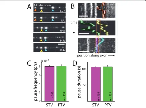
Figure 1STVs and PTVs move together.(A)Kymographs of an axon segment showing the movements of PTVs (GFP-bassoon; green) and STVs (synaptophysin-mcherry; magenta) in the same axon. Yellow lines underline the moving STVs and PTVs. Bottom panel: overlay of STV and
PTV fluorescence. On the ordinate axis, one pixel corresponds to 10 s. On the abscissa, the scale bar corresponds to 10μm.(B)Simulations of
model axons were performed by randomizing initial positions of vesicles while maintaining movement and pausing characteristics of the original imaged vesicles. Diagrams illustrate kymographs of three simulations of model axons. Movements of two vesicles are shown in each model
axon.(C)Plot showing the percentage of PTVs that move with STVs (green) and STVs that move with PTVs (magenta). PTV and STV simulations
(light green and light magenta, respectively) correspond to the predicted values from the simulations.(D)Quantification of the percentage of
3.4 s (n = 954 pauses; Figure 2D). Both measurements concur with previous observations [4]. As shown in Figure 2C,D, PTV pause frequency and duration in 7- to 8-DIV neurons were nearly identical to those in the STV data, with PTVs pausing at a frequency of 0.0082 ± 0.0003 pauses/s and average duration of 108.3 ± 3.5 s (n = 253 vesicles, n = 933 pauses). The similarities between the pausing behavior of STVs and PTVs raise the question of whether PTVs might pause with STVs at sites of eventual synapse formation. Like STVs [4], regulation of PTV paus-ing could be an important target of signals that control synapse assembly.
STVs and PTVs pause simultaneously at the same sites in the axon
For a synapse to develop, both active zone and synaptic vesicle proteins need to be recruited to the same site.
Sharing a pause site provides a potential mechanism for recruiting both STVs and PTVs to the same spot in the axon. Since places where STVs pause are preferred sites of synapse formation, it seemed likely that PTVs also pause at these sites. To test this hypothesis, STVs and PTVs were imaged and analyzed as described above. From the time-lapse panels and kymographs shown in Figure 3A,B, it is clear that both types of vesicles paused at sites where the other type of vesicle also paused.
To determine whether pausing of STVs and PTVs at the same sites was significantly greater than would be predicted by chance, pausing at the same sites was quantified in each axon then compared to simulations in model axons (Figure 3C). For STVs, 47.9% of pauses were at sites where a PTV also paused (n = 305 pauses; Figure 3D), compared to 24.5% using the same analysis of model axons (n = 33,967 pauses; Figure 3D). This
time
position along axon
C
STV
PTV
0 50 100
pause duration (s)
n = 954 n = 933
D
STV
PTV
0 2 4 6 8
x 10-3
pause fr
equenc
y (p/s)
n = 262 n = 253
B
GFP-bassoon
t = 0
15s
150s
345s
405s
A
Figure 2PTV pausing is qualitatively and quantitatively similar to STV pausing.(A)Time-lapse images of PTVs labeled with GFP-bassoon. Individual PTVs are tracked with red, yellow or blue arrows. Most PTVs paused, and pauses were of varied durations. Frames were collected at
the indicated times. Scale bars: 5μm.(B)Kymographs demonstrating the movements of PTVs in three axons. Individual PTVs paused repeatedly
at the same site (top panel), and multiple PTVs paused at the same site (middle and bottom panels). Pausing of multiple PTVs at a given site occurred both sequentially (middle panel) and simultaneously (bottom panel). Individual vesicles are highlighted in different colors for
visualization. Pause sites are outlined in orange. On the ordinate axis, one pixel corresponds to 10 s.(C,D)STVs (magenta) and PTVs (green)
t = 0
130s
150s
180s
190s
PTV
STV
0 20 40
% pause sit
es shar
ed
PTV
STV
0 20 40 60 80 100
% enc
ount
ers
resulting in pauses
position along axon
time
bassoon-GFP
B
D
E
A
overlay
1
2
3
STVs STV simulation
PTVs
PTV simulation
n = 90 n = 135 n = 615 n = 66582 n = 291 n = 32800
synaptophysin-mcherry
C
position along the axon (
P
m)
0 30 60 90 120 150 180
1
2
3
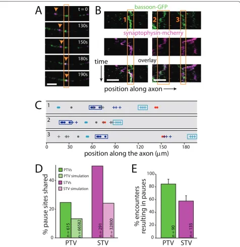
Figure 3STVs and PTVs pause at the same sites.(A)Time-lapse images of a segment of axon expressing both synaptophysin-mcherry (magenta) and GFP-bassoon (green). In each panel, fluorescence signals from the two channels are overlaid, and co-localization is indicated by
white. The orange box outlines a site where a PTV is paused. The arrow tracks a STV that then pauses at that site. Scale bars: 5μm.(B)
Kymographs showing two examples of STVs and PTVs that pause at the same sites. On the ordinate axis, one pixel corresponds to 10 s. Bottom
panel: overlay of STV and PTV fluorescence. Orange boxes outline three shared pause sites.(C)Simulations of model axons were performed by
randomizing initial positions of vesicles while maintaining movement and pausing characteristics observed for the original experimental vesicles. Diagrams show the superimposed locations of all pause sites for individual STVs and PTVs in model axons. Three simulations of the same experimental data are shown. STV pause sites are indicated with a dot while PTV pause sites are indicated with a plus sign. Each color represents an individual vesicle. Cyan and magenta boxes outline the pause sites of the same PTV and STV, respectively, in each model axon. For each axon imaged, 100 such simulations were performed, allowing estimation of the degree of co-transport and co-pausing expected from chance alone.
(D)Plot illustrating the percentage of PTV (green) and STV (magenta) pause sites that are shared with STVs and PTVs, respectively. The fraction of
shared sites is much higher than predicted by chance (via simulations, light green and light magenta).(E)A large majority of PTVs that
indicates that STVs preferentially pause at PTV pause sites. Likewise, 24.5% of all PTV pauses were at sites where an STV also paused (n = 620 pauses; Figure 3D), compared to 14.0% using the same analysis of model axons (n = 69,263 pauses; Figure 3D), indicating that PTVs preferentially pause at STV pause sites. Some STVs do not encounter PTV sites during our imaging
time window and vice versa. To account for this, we
also quantified pausing at the same site with the analysis limited to STVs and PTVs that had the opportunity to pause at PTV and STV sites, respectively (Figure 3E); 57.8% of STVs that encountered a PTV site paused (n =
135), and 84.4% of PTVs that encountered STV sites paused at those sites (n = 90). PTVs are more likely to pause at STV sites thanvice versa; however, this differ-ence appears to be an inherent property of the vesicles since it is also observed in model axons (Figure 4A,C). It has been shown previously that STV pause sites are preferred sites of synapse formation [4], and PTVs pause at these same sites; therefore, PTVs pause at sites of synapse formation.
Since both PTVs and STVs pause in the same places, the next question addressed was whether STVs and PTVs stop at these sites at the same time. Synapse
0 20 40 60 80 100
*
pause duration (s)
pr
esent
absent
PTV:
0 40 80 120
n = 42
n = 36
pr
esent
absent
PTV:
n = 2334
n = 55
STVs
STV simulation
n = 80 n = 5767
% enc
ount
ers
resulting in pauses
0 40 80
pr
esent
absent
n = 47 n = 1591 n = 43 n = 4906PTVs
PTV simulation
STV:
*
% enc
ount
ers
resulting in pauses
0 40 80 120
pr
esent
absent
STV:
n = 44 n = 32
pause duration (s)
A
B
C
D
20 60 100
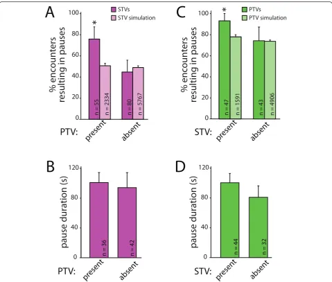
Figure 4STVs and PTVs preferentially pause at the same sites at the same time.(A)STVs (magenta) are significantly more likely to stop at a PTV pause site when PTVs are at the site. This preference cannot be accounted for by chance since no dependence on the presence of PTVs was seen in simulations (light magenta). The data presented correspond to the mean + 95% confidence intervals; *95% confidence intervals do
not overlap.(B)STVs pause for similar lengths of time regardless of whether a PTV is present at the pause site. Data are presented as mean +
standard error.(C)PTVs (green) are more likely to pause at a site when an STV is present. The same dependence was not observed in
simulations (light green).(D)PTVs pause for similar lengths of time, regardless of whether STVs are present at the pause sites. Data are the mean
assembly is a rapid process, and concurrent attraction of both STVs and PTVs could provide a mechanism for efficient assembly of presynaptic terminals. To deter-mine whether PTVs and STVs are simultaneously attracted to sites of synapse formation, we quantified whether STVs preferentially pause at PTV pause sites when a PTV is present. As shown in Figure 4, STVs were more likely to pause at a site that contained a PTV than a site that did not. When STVs encountered pause sites with PTVs present, 76.4 ± 11.2% of STVs paused with the PTV (n = 55; Figure 4A). In contrast, only 45.0 ± 10.9% of STVs paused at PTV pause sites when a PTV was not there (n = 80). This 1.7-fold difference in the likelihood of pausing with a PTV could not be accounted for by chance because there was no interac-tion between STVs and PTVs when the same analysis was performed on data obtained from simulations with model axons (Figure 4A). In the model, 52.4 ± 1.9% of STVs paused at PTV pause sites with a PTV present (n = 2,334), and 51.6 ± 1.3% of STVs paused at PTV pause sites without PTVs present (n = 5,767). The pause dura-tion of an STV stopped at a PTV pause site was also measured. In contrast to the likelihood of pausing, the pause duration of STVs is not affected by the presence or absence of PTVs (Figure 4B). The average pause duration for STVs paused at sites with PTVs present was 101.0 ± 13.4 seconds (n = 42 pauses), while the average pause duration for an STV paused at a site without a PTV present was 94.2 ± 20.1 seconds (n = 36).
It is not yet known whether STVs and PTVs must be recruited to nascent presynaptic terminals in a defined order, so we also tested whether PTVs are more likely to pause at sites that contain STVs. As shown in Figure 4C, PTVs paused at sites that contained STVs at a sig-nificantly higher rate (93.6 ± 7.0%, n = 47) than at sites where STVs had previously paused but were no longer present (74.4 ± 13.0%, n = 43). In contrast, this effect of STVs on PTV pausing behavior was not seen with ana-lysis of the randomized model: in the simulations, 76.0 ± 2.0% of PTVs paused at STV sites with an STV pre-sent (n = 1,591), and 74.7 ± 1.1% of PTVs paused at STV sites without STVs present (n = 4,906). There was an equal probability that either vesicle arrived first at a shared pause site (based on 95% confidence interval; data not shown). Similar to STVs pausing at PTV sites, the mean pause duration of PTVs pausing at STV pause sites is not affected by the presence of STVs (Figure 4D). The average pause duration for PTVs paused at sites with STVs present was 100.5 ± 12.4 s (n = 44 pauses), while the average pause duration for PTVs paused at sites without STVs present was 80.9 ± 15.3 s (n = 32 pauses). These data demonstrate that PTVs pre-fer to pause at sites that contain STVs andvice versa,
suggesting that STVs and PTVs are attracted to the same sites at the same time.
Recruitment of a PTV enhances accumulation of additional PTVs at sites of synapse formation
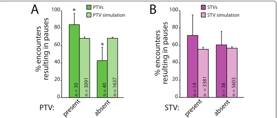
Assembly of presynaptic terminals involves recruitment of multiple PTVs [11]. Therefore, we wondered whether PTVs might also increase attraction of other PTVs to sites of synapse formation. To test this, we quantified the percentage of PTVs that paused at sites where another PTV was either present or had previously paused. When a PTV was already anchored at a particu-lar pause site, there was a high likelihood of an addi-tional PTV pausing at that site: 83.3 ± 13.3% of PTVs paused when they encountered sites with other PTVs (n = 30; Figure 5A). However, when a PTV was not present at the pause site, the likelihood that a PTV would pause was significantly lower (42.5 ± 15.3%, n = 40). This nearly two-fold increase in attraction of PTVs to sites containing other PTVs could not be explained by chance since the increase was not observed in our simu-lations. In model axons, PTVs paused at 67.1 ± 1.6% of sites with other PTVs (n = 1,267) and 68.1 ± 1.2% of sites without other PTVs (n = 5,827; Figure 5A). This indicates that the presence of a PTV at a pause site pro-motes recruitment of additional PTVs to that site.
Interestingly, the presence of an STV at a particular pause site did not significantly increase the probability of an additional STV pausing at that site (chance of pausing with another STV present = 71.4 ± 23.7%, n = 14; without another STV present = 60.5 ± 15.5%, n = 38; Figure 5B). As expected, no dependence of STV pausing on the presence of other STVs was observed in our randomized model axon (STV present = 56.8 ± 2.4%, n = 1,665; STV absent = 59.8 ± 1.6%, n = 3,443; Figure 5B). These data suggest that STVs do not inter-act with one another in a way that promotes recruit-ment to sites of synapse formation.
STV pausing is only mildly dependent on PTVs
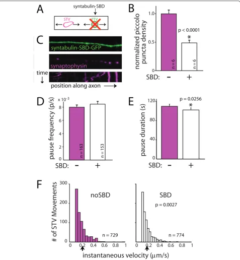
This construct mimics the syntaxin binding domain (SBD) of syntabulin and interferes with the syntaxin-syntabulin interaction. Expression of syntaxin-syntabulin-SBD has been shown to specifically prevent the majority of PTVs from being transported out of the cell body and into the axon without directly affecting STV transport or other KIF5 cargo [18,19] (Figure 6A). Imaging of our cortical cultures confirmed that the density of endogenous piccolo puncta is dramatically decreased in axons of neurons expressing syntabulin-SBD fused to GFP when compared to neurons expressing GFP alone (Figure 6B).
Consistent with the published data, transfection of neurons with the syntabulin-SBD construct did not interfere with STV transport in axons (Figure 6C): STVs still moved, and the mean distance moved over the course of imaging was unchanged (no SBD = 8.90 ± 0.93 μm, n = 163 vesicles; SBD = 9.30 ± 0.82 μm, n =
153 vesicles; P= 0.68). When STV pausing was
quanti-fied, expression of the syntabulin-SBD construct yielded no change in the pause frequency (no SBD = 0.0085 ± 0.0003 pauses/s, n = 163 vesicles; SBD = 0.0080 ± 0.0004 pauses/s, n = 153 vesicles; P = 0.62; Figure 6D) but did cause a slight decrease in the average duration of STV pauses (no SBD = 109.9 ± 4.4 s, n = 586 pauses;
SBD = 102.4 ± 4.3 s, n = 582 pauses;P = 0.03; Figure
6E). In addition, the instantaneous velocities of STVs were slightly reduced in SBD-transfected neurons (no
SBD = 0.208 ± 0.004μm/s, n = 729 movements; SBD =
0.193 ± 0.004μm/s, n = 774 movements; P= 0.003; Fig-ure 6F). Although there was a decrease in STV pause
duration when PTV transport was disrupted, the magni-tude of the effect and the lack of a change in the prob-ability of pausing argue against PTVs themselves controlling STV pausing. These data suggest that a direct interaction between STVs and PTVs is not responsible for the increased attraction of STVs to sites that contain PTVs.
Discussion
Synaptic vesicle and active zone proteins assemble rapidly at stable axo-dendritic contacts. Here, we hypothesized that trafficking of STVs and PTVs is coor-dinated even prior to synapse assembly, which could facilitate rapid assembly. By performing live multi-chan-nel fluorescence confocal imaging in neurons expressing both synaptophysin-mRFP and GFP-bassoon, we were able to record and analyze the movements of STVs and PTVs simultaneously in the same neuron. Our results indicate that STVs and PTVs are coordinated during transport and before stabilization since PTVs and STVs move together within the axon. We also find that STVs and PTVs pause at the same sites within the axon, parti-cularly when the other type of vesicle is also paused at that site. This attraction of STVs and PTVs to the same pause sites is not mediated by a direct interaction between STVs and PTVs since reducing the density of PTVs in the axon yielded only small changes in STV pausing. In summary, our data support a model of synapse formation in which STV and PTV trafficking is coordinated even prior to axo-dendritic adhesion. This coordination includes coincident stopping of STVs and
pr
esent
absent
pr
esent
absent
STV:
PTV:
*
n = 30 n = 3091 n = 40 n = 1637 n = 14 n = 3381
PTVs
PTV simulation
STVs
STV simulation
n = 38 n = 5603
% enc
ount
ers
resulting in pauses
% enc
ount
ers
resulting in pauses
0 20 40 60 80 100
0 20 40 60 80 100
A
B
*
Figure 5Multiple PTVs are attracted to the same sites.(A)PTVs are more likely to pause when other PTVs are present (green). Spatially
randomized simulations are shown in light green and cannot account for the tendency of PTVs to pause simultaneously with other PTVs.(B)
0 0.2 0.4 0.6 0.8 1 0
100 200 300
noSBD
# of ST
V M
o
v
ements
instantaneous velocity (
P
m/s)
0 0.2 0.4 0.6 0.8 1
SBD
p = 0.0027
n = 729 n = 774
syntabulin-SBD-GFP
position along axon
synaptophysin
A
B
SBD:
0 2 4 6 8 x 10⁻³
p
ause f
requenc
y (
p/s)
n = 163 n = 153
+
-D
F
time
C
STV PTV
syntabulin-SBD
E
0
0.5 1.0
normaliz
ed pic
colo
punc
ta densit
y
SBD:
-
+
*
p < 0.0001
n = 6 n = 6
0 40 80
120 p = 0.0256
*
pause duration (s)
SBD:
-
+
Figure 6A direct interaction between STVs and PTVs cannot account for the attraction of STVs to pause sites that contain PTVs.(A)
PTV transport was inhibited using dominant-negative syntabulin (syntaxin binding domain, SBD) fused to GFP.(B)Expression of
syntabulin-SBD-GFP decreases the density of PTV puncta in axons when compared to axons expressing syntabulin-SBD-GFP alone. PTVs were identified by immunofluorescent
labeling for endogenous piccolo.(C)Images and kymograph (bottom) showing that STVs (magenta) move and pause in SBD-expressing axons
(green).(D)The frequency of STV pausing was unchanged in the presence of SBD-GFP.(E)STV pause durations are shorter when PTV
localization in the axon is disrupted. The change in pausing upon expression of SBD-GFP is not sufficient to account for the coordinated pausing
of STVs and PTVs. In B, D and E, error bars correspond to the standard error. *, difference is significant.P-values are from Wilcoxon rank-sum test
and are indicated in the figure.(F)The instantaneous velocities of STVs are increased in SBD-expressing axons. Black arrow, mean instantaneous
PTVs at predefined sites of synapse formation, indepen-dent of a direct interaction between STVs and PTVs.
Is STV and PTV transport coordinated?
Although it is clear that both PTVs and STVs need to be recruited to the same sites in order for pre-synaptic terminals to develop, it is not immediately apparent as to how they both arrive at the same destinations. Pre-viously, STV and PTV dynamics have been imaged only separately, leaving it unknown whether they are trans-ported together [4,9,11-15,17,21,23]. Recent work showed that clear, synaptic vesicle protein-containing vesicles and dense-core, PTV-like vesicles can be seen apparently tethered together in electron micrographs of young neurons [16]. This was the first indication that PTVs and STVs might be transported together, but it remained unclear whether these vesicle aggregates cor-respond to either STVs and PTVs being transported together or paused together or a later stage in synapse development. Our analysis indicates that a sizable por-tion of STVs and PTVs move with each other and spend the majority of their time together. STVs and PTVs often moved separately before or after moving together, suggesting that STVs and PTVs can move with each other while also maintaining their separate identi-ties. This observation indicates that, although it is possi-ble that some STVs contained molecules of GFP-bassoon and/or a portion of PTVs contained molecules of synaptophysin-mRFP, missorting of STV and PTV marker proteins cannot account for the observed co-transport of STVs and PTVs. Coordinating STV and PTV movement could represent a mechanism for rapid synapse development, since a full complement of synap-tic proteins could immediately be delivered to a poten-tial synaptic site. It will be important in the future to determine whether co-transport is mediated by a direct interaction between STVs and PTVs or by other mechanisms.
Previously, it was shown that STV pause sites are pre-ferred sites of synapse formation [4], raising the ques-tion of whether PTVs also tend to pause at these same sites of synapse formation. By labeling both STVs and PTVs within the same axon, we were also able to com-pare spatial and temporal properties of pausing for both types of vesicles. STV and PTV pausing were qualita-tively and quantitaqualita-tively similar. Like STVs, multiple PTVs paused at the same sites, and the same PTVs returned repeatedly to a given site. The frequency of pausing and duration of pauses for STVs and PTVs were also similar. Importantly, STVs and PTVs paused at the same sites within the axon. This implies that PTVs also pause at predefined sites of synapse forma-tion. The propensity of STVs and PTVs to preferentially pause at the same sites within the axon suggests that
there is a mechanism that recruits both types of vesicles to the same site at the same time. These same mechan-isms could be utilized by axons to recruit the necessary proteins during synapse formation.
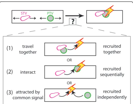
What causes STV and PTV pausing?
Our data indicate that PTVs preferentially pause at sites where an STV is present. Likewise, STVs preferentially pause at sites where a PTV is present. There are at least three potential hypotheses that could account for this phenomenon (Figure 7). First, pausing of STVs and PTVs at the same sites could be a result of STVs and PTVs traveling together and consequently being recruited together. Second, it is possible that a paused vesicle can interact with and stabilize a moving vesicle. In this scenario, a local signal could cause one type of vesicle to pause at a specific site within the axon. That paused vesicle could then interact with other vesicles and cause them to pause at the same site. Third, STVs and PTVs could be independently attracted to a com-mon signal within the axon. In this case, both STVs and PTVs would respond to that signal by preferentially pausing at the site of the signal, regardless of the pre-sence of other vesicles. This, in turn, would increase the probability that either vesicle was present at the site of the signal and, therefore, the chance STVs and PTVs are simultaneously paused at the same site. Each hypothesis represents a viable mechanism through which both synaptic vesicle and active zone proteins could be recruited to the same site in the axon.
STV PTV
?
recruited together
recruited sequentially
recruited independently travel
together
interact
attracted by common signal
OR OR
(1)
(2)
(3)
Our data are most consistent with the hypothesis that STVs and PTVs are recruited to the same sites at the same time as a consequence of responding to a common local signal. The first mechanism - simultaneous recruitment of STVs and PTVs that are transported together -cannot by itself explain coincident pausing since in many cases the STVs and PTVs that paused together paused sequentially rather than simultaneously (for example, Figure 3A,B(1 and 3)). To distinguish between the remaining two mechanisms, we reduced the density of PTVs within the axon, using a dominant negative construct corresponding to the SBD of syntabulin. If PTVs interact with STVs to influence STV pausing and recruitment to a given site, then decreasing PTV density should substantially alter STV pausing. However, in SBD-transfected axons, STVs displayed only small differ-ences in pause duration and no change in the frequency of pausing when compared to STVs in control axons. Although differences in pause duration and velocity were statistically significant, the relatively subtle change in STV pausing suggests that PTVs exert only a small influence on STV pausing. This argues against the sec-ond hypothesis, in which STV and PTV pausing is exclusively controlled by an interaction between STVs and PTVs. However, based on the data, it is possible that STVs and PTVs respond to both local signals and one another.
It will be important in the future to identify the sig-nals that recruit STVs and PTVs. It is not yet known which signals cause vesicles to pause. Potential signals include calcium, phosphorylation or small GTPase activ-ity. Although it was previously shown that calcium could increase the duration of pausing, the probability of pausing was unchanged by reducing intracellular cal-cium [4], suggesting that calcal-cium alone may not be responsible. Phosphorylation is known to regulate the activity of microtubule motor proteins and their associa-tion with microtubules or vesicles [24-28], raising the possibility that phosphorylation of motor proteins is the ultimate mechanism by which local signals cause STVs and PTVs to stop their transport. Small GTPases are well-known for their ability to control vesicle targeting [29-31], and recent work has shown that in C. elegans, arl-8, an Arf-like small G-protein, controls where along the axon synaptic proteins aggregate [32].
Why don’t all vesicles pause at any given site?
Although the majority of STVs and PTVs that encoun-ter pause sites will pause at them, many vesicles ignore the pause sites and continue their movements. This is even true for stopping and recruitment that occurs in response to synaptogenic adhesion or axo-dendritic con-tact [4]. It is not clear why. One possibility is that there could be a limited number of ‘docking sites’ at each
pause site. This seems unlikely since some vesicles pass by even while others can still pause. Alternatively, STVs and PTVs could exist in multiple states, at least one of which is unresponsive to local signals that cause paus-ing. PTVs and STVs display heterogeneity in apparent size, velocity, and pausing properties, which has been noted here and elsewhere [4,9,11,13,14,19,21]. These apparent differences may correlate with structural differ-ences [16]. In general, smaller/dimmer vesicles appear more likely to move quickly and pass by pause sites without pausing. Perhaps these apparently smaller vesi-cles are unable to respond to the cues that induce paus-ing. Such a state could be a result of differences in their motors or in other proteins associated with the STVs or PTVs. The idea of STVs and PTVs as heterogeneous subpopulations will be an interesting topic for further exploration.
Conclusions
For synapses to form, all of the components of presy-naptic terminals must be delivered rapidly to the site of synapse formation. Despite its importance, we are only beginning to understand how this occurs. Here we have proposed a model for presynaptic terminal assembly in which trafficking of synaptic vesicle and active zone pro-teins is coordinated even prior to axo-dendritic contact. This coordination occurs through a combination of co-transport, perhaps in aggregates of vesicles tethered together [16], and co-pausing in response to local sig-nals. This coordination then can facilitate presynaptic terminal assembly and contribute to the rapid recruit-ment of synaptic components that has been consistently observed. The same signals that induce co-pausing prior to transport may act downstream of synaptogenic adhe-sion to simultaneously attract STVs and PTVs to sites of synapse assembly.
Materials and methods
All studies were conducted with an approved protocol from the Case Western Reserve University Institutional Animal Care and Use Committee, in compliance with the National Institutes of Health guidelines for the care and use of experimental animals.
Neuronal cultures and transfection
Primary neuronal cultures were prepared from postnatal rat visual cortex essentially as described previously [4,5,15], except neurons were maintained in Neurobasal-A medium supplemented with glutamax and B27 (Invi-trogen, Carlsbad, CA, USA). Neurons were transfected with Lipofectamine 2000 (Invitrogen) 24 to 48 h before live imaging. Excluding GFP-bassoon transfection, 1 μg of DNA construct was combined with 1μg of
18-mm coverslip. Transfection of GFP-bassoon was
con-ducted in the same manner except 2 μg of DNA were
used to account for its large size. With double transfec-tion, localization of each protein appeared similar when expressed alone. Also, the distribution and movement of STVs labeled with synaptophysin-mcherry, synaptophy-sin-mRFP, and synaptophysin-GFP all appeared similar to one another. GFP-bassoon (GFP-Bsn 95-3938), synap-tophysin-mcherry, mRFP, synaptophysin-GFP and syntaxin-SBD-synaptophysin-GFP were generous gifts of Drs Thomas Dresbach (University of Heidelberg), Matthijs Verhage (Vrije Universiteit, Amsterdam), Jurgen Klin-gauf (University of Muenster), Jane Sullivan (University of Washington, Seattle) and Zu-Hang Sheng (National Institute of Neurological Disorders and Stroke, Bethesda). Each of these constructs has been shown pre-viously to be functional and properly localized [15,19,20,22,33,34].
Live imaging
Neurons were imaged at 7 to 8 DIV with a C1 Plus con-focal system with a Nikon Eclipse Ti-E microscope using a 40× Nikon Plan Apo 0.95 numerical aperture objective. Lasers were 488 nm argon and 543 nm helium-neon. Detection filters were 515/30 nm bandpass for GFP and 590/50 nm bandpass for mcherry/mRFP. Images were collected every 10 s, with scan times no greater than 3.3 s. This imaging interval was selected as a compromise between having a high temporal resolu-tion and minimizing the time the neurons were exposed to the laser to avoid any toxicity and photobleaching. A total of 45 images were collected for each time-lapse series. Imaging was conducted with constant perfusion with artificial cerebrospinal fluid (120 mM NaCl, 3 mM
KCl, 2 mM CaCl2, 2 mM MgCl2, 30 mM D-glucose, 20
mM HEPES, and 0.2% sorbitol, pH 7.3). Artificial cere-brospinal fluid perfusion was performed at 25°C since STV transport and pausing are not significantly different at ambient and physiological temperatures. Axons were identified using morphological criteria, as described pre-viously [4,15]. For dual-color imaging, channels were collected sequentially to eliminate bleed-through and neurons were imaged in which expression levels of both fusion proteins appeared comparable.
Immunofluorescence
Neurons were fixed for 15 minutes in 4% paraformalda-hyde in phosphate-buffered saline containing 4% sucrose, permeabilized for 5 minutes with 0.2% Triton X-100, and blocked with 10% horse serum. The primary antibody was piccolo (Synaptic Systems, Goettingen, Germany, and the secondary antibody was Alexa 633-conjugated goat anti-rabbit (Invitrogen). Coverslips were mounted in Fluoromount (Fisher Scientific, Pittsburgh,
PA, USA) containing DABCO (1,4-diazabicyclo[2.2.2] octane) (Sigma, St Louis, MO, USA).
Analysis and statistics
STV and PTV movements were tracked using ImageJ (NIH, Bethesda, MD, USA). To restrict the quantifica-tion of time-lapse movies to healthy neurons, axons were included in the analysis only if a least one vesicle moved within the field of view. Regions of axon with an intermediate STV or PTV density were imaged to allow us to track each vesicle reliably. STV and PTV move-ments were tracked independently while blind to the other channel. Movements were not analyzed if the axon moved significantly.
For quantification of movement and pausing, posi-tional data were imported into Matlab and analyzed with custom-written programs (Mathworks, Natick, MA, USA). For pause analysis, a pause was defined as a per-iod≥10 s, during which the velocity of the vesicle went to 0 ± 0.1 μm/s. The cutoff zero velocity (0.1μm/s) was chosen based on the average diameter of vesicles, which
was approximately 1 μm. Vesicles that never moved
were not included in the analysis. Therefore, vesicles which pause for very long durations might be underre-presented. Also, only vesicles that could be tracked for the entire imaging duration were included in the analy-sis. Vesicles that move at high velocities are more likely to leave the imaging area before the end of the movie. Therefore, these vesicles might also be underrepresented in the analysis.
Given the pixel density, scan speed and average vesicle size, a vesicle was typically imaged in approximately 10 to 50 ms, permitting reliable tracking of vesicles based on their apparent size, shape, and intensity, with rela-tively low influence of vesicle movements on these para-meters. The fastest STV movements, at a maximum approaching 1μm/s (comparable with the fastest veloci-ties that have been reported for STVs [4,9,17]), resulted in movements of 10μm during the imaging interval and were easily measured. Maximal velocities of PTVs are lower than maximal STV velocities [11,19] and were also easily recorded.
Data are presented cumulatively with 95% confidence intervals for binomially distributed data or as the mean ± standard error of the mean where appropriate. Confi-dence intervals were calculated based on a normal approximation, and data sets were considered signifi-cantly different if their 95% confidence intervals were non-overlapping. For data presented as means, signifi-cance was evaluated using the Wilcoxon rank sum test.
Randomized model
axon that was imaged, a model axon was generated with the same number of vesicles and axonal length as each experimental axon. In these models, the initial position of each vesicle was randomized. However, the number, timing and direction of movements, as well as the instantaneous velocity, and pause duration of each vesi-cle remained the same. The newly generated time-lapse series were then analyzed in the same manner as the experimental data. Each simulation was performed 100 times per axon to enhance statistical analysis.
Additional material
Additional file 1: Movie 1. Movie showing STV and PTV movements in the same axon. The top three panels show bassoon, synaptophysin and the transmitted light image, respectively. The bottom panel corresponds to the overlay of all three. STVs and PTVs can be seen moving together and pausing at the same sites. The yellow boxes highlight areas in which STVs and PTVs clearly move together. The axon itself does not move significantly during the imaging. Times are indicated at the bottom left.
Abbreviations
DIV: daysin vitro; GFP: green fluorescent protein; mRFP: monomeric red
fluorescent protein; PTV: piccolo-bassoon transport vesicle; SBD: syntaxin binding domain; STV: synaptic vesicle protein transport vesicle.
Acknowledgements
We thank Michael Sceniak for advice and help with Matlab programming, for help with design of the simulations and for helpful discussions about the project. We also thank Corbett Berry and Louie Zhou for technical assistance. We are grateful to Drs Thomas Dresbach (University of Heidelberg), Matthijs Verhage (Vrije Universiteit, Amsterdam), Jurgen Klingauf (University of Muenster), Jane Sullivan (University of Washington, Seattle) and Zu-Hang Sheng (National Institute of Neurological Disorders and Stroke, Bethesda) for GFP-bassoon (GFP-Bsn 95-3938), mcherry, synaptophysin-mRFP, synaptophysin-GFP and syntaxin-SBD-GFP constructs, respectively. This work was funded by a Research Starter grant from the PhRMA Foundation (SS), start-up funds from Case Western Reserve University (SS), and the NIH predoctoral Training Program in Molecular Therapeutics at Case Western Reserve University (LB).
Author details
1
Department of Pharmacology, Case Western Reserve University School of
Medicine, Cleveland, Ohio, 44106, USA.2Department of Neuroscience, Case
Western Reserve University School of Medicine, Cleveland, Ohio, 44106, USA.
Authors’contributions
LB and SS performed experiments and analysis and wrote the paper. Both authors read and approved the final manuscript.
Competing interests
The authors declare that they have no competing interests.
Received: 11 March 2011 Accepted: 10 May 2011 Published: 10 May 2011
References
1. Bury LA, Sabo SL:How it’s made: the synapse.Mol Interv2010,10:282-292.
2. McAllister AK:Dynamic aspects of CNS synapse formation.Annu Rev
Neurosci2007,30:425-450.
3. Friedman HV, Bresler T, Garner CC, Ziv NE:Assembly of new individual
excitatory synapses: time course and temporal order of synaptic molecule recruitment.Neuron2000,27:57-69.
4. Sabo SL, Gomes RA, McAllister AK:Formation of presynaptic terminals at
predefined sites along axons.J Neurosci2006,26:10813-10825.
5. Washbourne P, Bennett JE, McAllister AK:Rapid recruitment of NMDA
receptor transport packets to nascent synapses.Nat Neurosci2002,
5:751-759.
6. Niell CM, Meyer MP, Smith SJ:In vivoimaging of synapse formation on a
growing dendritic arbor.Nat Neurosci2004,7:254-260.
7. Ruthazer ES, Li J, Cline HT:Stabilization of axon branch dynamics by
synaptic maturation.J Neurosci2006,26:3594-3603.
8. Lee H, Dean C, Isacoff E:Alternative splicing of neuroligin regulates the
rate of presynaptic differentiation.J Neurosci2010,30:11435-11446.
9. Ahmari SE, Buchanan J, Smith SJ:Assembly of presynaptic active zones
from cytoplasmic transport packets.Nat Neurosci2000,3:445-451.
10. Lucido AL, Suarez Sanchez F, Thostrup P, Kwiatkowski AV, Leal-Ortiz S,
Gopalakrishnan G, Liazoghli D, Belkaid W, Lennox RB, Grutter P, Garner CC,
Colman DR:Rapid assembly of functional presynaptic boutons triggered
by adhesive contacts.J Neurosci2009,29:12449-12466.
11. Shapira M, Zhai RG, Dresbach T, Bresler T, Torres VI, Gundelfinger ED,
Ziv NE, Garner CC:Unitary assembly of presynaptic active zones from
Piccolo-Bassoon transport vesicles.Neuron2003,38:237-252.
12. Zhai RG, Vardinon-Friedman H, Cases-Langhoff C, Becker B,
Gundelfinger ED, Ziv NE, Garner CC:Assembling the presynaptic active
zone: a characterization of an active one precursor vesicle.Neuron2001,
29:131-143.
13. Kraszewski K, Mundigl O, Daniell L, Verderio C, Matteoli M, De Camilli P:
Synaptic vesicle dynamics in living cultured hippocampal neurons visualized with CY3-conjugated antibodies directed against the lumenal domain of synaptotagmin.J Neurosci1995,15:4328-4342.
14. Nakata T, Terada S, Hirokawa N:Visualization of the dynamics of synaptic
vesicle and plasma membrane proteins in living axons.J Cell Biol1998,
140:659-674.
15. Sabo SL, McAllister AK:Mobility and cycling of synaptic
protein-containing vesicles in axonal growth cone filopodia.Nat Neurosci2003,
6:1264-1269.
16. Tao-Cheng JH:Ultrastructural localization of active zone and synaptic
vesicle proteins in a preassembled multi-vesicle transport aggregate.
Neuroscience2007,150:575-584.
17. Kaether C, Skehel P, Dotti CG:Axonal membrane proteins are transported
in distinct carriers: a two-color video microscopy study in cultured hippocampal neurons.Mol Biol Cell2000,11:1213-1224.
18. Su Q, Cai Q, Gerwin C, Smith CL, Sheng ZH:Syntabulin is a
microtubule-associated protein implicated in syntaxin transport in neurons.Nat Cell
Biol2004,6:941-953.
19. Cai Q, Pan PY, Sheng ZH:Syntabulin-kinesin-1 family member
5B-mediated axonal transport contributes to activity-dependent presynaptic assembly.J Neurosci2007,27:7284-7296.
20. Dresbach T, Torres V, Wittenmayer N, Altrock WD, Zamorano P,
Zuschratter W, Nawrotzki R, Ziv NE, Garner CC, Gundelfinger ED:Assembly
of active zone precursor vesicles: obligatory trafficking of presynaptic cytomatrix proteins Bassoon and Piccolo via a trans-Golgi compartment.
J Biol Chem2006,281:6038-6047.
21. Dai Z, Peng HB:Dynamics of synaptic vesicles in cultured spinal cord
neurons in relationship to synaptogenesis.Mol Cell Neurosci1996,
7:443-452.
22. Dresbach T, Hempelmann A, Spilker C, tom Dieck S, Altrock WD,
Zuschratter W, Garner CC, Gundelfinger ED:Functional regions of the
presynaptic cytomatrix protein bassoon: significance for synaptic targeting and cytomatrix anchoring.Mol Cell Neurosci2003,
23:279-291.
23. Sytnyk V, Leshchyns’ka I, Delling M, Dityateva G, Dityatev A, Schachner M:
Neural cell adhesion molecule promotes accumulation of TGN organelles at sites of neuron-to-neuron contacts.J Cell Biol2002,
159:649-661.
24. Lee KD, Hollenbeck PJ:Phosphorylation of kinesinin vivocorrelates with
organelle association and neurite outgrowth.J Biol Chem1995,
270:5600-5605.
25. Hollenbeck PJ:Phosphorylation of neuronal kinesin heavy and light
chainsin vivo.J Neurochem1993,60:2265-2275.
26. Hamasaki T, Barkalow K, Richmond J, Satir P:cAMP-stimulated
swimming speed in Paramecium.Proc Natl Acad Sci USA1991,
88:7918-7922.
27. Dillman JF, Pfister KK:Differential phosphorylationin vivoof cytoplasmic
dynein associated with anterogradely moving organelles.J Cell Biol1994,
127:1671-1681.
28. Okada Y, Sato-Yoshitake R, Hirokawa N:The activation of protein kinase A
pathway selectively inhibits anterograde axonal transport of vesicles but not mitochondria transport or retrograde transportin vivo.J Neurosci
1995,15:3053-3064.
29. Itzen A, Goody RS:GTPases involved in vesicular trafficking: structures
and mechanisms.Semin Cell Dev Biol2011,22:48-56.
30. Segev N:GTPases in intracellular trafficking: an overview.Semin Cell Dev
Biol2011,22:1-2.
31. Chavrier P, Goud B:The role of ARF and Rab GTPases in membrane
transport.Curr Opin Cell Biol1999,11:466-475.
32. Klassen MP, Wu YE, Maeder CI, Nakae I, Cueva JG, Lehrman EK, Tada M,
Gengyo-Ando K, Wang GJ, Goodman M, Mitani S, Kontani K, Katada T,
Shen K:An Arf-like small G protein, ARL-8, promotes the axonal
transport of presynaptic cargoes by suppressing vesicle aggregation.
Neuron2010,66:710-723.
33. Hua Y, Sinha R, Martineau M, Kahms M, Klingauf J:A common origin of
synaptic vesicles undergoing evoked and spontaneous fusion.Nat Neurosci13:1451-1453.
34. de Wit J, Toonen RF, Verhage M:Matrix-dependent local retention of
secretory vesicle cargo in cortical neurons.J Neurosci2009,29:23-37.
doi:10.1186/1749-8104-6-24
Cite this article as:Bury and Sabo:Coordinated trafficking of synaptic
vesicle and active zone proteins prior to synapse formation.Neural
Development20116:24.
Submit your next manuscript to BioMed Central and take full advantage of:
• Convenient online submission • Thorough peer review
• No space constraints or color figure charges • Immediate publication on acceptance
• Inclusion in PubMed, CAS, Scopus and Google Scholar • Research which is freely available for redistribution