R E V I E W
Open Access
Microglia in the developing retina
Fenge Li
†, Danye Jiang
†and Melanie A. Samuel
*Abstract
Microglia are increasingly shown to be key players in neuron development and synapse connectivity.
However, the underlying mechanisms by which microglia regulate neuron function remain poorly understood
in part because such analysis is challenging in the brain where neurons and synapses are intermingled and
connectivity is only beginning to be mapped. Here, we discuss the features and function of microglia in the
ordered mammalian retina where the laminar organization of neurons and synapses facilitates such molecular
studies. We discuss microglia origins and consider the evidence for molecularly distinct microglia
subpopulations and their potential for differential roles with a particular focus on the early stages of retina
development. We then review the models and methods used for the study of these cells and discuss
emerging data that link retina microglia to the genesis and survival of particular retina cell subtypes. We also
highlight potential roles for microglia in shaping the development and organization of the vasculature and
discuss cellular and molecular mechanisms involved in this process. Such insights may help resolve the
mechanisms by which retinal microglia impact visual function and help guide studies of related features in
brain development and disease.
Keywords:
Microglia, Development, Retina, Synapse, Brain, Depletion models
Highlights
Microglia maturation is highly specified in the
retina.
Microglia play potential roles in vascularization,
neuron birth and survival, and synapse refinement.
Diverse microglia subpopulations found in retina
display distinct features.
Background
Microglia are the resident immune cells of the central
nervous system (CNS), and emerging work implicates
these cells in shaping diverse features of neural
devel-opment, connectivity, and homeostasis (reviewed in
[
1
–
4
]). However, whether and how particular neuron
or synapse types are targeted by microglia and the
functional consequences of these interactions are less
well described. It has been difficult to answer these
questions because circuits in the brain are complex
and we know relatively little about them. In this
review, we discuss known microglia interactions with
neurons in the accessible and well-mapped neural
cir-cuits of the mammalian retina. In the first part of the
review, we present an in-depth description of the
fea-tures of retina microglia and discuss their origins,
localization, and organization during development.
We also review evidence for microglia subpopulations
and present an atlas of microglial biomarkers over
de-velopment. In the second part, we discuss the
func-tions of microglia, with a focus on their roles in
modulating neurogenesis and development,
particu-larly regarding retinal ganglion cells and astrocytes. In
turn, these processes may influence novel roles for
microglia in modulating neurovascular organization.
Finally, we provide perspectives on key goals for
fu-ture research, which include potential roles for
micro-glia subpopulations and elucidation of mechanisms by
which particular synapses are spared or removed.
Continued study of microglia-specific functions in the
retina may help inform related studies in the brain
and provide unique opportunities to develop microglia
targeted treatment strategies in diverse neurological
diseases.
© The Author(s). 2019Open AccessThis article is distributed under the terms of the Creative Commons Attribution 4.0 International License (http://creativecommons.org/licenses/by/4.0/), which permits unrestricted use, distribution, and reproduction in any medium, provided you give appropriate credit to the original author(s) and the source, provide a link to the Creative Commons license, and indicate if changes were made. The Creative Commons Public Domain Dedication waiver (http://creativecommons.org/publicdomain/zero/1.0/) applies to the data made available in this article, unless otherwise stated. * Correspondence:[email protected]
†Fenge Li and Danye Jiang contributed equally to this work.
Main text
Part 1: features of retinal microglia
Microglia origin in the retina
Microglia originate from primitive yolk sac progenitors
[
5
,
6
]. Their development and survival are regulated by
several known transcription factors and cytokine
recep-tors (Table
1
). Among these, the transcription factor
PU.1 (also known as spleen focus forming proviral
inte-gration oncogene, SPI1) plays an important role in
microglia development in part through its binding
part-ner interferon regulatory factor 8 (IRF8) [
7
,
13
–
16
].
Pu.1-deficient mice lack microglia, circulating
mono-cytes, and tissue macrophages due to a reduction in early
myeloid progenitors, while IRF8-deficent mice display
defects in myeloid cell maturation [
13
,
14
]. Microglia
genesis is also regulated by the macrophage
colony-stimulating factor receptor CSF1R. CSF1R expression on
microglia is maintained throughout development.
Con-sistent with the requirement for CSF1R expression,
Csf1r
knockout mice lack microglia in addition to yolk sac
macrophages and osteoclasts [
8
–
10
]. Finally, animals
lacking toll-like receptor 4 (TLR4) display reduced
bipo-lar cell numbers and altered bipobipo-lar cell dendritic
dens-ity, in addition to loss of microglia in the retina. These
changes correlate with a significant reduction in retinal
function, suggesting a key role for TLR4 in mediating
visual function. However, whether microglia are causal
to these alterations remains unclear [
12
].
After they differentiate, microglia home to the CNS.
Microglia can be identified in mouse brain rudiment
as early as embryonic day (E)8.5 ~ E9.5. They are
thought to migrate to the CNS via the embryonic
cir-culatory system as mice that lack the sodium calcium
exchanger 1 (Ncx-1) have defective blood circulation
and microglia fail to enter the brain [
9
]. The origins
of microglia in the retina and their precise
develop-mental arrival have been less well studied. They are
present in human retina by 10 weeks gestation and in
mouse retina by E11.5, though it is likely they arrive
even earlier [
17
,
18
]. Similar timing has been
docu-mented in other species (E7 in quail, [
19
]; and at E12
in
rat,
[
20
]).
Two
waves
of
retinal
microglia
infiltration
have
been
proposed
based
on
the
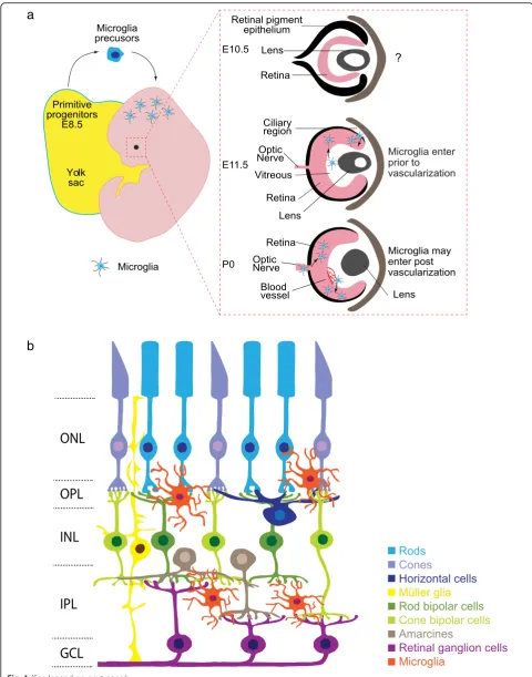
spatiotemporal localization of these cells. The first wave
hap-pens early in development prior to vascularization (Fig.
1
a).
At this time, microglia are thought to enter the retina by
ei-ther: 1) crossing the vitreal retina surface; or 2) migrating
from non-neural ciliary regions in the periphery [
17
,
18
,
21
,
22
]. A second wave of infiltration has been proposed after
blood vessels have formed through invasion from the optic
disc or via blood vessels themselves [
23
]. Since much of this
evidence is correlative, firm documentation of the timing
and routes by which microglia enter the retina awaits more
contemporary lineage tracing approaches.
Microglia location and lamination in the retina
Microglia entry into the retina coincides with retinal
neuron differentiation. Retinal neurons are derived from a
precursor pool of retinal progenitor cells (RPCs) that divide
to give rise to the five main types of retinal neurons:
photo-receptors, bipolars, amacrines, horizontal cells, and retinal
ganglion cells. As these neurons mature they become
or-dered into three cellular and two synaptic layers.
Photore-ceptors comprise the outer nuclear layer (ONL) and relay
information through synapses in the outer plexiform layer
(OPL) to inner retina neurons (horizontal, bipolar, and
amacrine cells). Bipolar and amacrine cells synapse with
retinal ganglion cells in the inner plexiform layer (IPL) (Fig.
1
b). Microglia comprise 0.2% of total retinal cells and are
found in addition to two other retina glia types, astrocytes
and Müller glia [
24
–
26
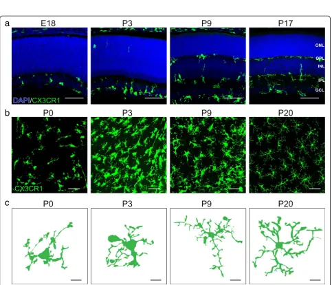
]. Interestingly, microglia are
pre-dominately located in the retinal synapse layers (Fig.
2
a).
The adult OPL contains ~ 47% of the microglial population,
while 53% are found in the IPL (Li and Samuel,
unpub-lished). It is perhaps telling that microglia localization
tracks the spatial distribution of developing retina synapses.
Synapses begin to emerge as early as E17 in the nascent
IPL, and at this time 99% of microglia localize to this
nar-row region [
27
,
28
]. This localization persists as synapses
mature and are refined. At postnatal day (P)3, ~ 80% of
microglia are localized to the developing IPL and ganglion
cell layer (GCL), and at P9, microglia become present
within the developing OPL. This pattern persists into
adult-hood, with microglia and their processes localizing
predom-inately to the inner retina and OPL, while the ONL is
largely devoid of these cells (Fig.
2
a) [
18
,
29
]. Thus,
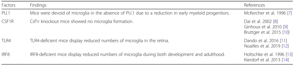
Table 1
Known factors that regulate microglia formation or survival
Factors Findings References
PU.1 Mice were devoid of microglia in the absence of PU.1 due to a reduction in early myeloid progenitors. McKercher et al. 1996 [7]
CSF1R Csf1r knockout mice showed no microglia formation. Dai et al. 2002 [8]
Ginhoux et al. 2010 [9] Bruttger et al. 2015 [10]
TLR4 TLR4-deficient mice display reduced numbers of microglia in the retina. Dando et al. 2016 [11]
Noailles et al. 2019 [12]
Fig. 2Spatiotemporal distribution of microglia in the developing mouse retina.a. Representative images showing distinct spatiotemporal localization patterns of microglia across retina development (E18, P3, P9, and P17) in CX3CR1GFP/+mice. Microglia are highly enriched at E18 and
P3 in the nascent IPL where synapses are developing. At P9, microglia also become present within the developing OPL. This pattern persists into adulthood. Blue, DAPI; green, microglia. Scale bar = 50μm.b-c. Representative images (b, scale bar = 50μm) and single cell reconstructions (c, scale bar = 10μm) of microglia in whole mount preparations of CX3CR1GFP/+retina across development (P0, P3, P9, and P20). At birth, retinal
microglia are amoeboid but become progressively ramified as the retina matures (See figure on previous page.)
microglia are at the right time and place to regulate retina
synapse refinement. In line with this idea, the absolute
number of retina microglia correlates with the peak of
ret-ina synapse pruning. The numbers of retret-ina microglia
in-crease over the first postnatal week, reaching twice that of
adult levels by P7 when outer and inner retina synapses
area actively refined. Microglia numbers then steadily
de-crease until the fourth postnatal week when they reach
steady state levels and the retina circuit is considered
ma-ture [
18
].
Microglia morphology
Morphological changes in microglia are thought to correlate
in part with their functional states [
30
–
32
]. Ramified
micro-glia are often referred to as
‘
resting
’
while amoeboid
micro-glia are often referred to as
‘
active
’
[
33
]. These terms can be
misleading, however, as live imaging suggests that microglia
are structurally dynamic in both ramified and amoeboid
morphologies, though the cellular functions they carry out
may differ. Ramified microglia actively retract and extend
their processes, monitor neurons, and are engaged in
me-tabolite removal and clearance in the CNS (reviewed in [
3
,
34
,
35
]). In contrast, amoeboid microglia contain numerous
lysosomes and phagosomes and are thought to be engaged
in synapse, axon, or cell engulfment [
36
,
37
]. Consistent
with this idea, microglia appear amoeboid in the brain
dur-ing development at the peak of cell and synapse remodeldur-ing
and then shift to a ramified state in the first two postnatal
weeks [
38
,
39
]. This developmental shift in microglia
morphology extends to the retina. At birth, retinal microglia
are amoeboid and extend their processes towards the basal
side of the retina but become progressively ramified as the
retina matures (Fig.
2
b, c) [
18
]. Shifts in microglia structure
also occur in response to CNS injury or pathogen invasion,
leading to the formation of reactive amoeboid microglia [
40
,
41
]. The mechanisms through which microglia alter their
structural states are not well understood. Koso et al.
re-ported that the zinc finger transcription factor Sall1 is
expressed specifically in amoeboid retina microglia and that
deleting Sall1 can cause ramified microglia to adopt a more
amoeboid appearance [
42
]. Continued efforts to understand
how microglia achieve different structural states and how
these states impact function may aid efforts to modulate
microglia activity in development or disease.
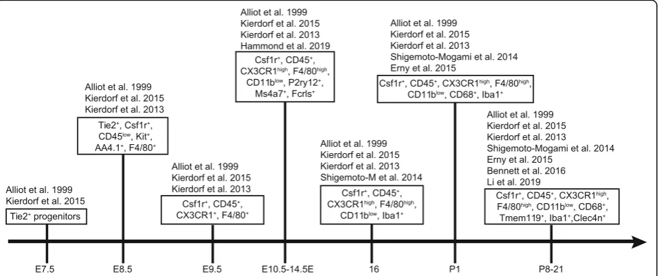
Microglia markers and subpopulations
All microglial precursors express the common
macro-phage markers CX3 chemokine receptor 1 (CX3CR1)
and ionized calcium binding adaptor molecule 1 (Iba1)
[
14
,
43
]. Microglia transiently express additional markers
during development, including F4/80, isolectin, CD45,
CD68, CD11b, and inducible nitric oxide synthase
(iNOS) that are typically lost or down regulated in adult
cells [
6
,
18
,
43
]. Common microglia biomarkers over
de-velopment are summarized in Fig.
3
[
6
,
14
,
44
–
47
].
Whether microglia can be considered a group of related
but distinct cell populations is an area of active
investi-gation. One possibility is that individual microglia can
display fluid cellular characteristics that vary according
to developmental or disease states. Alternately, microglia
may be comprised of physiologically distinct cell subsets.
Progress toward resolving these questions has been
somewhat challenging due to the dynamic nature of
microglia, their ability to migrate, and the potential for
molecular similarities between macrophages that may
enter the CNS under some conditions and resident
micro-glia populations [
48
,
49
]. However, it is clear that
anti-genic, structural, and transcriptional differences exist
between cohorts of microglia. For example, the cytokine
IL-34 appears to demark spatially distinct populations of
microglia in the retina. In normal adults, IL-34 negative
microglia are mainly localized to the OPL, while IL-34
positive microglia are located in the IPL [
50
]. In the
pres-ence of neuron degeneration, however, both populations
relocate to the retinal pigment epithelium (RPE) [
50
].
Ret-ina microglia also show different levels of CD11c, CD11b,
and TLR4 [
11
,
51
,
52
]. For example, CD11c appears more
abundant on microglia that are localized to compromised
retinal neurons [
53
]. Thus, it is tempting to speculate that
different subsets of microglia might be tuned to perform
niche specific functions or regulate specific neuron types
or geographic areas of the CNS.
Several recent molecular and sequencing based profiling
studies also support the presence of microglia
subpopula-tions in brain and retina. These populasubpopula-tions appear
dy-namic and vary with developmental time and the presence
or absence of disease [
54
–
56
]. But some common features
emerge: 1) microglia are among the most transcriptionally
diverse cell types in the brain; 2) their activation states can
be spatially distinct within both normal and abnormal
CNS environments; and 3) developing microglia can share
transcriptional similarities with those in aged or diseased
environments [
54
–
56
]. In a particularly thorough study,
Hammond et al. compared 76,000 individual microglia in
the brain at P5 and P30 to those derived from normal,
aged, and diseased adult brain [
54
]. This approach
identi-fied 9 transcriptional subpopulations of microglia that
remained consistent across all ages and disease states. In
addition, microglia derived from various regions of the
de-veloping brain showed more heterogeneity compared to
those in the adult brain [
54
,
55
]. Related studies in retina
show a similar trend. Profiling of retina microglia over
de-velopment revealed 6 microglia cell clusters and indicates
that retinal microglia have distinct transcriptional states
over development [
57
]. Comparison of retinal microglia to
transcriptional data from brain microglia showed that a
similar set of lineage specific factors are shared by both
populations, suggesting that developing retina and brain
microglia may be ontogenically similar. Finally, retinal
microglia early in development share many common
tran-scriptional features with retinal microglia in disease and
aging, suggesting some parallels between these conditions
[
54
]. Whether the microglia subsets in the retina and the
brain represent parallel groups is presently unclear.
Part 2: function of retinal microglia
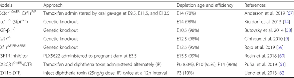
Methods to study microglia function
Developing good methods to specifically alter microglia
presence or function poses several challenges. First,
mol-ecules expressed on microglia are often found on
macro-phages
or
other
cell
types
making
cell-specific
approaches difficult to achieve. Second, genes required
for microglia development are often critically involved in
other aspects of animal maturation or survival. Third,
because microglia can migrate and are capable of
re-population or self-renewal, cell ablation approaches
often result in at least some residual microglia and
de-pletion drugs must be continuously administered. Due
to these issues, the interpretation of microglia functional
studies must take into account the models and methods
used. We thus will briefly discuss the pros and cons of
available microglia depletion models used to study retina
and brain microglia (Table
2
).
One category of microglia manipulation models
in-volves deleting various effector molecules, such as
com-plement, which are thought to alter microglia function
[
63
,
64
]. These types of models can be useful because
they have more limited developmental side effects and
are supported by correlative evidence implicating
micro-glia in the phenotypes observed. Yet, in many cases,
glo-bal knockouts are used that are not specific to microglia
and affect other cells and systems. Such approaches do
not prove the necessity and sufficiency of microglia in
the observed phenotypes. To overcome this, some
groups generate microglia effector molecule knockouts
by crossing a
Cx3cr1-Cre line [
65
] to conditional lines
Table 2
Microglia depletion models
Models Approach Depletion age and efficiency References
Cx3cr1CreER; Csf1rF/F Tamoxifen administered by oral gavage at E9.5, E11.5, and E13.5 E14 (70%) Anderson et al. 2019 [67]
Pu.1-/-(Sfpi−/−) Genetic knockout E14 (98%) Kierdorf et al. 2013 [14]
TGF-β−/− Genetic knockout E10.5 (98%) Butovsky et al. 2014 [58]
Csf1r-/- Genetic knockout E12.5 (98%) Ginhoux et al. 2010 [9]
Csf1rΔFIRE/ΔFIRE Genetic knockout E12.5 (95%) Rojo et al. 2019 [59]
CSF1R inhibitor PLX5622 administered to pregnant dam at E3.5 E15.5 (99%) Rosin et al. 2018 [60]
CX3CR1CreER-iDTR Tamoxifen and diphtheria toxin administered alternately (IP) P6 (60%), P10 (95%), P14 (98%) Puñal et al. 2019 [61]
[
65
–
68
] though it should be noted that other cell
popu-lations are also targeted in this approach [
65
].
Available models to deplete or delete microglia also have
important caveats. Three common approaches are used to
prevent microglia formation. Each of these involves
delet-ing or modifydelet-ing one of three genes required for lymphoid
or myeloid cell lineage cell development:
PU.1,
transform-ing growth factor beta (TGF-
β
), or
CSF1R. These
ap-proaches can achieve 98% microglia depletion in
embryonic brain [
9
,
14
,
58
]. However, knocking out any
one of these genes induces a host of additional physiologic
changes that cloud the interpretation of results. Pu.1
−/−null mice are born alive but die of severe septicemia
within days.
Pu.1
−/−mice are not only devoid of
parenchy-mal microglia in the brain, but also of circulating
mono-cytes and tissue macrophages [
14
]. TGF-
β
1
−/−mice
develop a lethal autoinflammatory syndrome shortly after
birth and die by 3
–
4 weeks of age [
69
].
Csf1r
null mice
(Csf1r
−/−), Csf1 homozygous mutant mice (Csf1
op/op) and
Csf1r specific osteoclast knockouts [TNF Receptor
Super-family Member 11a (Tnfrsf11a
cre):Csf1r
fl/fl] show a lack of
tooth eruption, have low body weight and growth rates,
misshapen skulls, and bone defects and usually die within
30 days after birth [
8
,
69
–
71
]. A new model of Csf1r
modulation in which a
Csf1r
enhancer is deleted
(Csf1r
ΔFIRE/ΔFIRE) appears to circumvent many of these
is-sues.
Csf1r
ΔFIRE/ΔFIREmice lack macrophages and brain
microglia and are healthy and fertile up to 9 months of
age without the growth and developmental abnormalities
reported in
Csf1r
−/−or
Csf1
op/oprodents [
59
].
Given these issues, many researchers have utilized
microglia depletion models. Two pharmacological
ap-proaches are commonly used. Drugs that inhibit CSF1R
(including PLX3397, PLX5562, GW2580, and BLZ945)
can be administered in chow, in water, or
intraperitone-ally to deplete microglia. In the brain, this can result in
90% microglia depletion in adults, and 99% depletion at
E15 when pregnant mice are fed inhibitor containing
chow [
60
,
72
]. Alternatively, liposomes containing
chlo-dronate can be administered in vivo or in vitro to kill
microglia that engulf them. While useful, this method
likely targets other phagocytes as well, and the efficiency
of microglia depletion is quite low (40~70%) [
73
,
74
].
Genetic models of microglia depletion are also widely used.
In these systems, depletion is achieved through targeted
expression of the diphtheria toxin receptor (iDTR)
primar-ily through crossing iDTR animals to CX3CR1-CreER
animals to generate CX3CR1-CreER-iDTR mice [
75
].
CX3CR1 is found on microglia, as well as all monocytes,
intestinal macrophages and dendric cells, some NK cells,
and activated T cells [
76
–
78
]. Thus, injecting this line with
alternating doses of tamoxifen and diphtheria toxin can
de-plete microglia but also affects subsets of other immune
cells [
43
]. When injections are initiated at P0, this model
achieves 70% microglia depletion in the retina by P6 and
98% depletion by P10
–
14 [
61
]. In brain, 99 and 85%
micro-glia depletion are achieved after drug administration in
young (~ 30 days) and adult animals (6
–
8 weeks),
respect-ively [
10
,
79
]. Following the same principle, the 10% of
microglia that are CD11b positive [
80
,
81
] can be depleted
using a CD11b-DTR model [
62
]. Though this approach
also targets other immune cell populations [
82
,
83
]. While
useful, it is important to note that these models do not
achieve complete ablation, and remaining numbers of
microglia can vary from animal to animal. Since it is
for-mally possible that a small fraction of microglia could
ac-complish the same task as many, it is difficult to interpret
negative data. Many of these models also do not allow the
study of early postnatal ages since high levels of microglia
depletion are not achieved for several days. Finally, these
models require continual drug administration to maintain
low levels of microglia since these cells can repopulate
lo-cally or from the periphery [
10
,
84
–
86
].
invading macrophages. Repopulating cells in which
CD11b
+microglia had been eliminated expressed high
levels of the peripheral macrophage markers CD45 and
CCR2 and appeared associated with blood vessels [
92
].
Further, evidence suggests there could be two sources of
repopulating retina microglia. In a CX3CR1-depletion
model, microglia that repopulated the central retina
appeard to be derived from residual microglia in the optic
nerve, while microglia that repopulated the peripheral
ret-ina were suggested to arise from macrophages in the
cil-iary body or iris [
91
]. Whether repopulating microglia are
transcriptionally, molecularly, or functionally similar to
the native population remains an open question.
Microglia and retinal vascularization
In mouse, as in human, there are two phases of vascular
growth in the eye. In the first phase, hyaloid vessels
ex-tend from the optic disk to the lens and supply blood
and nutrients to the developing eye [
93
,
94
]. Later in
velopment, hyaloid vessels regress, and the retina
de-velops its own independent vascular network [
93
].
Within the retina, three intraretinal vascular layers
inter-digitate distinct neural regions. The superficial plexus
interleaves the GCL, the intermediate plexus ascends
into the IPL, and the deep plexus is located within the
OPL [
93
]. Each of these vessel layers has a characteristic
location and branching pattern and thus are considered
somewhat independent neurovascular units [
95
,
96
].
Microglia have been implicated in both hyaloid vessel
re-gression and intraretinal vascular formation in the eye via
different mechanisms. Genetic or pharmacological ablation
of vitreal macrophages or microglia have been shown to
preserve the otherwise transient hyaloid vasculature. This
process is thought to involve microglia-mediated apoptosis
of vascular endothelial cells via WNT signaling [
97
]. After
hyaloid vessels regress, endothelial cells proliferate and
mi-grate radially into the retina from the center to the
periph-ery, and microglia are thought to play supportive and
guidance roles during this process [
98
]. Retinal microglia
are closely apposed to endothelial tip cell filopodia, which
guide blood vessel growth through the tissue [
99
–
102
]. In
supporting studies, either genetic ablation or depletion
of microglia reduces intraretinal vessel branching and
density, while patterning was restored by intravitreal
injec-tion of exogeneous microglia [
99
,
103
,
104
]. In addition,
microglia have recently been shown to regulate
develop-mental death of astrocytes [
61
]. Since astrocytes form a
re-ticular network that provides a substrate for angiogenesis
and vessel patterning [
105
–
107
], microglia may also
indir-ectly mediate vascular integrity through regulating
astro-cyte numbers [
61
]. However, it should be noted that the
effects on blood vessel patterning in these microglia
models are variable. In addition, there appear to be
redun-dant mechanisms that compensate when microglia are not
present which result in relatively normal adult blood vessel
patterning in microglia deletion models [
61
,
104
]. Finally,
microglia have also been implicated in pathogenic retina
angiogenesis. In diabetic retinopathy, abnormal intravitreal
neovascularization coincides with an elevation of
micro-glial TNF-
α
[
108
,
109
]. Similar results were reported in an
ischemic retinopathy model where activated retinal
micro-glia were found to produce IL-1
β
, which maintained
microglia activation and was associated with microvascular
injury [
110
]. Given these observations, it is clear that much
remains to be learned about the relationship between
microglia and vasculature in the eye, particularly as
micro-glia appear to alternatively promote developmental
vascu-lar regression, formation, or pathogenesis.
Microglia in neurogenesis and developmental cell death
Microglia have been implicated in developmental and
adult neurogenesis, though the evidence remains
some-what controversial. In the retina, neurons are generated
from RPCs at distinct ratios and times [
111
–
113
]. In
zebrafish, targeted knockdown of Csf1r with RNAi delayed
migration of microglia from the yolk sac to the retina and
was correlated with a withdrawal of RPCs from the cell
cycle, reduced neuron production, and microphthalmia
[
114
]. The data in mice, however, are less clear. While
ap-plication of a CSF1R inhibitor (PLX3397) [
115
] or
mino-cycline (thought to reduce microglia activation, [
116
])
reduced RPC proliferation and viability, respectively, the
numbers and gross organization of adult retina neuron
cell bodies appear intact in the absence of microglia [
61
].
Finally, in the adult brain, microglia have been suggested
to both enhance and inhibit neurogenesis, and results
ap-pear to vary depending on the model, brain region, disease
state, and inflammatory and cytokine milieu [
117
–
120
].
pathways that facilitate microglia-mediated phagocytosis
of neurons or CNS debris is quite extensive (Table
3
) and
includes synaptotagmin-11 (Syt11, [
125
]), G
protein-coupled receptor 34 (GPR34, [
126
]), Mer tyrosine kinase
(MerTK, [
127
–
129
]) and spleen tyrosine kinase (Syk,
[
130
]). It remains to be determined whether these
path-ways converge on a central microglia phagocytic process
or whether their use is context dependent.
Microglia and synapse refinement
Microglia play active roles in synapse pruning,
develop-ment, plasticity, and maintenance in the developing and
adult brain [
131
–
133
]. Recent data suggest that the
mech-anisms involved in this process may be region specific.
Microglia have been shown to regulate synapse refinement
in the developing retinogeniculate system via the classical
complement cascade proteins C1q and C3. Genetic
dele-tion of these complement components blocks the capacity
of microglia to properly remove synapses [
1
,
63
].
How-ever, in the developing barrel cortex, microglia appear to
eliminate synapses via CX3CR1/CX3CL1 and signaling
through a disintegrin and metalloproteinase
domain-containing protein 10 (ADAM10). This metalloprotease
cleaves CX3CL1 into a secreted form, and mice deficient
in ADAM10, CX3CR1, or CX3CL1 show decreased
apse elimination and display reduced engulfment of
syn-apse fragments by microglia [
134
]. Whether these
pathways and processes extend to the retina is not clear.
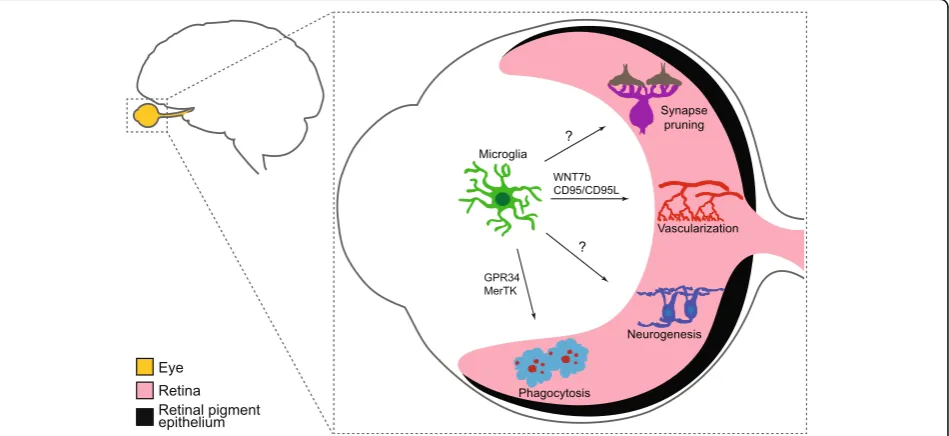
Fig. 4Proposed roles of microglia in the developing retina. Microglia play important roles in phagocytosis, vascularization, and neurogenesis through distinct mechanisms during retina development. These include modulation of both hyaloid vessel regression and intraretinal vascular patterning, regulation of the numbers of astrocytes and some RGC subsets, and RPC cycling. Roles for microglia in retina synapse pruning have also been proposed, though direct evidence for these pathways awaits further study
Table 3
Known pathways that contribute to cell and synapse engulfment
Pathways Findings References
C1q/C3 Mice deficient in complement protein C1q or the downstream complement protein C3 exhibit defects in CNS synapse elimination.
Stevens et al. 2007 [63]
C3/CR3 Microglia engulf presynaptic inputs during peak retinogeniculate pruning through complement receptor 3(CR3)/C3. Microglia also regulate retinal ganglion cell elimination by CR3-mediated engulfment of nonapoptotic neurons.
Schafer et al. 2012 [1] Anderson et al. 2019 [57]
Syt11 Syt11-knockdown increased cytokine secretion and nitric oxide release in primary microglia and enhanced microglial phagocytosis.
Du et al. 2017 [125]
GPR34 GPR34-deficient microglia showed reduced phagocytosis activity in both retina and acutely isolated cortical slices.
Preissler et al. 2015 [126]
MerTK Activated microglia release Gal-3 and a neuraminidase that desialylates microglial surfaces, enabling their phagocytosis via MerTK.
Grommes et al. 2008 [127] Caberoy et al. 2012 [128] Nomura et al. 2017 [129]
Syk Knock down of endogenous Syk decreased microglia phagocytosis of apoptotic neurons.
When microglia are depleted at P5, neuron and synapse
organization seem to be largely unaffected at P10 [
61
].
However, this negative data should be interpreted with
caution since: 1) a significant fraction of retina synapse
formation and remodeling occurs prior to P5; 2) the
models tested thus far still retained a small fraction of
microglia; and 3) visualizing microglia mediated synapse
pruning at single neuron resolution may show that only
particular subsets of neurons are affected. In line with
these ideas, adult retina depleted of microglia using the
CX3CR1-CreER-iDTR model show a loss of synapses in
the outer plexiform layer over time, resulting in decreased
retina function as measured by scotopic
electroretinogra-phy (ERG) recordings [
135
]. Thus, microglia may play
roles in maintaining synaptic integrity and function in the
adult retina. Continued efforts to understand the role of
microglia mediated synapse pruning in specific retinal
neuron subsets will help resolve whether microglia may
target specific cell types or synapses for removal.
Conclusions
Microglia are a fascinating cell type with the potential to
modulate or modify neuron development, survival,
con-nectivity, and vascularization (Fig.
4
). Studies in the
ret-ina and the brain are beginning to shed light on these
processes and the mechanisms involved, but this
enig-matic cell type still holds several key mysteries,
includ-ing: 1) how do microglia home to the CNS and monitor
and regulate their number and patterning; 2) do
micro-glia subpopulations play region or cell-type specific roles
in early neural development and neurodevelopmental
disorders; 3) what are the molecular mechanisms by
which microglia mediate synaptic refinement of specific
neurons or synapse types; and 4) what are the
interac-tions or signals that neurons provide to microglia that
encode neuron or synapse engulfment versus sparing?
Future studies that decipher these and related questions
will not only enable a better fundamental understanding
of neurobiology but also may provide untapped
oppor-tunities for treatment strategies aimed at preventing or
reversing diverse types of neural diseases.
Abbreviations
ADAM10:a disintegrin and metalloproteinase domain-containing protein 10; CNS: central nervous system; CSF1R: colony-stimulating factor one receptor; CX3CR1: CX3 chemokine receptor 1; DTR: Diphtheria toxin receptor; E: Embryonic day; ERG: Electroretinography; GCL: Ganglion cell layer; GPR34: G protein-coupled receptor 34; Iba1: Ionized calcium binding adaptor molecule 1; INL: Inner nuclear layer; IPL: Inner plexiform layer;
IRF-8: Interferon regulatory factor 8; MerTK: Mer tyrosine kinase; Ncx-1: Sodium calcium exchanger 1; ONL: Outer nuclear layer; OPL: Outer plexiform layer; P: Postnatal day; PU.1: PU box binding protein; RPCs: Retinal progenitor cells; RPE: Retinal pigment epithelium; SPI1: Spleen focus forming proviral integration oncogene; Syk: Spleen tyrosine kinase; Syt11: Synaptotagmin-11; TGF-β: Transforming growth factor beta; TLR4: Toll-like receptor 4; Tnfrsf11a: TNF Receptor Superfamily Member 11a
Acknowledgements
We thank members of our laboratory, Gretchen Diehl, and Wei Cao for scientific discussions and advice. This work was supported by the National Institutes of Health (NIH, R00AG044444, DP2EY02798, R56AG061808, and R01EY030458 to M.A.S.), the Cancer Prevention Research Institute of Texas (RR150005), the Brain Research Foundation, and the Ted Nash Foundation.
Authors’contributions
FL was the major contributor in designing the manuscript. DJ modified the manuscript and figures. MS edited the manuscript. All authors read and approved the final manuscript.
Funding
This work was supported by the National Institutes of Health (NIH, R00AG044444, DP2EY02798, R56AG061808, and R01EY030458 to M.A.S.), the Cancer Prevention Research Institute of Texas (RR150005), the Brain Research Foundation, and the Ted Nash Foundation.
Availability of data and materials
Not applicable.
Ethics approval and consent to participate
Not applicable.
Consent for publication
Not applicable.
Competing interests
The authors declare no competing interests.
Received: 18 September 2019 Accepted: 11 November 2019
References
1. Schafer DP, Lehrman EK, Kautzman AG, Koyama R, Mardinly AR, Yamasaki R, et al. Microglia sculpt postnatal neural circuits in an activity and
complement-dependent manner. Neuron. 2012;74(4):691–705. 2. Li Q, Barres BA. Microglia and macrophages in brain homeostasis and
disease. Nat Rev Immunol. 2018;18(4):225–42.
3. Colonna M, Butovsky O. Microglia function in the central nervous system during health and Neurodegeneration. Annu Rev Immunol. 2017;35:441–68. 4. Sierra A, Abiega O, Shahraz A, Neumann H. Janus-faced microglia: beneficial
and detrimental consequences of microglial phagocytosis. Front Cell Neurosci. 2013;7:6.
5. Ashwell K. Microglia and cell death in the developing mouse cerebellum. Brain Res Dev Brain Res. 1990;55(2):219–30.
6. Alliot F, Godin I, Pessac B. Microglia derive from progenitors, originating from the yolk sac, and which proliferate in the brain. Brain Res Dev Brain Res. 1999;117(2):145–52.
7. McKercher SR, Torbett BE, Anderson KL, Henkel GW, Vestal DJ, Baribault H, et al. Targeted disruption of the PU.1 gene results in multiple
hematopoietic abnormalities. EMBO J. 1996;15(20):5647–58. 8. Dai XM, Ryan GR, Hapel AJ, Dominguez MG, Russell RG, Kapp S, et al.
Targeted disruption of the mouse colony-stimulating factor 1 receptor gene results in osteopetrosis, mononuclear phagocyte deficiency, increased primitive progenitor cell frequencies, and reproductive defects. Blood. 2002; 99(1):111–20.
9. Ginhoux F, Greter M, Leboeuf M, Nandi S, See P, Gokhan S, et al. Fate mapping analysis reveals that adult microglia derive from primitive macrophages. Science. 2010;330(6005):841–5.
10. Bruttger J, Karram K, Wortge S, Regen T, Marini F, Hoppmann N, et al. Genetic cell ablation reveals clusters of local self-renewing microglia in the mammalian central nervous system. Immunity. 2015;43(1):92–106. 11. Dando SJ, Naranjo Golborne C, Chinnery HR, Ruitenberg MJ, McMenamin
PG. A case of mistaken identity: CD11c-eYFP(+) cells in the normal mouse brain parenchyma and neural retina display the phenotype of microglia, not dendritic cells. Glia. 2016;64(8):1331–49.
13. Holtschke T, Lohler J, Kanno Y, Fehr T, Giese N, Rosenbauer F, et al. Immunodeficiency and chronic myelogenous leukemia-like syndrome in mice with a targeted mutation of the ICSBP gene. Cell. 1996;87(2):307–17. 14. Kierdorf K, Erny D, Goldmann T, Sander V, Schulz C, Perdiguero EG, et al.
Microglia emerge from erythromyeloid precursors via Pu.1- and Irf8-dependent pathways. Nat Neurosci. 2013;16(3):273–80.
15. DeKoter RP, Walsh JC, Singh H. PU.1 regulates both cytokine-dependent proliferation and differentiation of granulocyte/macrophage progenitors. EMBO J. 1998;17(15):4456–68.
16. Beers DR, Henkel JS, Xiao Q, Zhao W, Wang J, Yen AA, et al. Wild-type microglia extend survival in PU.1 knockout mice with familial amyotrophic lateral sclerosis. Proc Natl Acad Sci U S A. 2006;103(43):16021–6.
17. Diaz-Araya CM, Provis JM, Penfold PL, Billson FA. Development of microglial topography in human retina. J Comp Neurol. 1995;363(1):53–68.
18. Santos AM, Calvente R, Tassi M, Carrasco MC, Martin-Oliva D, Marin-Teva JL, et al. Embryonic and postnatal development of microglial cells in the mouse retina. J Comp Neurol. 2008;506(2):224–39.
19. Martin-Estebane M, Navascues J, Sierra-Martin A, Martin-Guerrero SM, Cuadros MA, Carrasco MC, et al. Onset of microglial entry into developing quail retina coincides with increased expression of active caspase-3 and is mediated by extracellular ATP and UDP. PLoS One. 2017;12(8):e0182450. 20. Ashwell KW, Hollander H, Streit W, Stone J. The appearance and
distribution of microglia in the developing retina of the rat. Vis Neurosci. 1989;2(5):437–48.
21. Provis JM, Diaz CM, Penfold PL. Microglia in human retina: a heterogeneous population with distinct ontogenies. Perspect Dev Neurobiol. 1996;3(3):213–22.
22. Marin-Teva JL, Calvente R, Cuadros MA, Almendros A, Navascues J. Circumferential migration of ameboid microglia in the margin of the developing quail retina. Glia. 1999;27(3):226–38.
23. Chen L, Yang P, Kijlstra A. Distribution, markers, and functions of retinal microglia. Ocul Immunol Inflamm. 2002;10(1):27–39.
24. Perry VH. Evidence for an amacrine cell system in the ganglion cell layer of the rat retina. Neuroscience. 1981;6(5):931–44.
25. Schlamp CL, Montgomery AD, Mac Nair CE, Schuart C, Willmer DJ, Nickells RW. Evaluation of the percentage of ganglion cells in the ganglion cell layer of the rodent retina. Mol Vis. 2013;19:1387–96.
26. Macosko EZ, Basu A, Satija R, Nemesh J, Shekhar K, Goldman M, et al. Highly parallel genome-wide expression profiling of individual cells using Nanoliter droplets. Cell. 2015;161(5):1202–14.
27. Pei YF, Rhodin JA. The prenatal development of the mouse eye. Anat Rec. 1970;168(1):105–25.
28. Sernagor E, Eglen SJ, Wong RO. Development of retinal ganglion cell structure and function. Prog Retin Eye Res. 2001;20(2):139–74. 29. Silverman SM, Wong WT. Microglia in the retina: roles in development,
maturity, and disease. Annu Rev Vis Sci. 2018;4:45–77.
30. Kettenmann H, Hanisch UK, Noda M, Verkhratsky A. Physiology of microglia. Physiol Rev. 2011;91(2):461–553.
31. Harry GJ, Kraft AD. Microglia in the developing brain: a potential target with lifetime effects. Neurotoxicology. 2012;33(2):191–206.
32. Nayak D, Roth TL, McGavern DB. Microglia development and function. Annu Rev Immunol. 2014;32:367–402.
33. Ling EA, Wong WC. The origin and nature of ramified and amoeboid microglia: a historical review and current concepts. Glia. 1993;7(1):9–18. 34. Thomas WE. Brain macrophages: evaluation of microglia and their functions.
Brain Res Brain Res Rev. 1992;17(1):61–74.
35. Fetler L, Amigorena S. Neuroscience. Brain under surveillance: the microglia patrol. Science. 2005;309(5733):392–3.
36. Kaur C, Dheen ST, Ling EA. From blood to brain: amoeboid microglial cell, a nascent macrophage and its functions in developing brain. Acta Pharmacol Sin. 2007;28(8):1087–96.
37. Kettenmann H, Kirchhoff F, Verkhratsky A. Microglia: new roles for the synaptic stripper. Neuron. 2013;77(1):10–8.
38. Perez-Pouchoulen M, VanRyzin JW, McCarthy MM. Morphological and Phagocytic Profile of Microglia in the Developing Rat Cerebellum. eNeuro. 2015;2(4). 39. Okajima T, Tsuruta F. Microglial dynamics during brain development. Neural
Regen Res. 2018;13(2):222–3.
40. Davis EJ, Foster TD, Thomas WE. Cellular forms and functions of brain microglia. Brain Res Bull. 1994;34(1):73–8.
41. Kreutzberg GW. Microglia: a sensor for pathological events in the CNS. Trends Neurosci. 1996;19(8):312–8.
42. Koso H, Tsuhako A, Lai CY, Baba Y, Otsu M, Ueno K, et al. Conditional rod photoreceptor ablation reveals Sall1 as a microglial marker and regulator of microglial morphology in the retina. Glia. 2016;64(11):2005–24.
43. Schulz C, Gomez Perdiguero E, Chorro L, Szabo-Rogers H, Cagnard N, Kierdorf K, et al. A lineage of myeloid cells independent of Myb and hematopoietic stem cells. Science. 2012;336(6077):86–90.
44. Shigemoto-Mogami Y, Hoshikawa K, Goldman JE, Sekino Y, Sato K. Microglia enhance neurogenesis and oligodendrogenesis in the early postnatal subventricular zone. J Neurosci. 2014;34(6):2231–43.
45. Kierdorf K, Prinz M, Geissmann F, Gomez PE. Development and function of tissue resident macrophages in mice. Semin Immunol. 2015;27(6):369–78. 46. Erny D, Hrabe de Angelis AL, Jaitin D, Wieghofer P, Staszewski O, David E,
et al. Host microbiota constantly control maturation and function of microglia in the CNS. Nat Neurosci. 2015;18(7):965–77.
47. Bennett ML, Bennett FC, Liddelow SA, Ajami B, Zamanian JL, Fernhoff NB, et al. New tools for studying microglia in the mouse and human CNS. Proc Natl Acad Sci U S A. 2016;113(12):1738–46.
48. Yin J, Valin KL, Dixon ML, Leavenworth JW. The role of microglia and macrophages in CNS homeostasis, autoimmunity, and Cancer. J Immunol Res. 2017;2017:5150678.
49. Martin E, Boucher C, Fontaine B, Delarasse C. Distinct inflammatory phenotypes of microglia and monocyte-derived macrophages in Alzheimer's disease models: effects of aging and amyloid pathology. Aging Cell. 2017;16(1):27–38.
50. O'Koren EG, Yu C, Klingeborn M, Wong AYW, Prigge CL, Mathew R, et al. Microglial function is distinct in different anatomical locations during retinal homeostasis and degeneration. Immunity. 2019;50(3):723–37 e7.
51. Huang W, Chamberlain CG, Sarafian RY, Chan-Ling T. MHC class II expression by beta2 integrin (CD18)-positive microglia, macrophages and macrophage-like cells in rabbit retina. Neuron Glia BiolNeuron Glia Biol. 2008;4(4):285–94.
52. M. K. Ko; S. Saraswathy; J. G. Parikh; G.-S. Wu; N. A. Rao. Toll-like receptor 4 in the retinal microglia leads to photoreceptor oxidative stress. ARVO Annual Meeting Abstract, April 2009.
53. Tang PH, Pierson MJ, Heuss ND, Gregerson DS. A subpopulation of activated retinal macrophages selectively migrated to regions of cone photoreceptor stress, but had limited effect on cone death in a mouse model for type 2 Leber congenital amaurosis. Mol Cell Neurosci. 2017;85:70–81. 54. Hammond TR, Dufort C, Dissing-Olesen L, Giera S, Young A, Wysoker A,
et al. Single-cell RNA sequencing of microglia throughout the mouse lifespan and in the injured brain reveals complex cell-state changes. Immunity. 2019;50(1):253–71 e6.
55. Li Q, Cheng Z, Zhou L, Darmanis S, Neff NF, Okamoto J, et al.
Developmental heterogeneity of microglia and brain myeloid cells revealed by deep single-cell RNA sequencing. Neuron. 2019;101(2):207–23 e10. 56. Ronning KE, Karlen SJ, Miller EB, Burns ME. Molecular profiling of resident
and infiltrating mononuclear phagocytes during rapid adult retinal degeneration using single-cell RNA sequencing. Sci Rep. 2019;9(1):4858. 57. Anderson SR, Roberts JM, Zhang J, Steele MR, Romero CO, Bosco A, et al.
Developmental apoptosis promotes a disease-related gene signature and Independence from CSF1R signaling in retinal microglia. Cell Rep. 2019; 27(7):2002–13 e5.
58. Butovsky O, Jedrychowski MP, Moore CS, Cialic R, Lanser AJ, Gabriely G, et al. Identification of a unique TGF-beta-dependent molecular and functional signature in microglia. Nat Neurosci. 2014;17(1):131–43. 59. Rojo R, Raper A, Ozdemir DD, Lefevre L, Grabert K, Wollscheid-Lengeling E, et al.
Deletion of a Csf1r enhancer selectively impacts CSF1R expression and development of tissue macrophage populations. Nat Commun. 2019;10(1):3215. 60. Rosin JM, Vora SR, Kurrasch DM. Depletion of embryonic microglia using the
CSF1R inhibitor PLX5622 has adverse sex-specific effects on mice, including accelerated weight gain, hyperactivity and anxiolytic-like behaviour. Brain Behav Immun. 2018;73:682–97.
61. Puñal V, Paisley C, Brecha F, Lee M, Perelli R, Wang J et al. Large-scale death of retinal astrocytes during normal development is non-apoptotic and implemented by microglia. PLoS Biology, 2019:17(10).
62. Ueno M, Fujita Y, Tanaka T, Nakamura Y, Kikuta J, Ishii M, et al. Layer V cortical neurons require microglial support for survival during postnatal development. Nat Neurosci. 2013;16(5):543–51.
64. Fonseca MI, Chu SH, Hernandez MX, Fang MJ, Modarresi L, Selvan P, et al. Cell-specific deletion of C1qa identifies microglia as the dominant source of C1q in mouse brain. J Neuroinflammation. 2017;14(1):48.
65. Yona S, Kim KW, Wolf Y, Mildner A, Varol D, Breker M, et al. Fate mapping reveals origins and dynamics of monocytes and tissue macrophages under homeostasis. Immunity. 2013:79–91.
66. Giera S, Luo R, Ying Y, Ackerman SD, Jeong SJ, Stoveken HM, et al. Microglial transglutaminase-2 drives myelination and myelin repair via GPR56/ADGRG1 in oligodendrocyte precursor cells. Elife. 2018;7:e33385. 67. Anderson SR, Zhang J, Steele MR, Romero CO, Kautzman AG, Schafer DP,
et al. Complement targets newborn retinal ganglion cells for phagocytic elimination by microglia. J Neurosci. 2019;39(11):2025–40.
68. Sato-Hashimoto M, Nozu T, Toriba R, Horikoshi A, Akaike M, Kawamoto K, et al. Microglial SIRPαregulates the emergence of CD11c+ microglia and demyelination damage in white matter. Elife. 2019;8:e42025.
69. Kulkarni AB, Huh CG, Becker D, Geiser A, Lyght M, Flanders KC, et al. Transforming growth factor beta 1 null mutation in mice causes excessive inflammatory response and early death. Proc Natl Acad Sci U S A. 1993;90(2):770–4. 70. Marks SC Jr, Lane PW. Osteopetrosis, a new recessive skeletal mutation on
chromosome 12 of the mouse. J Hered. 1976;67(1):11–8.
71. Jacome-Galarza CE, Percin GI, Muller JT, Mass E, Lazarov T, Eitler J, et al. Developmental origin, functional maintenance and genetic rescue of osteoclasts. Nature. 2019;568(7753):541–5.
72. Han J, Harris RA, Zhang XM. An updated assessment of microglia depletion: current concepts and future directions. Mol Brain. 2017;10(1):25.
73. Drabek T, Janata A, Jackson EK, End B, Stezoski J, Vagni VA, et al. Microglial depletion using intrahippocampal injection of liposome-encapsulated clodronate in prolonged hypothermic cardiac arrest in rats. Resuscitation. 2012;83(4):517–26. 74. Han X, Li Q, Lan X, El-Mufti L, Ren H, Wang J. Microglial depletion with
Clodronate liposomes increases Proinflammatory cytokine levels, induces astrocyte activation, and damages blood vessel integrity. Mol Neurobiol. 2019;56(9):6184–96.
75. Goldmann T, Wieghofer P, Muller PF, Wolf Y, Varol D, Yona S, et al. A new type of microglia gene targeting shows TAK1 to be pivotal in CNS autoimmune inflammation. Nat Neurosci. 2013;16(11):1618–26.
76. Geissmann F, Manz MG, Jung S, Sieweke MH, Merad M, Ley K. Development of monocytes, macrophages, and dendritic cells. Science. 2010;327(5966):656–61. 77. Jung S, Aliberti J, Graemmel P, Sunshine MJ, Kreutzberg GW, Sher A, et al.
Analysis of fractalkine receptor CX(3)CR1 function by targeted deletion and green fluorescent protein reporter gene insertion. Mol Cell Biol. 2000;20(11):4106–14. 78. Niess JH, Brand S, Gu X, Landsman L, Jung S, McCormick BA, et al.
CX3CR1-mediated dendritic cell access to the intestinal lumen and bacterial clearance. Science. 2005;307(5707):254–8.
79. Parkhurst CN, Yang G, Ninan I, Savas JN, Yates JR 3rd, Lafaille JJ, et al. Microglia promote learning-dependent synapse formation through brain-derived neurotrophic factor. Cell. 2013;155(7):1596–609.
80. Murinello S, Moreno SK, Macauley MS, Sakimoto S, Westenskow PD, Friedlander M. Assessing retinal microglial phagocytic function in vivo using a flow Cytometry-based assay. J Vis Exp. 2016;116.
81. Greenhalgh AD, Zarruk JG, Healy LM, Baskar Jesudasan SJ, Jhelum P, Salmon CK, et al. Peripherally derived macrophages modulate microglial function to reduce inflammation after CNS injury. PLoS Biol. 2018;16(10):e2005264. 82. Dziennis S, Van Etten RA, Pahl HL, Morris D, Rothstein TL, Blosch CM,
Perlmutter RM, Tenen DG. The CD11b promoter directs high-level expression of reporter genes in macrophages in transgenic mice. Blood. 1995;85(2):319–29.
83. Duffield JS, Forbes SJ, Constandinou CM, Clay S, Partolina M, Vuthoori S, Wu S, Lang R, Iredale JP. Selective depletion of macrophages reveals distinct, opposing roles during liver injury and repair. J Clin Invest. 2005;115(1):56–65. 84. Yao Y, Echeverry S, Shi XQ, Yang M, Yang QZ, Wang GY, et al. Dynamics of spinal
microglia repopulation following an acute depletion. Sci Rep. 2016;6:22839. 85. Rice RA, Pham J, Lee RJ, Najafi AR, West BL, Green KN. Microglial
repopulation resolves inflammation and promotes brain recovery after injury. Glia. 2017;65(6):931–44.
86. Huang Y, Xu Z, Xiong S, Sun F, Qin G, Hu G, et al. Repopulated microglia are solely derived from the proliferation of residual microglia after acute depletion. Nat Neurosci. 2018;21(4):530–40.
87. Ortega-Martinez S, Palla N, Zhang X, Lipman E, Sisodia SS. Deficits in enrichment-dependent neurogenesis and enhanced anxiety behaviors mediated by expression of Alzheimer's disease-linked Ps1 variants are rescued by microglial depletion. J Neurosci. 2019;39(34):6766–80.
88. De Luca SN, Sominsky L, Soch A, Wang H, Ziko I, Rank MM, et al. Conditional microglial depletion in rats leads to reversible anorexia and weight loss by disrupting gustatory circuitry. Brain Behav Immun. 2019; 77:77–91.
89. Elmore MR, Najafi AR, Koike MA, Dagher NN, Spangenberg EE, Rice RA, et al. Colony-stimulating factor 1 receptor signaling is necessary for microglia viability, unmasking a microglia progenitor cell in the adult brain. Neuron. 2014;82(2):380–97.
90. Zhang Y, Zhao L, Wang X, Ma W, Lazere A, Qian HH, et al. Repopulating retinal microglia restore endogenous organization and function under CX3CL1-CX3CR1 regulation. Sci Adv. 2018;4(3):eaap8492.
91. Huang Y, Xu Z, Xiong S, Qin G, Sun F, Yang J, et al. Dual extra-retinal origins of microglia in the model of retinal microglia repopulation. Cell Discov. 2018;4:9.
92. Varvel NH, Grathwohl SA, Baumann F, Liebig C, Bosch A, Brawek B, et al. Microglial repopulation model reveals a robust homeostatic process for replacing CNS myeloid cells. Proc Natl Acad Sci U S A. 2012;109(44):18150–5.
93. Stahl A, Connor KM, Sapieha P, Chen J, Dennison RJ, Krah NM, et al. The mouse retina as an angiogenesis model. Invest Ophthalmol Vis Sci. 2010; 51(6):2813.
94. McLeod DS, Hasegawa T, Baba T, Grebe R, Galtier d'Auriac I, Merges C, et al. From blood islands to blood vessels: morphologic observations and expression of key molecules during hyaloid vascular system development. Invest Ophthalmol Vis Sci. 2012;53(13):7912–27.
95. Rhodes JM, Simons M. The extracellular matrix and blood vessel formation: not just a scaffold. J Cell Mol Med. 2007;11(2):176–205.
96. Iadecola C. The neurovascular unit coming of age: a journey through neurovascular coupling in health and disease. Neuron. 2017;96(1):17–42. 97. Lobov IB, Rao S, Carroll TJ, Vallance JE, Ito M, Ondr JK, et al. WNT7b
mediates macrophage-induced programmed cell death in patterning of the vasculature. Nature. 2005;437(7057):417–21.
98. Diez-Roux G, Lang RA. Macrophages induce apoptosis in normal cells in vivo. Development. 1997;124(18):3633–8.
99. Checchin D, Sennlaub F, Levavasseur E, Leduc M, Chemtob S. Potential role of microglia in retinal blood vessel formation. Invest Ophthalmol Vis Sci. 2006;47(8):3595–602.
100. Fantin A, Vieira JM, Gestri G, Denti L, Schwarz Q, Prykhozhij S, et al. Tissue macrophages act as cellular chaperones for vascular anastomosis downstream of VEGF-mediated endothelial tip cell induction. Blood. 2010:829–40. 101. Chen S, Tisch N, Kegel M, Yerbes R, Hermann R, Hudalla H, et al. CNS
macrophages control neurovascular development via CD95L. Cell Rep. 2017; 19(7):1378–93.
102. Penfold PL, Provis JM, Madigan MC, van Driel D, Billson FA. Angiogenesis in normal human retinal development: the involvement of astrocytes and macrophages. Graefes Arch Clin Exp Ophthalmol. 1990;228(3):255–63. 103. Ritter MR, Banin E, Moreno SK, Aguilar E, Dorrell MI, Friedlander M. Myeloid
progenitors differentiate into microglia and promote vascular repair in a model of ischemic retinopathy. J Clin Invest. 2006;116(12):3266–76. 104. Kubota Y, Takubo K, Shimizu T, Ohno H, Kishi K, Shibuya M, et al. M-CSF
inhibition selectively targets pathological angiogenesis and lymphangiogenesis. J Exp Med. 2009;206(5):1089–102.
105. O'Sullivan ML, Punal VM, Kerstein PC, Brzezinski JA, Glaser T, Wright KM, et al. Astrocytes follow ganglion cell axons to establish an angiogenic template during retinal development. Glia. 2017;65(10):1697–716. 106. Tao C, Zhang X. Retinal proteoglycans act as cellular receptors for basement
membrane assembly to control astrocyte migration and angiogenesis. Cell Rep. 2016;17(7):1832–44.
107. Selvam S, Kumar T, Fruttiger M. Retinal vasculature development in health and disease. Prog Retin Eye Res. 2018;63:1–19.
108. Yossuck P, Yan Y, Tadesse M, Higgins RD. Dexamethasone alters TNF-alpha expression in retinopathy. Mol Genet Metab. 2001;72(2):164–7.
109. Yoshida S, Yoshida A, Ishibashi T. Induction of IL-8, MPC-1, and bFGF by TNF-α in retinal glial cells: implications for retinal neovascularization during post-ischemic inflammation. Graefes Arch Clin Exp Ophthalmol. 2004;242(5):409–13. 110. Rivera JC, Sitaras N, Noueihed B, Hamel D, Madaan A, Zhou T, et al.
Microglia and interleukin-1beta in ischemic retinopathy elicit microvascular degeneration through neuronal semaphorin-3A. Arterioscler Thromb Vasc Biol. 2013;33(8):1881–91.
112. Belliveau MJ, Cepko CL. Extrinsic and intrinsic factors control the genesis of amacrine and cone cells in the rat retina. Development. 1999;126(3):555–66. 113. Kay JN, Link BA, Baier H. Staggered cell-intrinsic timing of ath5 expression
underlies the wave of ganglion cell neurogenesis in the zebrafish retina. Development. 2005;132(11):2573–85.
114. Huang T, Cui J, Li L, Hitchcock PF, Li Y. The role of microglia in the neurogenesis of zebrafish retina. Biochem Biophys Res Commun. 2012; 421(2):214–20.
115. Kuse Y, Ohuchi K, Nakamura S, Hara H, Shimazawa M. Microglia increases the proliferation of retinal precursor cells during postnatal development. Mol Vis. 2018;24:536–45.
116. Ferrer-Martin RM, Martin-Oliva D, Sierra-Martin A, Carrasco MC, Martin-Estebane M, Calvente R, et al. Microglial activation promotes cell survival in Organotypic cultures of postnatal mouse retinal explants. PLoS One. 2015;10(8):e0135238. 117. Monje ML, Toda H, Palmer TD. Inflammatory blockade restores adult
hippocampal neurogenesis. Science. 2003;302(5651):1760–5. 118. Cunningham C. Microglia and neurodegeneration: the role of systemic
inflammation. Glia. 2013;61(1):71–90.
119. Ramesh G, MacLean AG, Philipp MT. Cytokines and chemokines at the crossroads of neuroinflammation, neurodegeneration, and neuropathic pain. Mediat Inflamm. 2013;2013:480739.
120. Wang WY, Tan MS, Yu JT, Tan L. Role of pro-inflammatory cytokines released from microglia in Alzheimer's disease. Ann Transl Med. 2015;3(10):136. 121. Young RW. Cell death during differentiation of the retina in the mouse. J
Comp Neurol. 1984;229(3):362–73.
122. Vecino E, Acera A. Development and programed cell death in the mammalian eye. Int J Dev Biol. 2015;59(1–3):63–71.
123. Marín-Teva JL, Dusart I, Colin C, Gervais A, van Rooijen N, Mallat M. Microglia promote the death of developing Purkinje cells. Neuron. 2004; 41(4):535–47.
124. Casano AM, Albert M, Peri F. Developmental apoptosis mediates entry and positioning of microglia in the Zebrafish brain. Cell Rep. 2016;16(4):897–906. 125. Du C, Wang Y, Zhang F, Yan S, Guan Y, Gong X. Synaptotagmin-11 inhibits
cytokine secretion and phagocytosis in microglia. Glia. 2017;65(10):1656–67. 126. Preissler J, Grosche A, Lede V, Le Duc D, Krügel K, Matyash V, Szulzewsky F,
Kallendrusch S, Immig K, Kettenmann H, Bechmann I, Schöneberg T, Schulz A. Altered microglial phagocytosis in GPR34-deficient mice. Glia. 2015;63(2): 206–15.
127. Grommes C, Lee CY, Wilkinson BL, Jiang Q, Koenigsknecht-Talboo JL, Varnum B, Landreth GE. Regulation of microglial phagocytosis and inflammatory gene expression by Gas6 acting on the Axl/Mer family of tyrosine kinases. J NeuroImmune Pharmacol. 2008;3(2):130–40. 128. Caberoy NB, Alvarado G, Li W. Tubby regulates microglial phagocytosis
through MerTK. J Neuroimmunol. 2012;252(1–2):40–8.
129. Nomura K, Vilalta A, Allendorf DH, Hornik TC, Brown GC. Activated microglia Desialylate and Phagocytose cells via neuraminidase, Galectin-3, and Mer tyrosine kinase. J Immunol. 2017;198(12):4792–801.
130. Scheib JL, Sullivan CS, Carter BD. Jedi-1 and MEGF10 signal engulfment of apoptotic neurons through the tyrosine kinase Syk. J Neurosci. 2012;32(38): 13022–31.
131. Schafer DP, Stevens B. Phagocytic glial cells: sculpting synaptic circuits in the developing nervous system. Curr Opin Neurobiol. 2013;23:1034–40. 132. Schafer DP, Stevens B. Microglia function in central nervous system
development and plasticity. Cold Spring Harb Perspect Biol. 2015;7(10): a020545.
133. Hong S, Dissing-Olesen L, Stevens B. New insights on the role of microglia in synaptic pruning in health and disease. Curr Opin Neurobiol. 2016;36: 128–34.
134. Gunner G, Cheadle L, Johnson KM, Ayata P, Badimon A, Mondo E, Nagy MA, Liu L, Bemiller SM, Kim KW, Lira SA, Lamb BT, Tapper AR, Ransohoff RM, Greenberg ME, Schaefer A, Schafer DP. Sensory lesioning induces microglial synapse elimination via ADAM10 and fractalkine signaling. Nat Neurosci. 2019;22(7):1075–88.
135. Wang X, Zhao L, Zhang J, Fariss RN, Ma W, Kretschmer F, Wang M, Qian HH, Badea TC, Diamond JS, Gan WB, Roger JE, Wong WT. Requirement for microglia for the maintenance of synaptic function and integrity in the mature retina. J Neurosci. 2016;36(9):2827–42.
Publisher
’
s Note