R E S E A R C H A R T I C L E
Open Access
Dynamic regulation of mRNA decay during neural
development
Dana A Burow
†, Maxine C Umeh-Garcia
†, Marie B True, Crystal D Bakhaj, David H Ardell and Michael D Cleary
*Abstract
Background:Gene expression patterns are determined by rates of mRNA transcription and decay. While transcription is known to regulate many developmental processes, the role of mRNA decay is less extensively defined. A critical step toward defining the role of mRNA decay in neural development is to measure genome-wide mRNA decay rates in neural tissue. Such information should reveal the degree to which mRNA decay contributes to differential gene expression and provide a foundation for identifying regulatory mechanisms that affect neural mRNA decay. Results: We developed a technique that allows genome-wide mRNA decay measurements in intactDrosophila embryos, across all tissues and specifically in the nervous system. Our approach revealed neural-specific decay kinetics, including stabilization of transcripts encoding regulators of axonogenesis and destabilization of transcripts encoding ribosomal proteins and histones. We also identified correlations between mRNA stability and physiologic properties of mRNAs; mRNAs that are predicted to be translated within axon growth cones or dendrites have long half-lives while mRNAs encoding transcription factors that regulate neurogenesis have short half-lives. A search for candidate cis-regulatory elements identified enrichment of the Pumilio recognition element (PRE) in mRNAs encoding regulators of neurogenesis. We found that decreased expression of the RNA-binding protein Pumilio stabilized predicted neural mRNA targets and that a PRE is necessary to trigger reporter-transcript decay in the nervous system.
Conclusions:We found that differential mRNA decay contributes to the relative abundance of transcripts involved in cell-fate decisions, axonogenesis, and other critical events duringDrosophilaneural development. Neural-specific decay kinetics and the functional specificity of mRNA decay suggest the existence of a dynamic neurodevelopmental mRNA decay network. We found that Pumilio is one component of this network, revealing a novel function for this RNA-binding protein.
Keywords:Gene expression, mRNA decay, Transcriptome, Neurogenesis, Pumilio
Background
Transcription regulates neural developmental processes ranging from cell-fate specification [1] to synapse forma-tion [2]. However, differential transcripforma-tion alone is an ineffective method of regulating gene expression; math-ematical models demonstrate that mRNA decay is essen-tial for precise temporal and spaessen-tial control of mRNA abundance, particularly during developmental transitions [3]. Multiple mechanisms determine mRNA half-life [4]. One common mechanism involves the recognition of
cis-elements by RNA-binding proteins (RBPs).
RBP-mRNA interactions can activate or inhibit RBP-mRNA decay
by affecting the recruitment or activity of RNA degrad-ation complexes. Additional mRNA decay mechanisms include targeting by microRNAs and the nonsense-mediated decay (NMD) pathway. The stability of an in-dividual transcript may differ depending on cell type or the activation of signaling pathways. These mechanisms establish mRNA decay networks in which mRNA sta-bility is genetically programmed, tunable, and tightly regulated.
Regulation of mRNA decay appears to be particularly important for nervous system development [5]. This is partly due to the unique architecture of neurons; mRNAs exit the nucleus far from axon growth cones and dendrites where some transcripts are translated. The local concentrations of many mRNAs involved in axon pathfinding and synapse function are controlled * Correspondence:mcleary4@ucmerced.edu
†Equal contributors
Quantitative and Systems Biology Graduate Program, University of California, 5200 N. Lake Rd, Merced, CA, USA
via mRNA decay. For example, growth cone-localized
Robo3.2mRNA is degraded by the NMD pathway when axons encounter the spinal cord floor plate [6]. This
compartmentalized degradation of Robo3.2 is necessary
for the proper decussation of neurons in the spinal cord. Regulation of mRNA decay is also important for the proper proliferation and differentiation of neural progen-itors. Mouse neural progenitors lacking the RBP HuD have increased rates of self-renewal [7], and a circuitry involving the NMD pathway and neural miRNAs con-trols the balance between stem-cell proliferation and neural differentiation [8]. A role for mRNA decay in regulating cell-fate specification has been identified for the glial cells missing (gcm) gene in Drosophila [9]. Expression of agcmtransgene lacking a destabilizingcis -element causes neural progenitors to produce excess glia. Numerous examples of microRNAs, nonsense-mediated decay, and RBPs regulating the decay of target transcripts during neural development have been de-scribed, but the degree to which differential mRNA
decay globally affects mRNA abundance during in vivo
neurogenesis is not completely defined.
The degradation of maternally deposited mRNAs in
early Drosophila embryos has provided valuable
infor-mation about the role of mRNA decay in animal devel-opment [10]. However, analysis of zygotic mRNA decay during later stages of Drosophila development, specific-ally in the nervous system, presents several technical challenges. First, it requires a method to measure mRNA decay that does not interfere with gene expression or de-velopment. Traditional approaches for measuring mRNA decay rely on transcription inhibition (using drugs or temperature-sensitive mutations that inhibit RNA poly-merase II) and may have unwanted side effects [11]. Sec-ond, it requires a method to measure neural-specific mRNA decay in intact embryos. Many genes with neural development functions are expressed in multiple tissues, and the same transcript may have different half-lives in
neural versus non-neural cells. Whole embryo mRNA
decay measurements will therefore represent the aggre-gate half-life of an mRNA across multiple tissues.
A potential solution to the above mentioned chal-lenges is to use a pulse-chase approach to ‘tag’ nascent mRNAs in specific cell types then follow the decay of tagged mRNAs over time. Tissue-specific expression of the Toxoplasma gondii uracil phosphoribosyltransferase
(T.g.UPRT) enzyme in Drosophila allows tagging of
nascent mRNAs with 4-thiouracil and subsequent purification of the tagged mRNA (a technique known as TU-tagging) [12]. Variations of TU-tagging, in which pulse-labeling is followed by a “chase” in media lacking tagged uracil, have been used to obtain genome-wide mRNA decay measurements in yeast [13] and mammalian cell lines [14]. Here we combined TU-tagging with a
pulse-chase approach to obtain genome-wide
measure-ments of mRNA decay across all tissues of Drosophila
embryos and genome-wide measurements of mRNA decay specifically in the nervous system. This approach identified key components of a neural development mRNA decay network, including the differential decay of mRNAs within distinct functional classes and the role of the RBP Pumilio in regulating neural mRNA decay.
Results
TU-decay allows mRNA decay measurements in intact
Drosophilaembryos
To measure zygotic mRNA decay inDrosophilaembryos,
we developed a pulse-chase approach termed‘TU-decay’. We first used the nucleoside 4-thiouridine (4sUd) to tag mRNAs in all embryonic tissues (independent of
T.g.UPRT). Stage 12 to 15 embryos were incubated in 1
mM 4sUd during a 1-hour ‘pulse’ then transferred to
‘chase’media containing 10 mM uridine. 4sUd-RNA
(TU-RNA) was purified after the 1-hour pulse, a 1-hour chase, and a 3-hour chase. A potential limitation of this approach is continued incorporation of 4sUd during the chase, ei-ther due to incomplete saturation with uridine or
recyc-ling of 4sUd from degraded transcripts. 4sUd
incorporation during the chase could result in artificially long half-life calculations. To test for such an effect, we performed parallel experiments in which the transcription inhibitor actinomycin D (actD) was included in the chase media. Efficient transcription arrest upon actD exposure was confirmed by the lack of 4sUd incorporation in actD-treated embryos (Additional file 1).
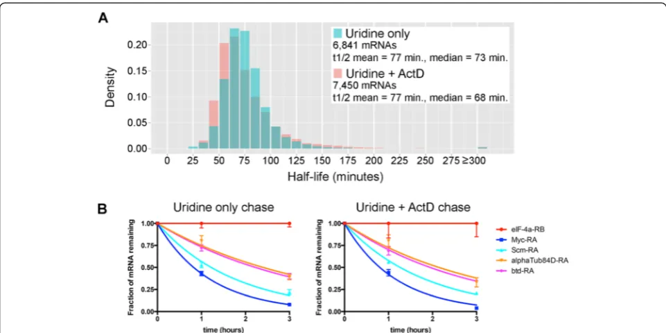
TU-RNA abundance at each time point was measured by microarray analysis, and mRNA half-life was calcu-lated by fitting measurements to a single exponential decay equation. Data for the majority of mRNAs fit this model; 79% of mRNAs from the uridine-only data (UO), and 87% of mRNAs from the uridine plus actD data
(U + actD) fit with R2≥0.75. The UO and U + actD
data were first used to test for potential biases in decay measurements. We did not detect any correl-ation between uridine number and mRNA half-life (Additional file 2), demonstrating that uridine frequency does not affect decay measurements. We also found no correlation between transcript abundance and mRNA half-life, demonstrating that there is no bias in the decay measurements for low-abundance versus high-abundance transcripts (Additional file 2).
The distribution of mRNA half-lives was similar for pulse-chase experiments performed with or without actD (Figure 1A), and decay kinetics for the majority of mRNAs were highly reproducible; only 11.6% of mRNAs
had standard deviations in half-life ≥30% of the mean
(Figure 1B), including orthologs of known mammalian
high-stability mRNAs (for example,eIF4a[15]) and
low-stability mRNAs (for example, Myc [16]). An additional
indication of the reproducibility of these measurements is provided by gene ontology (GO) analysis of the UO and U + actD datasets. The top 1,000 most stable transcripts and the bottom 1,000 least stable transcripts from both datasets yielded very similar GO results (Additional file 3). Given the reproducibility and simi-larity of decay measurements obtained with or without actD, we conclude that TU-decay effectively measures mRNA decay without the need to use pharmacological inhibitors of RNA polymerase II or other methods of arresting transcription.
Gene ontology categories identified by combining the UO and U + actD datasets are summarized in Table 1, and a complete list of half-lives is provided in Additional file 4. The results are similar to category enrichments
seen for stable versus unstable mRNAs in studies of
Drosophila maternal mRNA decay [10], yeast mRNA decay [17], and mammalian mRNA decay [18]. High-stability transcripts had a significant enrichment of mRNAs encoding ribosomal proteins, cytoskeletal pro-teins, chromatin-associated propro-teins, electron transport proteins, and other mRNAs involved in constitutive or ‘housekeeping’functions. Low-stability transcripts had a significant enrichment of mRNAs encoding transcription
factors, kinases, and other mRNAs associated with cell-fate decisions and organ development, including regula-tors of neuron projection morphogenesis. This agreement with gene ontology results obtained using alternative model systems further supports the validity of our pulse-chase approach. We conclude that TU-decay is an effective
technique for measuring mRNA decay in Drosophila
embryos.
TU-tagging purifies neural-transcribed mRNAs from whole embryos
We previously used TU-tagging to identify taranis as a
nervous system-expressed gene in Drosophila embryos
[19]. These experiments used prospero-GAL4 to drive
UAS-T.g.UPRT expression (pros > UPRT). Embryos at 0 to 16 h of development were exposed to 4-thiouracil (4TU) for 2 h and TU-tagged RNA was isolated from whole embryo lysates. Purified TU-RNA was compared to non-tagged RNA from the same embryos in
micro-array experiments. The prospero-GAL4 construct
ex-presses GAL4 in neuroblasts, ganglion mother cells, and glia of the central nervous system (CNS) (Figure 2A) in
addition to peripheral sensory neurons. Prospero-Gal4
driving UAS-T.g.UPRTalso results in mRNA tagging in
post-mitotic CNS neurons since the UPRT (and likely some converted 4-thio-UMP) is transferred from pro-genitors to neurons [20].
Analysis of the complete pros > UPRT data (not previ-ously described) identified 148 genes enriched 1.5-fold or
greater compared to untagged mRNA, includingprospero
itself (Additional file 5). Published literature and the
BerkeleyDrosophilaGenome Project (BDGP)in situ
data-base provided embryonic expression patterns for 108 of the 148 enriched genes. Of these, 94 (87%) are expressed in the nervous system. To further test the identification of
neural mRNAs, we performed in situ hybridizations for
ten enriched mRNAs whose expression patterns had not previously been characterized. We found that all ten were expressed in the nervous system, with eight specifically expressed in the CNS (Figure 2B). In total, we verified that 104 out of 118 (88%) genes identified bypros > UPRT
TU-tagging were expressed in the nervous system.
We used the same pros > UPRTgenotype to perform
nervous system-specific TU-decay analysis. Stage 12 to
15 embryos were exposed to the pulse-chase time course described for whole embryos. To confirm enrichment of neural mRNAs in these experiments, we compared
the pulse-labeled TU-RNA from pros > UPRT embryos
to the pulse-labeled TU-RNA from whole embryos. Neural-specific TU-RNA had a modest enrichment of known nervous system-expressed mRNAs, as expected given the presence of these transcripts in both stage-matched samples (Figure 2C). Neural-specific TU-RNA was significantly depleted of many mRNAs primarily expressed in the mesoderm and muscle (Figure 2C). We also performed gene ontology analysis of transcripts that were absent from neural-specific TU-RNA but present in whole embryo TU-RNA. This revealed a sig-nificant depletion of transcripts associated with the de-velopment or function of tissues outside the nervous system (Figure 2D). Based on the above indicators of
Table 1 Whole embryo mRNA decay analysis: functional annotation of high-stability and low-stability mRNAs
Whole embryo measurements: high-stability mRNAs
GO term Definition Pvalue FDR Count Fold enrichment
0022626 Cytosolic ribosome 2 × 10−57 5 × 10−56 67 10.3
RpL4,RpL23,RpS16,RpS24
0000022 Microtubule cytoskeleton 8 × 10⁻¹⁷ 3 × 10⁻¹⁴ 57 3.5
Tub84b,Tub56D,ssp4,robl
0022900 Electron transport chain 1 × 10⁻¹⁴ 2 × 10⁻¹² 29 5.9
mt:Cyt-b, mt:CoI,mt:CoII,mt:ND1
0031497 Chromatin assembly 2 × 10⁻⁹ 2 × 10⁻⁷ 15 7.8
His1,His2A,His2Av,His2B,His3
0045941 Positive regulation of transcription 4 × 10⁻³ .14 15 2.4
DSIF,HP1,Trl,ash2,lid,arm,btd
0048024 Regulation of mRNA splicing 7 × 10⁻³ .19 10 2.9
Hrb27c,Hrb87f,B52,SC35,snf
Whole embryo measurements: low-stability mRNAs
GO term Definition Pvalue FDR Count Fold enrichment
0004672 Protein kinase activity 2 × 10⁻⁹ 1 × 10⁻⁶ 41 2.6
S6kII,BubR1,mkk4,dTOR,wts,htl
0048812 Neuron projection morphogenesis 2 × 10⁻⁸ 2 × 10⁻⁶ 40 2.7
Con,trio,Ephrin,Eph,otk,drl,rl
0006351 Transcription 1 × 10⁻⁵ 4 × 10⁻⁴ 47 1.8
svp,dpn,Myc,grn,kn,Trx,eve
0019226 Transmission of nerve impulse 3 × 10⁻⁵ 9 × 10⁻⁴ 26 2.6
CASK,cha,synaptogyrin,cpx,shi
0014706 Striated muscle tissue development 3 × 10⁻⁵ 1 × 10⁻³ 11 5.3
Hem,Mef2,Mlp84B,flw,mbc
0051674 Localization of cell 5 × 10⁻⁴ .01 23 2.3
cbl,APC,Dl,Mhc,baz,dome,cv-c
nervous system specificity, we proceeded to use the
pros > UPRT pulse-chase data to calculate neural mRNA half-lives.
Functionally related mRNAs have neural-specific decay kinetics
Neural mRNA half-lives were calculated as described for whole embryos. Seventy-three percent of mRNAs fit the exponential decay equation with an R2≥0.75. The repro-ducibility of neural-specific measurements was similar to
that observed for whole embryos; between biological replicates, only 13.1% of mRNAs had standard devia-tions in half-life ≥30% of the mean for that transcript. The distribution of mRNA half-lives based on neural-specific and whole embryo measurements was very similar (Figure 3A). This suggests that the general mRNA-decay machinery and kinetics of turnover are not substantially different in the nervous system com-pared to other tissues. Genome-wide neural-specific mRNA half-lives are provided in Additional file 6.
Figure 2Pros > UPRTTU-tagging purifies neural mRNAs from embryos.(A)Prospero-GAL4drivingUAS-GFP::lacZ.nls(encoding a nuclear-localized GFP-β-galactosidase fusion) in the embryonic central nervous system shows the expression pattern ofprospero-GAL4. Anti-Prospero =magenta, anti-β-galactosidase =green.(B)RNAin situhybridization for eight genes identified as nervous system-enriched by TU-tagging. Gene annotation symbols are listed in the top panel. Top panel = ventral view. Bottom panel = lateral view. Developmental stage is listed in thelower right corner
Gene ontology analysis of low-stability versus high-stability neural mRNAs identified several differences compared to the whole embryo dataset (Table 2). While both datasets had the category ‘transcription’ enriched among low-stability mRNAs, the neural-specific mea-surements had a unique enrichment of mRNAs involved in neuron differentiation, mitosis, histone methylation, endocytosis, and ribosome biogenesis. Differences be-tween the two datasets are even more striking for high-stability mRNAs. The most significantly enriched category for neural high-stability mRNAs is‘neuron pro-jection morphogenesis’which includes regulators of axo-nogenesis and axon pathfinding. In contrast, mRNAs in this category were among the least stable mRNAs in whole embryos, suggesting differential regulation of some transcripts within this GO category. Examples of ‘neuron
projection morphogenesis’ mRNAs with inverse
stabili-ties based on neural-specific versus whole embryo
mea-surements includeAPC-like, Abelson interacting protein,
and off-track.Other high-stability mRNAs unique to the neural-specific dataset include those encoding small GTPases that regulate vesicle trafficking and transcripts encoding regulators of programmed cell death.
Genome-wide comparison of mRNA half-lives be-tween the neural-specific and whole embryo datasets re-vealed that 24% of mRNAs had statistically-significant
half-life differences of ≥1.5-fold (484 mRNAs had
de-creased stability based on neural-specific measurements, and 409 mRNAs had increased stability based on neural-specific measurements, out of 3,776 transcripts) (Figure 3B and Additional file 7). Importantly, these differences do not randomly occur across the transcrip-tome; mRNAs with different half-lives based on
neural-specific versus whole embryo measurements fall into
specific gene ontology categories (Table 3). mRNAs with shorter half-lives based on neural-specific measurements include those encoding ribosomal proteins (26 ribosomal protein mRNAs), translation regulators (EF2, eIF-5A),
Figure 3Comparison of neural-specific and whole embryo decay measurements suggests many transcripts have neural-specific half-lives.
histones (7 histone mRNAs), and metabolic enzymes (Gapdh1, multiple ATP synthetases). mRNAs with longer half-lives based on neural-specific measurements include those involved in neurotransmission (CASK, stj, homer,
Dap160, Rop), axonogenesis (pk, Appl, miple, galectin), axonal transport (Klp64D,TBCB), and establishment or maintenance of neuroblast polarity (baz,par-6,Lgl,pins,
Gαo). Each of these genes is transcribed in the nervous sys-tem plus additional tissues (based on FlyBase annotations and/or BDGPin situ data), suggesting that half-life differ-ences between whole embryo and neural-specific measure-ments are due to neural-specific mRNA decay kinetics.
Neural mRNA stability correlates with predicted transcript localization and function
Gene ontology analysis of neural mRNA-decay data re-vealed that mRNAs encoding transcription factors tend
to be rapidly decayed, and mRNAs encoding proteins in-volved in neuron projection morphogenesis and neuro-transmission tend to be stable. The rapid decay of transcription-factor mRNAs could be related to transient protein expression during cell-fate specification. To test this hypothesis, we identified 39 mRNAs encoding tran-scription factors that are involved in CNS neurogenesis (based on Interactive Fly annotations [21]). The stability of mRNAs associated with neuron projection morpho-genesis and neurotransmission could be related to their localization in axon growth cones or dendrites. To test this hypothesis, we identified 35Drosophilaorthologs of mRNAs that are localized to axon growth cones [22] or dendrites [23] in mammals.
We compared the half-lives of the neurogenesis transcription factor mRNAs (which we refer to as ‘neural fate’ mRNAs) and the half-lives of the predicted
Table 2 Neural-specific mRNA decay analysis: functional annotation of high-stability and low-stability mRNAs
Neural-specific measurements: low-stability mRNAs
GO term Definition Pvalue FDR Count Fold enrichment
0006351 Transcription 4 × 10⁻¹⁰ 2 × 10⁻⁷ 58 2.5
svp,dpn,pros,vvl,grh,gsb,onecut
0030182 Neuron differentiation 2 × 10⁻⁷ 4 × 10⁻⁵ 48 2.3
chinmo,Dl,tup,Ephrin,Eph,fax,shot
0007067 Mitosis 6 × 10⁻⁵ 3 × 10⁻³ 21 2.9
cycB3,cdc27,Sse,cnn,glu,mts
0018024 Histone methyltransferase activity 2 × 10⁻⁴ .02 7 6.9
G9a,Set1,Set2,Su(z)12,ash1,egg
0006897 Endocytosis 4 × 10⁻³ .08 25 1.9
Arpc4,shi,CalpA,pat1,faf,scb,Rac1
0005840 Ribosome 2 × 10⁻³ .06 21 2.0
RpL17,RpL35,mRpL11,mRpS35
Neural-specific measurements: high-stability mRNAs
GO term Definition Pvalue FDR Count Fold enrichment
0048812 Neuron projection morphogenesis 5 × 10⁻⁶ 6 × 10⁻⁴ 33 2.4
trc,fusl,Fas2,Appl,Fmr1,otk,ben
0003924 GTPase activity 2 × 10⁻⁴ .02 17 2.9
Rab23,Rab30,Rab11,Rho1,Ras64B
0012501 Programmed cell death 2 × 10⁻³ .06 17 2.4
grim,pten,scyl,egr,mod(mdg4)
0004672 Protein kinase activity 9 × 10⁻³ .24 23 1.8
CamKI,Cdk4,Ror,ald,aur,nmo,trbl
0019226 Transmission of nerve impulse .02 .25 17 1.8
CASK,CanB,Dap160,cpx,Sap47
0051674 Localization of cell .01 .16 18 1.9
cbl,APC,cortactin,par-6,baz,spri
axon or dendrite-localized mRNAs (which we refer to
as ‘localized’ mRNAs) to the half-lives of all neural
mRNAs. This revealed a significant trend of below average stability for neural fate mRNAs and above average stability for localized mRNAs (Figure 4A, Additional file 8). Rapidly decayed neural fate mRNAs include those encoding proteins whose expression is tightly coupled to the timing of neuroblast cell divi-sions (cas, svp, pdm-1) [24]; proteins that are differen-tially expressed within neuroblast lineages (Hey,Dl) [25]; and proteins that are differentially expressed among neu-roblasts, GMCs, and neurons (dpn,pros) [26] (Figure 4B). Thus, rapid mRNA decay correlates with dynamic tem-poral or spatial patterns of protein expression. Additional transcription factor mRNAs that are rapidly decayed but were not included in the list of neurogenesis transcrip-tion factors includefkh,pnt, glu,BarH1, and others that may have functions in neural development that require precise regulation of mRNA abundance.
Stable mRNAs predicted to localize to axon growth
cones or dendrites include a known Drosophila
dendrite-localized mRNA (CamKII [27]) and two orthologs of
mammalian mRNAs that are locally translated in dendrites (Arc1[28] andEf1α100E[29]) (Figure 4C). Stable predicted localized mRNAs also include transcripts encoding proteins that localize to pre- or post-synaptic sites in
Drosophila neurons (homer [30], CASK [31], and
synaptogyrin [32]), although the site of translation of these mRNAs is unknown. We conclude that the ele-vated stability of these mRNAs may be related to evolutionarily conserved patterns of subcellular mRNA localization.
Pumilio regulates neural mRNA decay
To identify sequence elements that might target neural
fate mRNAs for rapid decay, we searched for knowncis
-regulatory elements in the 3’ UTRs of this group of
transcripts. As a first step, we tested for any correlation
between 3’ UTR length and mRNA stability, since
shorter 3’ UTRs could contain fewer cis-elements. We
found no correlation between 3’ UTR length and stability
and no bias toward short or long 3’ UTRs in the stable
localized versus unstable neural fate mRNAs (or in the
entire neural mRNA decay dataset) (Additional file 9).
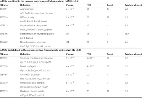
Table 3 Functional annotation of mRNAs with altered stability based on neural-specific versus whole embryo measurements
mRNAs stabilized in the nervous system (neural/whole embryo half-life >1.5)
GO term Definition Pvalue FDR Count Fold enrichment
007409 Axonogenesis 2 × 10⁻³ .06 13 2.8
APC,CadN,trio,caps,fray,otk,daw
0003924 GTPase activity 2 × 10⁻³ .31 10 3.5
Rab21,Rab30,Ras64B,RabX1
0009312 Oligosaccharide biosynthesis 9 × 10⁻³ .15 4 9.2
mgat2,GalNAc-T1,pgant2,pgant5
0045196 Establishment of neuroblast polarity .01 .16 3 18.7
par-6,baz,Lgl
0007269 Neurotransmitter secretion .04 .56 6 2.6
CanB,Lgl,CASK,Dap160,cpx,rop
mRNAs destabilized in the nervous system (neural/whole embryo half-life <0.6)
GO term Definition Pvalue FDR Count Fold enrichment
0003735 Structural constituent of ribosome 5 × 10⁻¹⁷ 5 × 10⁻¹⁵ 26 9.2
RpL11,RpL30,RpL6,RpS16,RpS23
0000278 Mitotic cell cycle 4 × 10⁻⁸ 4 × 10⁻⁵ 30 3.2
dap,cycB3,ball,glu,eff,ncd,mts
0031497 Chromatin assembly 3 × 10⁻⁴ .02 7 7.5
H2B,H3,H3.3A/B,HP2,Df31,Set
0005839 Proteasome core complex 8 × 10⁻³ .07 5 6.0
Pros26,Prosα7,Prosβ2,Prosβ7
0006119 Oxidative phosphorylation 6 × 10⁻⁴ .03 12 3.5
ATPsynB,ATPsynD,mt:CoIII
Candidatecis-elements were selected based on known roles in maternal mRNA decay, known or predicted ex-pression of the corresponding trans-acting factor in the nervous system, and the availability of databases that identify transcripts containing thecis-element. Based on these criteria, we tested for enrichment or depletion of AU-rich elements (AREs) [33], miR-124 seed sequences (miR-124 is a nervous system-specific microRNA [34]), and Pumilio recognition elements (PREs) [35]. Of these candidates, only PREs were significantly enriched among the low-stability neural fate mRNAs (20 out of 36
mRNAs with half-life ≤75 min contain a PRE) and
de-pleted from high-stability localized mRNAs, although not at a statistically-significant level (1 out of 29 mRNAs
with half-life ≥80 min contain a PRE) (Figure 5A,
Additional file 8). AREs and miR124 seed sequences had no significant enrichment or depletion in neural fate or localized mRNAs (Figure 5A) or in the entire set of low or high-stability neural mRNAs.
For the PRE analysis, we used all mRNAs that contain the 8-nucleotide motif UGUA(A/U/C)AUA at least
once in their 3’ UTR, as identified by Gerber et al.
[35]. Of the 1,431 mRNAs that contain this motif, 439 were present in our neural-specific dataset. PREs do not appear to be a general determinant of neural
mRNA stability; there was no significant enrichment of PRE-containing transcripts among all low-stability or high-stability neural mRNAs (8.5% of mRNAs with
half-life ≤75 min contain a PRE, and 8.8% of mRNAs
with half-life ≥80 min contain a PRE). In addition to
mRNAs containing the PRE motif, Gerber et al.
identi-fied 156 potential Pumilio targets using a tandem-affinity purification (TAP)-tagged Pumilio approach [35]. The list of Pumilio targets identified by TAP does not include any transcripts from our neural fate or localized mRNA classes. Similar to the PRE data, the Pumilio TAP data were not a predictor of mRNA sta-bility among all neural mRNAs (0.9% of mRNAs with
half-life ≤75 min were predicted targets by TAP, and
1.5% of mRNAs with half-life ≥80 min were predicted
targets by TAP). We conclude that the presence of a PRE motif (and not identification as a Pumilio target by TAP) correlates with low mRNA stability only in the neural fate mRNA class.
The significant enrichment of PREs among low-stability neural fate mRNAs suggested a novel role for Pumilio in neural development. Pumilio has been impli-cated in the regulation of pre- and post-synaptic morph-ology and function [36] and memory formation [37] in
Drosophila. We investigated the expression of Pumilio in
Figure 4mRNA stability correlates with function and predicted localization.(A)mRNA half-life distributions of all neural mRNAs, the neural fate transcription factor subset (‘neural fate’), and the predicted axon growth cone or dendrite-localized mRNA subset (‘localized’). Thehorizontal line
the embryonic nervous system and found widespread localization of Pumilio in the cytoplasm of neural pro-genitors, neurons, and certain axon projections through-out the ventral nerve cord (Figure 5B), similar to Pumilio expression patterns in the larval central nervous system [36].
To test if Pumilio affects neural mRNA decay, we used neural-specific RNA interference [38] to de-crease Pumilio expression. Western blot analysis showed that Pumilio RNAi decreased Pumilio protein to approximately 50% of wild-type levels (Additional
file 10). Knockdown of Pumilio using pros-Gal4 X
UAS-pumilio{RNAi} did not cause detectable neural
development defects, based on analysis of Fas2+ axon
projection patterns and production of Eve+ neurons.
We conclude that partial knockdown of Pumilio has no effect or subtle effects on neural development. However, we predicted that partial knockdown of Pumilio should be sufficient to induce changes in mRNA stability if Pumilio regulates mRNA decay.
We performed TU-decay experiments in Pumilio
knockdown embryos, with pros-GAL4 driving
UAS-Pumilio{RNAi}and UAS-T.g.UPRT. Neural mRNA half-lives in the Pumilio knockdown embryos were com-pared to control pros > UPRT half-lives. Reproducible half-life measurements were obtained for 3,008 tran-scripts, and predicted Pumilio targets (PRE-containing transcripts) showed a significant trend of stabilization
compared to non-targets (Figure 5C, Additional
file 11). Data were obtained for 265 predicted
Pumi-lio targets, and 132 (50%) had a
statistically-significant increase in mRNA half-life of≥1.3-fold. As expected, many of the stabilized Pumilio targets encode transcription factors and other cell-fate determinants (Figure 5D). Stabilized Pumilio targets also include regu-lators of axon pathfinding (Eph receptor tyrosine kinase,
off-track, Sema-1a) that are not predicted to localize in axon growth cones. These experiments did not yield re-producible data for the one stable Pumilio target pre-dicted to localize to axon growth cones or dendrites,
Tetraspanin 42Ee. However, we did obtain measurements for another predicted localized Pumilio target,discs large 1(dlg1).dlg1mRNA was previously identified as a target of translation repression by Pumilio [39], and its
Figure 5Pumilio regulates neural mRNA stability.(A)Enrichment or depletion of candidatecis-regulatory elements as follows: AREs, miR-124 target sites, and PREs. Odds ratios were calculated as the frequency of each element within the low-stability (t1/2≤75 min)‘neural fate’mRNAs or high-stability (t1/2≥80 min)‘localized’ mRNAsversusthe frequency of that element in all low-stability or high-stability neural mRNAs. ** =Pvalue < 1.0 × 10⁻¹⁵(Fisher’s test).(B)Pumilio expression in a stage 15 embryo detected with an anti-Pumilio antibody (green). Anti-Fas2 antibody stain (magenta) shows a subset of longitudinal axon fiber tracts and axons leaving the ventral nerve cord.(C)Cumulative distribution function plots of mRNA half-life changes inPumilioRNAiversuscontrol embryos (PumilioRNAi/control ratio). The distribution for all neural mRNAs is plotted inblue. The distribution for predicted Pumilio targets is plotted inred. The shift to the right for predicted Pumilio targets indicates a trend of stabilization inPumilio
ortholog, PSD-95, is localized to dendrites in
mamma-lian neurons [40]. dlg1 mRNA is relatively unstable in
control embryos (half-life =65 min) and is significantly stabilized inPumilio{RNAi}embryos (half-life =127 min).
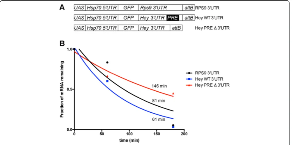
To test if a 3’UTR PRE is necessary for destabilization of a predicted Pumilio target in the nervous system, we
constructed reporter transcripts composed of the hsp70
5’ UTR, the green fluorescent protein (GFP) open
read-ing frame, and one of three 3’ UTR sequences
contain-ing an endogenous transcription termination signal; the
RPS9 3’ UTR (without any PRE), the Hey 3’ UTR
(containing a single naturally occurring PRE), and aHey
3’UTR with the PRE deleted (Figure 6A). These reporter
constructs were placed under UAS control and
inte-grated into the same chromosome locus in transgenic fly lines.Pros-Gal4was used to activate transcription of the
reporter and UAS-UPRT, thus allowing neural-specific
measurement of reporter stability. The reporter contain-ing the RPS9 3’UTR had slightly above average stability, similar to the endogenous RPS9 transcript (Figure 6B). In contrast, the reporter containing the Hey 3’UTR was
rapidly decayed. Deletion of the PRE from the Hey 3’
UTR caused a dramatic increase in stability compared to the unmodified Hey 3’UTR reporter. These results agree with the increased stability of the endogenous Hey tran-script inPumilio{RNAi}embryos and the lack of any sig-nificant change in endogenous RPS9 transcript stability
inPumilio{RNAi}embryos (Additional file 11). We con-clude that the single PRE within the Hey 3’UTR targets this transcript for degradation in the nervous system.
Discussion
mRNA abundance during neural development is deter-mined by the rate of gene transcription and the rate of mRNA decay. We addressed the role of mRNA decay in neural development using the TU-decay technique. Our
results provide in vivo measurements of zygotic mRNA
decay in Drosophila embryos and allow investigation of
neural-specific mRNA decay properties. It is difficult to determine how closely half-lives calculated by TU-decay reflect endogenous decay rates, since any technique used to quantify mRNA turnover may introduce biases. How-ever, the global trends of mRNA decay identified in this work are similar to those described for other systems (for example, gene ontology categories for low-stability
versus high-stability mRNAs), suggesting that TU-decay provides accurate measurements of relative mRNA stability.
Our comparison of whole embryo and neural-specific mRNA decay data revealed unique neural mRNA decay properties for 893 transcripts. While we have not checked the expression patterns of all 893 transcripts, many are expressed in the nervous system and add-itional tissues. We conclude that the different half-life
calculations for these transcripts are due to heteroge-neous transcript decay measurements in whole embryo
experiments versusmore homogeneous transcript decay
measurements in the neural-specific TU-decay experi-ments. Neural-specific decay properties likely enable a single mRNA to have distinct expression dynamics in the nervous system compared to other tissues.
Many transcripts with decreased stability in the ner-vous system encode proteins involved in cell growth and proliferation, such as ribosomal proteins and histones. This may relate to the transition from proliferation to differentiation that occurs in the nervous system at the embryonic stages we analyzed. Early-born neuroblast progeny differentiate into neurons and glia at stage 12, and progenitor proliferation significantly decreases by stage 15 [41]. A similar decrease in proliferation occurs in yeast shifted to nutrient-limited media, and this tran-sition causes a dramatic destabilization of ribosomal pro-tein mRNAs [13]. Lower histone mRNA stability has been described for human fibroblasts compared to their corresponding induced pluripotent stem cells (iPSCs) [42]. Histone mRNAs are more stable in iPSCs, presum-ably to support chromosome replication and the produc-tion of differentiated progeny. Similar destabilizaproduc-tion of histone mRNAs in the differentiating embryonic nervous system may be related to the decreased mitotic potential of progenitors and the post-mitotic state of neurons.
Many transcripts with increased stability in the ner-vous system encode proteins with known neural-specific functions, including neurotransmission, axonogenesis, and maintenance of neuroblast polarity. Many of these genes have widespread expression patterns but elevated levels of mRNA or protein in the nervous system.
Exam-ples include Cadherin-N (CadN), Apc-like (Apc), and
bazooka(baz). CadN is expressed in embryonic muscles and neurons, but the protein is detected at much higher levels in neurons [43]. Apc is detected ubiquitously at low levels in embryos, with high levels of protein
con-centrated in the central nervous system [44]. Bazooka
mRNA is detected in epithelial cells and neuroblasts, where Bazooka regulates spindle orientation and cell po-larity [45]. Multiple mRNAs encoding regulators of neuroblast polarity had increased stability based on
neural-specific measurements as follows: baz, pins,
par-6, Lgl, and Gαo. The stability of these transcripts may support appropriate levels of protein production in neuroblasts without the need for high rates of transcrip-tion. Baz, Par-6, and Lgl also have functions in post-mitotic neurons [46,47] and the increased stability of these mRNAs could be due to stabilization in neurons.
Genome-wide studies of mRNA decay in other sys-tems support a model in which mRNA half-life is linked to the function of the encoded protein [10,48]. We found that this model is applicable to mRNAs encoding
transcription factors that regulate neural cell-fate deci-sions. These transcription factors tend to act transiently in neural progenitors or their progeny and have precise spatial or temporal expression patterns. While the ex-pression of these genes likely involves transcriptional in-duction and repression, a short mRNA half-life will ensure robust expression patterns. Many cell-fate deter-minant transcription factors appear to be transcribed at low levels and rapidly decayed, since 1-hour pulse mea-surements yielded weak signals, and we could not detect any signal above background in subsequent chase mea-surements. Examples include cell-fate determinants that are transiently expressed in neural progenitors, such as
gcm [49], ac [50], and Kr [51]. Future work could
im-prove the analysis of weakly transcribed, rapidly decayed mRNAs by including earlier chase time points and using more sensitive detection methods (such as RNA-sequencing).
We also identified a potential relationship between mRNA half-life and mRNA localization in neurons. While the predicted localization of candidate mRNAs re-mains to be confirmed, several of the corresponding proteins are known to localize to axon terminals or den-drites inDrosophila. It will be interesting to test if a long mRNA half-life is predictive of subcellular localization in neurons, particularly for transcripts whose mammalian orthologs have not previously been identified in growth
cones or dendrites. For example, Stretchin-Mlck and
Fuseless are among the most stable neural mRNAs, and their proteins have known roles in axon guidance [52] and synaptic vesicle exocytosis [53], respectively. Stretchin-Mlck is predicted to act at axon growth cones, and Fuseless is concentrated in presynaptic sites of neuromuscular junctions, but their mRNA localization and site of translation is unknown.
While the data generated in this study may be useful forde novoidentification ofcis-regulatory elements, here we chose to focus on candidate elements that might determine the decay rate of neural cell-fate mRNAs. Pumilio recognition elements were the only significantly enriched element among this class of mRNAs. Trad-itional models of Pumilio function emphasize its role as a negative regulator of translation, but recent work has
shown that Drosophila Pumilio and human orthologs
promote deadenylation that is likely to subsequently trigger transcript decay [54]. In addition, an mRNA decay function for Pumilio was predicted based on the
enrichment of PREs inDrosophilamaternal mRNAs
de-graded by zygotic factors [10]. In this study, we identi-fied a novel mRNA decay function for Pumilio during neural development. We found that an 8-nucleotide PRE
motif defined by Gerber et al. [35] is significantly
PRE-containing neural fate mRNAs were not previously identified as Pumilio targets by tandem-affinity purifica-tion using a TAP-tagged Pumilio fragment [35]. The ab-sence of these transcripts from the TAP dataset may be due to differences in experimental design. For example, the TAP experiments used a maternal GAL4 to drive ex-pression of TAP-Pumilio ubiquitously in early embryos while our TU-decay data are neural-specific. These re-sults may also indicate that Pumilio-dependent mRNA decay involves co-factors that are neural-specific. The increased stability of neural fate mRNAs in Pumilio knockdown embryos supports our model and suggests that Pumilio influences critical programs of gene expres-sion during neural development. The regulation of Pumilio-dependent mRNA decay in the developing nervous system will be an important area of future investigation.
Conclusions
TU-decay experiments revealed dynamic regulation of mRNA stability within the developing nervous system. These results suggest that differential mRNA decay con-tributes to the spatiotemporal gene expression patterns for many important regulators of neural development. These data provide a useful resource for defining com-ponents of the neural mRNA decay regulatory network, as demonstrated by our identification of Pumilio as an mRNA stability determinant.
Methods
Fly lines
The following fly lines were used: UAS-T.g.UPRT (3rd
chromosome), UAS-T.g.UPRT (2nd chromosome,
pro-vided by M. O’Connor), prospero-Gal4, and
UAS-GFP::lacZ.nls (Bloomington Drosophila Stock Center
(BDSC)). Whole embryo TU-decay experiments were
performed using embryos of either UAS-T.g.UPRT line
without any Gal4 driver. Pumilio knockdown was per-formed using the TRiP project line HMS01685 (BDSC
stock number 38241). UAS-GFP-3’UTR reporter
con-structs were made by modifying pUAST-3XemGFP
(Drosophila Genetics Resource Center) to remove two
copies of emGFP and replace the SV40 terminator with
theDrosophila Hsp70terminator and anattBsite.RPS9,
Heywild type, andHeyPRE deletion 3’UTR DNA were
chemically synthesized (Integrated DNA Technologies)
and ligated into the modified pUAST-emGFP-attB
back-bone. Constructs were injected into
P{nos-phi-C31\int.NLS}X, P{CaryP}attP40 embryos for PhiC31-mediated integration (Rainbow Transgenic Flies, Inc.).
TU-tagging
Stage 12 to 15 embryos were used for pulse-chase label-ing of RNA. Embryos were permeabilized as previously
described [12] and pulse-labeled in tagging media [D22 insect media, 5% FBS, and either 1 mM 4-thiouridine (whole embryo tagging) or 1 mM 4-thiouracil (neural-specific tagging)] for 1 h at 30°C. Pulse-labeled embryos were then transferred for 1 or 3 h to chase media containing uridine [D22 insect media, 5% FBS, 10 mM uridine] at 30°C. Embryos were homogenized for 30 sec and frozen in Trizol (Invitrogen) at −80°C. Multiple sam-ples for each time point were pooled for RNA extraction and TU-RNA purification, as previously described [12].
Microarray analysis
Biological replicates (independent RNA collections, TU-RNA purifications, and microarray hybridizations) were performed for all experiments. Purified TU-RNA plus a spike-in control (One-Color RNA Spike-In Kit, Agilent) was used to generate Cy3 labeled cRNA (Low Input Quick Amp Labeling Kit, Agilent) and hybridized to
Agilent 4x44K Drosophila gene expression microarrays.
Signal intensities within a pulse-chase series were nor-malized using the spike-in control. For comparisons be-tween pulse-chase series, 1-hour chase/1-hour pulse and 3-hour chase/1-hour pulse ratios were normalized so that the mean of the ratios at each time-point was the same across experiments (the top and bottom 10% ratio values were excluded from normalization factor calculations). For comparisons of signal intensities in 1-hour pulse samples (Figure 2C), spots with values below background were excluded from mean normalization calculations but were included in calculations of average signal intensity per gene.
Data access
The microarray data were deposited in the NCBI Gene Expression Omnibus (GEO) (http://www.ncbi.nlm.nih. gov/geo/) under accession numbers GSE67435 (whole embryo data) and GSE67512 (neural-specific data).
Statistical analysis
Statistical analyses of genome-wide mRNA levels and genome-wide half-life values were made using mean-normalized ratio values for each dataset and absolute difference calculations using the significance analysis of microarrays (SAM) program [55]. Two-class, unpaired sample analysis and the T-statistic were used to calculate
qvalues (a multiple testing corrected false discovery rate
(FDR)). FDR values listed in figure legends are the q
value (for statistical analyses performed using SAM). All other statistical analyses were made using the R software environment. For comparisons of half-life values
be-tween samples (neural-specific versus whole embryo,
as those with standard deviation in half-life≤60% of the mean half-life.
mRNA half-life calculations
Normalized signal intensities for time 0, 1, and 3 h were fitted to the linear transform of a single order exponen-tial decay model, ln(Ε(t)) =Ε0kt, by least squares
regres-sion analysis in R. E0is the transcript expression value
for the pulse-labeled sample andkis the decay rate con-stant. Fit of the decay model was assessed by an adjusted
R2 value. Half-life was determined by the equation,
t1/2= ln2/k. Many mRNAs whose levels decreased
slightly or not at all during the chase had R2≤0.75.
These high-stability mRNAs (typically with half-lives
estimated at ≥300 min) were excluded from calculations
of mean and median half-life for all datasets. mRNAs in this category that had reproducible half-life measure-ments across biological replicates (standard deviation/
mean≤60%) were included as high-stability mRNAs for
gene ontology analyses and comparisons of whole em-bryoversusneural mRNA half-lives.
Gene ontology analysis
Gene ontology analysis was performed using DAVID functional annotation clustering [56,57], with default set-tings and high classification stringency. Unnamed genes (those identified by CG number only) were excluded from GO analysis. Representative GO terms for each annotation cluster are listed in the results tables. Func-tionally redundant annotation clusters were excluded.
Immunofluorescent imaging andin situRNA hybridization
Embryo fixation and antibody staining were performed using standard methods. The following antibodies were used: rat anti-Pumilio (1:200, provided by Robin Wharton), mouse anti-Fas2 (1:100, Developmental Studies Hybridoma Bank), mouse anti-prospero (1:20, Developmental Studies Hybridoma Bank), rabbit anti-β-gal (1:1000, Abcam). RNA probes were made using cDNA clones obtained from the
Drosophila Genome Resource Center, Indiana University, USA. Digested plasmids were used as template forin vitro
transcription of DIG-labeled RNA probes (Roche Applied Science).In situhybridization was performed using stand-ard methods.
Reverse transcriptase quantitative PCR
Purified TU-RNA quality was assessed by Bioanalyzer RNA Pico Eukaryotic total RNA assay (Agilent) prior to whole transcriptome amplification and cDNA synthesis (MessageBOOSTER Whole-TranscriptomecDNA Synthe-sis Kit for qPCR, Epicentre). Real-time PCR quantitation was performed on a Rotor-Gene Q (Qiagen) in 20 uL reac-tions using QuantiTect Primer Assays (Qiagen) or custom designed primers (table), and SYBR green detection (SYBR
Select Master Mix, Applied Biosystems). Ct values
were normalized to an RpL32 internal reference and
relative abundance calculated by the equation,
fold-change = 2Δ(ΔCt). Transcript decay was modeled as
described above using relative abundance at time 0, 1 and 3 h.
Additional files
Additional file 1:Transcription arrest in actinomycin D treated embryos.TU-RNA blot for control and ActD-treated embryos.
Additional file 2:Uracil number and transcript abundance do not bias mRNA decay measurements.Scatter plots of uracil number versus mRNA half-life (A) and transcript abundance in the 1-hour pulse sample
versusmRNA half-life (B).
Additional file 3:Gene ontology categories of low-stability and high-stability mRNAs from UO and U + actD experiments.Table listing the gene ontology category enrichment for the 1,000 least stable and 1,000 most stable mRNAs in each dataset.
Additional file 4:Whole embryo mRNA 1-hour pulse expression levels and transcript half-lives.Table listing normalized 1-hour pulse (t0) microarray signal intensity and normalized transcript half-life. Values are the mean per transcript calculated by combining the UO and U + actD experiments. Standard deviation (SD) is listed for the 1-hour pulse and half-life values.
Additional file 5:Neural-enriched mRNAs identified by Pros > UPRT TU-tagging.Table listing log2ratio of normalized microarray signals for TU-RNA purified from Pros > UPRT embryos divided by untagged RNA signals from the same embryos (pros UPRT log24TU/untag). Tissue expression pattern is summarized for all mRNA with log2value >0.52 in the‘Expression’column, and the evidence for the tissue expression is referenced in the‘Source’column. BDGP = Berkeley Drosophila Genome Projectin situhybridization database.
Additional file 6:Neural-specific mRNA 1-hour pulse expression levels and transcript half-lives.Table listing normalized 1-hour pulse (t0) microarray signal intensity and normalized transcript half-life for neural-specific measurements. Values are the mean per transcript calculated by combining duplicate Pros > UPRT TU-Decay experiments. Standard deviation (SD) is listed for the 1-hour pulse and half-life values.
Additional file 7:Neural versus whole embryo half-lives.Table listing neural-specific and whole embryo half-lives and the ratio of neural/whole embryo half-life. The statistical significance of half-life differences was assessed by SAM analysis, and theqvalue (false discovery rate) is listed for each transcript (ns = not significant).
Additional file 8:Half-life of neural fate and localized mRNAs.Table listing 1-hour pulse (t0) and half-life for the‘neural fate’and‘localized’ categories of mRNAs. Transcripts that contain a PRE motif in the 3’UTR are noted in the‘Contains PRE’column.
Additional file 9:There is no correlation between 3’UTR length and mRNA half-life.Scatter plot comparing 3’UTR length and mRNA half-life for whole embryo and neural-specific datasets.
Additional file 10:Decreased Pumilio expression in Pumilio RNAi embryos.Anti-Pumilio western blot of control and Pumilio RNAi embryos.
Additional file 11:Pumilio RNAiversuscontrol half-lives.Table listing Pumilio RNAi half-lives (average per transcript across three replicate experiments) and control half-lives (average per transcript across two replicate experiments) and the ratio of Pumilio RNAi/control half-life. The statistical significance of half-life differences was assessed by SAM analysis and theqvalue (false discovery rate) is listed for each transcript (ns = not significant).
Abbreviations
rate; GFP: green fluorescent protein; miR: microRNA; NMD: nonsense-mediate decay; PRE: Pumilio recognition element; RBP: binding protein; RNAi: RNA-interference; SAM: significance analysis of microarrays; T.g.UPRT: Toxoplasma gondii uracil phosphoribosyltransferase; t1/2: half-life; TU: thiouracil; UTR: untranslated region.
Competing interests
The authors declare that they have no competing interests.
Authors’contributions
DAB, MCUG, and MDC designed all the experiments. MCUG developed the TU-decay technique, and DAB and MCUG performed all the TU-decay experiments with essential assistance from MBT. DAB and CDB performed the analysis of the 3’UTR reporter transcripts. DHA developed methods for calculating mRNA half-lives. DAB, MCUG, and MDC performed data analyses and statistical tests. The manuscript was written by MDC with feedback from all authors. All authors approved the final manuscript.
Acknowledgements
We thank Michael O’Connor and Robin Wharton for reagent gifts. The TRiP at Harvard Medical School (NIH/NIGMS R01-GM084947) provided the transgenic RNAi fly stock used in this study. The Drosophila Genomics Resource Center (NIH 2P40OD010949-10A1) provided the plasmid used in this study, and fly stocks were obtained from the Bloomington Drosophila Stock Center (NIH P40OD018537). Monoclonal antibodies used in this study were obtained from the Developmental Studies Hybridoma Bank, created by the NICHD of the NIH. This work was supported by a grant from the California Institute for Regenerative Medicine to DAB (CIRM TG2-01163) and grants from the National Institutes of Health to MDC (R21NS071452 and R01HD076927).
Received: 24 February 2015 Accepted: 7 April 2015
References
1. Guillemot F. Spatial and temporal specification of neural fates by transcription factor codes. Development. 2007;134:3771–80.
2. Polleux F, Ince-Dunn G, Ghosh A. Transcriptional regulation of vertebrate axon guidance and synapse formation. Nat Rev Neurosci. 2007;8:331–40. 3. Alonso CR. A complex‘mRNA degradation code’controls gene expression
during animal development. Trends Genet. 2012;28:78–88.
4. Schoenberg DR, Maquat LE. Regulation of cytoplasmic mRNA decay. Nat Rev Genet. 2012;13:246–59.
5. Perrone-Bizzozero N, Bolognani F. Role of HuD and other RNA-binding proteins in neural development and plasticity. J Neurosci Res. 2002;68:121–6. 6. Colak D, Ji SJ, Porse BT, Jaffrey SR. Regulation of axon guidance by compartmentalized nonsense-mediated mRNA decay. Cell. 2013;153:1252–65. 7. Akamatsu W, Fujihara H, Mitsuhashi T, Yano M, Shibata S, Hayakama Y, et al.
The RNA-binding protein HuD regulates neuronal cell identity and maturation. Proc Natl Acad Sci U S A. 2005;102:4625–30. 8. Lou CH, Shao A, Shum EY, Espinoza JL, Huang L, Karam R, et al.
Posttranscriptional control of the stem cell and neurogenic programs by the nonsense-mediated RNA decay pathway. Cell Rep. 2014;6:748–64. 9. Soustelle L, Roy N, Ragone G, Giangrande A. Control of gcm RNA stability
is necessary for proper glial cell-fate acquisition. Mol Cell Neurosci. 2008;37:657–62.
10. Thomsen S, Anders S, Janga SC, Huber W, Alonso CR. Genome-wide analysis of mRNA decay patterns during early Drosophila development. Genome Biol. 2010;11:R93.
11. Friedel CC, Dölken L, Ruzsics Z, Koszinowski UH, Zimmer R. Conserved principles of mammalian transcriptional regulation revealed by RNA half-life. Nucleic Acids Res. 2009;37:e115.
12. Miller MR, Robinson KJ, Cleary MD, Doe CQ. TU-tagging: cell type-specific RNA isolation from intact complex tissues. Nat Methods. 2009;6:439–41. 13. Munchel SE, Shultzaberger RK, Takizawa N, Weis K. Dynamic profiling of
mRNA turnover reveals gene-specific and system-wide regulation of mRNA decay. Mol Biol Cell. 2011;22:2787–95.
14. Goodarzi H, Najafabadi HS, Oikonomou P, Greco TM, Fish L, Salavati R, et al. Systematic discovery of structural elements governing stability of mammalian messenger RNAs. Nature. 2012;485:264–8.
15. Galicia-Vázquez G, Cencic R, Robert F, Agenor AQ, Pelletier J. A cellular response linking eIF4AI activity to eIF4AII transcription. RNA. 2012;18:1373–84.
16. Dani C, Blanchard JM, Piechaczyk M, El Sabouty S, Marty L, Jeanteur P. Extreme instability of myc mRNA in normal and transformed human cells. Proc Natl Acad Sci U S A. 1984;81:7046–50.
17. Miller C, Schwalb B, Maier K, Schulz D, Dumcke S, Zacher B, et al. Dynamic transcriptome analysis measures rates of mRNA synthesis and decay in yeast. Mol Syst Biol. 2011;7:458.
18. Tani H, Mizutani R, Salam KA, Tano K, Ijiri K, Wakamatsu A, et al. Genome-wide determination of RNA stability reveals hundreds of short-lived noncoding transcripts in mammals. Genome Res. 2012;22:947–56.
19. Manansala MC, Min S, Cleary MD. The Drosophila SERTAD protein Taranis determines lineage-specific neural progenitor proliferation patterns. Dev Biol. 2013;376:150–62.
20. Lai SL, Miller MR, Robinson KJ, Doe CQ. The Snail family member Worniu is continuously required in neuroblasts to prevent Elav-induced premature differentiation. Dev Cell. 2012;23:849–57.
21. Brody T. The Interactive Fly. www.sdbonline.org/sites/fly/aimorph/cns.htm 22. Zivraj KH, Tung YC, Piper M, Gumy L, Fawcett JW, Yeo GS, et al. Subcellular
profiling reveals distinct and developmentally regulated repertoire of growth cone mRNAs. J Neurosci. 2010;30:15464–78.
23. Zhong J, Zhang T, Bloch LM. Dendritic mRNAs encode diversified functionalities in hippocampal pyramidal neurons. BMC Neurosci. 2006;7:17.
24. Li X, Chen Z, Desplan C. Temporal patterning of neural progenitors in Drosophila. Curr Top Dev Biol. 2013;105:69–96.
25. Monastirioti M, Giagtzoglou N, Koumbanakis KA, Zacharioudaki E, Deligiannaki M, Wech I, et al. Drosophila Hey is a target of Notch in asymmetric divisions during embryonic and larval neurogenesis. Development. 2010;137:191–201.
26. Doe CQ. Neural stem cells: balancing self-renewal with differentiation. Development. 2008;135:1575–87.
27. Ashraf SI, McLoon AL, Sclarsic SM, Kunes S. Synaptic protein synthesis associated with memory is regulated by the RISC pathway in Drosophila. Cell. 2006;124:191–205.
28. Farris S, Lewandowski G, Cox CD, Steward O. Selective localization of arc mRNA in dendrites involves activity- and translation-dependent mRNA degradation. J Neurosci. 2014;34:4481–93.
29. Huang F, Chotiner JK, Steward O. The mRNA for elongation factor 1alpha is localized in dendrites and translated in response to treatments that induce long-term depression. J Neurosci. 2005;25:7199–209.
30. Rolls MM, Satoh D, Clyne PJ, Henner AL, Uemura T, Doe CQ. Polarity and intracellular compartmentalization of Drosophila neurons. Neural Dev. 2007;2:7.
31. Chen K, Featherstone DE. Pre and postsynaptic roles for Drosophila CASK. Mol Cell Neurosci. 2011;48:171–82.
32. Stevens RJ, Akbergenova Y, Jorquera RA, Littleton JT. Abnormal synaptic vesicle biogenesis in Drosophila synaptogyrin mutants. J Neurosci. 2012;32:18054–67.
33. Cairrao F, Halees AS, Khabar KS, Morello D, Vanzo N. AU-rich elements regulate Drosophila gene expression. Mol Cell Biol. 2009;29:2636–43. 34. Sun K, Westholm JO, Tsurudome K, Hagen JW, Lu Y, Kohwi M, et al.
Neurophysiological defects and neuronal gene deregulation in Drosophila mir-124 mutants. PLoS Genet. 2012;8, e1002515.
35. Gerber AP, Luschnig S, Krasnow MA, Brown PO, Herschlag D. Genome-wide identification of mRNAs associated with the translational regulator PUMILIO in Drosophila melanogaster. Proc Natl Acad Sci U S A. 2006;103:4487–92.
36. Menon KP, Sanyal S, Habara Y, Sanchez R, Wharton RP, Ramaswami M, et al. The translational repressor Pumilio regulates presynaptic morphology and controls postsynaptic accumulation of translation factor eIF-4E. Neuron. 2004;44:663–76.
37. Dubnau J, Chiang AS, Grady L, Barditch J, Gossweiler S, McNeil J, et al. The staufen/pumilio pathway is involved in Drosophila long-term memory. Curr Biol. 2003;13:286–96.
38. Ni JQ, Zhou R, Czech B, Liu LP, Holderbaum L, Yang-Zhou D, et al. A genome-scale shRNA resource for transgenic RNAi in Drosophila. Nat Methods. 2011;8:405–7.
39. Chen G, Li W, Zhang QS, Regulski M, Sinha N, Barditch J, et al. Identification of synaptic targets of Drosophila Pumilio. PLoS Comput Biol. 2008;4, e1000026.
41. Li L, Vaessin H. Pan-neural Prospero terminates cell proliferation during Drosophila neurogenesis. Genes Dev. 2000;14:147–51.
42. Neff AT, Lee JY, Wilusz J, Tian B, Wilusz CJ. Global analysis reveals multiple pathways for unique regulation of mRNA decay in induced pluripotent stem cells. Genome Res. 2012;22:1457–67.
43. Iwai Y, Usui T, Hirano S, Steward R, Takeichi M, Uemura T. Axon patterning requires DN-cadherin, a novel neuronal adhesion receptor, in the Drosophila embryonic CNS. Neuron. 1997;19:77–89.
44. Ahmed Y, Nouri A, Wieschaus E. Drosophila Apc1 and Apc2 regulate wingless transduction throughout development. Development. 2002;129:1751–62.
45. Kuchinke U, Grawe F, Knust E. Control of spindle orientation in Drosophila by the Par-3-related PDZ-domain protein Bazooka. Curr Biol. 1998;8:1357–65. 46. Rolls MM, Doe CQ. Baz, Par-6 and aPKC are not required for axon or
dendrite specification in Drosophila. Nat Neurosci. 2004;7:1293–5. 47. Staples J, Broadie K. The cell polarity scaffold lethal giant larvae regulates
synapse morphology and function. J Cell Sci. 2013;126:1992–2003. 48. Wang Y, Liu CL, Storey JD, Tibshirani RJ, Herschlag D, Brown PO. Precision
and functional specificity in mRNA decay. Proc Natl Acad Sci U S A. 2002;99:5860–5.
49. Jones BW, Fetter RD, Tear G, Goodman CS. glial cells missing: a genetic switch that controls glial versus neuronal fate. Cell. 1995;82:1013–23. 50. Skeath JB, Carroll SB. Regulation of proneural gene expression and cell fate
during neuroblast segregation in the Drosophila embryo. Development. 1992;114:939–46.
51. Cleary MD, Doe CQ. Regulation of neuroblast competence: multiple temporal identity factors specify distinct neuronal fates within a single early competence window. Genes Dev. 2006;20:429–34.
52. Kim YS, Fritz JL, Seneviratne AK, VanBerkum MF. Constitutively active myosin light chain kinase alters axon guidance decisions in Drosophila embryos. Dev Biol. 2002;249:367–81.
53. Long AA, Kim E, Leung HT, Woodruff 3rd E, An L, Doerge RW, et al. Presynaptic calcium channel localization and calcium-dependent synaptic vesicle exocytosis regulated by the Fuseless protein. J Neurosci. 2008;28:3668–82.
54. Weidmann CA, Raynard NA, Blewett NH, Van Etten J, Goldstrohm AC. The RNA binding domain of Pumilio antagonizes poly-adenosine binding protein and accelerates deadenylation. RNA. 2014;20:1298–319. 55. Tusher VG, Tibshirani R, Chu G. Significance analysis of microarrays
applied to the ionizing radiation response. Proc Natl Acad Sci U S A. 2001;98:5116–21.
56. da Huang W, Sherman BT, Lempicki RA. Systematic and integrative analysis of large gene lists using DAVID bioinformatics resources. Nat Protoc. 2009;4:44–57.
57. da Huang W, Sherman BT, Lempicki RA. Bioinformatics enrichment tools: paths toward the comprehensive functional analysis of large gene lists. Nucleic Acids Res. 2009;37:1–13.
Submit your next manuscript to BioMed Central and take full advantage of:
• Convenient online submission
• Thorough peer review
• No space constraints or color figure charges
• Immediate publication on acceptance
• Inclusion in PubMed, CAS, Scopus and Google Scholar
• Research which is freely available for redistribution