R E S E A R C H A R T I C L E
Open Access
APOE4 enhances age-dependent decline in
cognitive function by down-regulating an
NMDA receptor pathway in EFAD-Tg mice
De-shan Liu
2,3†, Xiao-dong Pan
1,2,3†, Jing Zhang
2,3, Hui Shen
2, Nicole C Collins
4, Arron M Cole
4, Kevin P Koster
4,
Manel Ben Aissa
4, Xiao-man Dai
2,3, Meng Zhou
4, Leon M Tai
4, Yuan-gui Zhu
2,3, Mary Jo LaDu
4*and Xiao-chun Chen
1,2,3*Abstract
Background:Alzheimer’s disease (AD) causes progressive loss of memory and cognition, exacerbated byAPOE4,the greatest genetic risk factor for AD. One proposed mechanism for apolipoprotein E (apoE) effects on cognition is via NMDAR-dependent signaling.APOEgenotype-specific effects on this pathway were dissected using EFAD-transgenic (Tg) mice (5xFAD mice, that over-express human amyloid-beta (Aβ) via 5 familial-AD (FAD) mutations, and express human apoE), and 5xFAD/APOE-knockout (KO) mice. Previous data from EFAD-Tg mice demonstrate age-dependent (2-6 months), apoE-specific effects on the development of Aβpathology. This study tests the hypothesis that apoE4 impairs cognition via modulation of NMDAR-dependent signaling, specifically via a loss of function by comparison of E4FAD mice with 5xFAD/APOE-KO mice, E3FAD and E2FAD mice.
Results:Using female E2FAD, E3FAD, E4FAD and 5xFAD/APOE-KO mice aged 2-, 4-, and 6-months, the Y-maze and Morris water maze behavioral tests were combined with synaptic protein levels as markers of synaptic viability. The results demonstrate a greater age-induced deficit in cognition and reduction in PSD95, drebrin and NMDAR subunits in the E4FAD and 5xFAD/APOE-KO mice compared with E2FAD and E3FAD mice, consistent with an apoE4 loss of function. Interestingly, for NMDAR-mediated signaling, the levels of p-CaMK-II followed this same apoE-specific pattern as cognition, while the levels of p-CREB and BDNF demonstrate an apoE4 toxic gain of function: E2FAD > E3FAD > 5xFAD/APOE-KO > E4FAD.
Conclusion:These findings suggest that compared with E2FAD and E3FAD, E4FAD and 5xFAD/APOE-KO mice exhibit enhanced age-induced reductions in cognition and key synaptic proteins via down-regulation of an NMDAR signaling pathway, consistent with an apoE4 loss of function. However, levels of p-CREB and BDNF, signaling factors common to multiple pathways, suggest a gain of toxic function. Publications in this field present contradictory results as to whether APOE4imparts a loss or gain of function. As with the results reported herein, the overall effect ofAPOE4on a given CNS-specific measure will be the product of multiple overlapping mechanisms. Thus, caution remains critical in determining whetherAPOEgene inactivation or therapies that correct the loss of positive function related to apoE4, are the appropriate therapeutic response.
Keywords:Alzheimer’s disease, Apolipoprotein E, Behavior, Synaptic proteins, Signaling pathways, Transgenic mice
* Correspondence:[email protected];[email protected]
†Equal contributors 4
Department of Anatomy and Cell Biology, University of Illinois at Chicago, 808 S.Wood St., M/C 512, 60612 Chicago, IL, USA
1
Department of Neurology, Fujian Medical University Union Hospital, 29 Xinquan Road, 350001 Fuzhou, China
Full list of author information is available at the end of the article
© 2015 Liu et al.; licensee BioMed Central. This is an Open Access article distributed under the terms of the Creative Commons Attribution License (http://creativecommons.org/licenses/by/4.0), which permits unrestricted use, distribution, and
reproduction in any medium, provided the original work is properly credited. The Creative Commons Public Domain Dedication waiver (http://creativecommons.org/publicdomain/zero/1.0/) applies to the data made available in this article, unless otherwise stated.
Liuet al. Molecular Neurodegeneration (2015) 10:7
Introduction
Alzheimer’s disease (AD) is a progressive neurodegener-ative disease that causes loss of memory and cognitive function, and is the most common cause of dementia in individuals over the age of 60.APOE4,the greatest gen-etic risk factor for sporadic Alzheimer’s disease (AD), in-creases risk ~3- and 15-fold with a single or double allele [1-11] compared to APOE3, whereas APOE2 de-creases AD risk ~2-fold per allele [12-16]. The multifac-torial mechanisms through which apolipoprotein E (apoE) affects AD risk ultimately converge on modula-tion of cognitive funcmodula-tion. As well, the amyloid-βpeptide (Aβ) [17-19], the proposed proximal neurotoxin in AD, is a major cause of impaired synaptic function, particu-larly soluble oligomeric forms of the peptide (oAβ) [20-23]. However, how human (h)-apoE interacts with Aβto affect cognitive function, and the potential under-lying neuronal signaling pathways, remains unclear, in part due to the lack of a tractable familial AD (FAD)-Tg mouse model. In addition, debate continues on whether apoE4 represents an overall loss of positive function or gain of toxic function, a distinction that significantly impacts therapeutic approaches for targeting not only APOE4-induced AD risk, but for the effects on all h-APOEgenotypes.
In AD patients, APOE4 is associated with an earlier age of onset for cognitive deficits than APOE3 [6-11], and possibly a faster rate of cognitive decline [24,25], though results are conflicting regarding the latter. How-ever, even in the absence of AD, older APOE4 carriers (60+ years of age) exhibit deficits in episodic memory and higher rates of cognitive decline compared to APOE3 carriers [26-29]. Although these data demon-strate greater apoE4-induced cognitive impairment com-pared to apoE3, it remains unclear whether this is a loss of positive function or gain of toxic function. This issue is highlighted by a recent case report of a 40-year-old male patient with an ablative frame shift mutation that results in a complete lack of apoE [30]. The patient is described as cognitively normal on gross functional tests (MMSE), raising the hypothesis that all the h-APOE ge-notypes are either a gain of toxic function, or are not re-quired for cognitive function. However, sub-domain tests indicate deficits in memory, language, visual-spatial abilities and executive function, in addition to signs of dyslexia [30], supporting the loss of function hypothesis. Data from Tg mouse models on the role of apoE on cognitive decline are primarily derived from models that express h-apoE, but without h-Aβ pathology. As with non-AD patients, inAPOE-TR mice, apoE4 is associated with cognitive deficits in both young (Morris water maze, Barnes maze) [31,32] and older mice (Morris water maze, Y-maze) [33,34]. Similar data were also observed in mice expressing h-apoE under the control of heterologous
promoters (reviewed [35]). In FAD-Tg mouse models ex-pressing h-apoE under the control of the NSE promoter, behavioral performances (water maze) follow the pattern apoE3>apoE4 = apoE-knockout (KO), consistent with a loss of positive function for apoE4 [36]. However, as apoE is physiologically expressed by glia, the relevance of these data is unclear.
At the synaptic level, AD patients exhibit decreased levels of postsynaptic intracellular scaffold proteins, in-cluding postsynaptic density protein 95 (PSD95) and drebrin, suggesting post-synaptic disruption precedes loss of pre-synaptic proteins to initiate the cognitive def-icits characteristic of the disease (reviewed in [37-39]). Importantly, decreased levels of PSD95 and drebrin can lead to decreased expression of N-methyl-D-aspartate receptor (NMDAR) subunits (N1, NR2A and NR2B) [37,38]. Clinically, in vivo and in vitroevidence indicate that AD, Aβ, inflammation and chronic vasculitis can re-sult in chronic NMDAR activation, disrupting postsyn-aptic ionic gradients, long-term potentiation (LTP) and cognition [37-39]. Further, lower NDMA receptor levels may result in a decreased Ca2+-dependent activation of the calcium-calmodulin-II (CaMK-II)/cAMP response binding element peptide (CREB) pathway, leading to de-creased production of the brain derived neurotropic fac-tor (BDNF), critical for synaptic function and for increasing NMDAR levels via positive feedback [39-43]. Mechanistically, an apoE4-induced reduction in post-synaptic proteins may disrupt CaMK-II/CREB/BDNF sig-naling to impair cognitive function [44]. Similar effects are observed in long-term primary neuron-glia co-cultures, as apoE4 accelerates the loss of GluN1 levels and mature spines compared to apoE3 [45]. Further, by inducing intra-cellular sequestration, apoE4 reduces neuronal cell-surface expression of NMDA receptorsin vitro[46]. However, lit-tle is known about theAPOEgenotype-specific effects on these processes in combination with AD pathology.
To assess whether apoE4 imparts a loss or gain of function requires a comparison to the absence of apoE (APOE-KO), not simply a comparison to apoE2/apoE3. For example,in vivostudies demonstrate that with LPS-induced inflammation and amyloid deposition, apoE4 is anti-inflammatory [47] and anti-amyloidigenic [35,48] compared to apoE-KO, though apoE3 is better than apoE4. In other data more directly related to synaptic dysfunction, no differences were observed between apoE4 and apoE-KO in measures including spine density and LTP [49,50], with apoE3 higher than both. Finally, apoE4 exhibits a gain of toxic function compared to apoE-KO for oAβ42-dependent attenuation of LTP [51] and oAβ42-induced neurotoxicity in neuron/glial co-cultures [52]. Thus, it is critical to determine the effect of h-apoE on postsynaptic protein expression and signal-ing in the EFAD and 5xFAD/APOE-KO mice.
As data indicate that the APOE4-induced risk for AD is significantly greater in females compared to males in both humans and APOE-TR mice [35,53-55], female EFAD-Tg mice [48] were used in this study to identify the effects of Aβ pathology on APOE genotype-specific modulation of behavior. EFAD mice are an AD-Tg mouse model with h-apoE expressed under the regulated control of the endogenous mouse (m)-apoE promoter (APOE-TR) [56] and h-Aβ42 over-expressed via the 5xFAD-Tg mice, an FAD-Tg mouse model [57]. In addition, E4FAD mice were compared to 5xFAD/APOE-KO to address whether apoE4 imparts a loss of positive or gain of toxic function. Finally, E2FAD, E3FAD, E4FAD and 5xFAD/APOE-KO mice at 2-, 4-, and 6-months of age were used as previous data demonstrated significant age-dependent (2-6 months), apoE isoform-specific (apoE4 > apoE3 = apoE2) effects on the development of Aβ path-ology in EFAD mice [48,58,59]. Therefore, from a transla-tional perspective, it is important to incorporate sex, APOEgenotype, and Aβpathology in a preclinical model. Using the recently developed, tractable EFAD-Tg mice and 5XFAD/APOE-KO mice, age-dependent changes in spatial recognition memory (Y-maze and Morris water maze), pre-synaptic (synaptophysin) and post-synaptic (PSD95 and drebrin) protein levels, and the NMDAR subunits levels and activation of the CaMK-II-CREB-BDNF path-way were measured in 2-, 4- and 6-month female mice. The results demonstrate a greater age-induced deficit in behavior and reduction in postsynaptic proteins in the E4FAD and 5XFAD/APOE-KO mice compared with E2FAD and E3FAD mice, consistent with an apoE4 loss of function. However, further results demonstrate that while phosphorylated CaMK-II (p-CaMK-II) followed the same apoE-specific pattern as cognition and synaptic protein levels, levels of phosphorylated CREB (p-CREB) and BDNF demonstrate an apoE4 toxic gain of function.
Results
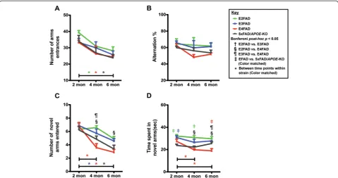
Age-dependent decline in E4FAD mice in Y-maze spatial recognition memory test and deficits in E4FAD and 5xFAD/ APOE-KO mice compared to E3FAD and E2FAD mice To determine the effect of APOE genotype and age on cognitive function, spatial recognition memory was assessed via Y-maze in 2-, 4-, and 6-month EFAD and 5xFAD/APOE-KO mice. There were no significant dif-ferences in the number of arm entrances (baseline-line exploratory activity, Figure 1A) or spontaneous alterna-tion (Figure 1B) by APOE genotype, however there was significance in both tests between age groups (two-way ANOVA, Additional file 1). Bonferroni post-hoc analysis demonstrated significantly lower arm entrances between 2 and 6 month old mice for E2FAD, E4FAD, and 5×FAD/ APOE-KO (p > 0.05) (Figure 1A). Two-way ANOVA ana-lysis showed a significant age effect for spontaneous
alternation, however Bonferroni post-hoc analysis re-vealed no significance (Figure 1B). Thus, subsequent ef-fects (Figure 1C,D) were not significantly influenced by differences in spontaneous exploratory spatial navigation.
Spatial recognition memory was assessed using the nat-ural tendency of mice to preferentially explore novel over familiar spatial environments in a two-trial Y-maze test, measuring the number of novel arms entered (Figure 1C) and time spent in novel arms (Figure 1D). Two-way ANOVA demonstrated a genotype and age effect but not an age X genotype effect for both number of novel arms entered and time in novel arms (Additional file 1). Bonfer-ronipost-hoc analysis revealed that a significant age effect was observed for the E4FAD mice from 2-4 months and from 2-6 months (Figure 1C,D), while E3FAD and 5xFAD/APOE-KO also decreased significantly form 2-6 in number of arms entered (1C). In comparisons among the genotypes at each age, E4FAD mice displayed deficits in spatial cognition (fewer novel arm entries) compared to E2FAD and E3FAD mice at 4 months, and compared to E2FAD mice at 6 months (Figure 1C), with no difference between E4FAD and 5xFAD/APOE-KO mice. Results for the time spent in the novel arms (Figure 1D) suggest that both E4FAD and 5xFAD/APOE-KO mice spent consist-ently less time in novel arms than E2FAD and E3FAD mice. Of interest, time in novel arms for E4FAD mice at 6 months is significantly lower than 5xFAD/APOE-KO mice, the only example of an apoE4 gain of toxic function for the Y-maze (Figure 1D). Together these results are consist-ent with E2FAD≥ E3FAD > 5xFAD/APOE-KO≥ E4FAD for spatial recognition memory as assessed by Y-maze.
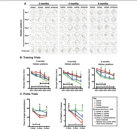
Deficits in spatial and learning and memory in the Morris water maze are greater in E4FAD and 5xFAD/APOE-KO mice compared to E3FAD and E2FAD mice
Cognition was further assessed for spatial reference and working memory using the MWM (Figure 2). Two MWM tests were utilized to assess the capacity of mice to learn the location of a hidden platform using relevant visual cues. A 5-day training phase was used as a meas-ure of spatial learning and memory, followed by removal of the platform for two probe trials (Figure 2C) to assess retrieval of spatial reference memory. All genotypes at each age exhibited comparable swimming speed and sensory motor functions, as determined by a visual cue test (data not shown). Thus, sensory motor or motiv-ational effects on learning and memory performance were considered comparable. Swimming tracks for train-ing and the probe trials were recorded for each day for each genotype and 2-, 4- and 6-month (representative example, Figure 2A).
For the 5-day training phase, the time to find the hid-den platform was recorded and plotted against trial date at 2-, 4- and 6-month (Figure 2B). There was a genotype
and training day effect for all age groups (two-way ANOVA, Additional file 1). Bonferronipost-hoc analysis revealed that for all the genotypes at each age, the time to reach the hidden platform decreased from 1 to 5 days in training phase, indicating that the mice were able to learn the task, with the exception of 5xFAD/APOE-KO mice at 4 months. The escape latency for the E2FAD and E3FAD mice decreased significantly from 1 to 3 days at both 4 and 6 months, while the E4FAD and 5xFAD/APOE-KO mice required the full 5 days for a sig-nificant learning effect at 4 and 6 months. It is also inter-esting to note that from 2-6 months, the escape latency, measured as the slope of the learning curve, increased from 2 to 6 months for the E4FAD (-4.26 to -2,30) and 5xFAD/APOE-KO mice (-5.13 to -3.35), suggesting fail-ure of some compensatory effect over time. In compar-isons among the genotypes at each age, the escape latency was longer for E4FAD compared to E2FAD mice at several training days for 2-, 4-, and 6-months (Figure 2B). This result suggests that on a given day, E4FAD mice showed delayed acquisition and poor re-tention of spatial information from the day before and, therefore, took longer to reach the position of the plat-form than the E2FAD mice. In general, the results for
training trials of E4FAD were comparable to 5xFAD/ APOE-KO mice, while E2FAD and E3FAD mice were comparable.
After 5 days of training, the platform was removed and the number of times the mice crossed the previous plat-form location and the time spent in the target quadrant searching for the platform were recorded (Figure 2C). There was a genotype and age effect, but not a genotype X age effect, for both probe trials (two-way ANOVA, Additional file 1). Post-hoc analysis by Bonferroni re-vealed a significant age effect for E4FAD mice for both measures, with a similar trend for the 5×FAD/APOE -KO. This decline is particularly dramatic for platform crosses at 6 months (Figure 2C). In comparisons among the genotypes at each age, there were no genotype ef-fects at 2 months in either probe trial (Figure 2C). In comparisons among the genotypes at each age, the E4FAD mice spent less time in the target quadrant than both E2FAD and E3FAD mice at 4 months, and less than E2FAD at 6 months. For the number of platform crosses, the only significant difference was between E2FAD and E4FAD mice at 6 months.
The results for the MWM indicate that recently ac-quired spatial learning and working memory, and long-Figure 1Age-dependent decline in Y-maze of performance in E4FAD mice compared to E3FAD and E2FAD mice.Results at 2-, 4-, and 6-months of age for E2FAD, E3FAD, E4FAD, and 5×FAD/APOE-KO mice: Y-maze results for(A)the total number of arm entries,(B)percent alternation,(C)novel arm recognition, and(D)time spent in novel arms. N≥6 per group, expressed as means ± S.E.M. Significant differences at
p< 0.05 via two-way ANOVA, Bonferronipost-hoctest identified by:†between E2FAD and E3FAD, § between E2FAD and E4FAD,¶between
E3FAD and E4FAD. Color matched‡(green = E2FAD, blue = E3FAD, red = E4FAD, grey = 5×FAD/APOE-KO) between EFAD strain and 5×FAD/
APOE-KO.Along the x-axis, color matched *indicates significant differences between time points within a mouse strain. There is no significant change with age unless marked.
term reference memory, are impaired in E4FAD mice compared to E3FAD and E2FAD mice. These data do not support a difference between E4FAD and 5×FAD/ APOE-KO mice (E2FAD ≥ E3FAD > E4FAD = 5×FAD/ APOE-KO). As with Y-maze, the conclusion is thatAPOE4 presents primarily as loss of function.
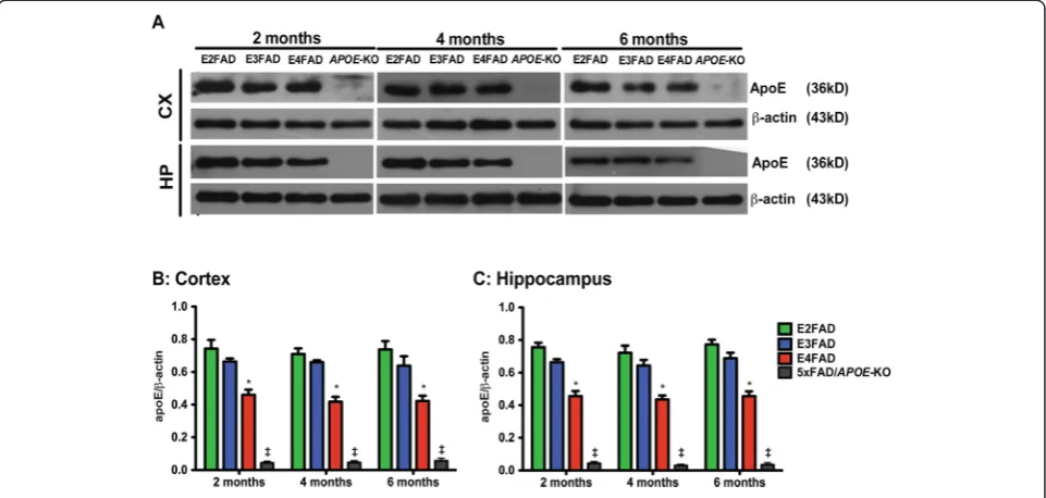
Total apoE levels are lower in E4FAD mice compared to E3FAD and E2FAD mice
Total apoE (Figure 3) levels in the cortex and hippocam-pus of 2-, 4-, 6-month EFAD and 5xFAD/APOE-KO mice were measured by Western blot (representative blot Figure 3A) and normalized to β-actin. Two-way Figure 2Age-dependent decline in Morris water maze training and performance is exacerbated in E4FAD and 5xFAD/APOE-KO mice compared to E3FAD and E2FAD mice.Results at 2-, 4-, and 6-months of age for E2FAD, E3FAD, E4FAD, and 5xFAD/APOE-KO mice:(A)Representative swimming tracks in Morris water maze for days 1-5 of training (APOE-KO = 5xFAD/APOE-KO).(B)Training trials: Escape latency for hidden platform.(C)
Probe trials: time spent in target quadrant and number of platform crossings. N≥6 per group, expressed as means ± S.E.M. Significant differences atp< 0.05 via two-way ANOVA, Bonferronipost-hoctest identified by:†between E2FAD and E3FAD, § between E2FAD and E4FAD,¶between E3FAD and E4FAD.
Color matched‡(green = E2FAD, blue = E3FAD, red = E4FAD, grey = 5xFAD/APOE-KO) between EFAD strain and 5xFAD/APOE-KO. Along the x-axis, color matched *indicates significant differences between time points within a mouse strain. There is no significant change with age unless marked.
ANOVA showed only a genotype effect for apoE levels (Additional file 1). Age had no effect on apoE levels in any genotype in either brain region at any age (Figure 3B, C). However Bonferronipost-hoc analysis, for both brain regions at each age, total apoE4 levels were significantly lower than apoE2 and apoE3 (Figure 3B,C). These data are consistent with previous studies comparing apoE4 levels with apoE3 in humans and mice [48,60-64] and support the few published studies comparing apoE4 with apoE2 levels [48,60]. As would be expected, the levels of apoE in the 5xFAD/APOE-KO mice were just above level of detection and significantly lower than any of the apoE isoforms.
Age-dependent decline in post-synaptic-related protein levels is exacerbated in E4FAD and 5xFAD/APOE-KO mice compared to E3FAD and E2FAD mice
To begin to dissect potential pathways for apoE4 modu-lation of cognitive deficits, levels of presynaptic (synap-tophysin) and postsynaptic (PSD95, drebrin) proteins were measured in the hippocampus by Western blot (representative blot Figure 4A).
There were no age or genotype effects on the levels of synaptophysin, a presynaptic protein (Figure 4B; two-way ANOVA, Additional file 1). Two-two-way ANOVA of PSD95 (4C) and drebrin (4C) revealed a significant ef-fect for genotype, age and genotype X age (Additional file 1). Although post-synaptic proteins PSD95 and
drebrin levels were equal among genotypes at 2 months, Bonferroni post-hoc analysis showed significant age ef-fects for both proteins in all genotypes from 2-6 months with the exception of drebrin levels in the E2FAD mice. It is also interesting to note that the decrease in both PSD95 and drebrin for E4FAD and 5xFAD/APOE-KO were significant from 2-4 months, while E3FAD de-creased significantly from 4-6 months, and PSD95 levels in E2FAD mice decreased minimally and only from 2-6 months.
In comparisons among the genotypes at each age, both PSD95 and drebrin levels in E2FAD mice were significantly higher than E4FAD and 5xFAD/APOE-KO at 4 and 6 months. Comparisons among genotypes demonstrate that at 4 and 6 months, PSD95 levels were E2FAD = E3FAD > E4FAD > 5xFAD/APOE-KO, evidence for apoE as a loss of function, although there was no difference in drebrin levels between E4FAD and 5xFAD/APOE-KO mice, and these drebrin levels were significantly lower than the drebrin levels in E2FAD and E3FAD mice, with the resulting sum-mary for drebrin: E2FAD = E3FAD > E4FAD = 5xFAD/ APOE-KO.
Collectively these data support the observation that postsynaptic proteins are affected prior to presynaptic proteins [38,39,65-67] and this effect may underlie apoE-modulated cognitive deficits. Further, as with cog-nitive dysfunction, apoE represents primarily a loss of positive function.
Figure 3ApoE levels are lower in E4FAD mice compared to E3FAD and E2FAD mice.Results at 2-, 4-, and 6-months of age for E2FAD, E3FAD, E4FAD, and 5×FAD/APOE-KO mice:(A)Representative Western blot for apoE protein in cortex (CX) and hippocampus (HP) withβ-actin as a control for protein loading (APOE-KO = 5×FAD/APOE-KO). Relative apoE protein levels in(B)CX and(C)HP. N = 6 per group, expressed as means ± S.E.M. Significant difference atp< 0.05 via two-way ANOVA, Bonferronipost-hoctest identified by *for E4FAD compared to E2FAD and E3FAD. No significant change between time points.‡ApoE levels in 5xFAD/APOE-KO mice were≥10-fold lower than E4FAD,p< 0.000001.
Age-dependent decline in NMDAR subunits levels is exacerbated in E4FAD and 5xFAD/APOE-KO mice compared to E3FAD and E2FAD mice
Evidence indicates that reduced postsynaptic NMDAR levels are involved in cognitive dysfunction in AD [37-39]. Therefore, levels of the NMDAR subunits NMDAR1 (Figure 5B), NMDAR2A (Figure 5C) and NMDAR2B (Figure 5D) were measured in the hippocampus by West-ern blot (representative blot Figure 5A).
Two-way ANOVA of NMDAR results show a signifi-cant genotype and age, but no genotype X age effect (Additional file 1). Further Bonferroni post-hoc analysis revealed that at 2 months, the three NMDAR subunits levels were equal among genotypes except for lower levels of NMDAR1 in 5xFAD/APOE-KO mice, indicating a loss of apoE4 positive function compared to apoE-KO. After 2 months, all three NMDAR subunits in all geno-types decreased from 2-6 months (Figure 5B,C,D), with the exception of, again, E2FAD, consistent with the re-sults for NMDAR2A (Figure 4C). Comparisons among genotypes demonstrate the NMDAR1 levels are consist-ently higher in E2FAD mice compared to the other geno-types, a trend is also observed for the levels of NMDAR2A and NMDAR2B. While the general trend for the NMDAR subunits is E2FAD and E3FAD being higher than E4FAD
and 5xFAD/APOE-KO, it is significant to note that NMDAR2B levels are significantly lower in E4FAD com-pared to 5xFAD/APOE-KO mice at 4 months, with the trend continuing to 6 months, an example of apoE4 gain of toxic function (Figure 5D).
Overall, consistent with cognition and levels of post-synaptic proteins, these data indicates that apoE medi-ates primarily a loss of positive function with NDMAR subunits levels: E2FAD ≥ E3FAD > E4FAD ≈ 5xFAD/ APOE-KO.
Age dependent decline in p-CaMK-II levels is significant in E3FAD, E4FAD and 5xFAD/APOE-KO mice compared to E2FAD mice; age dependent decline in p-CREB and BDNF is exacerbated in E4FAD > 5xFAD/APOE-KO≥E3FAD > E2FAD The NDMAR subunit levels were significantly lower in E4FAD, with a trend in 5xFAD/APOE-KO mice, com-pared to E2FAD and E3FAD mice. As reduced levels/ activation of the NMDAR pathway CaMK-II/CREB/ BDNF [42,68,69] are observed in AD patients and asso-ciated with impaired neuronal function in vitro and in vivo[70-73], these downstream signaling molecules, specifically the levels of BDNF (Figure 6D) and activated p-CaMK-II (Figure 6B) and pCREB (Figure 6C), were measured in the hippocampus by Western blot Figure 4Age-dependent decline in post-synaptic protein levels is exacerbated in 5xFAD/APOE-KO≥E4FAD mice compared to E3FAD and E2FAD mice.Results at 2-, 4-, and 6-months of age for E2FAD, E3FAD, E4FAD, and 5xFAD/APOE-KO mice:(A)Representative Western blot images for PSD95, drebrin and synaptophysin proteins in HP withβ-actin as a control for protein loading (APOE-KO = 5xFAD/APOE-KO). Relative protein levels of(B)synaptophysin,(C)PSD95 and(D)drebrin. N = 6 per group, expressed as means ± S.E.M. Significant differences atp< 0.05 via two-way ANOVA, Bonferroni post-hoc test identified by:†between E2FAD and E3FAD, § between E2FAD and E4FAD,¶between E3FAD and
E4FAD. Color matched‡(green = E2FAD, blue = E3FAD, red = E4FAD, grey = 5xFAD/APOE-KO) between EFAD strain and 5xFAD/APOE-KO. Along the X-axis, color matched *indicates significant differences between time points within a mouse strain. There is no significant change with age unless marked.
(representative blot Figure 6A). Two-way ANOVA on the p-CaMK-II, p-CREB, and BDNF results showed a signifi-cant effect genotype, age and genotype X age interaction (Additional file 1), Bonferroni post-hoc analysis are de-tailed below.
p-CaMK-II. No differences in p-CaMK-II levels were observed among the genotypes at 2 months (Figure 6B). After 2 months, p-CaMK-II levels decreased from 2-6 months in all the genotypes, although the decrease from 4-6 months was not significant in only E2FAD mice. Comparison among the genotypes at 4 and 6 months, revealed p-CaMK-II levels: E2FAD > E3FAD = E4FAD = 5xFAD/APOE-KO.
p-CREB. At 2 months, p-CREB levels were higher in E2FAD and E3FAD mice compared to 5xFAD/APOE -KO mice, and E4FAD levels were lower E2FAD mice (Figure 6C). From 2-6 months, p-CREB levels decreased significantly in all the genotypes but E2FAD. Compari-son among the genotypes revealed at 6 months, with a similar trend at 4 months, the levels of p-CREB for the ge-notypes was E2FAD > E3FAD > 5xFAD/APOE-KO > E4FAD, consistent with a gain of toxic function for apoE4.
BDNF. As observed for p-CaMK-II, BDNF levels were not different among the genotypes at 2 months Figure 6D).
After 2 months, BDNF levels decreased with age in the E3FAD, E4FAD and 5xFAD/APOE-KO mice, while levels in E2FAD mice did not change. Comparison among the ge-notypes revealed that at both 4 and 6 months, the levels of BDNF for the genotypes was E2FAD > E3FAD ≥5xFAD/ APOE-KO > E4FAD. As with p-CREB, levels of BDNF are consistent with a toxic gain of function for apoE4.
Thus, in contrast to cognitive dysfunction, postsynatic protein levels (PSD95, drebrin, NDMAR), and p-CaMK-II where apoE4 appears to be a loss of function compared to apoE-KO, apoE4 demonstrates a toxic gain of function with p-CREB and BDNF levels.
Discussion
APOE4-induced AD risk is likely the result of multiple, overlapping mechanisms, both Aβ-dependent and Aβ in-dependent (for review [74]). One challenge in understand-ing the effect of APOE genotype on various mechanistic readouts is determining whether apoE4 represents a loss of positive function or a gain of toxic function. Thus, we investigated the early, age-dependent APOE genotype-specific effects on cognitive functions and synaptic viabil-ity in EFAD-Tg mice [48,58,59], specifically female mice based on data in both humans [53-55] and Tg mice Figure 5Age-dependent decline in NMDAR subunit protein levels are exacerbated in E4FAD and 5xFAD/APOE-KO compared to E3FAD and E2FAD mice.Results at 2-, 4-, and 6-months of age for E2FAD, E3FAD, E4FAD, and 5xFAD/APOE-KO mice:(A)Representative Western blot for NMDAR1, NMDAR2A and NMDAR2B proteins in HP withβ-actin as a control for protein loading (APOE-KO = 5xFAD/APOE-KO). Relative protein levels of(B)NMDAR1,(C)NMDAR2A, and(D)NMDAR2B. N = 6 per group, expressed as means ± S.E.M. Significant differences atp< 0.05 via two-way ANOVA, Bonferroni post-hoc test identified by:†between E2FAD and E3FAD, § between E2FAD and E4FAD,¶between E3FAD and
E4FAD. Color matched‡(green = E2FAD, blue = E3FAD, red = E4FAD, grey = 5xFAD/APOE-KO) between EFAD strain and 5xFAD/APOE-KO. Along the x-axis, color matched *indicates significant differences between time points within a mouse strain. There is no significant change with age unless marked.
[33,35,36,75,76] that APOE4 females exhibit significantly increased cognitive impairment compared to APOE4 males and APOE3 females. In the Y-maze, a significant age-dependent decline in spatial recognition memory was observed only for the E4FAD mice from 2-4 months, indi-cating a more rapid decline at earlier stages of Aβ depos-ition compared to other genotypes (Figure 1). In the MWM, a measure of spatial learning and memory, the E4FAD and 5xFAD/APOE-KO mice were both slower to learn than the E2FAD and E3FAD mice during the 5-day training phase (Figure 2B). In addition, both the E4FAD and 5xFAD/APOE-KO mice exhibited an age-related in-crease in escape latency from 2-6 months during the train-ing trials, suggesttrain-ing the failure of some compensatory effect over time. As this is consistent with previous studies demonstrate higher anxiety levels in APOE4-TR and APOE-KO mice [76,77], we hypothesize that this elevated stress response may facilitate spatial learning in young E4FAD mice and mask adverse effects of apoE4 on spatial cognition. Indeed, it has been shown that normal aging can counteract stress-induced facilitation of cognitive pro-cessing inAPOE4-TR mice, as measured by MWM, mak-ing phenotypic differences easier to detect in older mice [33,75]. This apoE4 effect in the EFAD mice is amplified by the overproduction of Aβ42 driven by the presence of the 5-FAD mutations. Indeed, the 5xFAD mice show
progressive learning and memory deficits tasks as early as 3 months [78-82]. As deficits in spatial learning and mem-ory due to apoE4 have mainly been reported in older and non-AD mice [33,75], our findings are consistent with synergistic effects between apoE4 and the aggressive Aβ42 pathology characteristic of the EFAD mice [48]. In addition, the use of only female EFAD mice also optimized the risk of cognitive deficits in the E4FAD mice. Indeed, sex interacts withAPOEto affect cognitive function. Clin-ical data indicate that the APOE4-induced risk for AD is significantly greater, perhaps exclusive to, females [53-55]. These data are consistent with the greater cognitive im-pairment in female APOE4-TR mice compared to female APOE3-TR mice, and with bothAPOE3- andAPOE4-TR females compared toAPOEgenotype-matched males (re-view [35]). Overall, as measured in this study, behavior ap-peared to be primarily an apoE4 loss of function, specifically: E2FAD = E3FAD > E4FAD ≈ 5xFAD/APOE -KO. However, further studies in humans and Tg-mouse models are critical to determine the role of potential inter-active effects among Aβ pathology, APOE genotype and sex on memory and cognitive decline.
Spatial and learning memory performances are directly linked to synaptic function. ApoE4 is associated with pro-gressive synaptic deficits in both AD patients and h-APOE-Tg mouse models [83-87]. Consistent with previous Figure 6Age-dependent decline in NMDAR-mediated signaling proteins is exacerbated in E4FAD≥5xFAD/APOE-KO≥E3FAD > E2FAD mice.Results at 2-, 4-, and 6-months of age for E2FAD, E3FAD, E4FAD, and 5xFAD/APOE-KO mice:(A)Representative Western blot images for p-CaMK-II, p-CREB, and BDNF proteins in HP withβ-actin/CREB,β-actin/CaMK-II orβ-actin as a control for protein loading, respectively (APOE-KO = 5xFAD/APOE-KO). Relative protein levels of(B)p-CaMK-II,(C)p-CREB and(D)BDNF. N = 6 per group, expressed as means ± S.E.M. Significant differences atp< 0.05 via two-way ANOVA, Bonferronipost-hoctest identified by:†between E2FAD and E3FAD,§between E2FAD and E4FAD,
¶
between E3FAD and E4FAD. Color matched‡(green = E2FAD, blue = E3FAD, red = E4FAD, grey = 5xFAD/APOE-KO) between EFAD strain and 5xFAD/APOE-KO. Along the x-axis, color matched *indicates significant differences between time points within a mouse strain. There is no significant change with age unless marked.
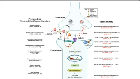
reports, age-induced reductions in synaptic proteins pref-erentially occurred in post-synaptic proteins compared particularly to synaptophysin, a pre-synaptic protein (Figure 4) [81,88-90]. In the current model, levels of drebrin and PSD95 were lower in E4FAD and 5xFAD/ APOE-KO mice compared to E2FAD and E3FAD mice, consistent with an apoE4 loss of function. ApoE is also linked to long-term potentiation (LTP) and NMDAR-mediated signaling [50,51]. As the NMDAR component of synaptic transmission has been shown to decline during aging [91,92], NMDAR activation may provide a mechan-istic pathway for understanding apoE-related memory im-pairment. Indeed, in this study apoE-related cognitive impairment correlates with a decrease in the levels of NMDR subunits and components of the signaling pathway (p-CaMK-II/p-CREB/BDNF). Levels of all three NMDAR subunits were reduced with age for all the genotypes with reductions greater in the E4FAD and 5xFAD/APOE-KO mice compared to E2FAD and E3FAD mice, consistent with an apoE4 loss of function (Figure 5). These apoE4-related deficits in NMDAR-dependent functions likely re-flect changes to neuronal networks contributing to short and long-term memory, and their contribution to memory consolidation [93-96]. Reduction of BDNF levels through either genetic or pharmacological means not only im-paired LTP and reduced the number of synapses, but also caused deficits in the formation and consolidation of memory [97-99]. However, the effects of APOEgenotype on the p-CaMK-II, p-CREB, and BDNF signaling cascade are not consistent. Again, all the signaling components were reduced with age for all the genotypes (Figure 6). While the greater reduction in p-CaMK-II in E4FAD and 5xFAD/APOE-KO mice compared to E2FAD and E3FAD mice is consistent with anAPOE4loss of function,apoE4 represents a gain of toxic function for both the activated p-CREB transcription factor and its downstream target protein BDNF, as the reductions in p-CREB and BDNF levels are greater in E4FAD compared to 5xFAD/APOE-KO mice (summarized in Figure 7, right side). This gain of toxic function does not translate directly to the behavioral measures used in this study. One likely possibility for this seeming disconnect is that a number of signaling pathways are activated by changes in synaptic proteins, particularly the NMDAR, that eventually converge to modulate behav-ioral readouts. Thus, the final effect on behavior will be a composite of a number of signaling pathways (reviewed in [100]). Taken together, our findings support the notion that apoE4 exacerbates behavioral deficits in EFAD mice by decreasing synapse viability by reducing synapse-related proteins, particularly via down-regulation of NMDAR and NMDAR-mediated signaling via CaMK-II, CREB, and BDNF (Figure 7). Consistent with theAPOE2 protective effect for AD risk [1-11], these results also dem-onstrate that E2FAD mice are consistently less susceptible
to age-induced changes in the components of this cascade, from signaling to behavior.
ApoE is the primary ligand for the low-density lipo-protein (LDL) receptor (LDLR) family (apoE receptors), although Reelin is the primary ligand for ApoE-receptor 2 (ApoER2). ApoER2 and Reelin are important modula-tors of synaptic plasticity and NMDAR functionsin vitro and in vivo [106-108]. Thus, the association between ApoER2, Reelin and NMDAR are critical for LTP, mem-ory formation and retrieval. ApoE4 has been demon-strated to reduce the cell-surface levels of both ApoER2 and NMDAR via intracellular sequestration, thus inhibit-ing the ability of Reelin to facilitate glutamate-mediated synaptic plasticity [46]. The impaired recycling of apoE4 may contribute to this reduction in receptors at the cell surface [105]. Loss of ApoER2 reduces Reelin binding, thus further reducing activation of NMDAR via signaling by the Src family kinases [46,105].
While a number of Aβ-independent mechanisms likely contribute to the APOE-associated risk for AD [109], oAβhas been demonstrated to be preferentially synapo-toxic (for review [37,110]). We have published the effects of APOE genotype on Aβ accumulation in the EFAD mice at 6-months of age, the age of the mice used for this study [48,58,59,111,112]. These results demonstrate amyloid deposition by IHC and total brain Aβ42 by ELISA is: 5xFAD > E4FAD > E3FAD = E2FAD. A three-step sequential protein extraction protocol using TBS (soluble), TBS + Triton X-100 (TBSX, detergent), and formic acid (FA, insoluble) was used for the hippocam-pus and cortex. In the soluble fractions of both brain re-gions, both Aβ42 and oAβ are: E4FAD > E3FAD = E2FAD. There is no APOE genotype difference in the levels of Aβ42 in the detergent fraction. In the insoluble fraction, Aβ42 is: E4FAD > E3FAD = E2FAD. As the EFAD mice are on the 5xFAD background, the amount of Aβ40 is difficult to detect; the primary species is Aβ42. Thus, Aβ levels (amyloid, soluble and insoluble) are greatest in the E4FAD mice. This association be-tweenAPOEand Aβaccumulation is consistent with the functional changes reported herein. Therefore, a particu-larly relevant approach to interpreting the results of this study is to consider APOE modulation of soluble Aβ levels at the synapse (Figure 7, left side). Previous publi-cations from our group and others demonstrate that apoE isoform-specific effects on Aβclearance and inter-actions with apoE receptors likely play a role in this process at several levels. It has been specifically demon-strated that apoE4 both increases the levels of oAβand directs it to the synapse [5]. ApoE isoforms may modu-late oAβ levels through differential apoE/Aβ complex levels [113]. However, as isolation and analysis of the apoE/Aβ complex in vivois technically challenging, data are conflicting as to the significance or even the existence
of this complex [114-116]. Nevertheless, it is interesting to note that the levels of soluble apoE4/Aβ complex are lower than apoE3/Aβand decrease in AD in human syn-aptosomes, CSF and EFAD-Tg mouse brains, the reverse of soluble oAβlevels [48,58,59,113]. ApoE receptors also play a key role, particularly ApoER2, as Reelin signaling can prevent the oAβ-induced inhibition of NMDAR at the synapse [117]. As well, neuronal LRP1 provides a signifi-cant mechanism for the clearance Aβ [101,102] and in vitro, Aβ clearance is impaired with apoE4, compared to apoE3 [103], consistent with a greater accumulation of intraneuronal Aβ[104]. To connect these Aβ-dependent processes to decreased synaptic function and impaired cognition requires, in part, a return to ApoER2, NMDAR and Reelin. ApoE4 induces deficits in ApoER2 and NMDAR signaling and recycling, as well as reducing Reelin binding to both receptors [46,105]. Ultimately, these apoE-mediated differential effects on apoE receptors and Aβaccumulation could contribute to the mechanisms responsible for synaptic dysfunction and cognitive decline
characteristic of observed in the EFAD mice that, ultim-ately, will translate to AD patients.
Conclusions
Herein we provide evidence that the APOE4 genotype constitutes a loss of positive function contributing to age-related deficits in behavioral performances in the EFAD and 5xFAD/APOE-KO mice (Figure 7-right side; Table 1A). This loss of positive function with apoE4 was related to a decrease in post-synaptic proteins, including NMDAR subunits, leading to impaired NMDAR-related signaling. However, apoE4 represents a gain of toxic func-tion for the final components measured in this pathway, activated p-CREB and its downstream target protein BDNF. It is our interpretation that multiple signaling path-ways converge to determine the final synaptic transmission impairment and learning and memory deficits associated with apoE4. This conclusion requires further study to de-termine the potential contributions of other signaling com-ponents to either an apoE4-mediated loss or gain of Figure 7Diagram of potentialAPOE4-induced decrease in synapse-related proteins and disruption of NMDAR-mediated signaling, resulting in impaired learning and memory.WithAPOE4, the levels of key post-synaptic proteins are reduced, including the subunits of NMDAR. NMDAR signaling via activation by phosphorylation of CaMK-II and CREB to increase expression of BDNF is also disrupted byAPOE4, compromising synaptic function and impairing learning and memory. Previous work has demonstrated that apoE isoform-specific effects on Aβ clearance and interactions with apoE receptors likely play a role in this process at several levels. Soluble levels of Aβare lower with apoE3 and inversely correlated with levels of apoE3/Aβcomplex [59], suggesting a potential clearance mechanism. LRP mediates Aβuptake by neurons [101,102], and,in vitro, Aβclearance is impaired with apoE4 [103], consistent with a greater accumulation of intraneuronal Aβ[104] compared to apoE3. For ApoER2, ligand recycling is impaired with apoE4 compared to apoE3 [46]. Loss of ApoER2 reduces Reelin binding, thus reducing activation of NMDAR via signaling by the Src family kinases [46,105], leading to decreased synaptic function and therefore decreased learning and memory.
function, data of high therapeutic significance. Table 1B provides a representative summary of CNS-relevant func-tions modulated by apoE4 that can be attributed to a loss of positive or gain of toxic function. Again, it is important to assess whether apoE4 imparts a loss of positive or gain of toxic function in comparison to the absence of apoE (APOE-KO), not simply a comparison to apoE2/apoE3. Table 1B provides a larger perspective on the interplay between among multiple functions that exhibit a loss of positive (for example, anti-inflammatory properties and amyloid deposition) or gain of toxic function (for
example, toxic proteolytic fragments of apoE and oAβ -induced neurotoxicityin vitroandex vivo). Only an un-derstanding of the relative contribution of the functions measured in this paper, listed in Table 1A and the many others as yet undefined will enable a confident identifi-cation of APOE4-induced AD risk as a loss of positive or gain of toxic function.
Targeting the most potent genetic risk factor for AD appears a very attractive strategy and is still under in-tense study. If the hypothesis is that all apoE isoforms, particularly apoE4, represent a toxic gain of function, Table 1 Evidence thatAPOE4is a loss of positive or gain of negative function: comparison to absence ofAPOE
A. Within manuscript
Measure apoE4 represents: Mouse model
Behavior/cognition Loss of function 5xFAD/APOE-KO, EFAD
Postsynaptic protein levels Loss of function
NMDAR subunits Loss of function
p-CaMK-II levels Loss of function
p-CREB levels Gain of toxic function
BDNF levels Gain of toxic function
B. Literature overview
Measure apoE4 represents: Mouse model Reference
Anti-inflammatory Loss of function APOE-KO,APOE-TR [47]
Baseline LTP Loss of function WT,APOE-KO,APOE-TR
(Ex vivohippocampal slice cultures)
[50]
Amyloid deposition Loss of function APOE-KO x APPV717+/- [118],
APPV717+/-x GFAP-apoE+/- [119,120],
5xFAD, EFAD [48]
for review, [35]
ApoE lipidation Loss of function APOE-TR [121]
EFAD [58]
Dendritic spine density Loss of function WT,APOE-KO, GFAP-apoE+/+ [84]
BBB integrity Loss of function WT,APOE-KO,APOE-TR, GFAP-APOE [122]
Aβclearance across the BBB Loss of function WT, APOE+/-, APOE-KO [123]
Behavior/cognition Gain of toxic function WT,APOE-KO,APOE-TR [33]
WT,APOE-KO,APOE-TR [34]
WT,APOE-KO, GFAP-apoE [124]
WT,APOE-KO, GFAP-apoE (female) [125]
WT,APOE-KO, NSE-apoE [124,126]
Accumulation of intraneuronal oAβ Gain of toxic function APOE-KO,APOE-TR [127]
oAβ-induced neurotoxicity Gain of toxic function WT,APOE-KO,APOE-TR [52]
In vitroneuron/glial co-cultures
oAβ-dependent inhibition of LTP Gain of toxic function APOE-KO,APOE-TR [51]
Neurotoxicity of apoE proteolytic fragments Gain of toxic function Variety withAPOE-KO control [75,128] for review, [129]
Neurite outgrowth Loss of function APOE-KO olfactory epithelia cultures
(exogenous apoE added)APOE-KO
[130]
Gain of toxic function cortical neuron cultures (exogenous apoE added) [131]
then reducing APOE expression and/or apoE levels is one therapeutic approach for AD. However, the potential dangers of this approach in the human brain are still subjected to debate [30]. Here, we provide additional insight into the mechanism by which APOE4increases AD risk, in which apoE4 mainly appears as a loss of positive function. Accordingly, rather than APOE gene inactivation, therapies that correct the loss of positive function related to apoE4, such as increasing the lipida-tion of apoE4 containing lipoproteins [58] appear to be more appropriate.
Methods Animals
All experiments were conducted in accordance with the rules and regulations of the Institutional Animal Care and Use Committee protocols at Fujian Medical Univer-sity, in conformance with international guidelines for the ethical use of animals. Investigators conducting the sam-ple processing and analyses were blinded for APOE genotype and age. The 5xFAD/APOE-KO and EFAD mice (E2FAD, E3FAD, and E4FAD) were supplied by the LaDu lab. The EFAD mice [48] were originally generated by crossing 5xFAD mice [57] and h-APOE-TR mice [56]. 5xFAD mice express APP K670N/M671L + I716V + V717I and PS1 M146L + L286V under the control of the neuron-specific mouse Thy-1 promoter, resulting in the overproduction of specifically Aβ42 [57]. In APOE-TR mice, the coding domain of m-apoE is replaced by h-apoE2, apoE3 or apoE4 [56]. Thus, EFAD mice are APOE-TR+/+/5xFAD+/- [48]. The 5xFAD/APOE-KO mice were made by knocking-out m-APOEfrom the 5xFAD mice.
Behavioral tests
Spatial/reference memory was assessed in EFAD mice first using the Y-maze test, followed by the Morris water maze (MWM) test, as previous described [90,132]. Y-maze. Spontaneous alteration including total activity and per-centage spontaneous alternation/exploration was initially determined as a measure of normal spatial navigation. Short-term spatial recognition memory was then assessed using a two-trial protocol with 10 minute (min) training (trial 1), 4 hour (hr) inter-trial interval and a 5 min reten-tion trial (trial 2) for number of entries and time spent in each arm.MWM.Acquisition trials (training) consisted of 4 trials (maximum 1 min) a day for 5 consecutive days with escape latency recorded for each trial. Reference memory was assessed on the sixth day in a one trial test for time spent in the target quadrant and the number of times the original area of the platform was crossed.
Tissue harvest and western blotting
2-, 4- and 6-month EFAD mice were anesthetized with sodium pentobarbital (50 mg/kg), transcardially perfused
with ice-cold PBS, brains removed and dissected into cortex and hippocampus, snap-frozen in liquid nitrogen and stored at -80°C, as previous described [133]. Dis-sected brains were homogenized in lysis buffer [90,132] (50 mM Tris-HCl, 150 mM NaCl, pH7.4, 1% Triton X-100, 1x protease inhibitor cocktail) and 40 μg of total protein (BCA protein assay kit; Pierce, Rockford, IL) was separated on 4–12% gradient Bis-Tris gels (Invitrogen) under reducing conditions, and transferred to PVDF membranes [47]. The following primary antibodies were used: rabbit PSD95 (1:3000, Abcam), mouse anti-synaptophysin (1:2000, Abcam), mouse/rabbit anti-β -actin (1:2000; Abcam), rabbit anti-drebrin antibody (1:1000; Abcam), rabbit anti-NMDAR1/anti-NMDAR2B (1:1000; Millipore), anti-NMDAR2A (1:500; Millipore), mouse anti-apoE (1:600; Santa Cruz), rabbit anti-BDNF (1:200; Santa Cruz), rabbit anti-p-CaMK-II (1:1000; Santa Cruz) and rabbit anti-p-CREB (1:1000; Cell Signal-ing) [90,132]. HRP-conjugated secondary antibodies, en-hanced chemiluminescence (Amersham, Piscataway, NJ) and Image J software were used to quantify densities of the immunoreactive bands relative toβ-actin.
Statistical analysis
Data are expressed as mean ± standard error mean (S.E. M.). Data were analyzed by two-way analysis of variance (ANOVA), followed by Bonferroni post-hoc using Graph-Pad Prism version 4 for Macintosh. The 2-way ANOVA ta-bles for each Figure have been added as Additional file 1. Differences for age and genotype were considered signifi-cant forp< 0.05; n≥6.
Additional file
Additional file 1:Two-way ANOVA Tables.
Abbreviations
Aβ:Amyloid-β; AD: Alzheimer’s disease; apoE: Apolipoprotein E; ApoER2: ApoE receptor 2; BDNF: Brain derived neurotropic factor; CaMK-II: Calcium/calmodulin-dependent protein kinase-II; CREB: CAMP-response element binding protein; EFAD mice: Tg-mice expressing 5xFAD mutations and humanAPOE2,APOE3, or
APOE4; FAD: Familial-AD; h: Human; KO: Knockout; LTP: Long-term potentiation; m: Mouse; min: Minute; MWM: Morris water maze; NMDAR: N-methyl-D-aspartate receptor; oAβ: Oligomeric amyloid-β; p: Phosphorylated; p-CREB: Phosphorylated cAMP-response element binding protein; p-CaMK-II: Phosphorylated calcium/ calmodulin-dependent protein kinase-II; PSD95: Postsynaptic density protein 95; Tg: Transgenic; TR: Targeted replacement; 5xFAD: Tg mice expressing 5 FAD mutations; 5xFAD/APOE-KO: 5xFAD mice with m-APOE-KO.
Competing interests
The authors declare that they have no competing interests.
Authors’contributions
D-s L: Provided substantial contributions to project conception, experimental design and manuscript preparation. X-d P: Provided substantial contributions to project conception, experimental design and manuscript preparation. JZ: Preformed Y-maze behavior tests and complied data for Figure 1. HS: Preformed MWM behavior tests and complied data for Figure 2. NCC: Generated Figures 1, 2, 3, 4, 5, 6 and 7 in collaboration with AMC and MJLD and contributed
to interpretation of the results and preparation of the manuscript. AMC: Generated Figure 7 in collaboration with NCC and contributed to the preparation of the manuscript. KPK: Generated Table 1 and contributed to interpretation of literature for the manuscript. MBA: Contributed to interpretation of the results and preparation of the manuscript. X-m D: Generated Western Blots and analysis for Figures 3, 4, 5, and 6 in collaboration with MZ. MZ: Generated Western Blots and analysis for Figures 3, 4, 5, and 6 in collaboration with X-m D. LMT: Contributed to interpretation of the results and prepared the manuscript in collaboration with MJLD. Y-g Z: Tissue harvest and homogenization for Figures 3, 4, 5 and 6. MJLD: Provided fundamental contributions to interpretation of results, writing and revision of the manuscript. X-c C: Provided substantial contributions to project conception, experimental design and manuscript preparation and revision. All authors read and approved the final manuscript.
Acknowledgements
These studies were funded by National Natural Science Foundation of China 81171216 (X-cC); National Natural Science Grant of China No. 81200991 and Fujian Province New Century Excellent Talents Support Program, China JA13131 (X-dP); National Natural Science Foundation of China No. 81071007 (Y-gZ); and Alzheimer’s Drug Discovery Foundation Grant, NIH/NIA P01AG030128, NIH/NIA R21AG048498, and University of Illinois at Chicago Center for Clinical and Translational Science Grant UL1RR029879 (MJL).
Author details
1Department of Neurology, Fujian Medical University Union Hospital, 29
Xinquan Road, 350001 Fuzhou, China.2Fujian Institute of Geriatrics, Fujian Medical University Union Hospital, 29 Xinquan Road, 350001 Fuzhou, China. 3
Key Laboratory of Brain Aging and Neurodegenerative Disease, Fujian Key Laboratory of Molecular Neurology, Fujian Medical University, 29 Xinquan Road, 350001 Fuzhou, China.4Department of Anatomy and Cell Biology, University of Illinois at Chicago, 808 S.Wood St., M/C 512, 60612 Chicago, IL, USA.
Received: 26 November 2014 Accepted: 26 January 2015
References
1. Corder EH, Saunders AM, Strittmatter WJ, Schmechel DE, Gaskell Jr PC, Rimmler JB, et al. Apolipoprotein E, survival in Alzheimer’s disease patients, and the competing risks of death and Alzheimer’s disease.
Neurology. 1995;45:1323–8.
2. Roses AD. Apolipoprotein E genotyping in the differential diagnosis, not prediction, of Alzheimer’s disease. Ann Neurol. 1995;38:6–14.
3. Kim D, Tsai LH. Bridging physiology and pathology in AD. Cell. 2009;137:997–1000.
4. Leduc V, Jasmin-Belanger S, Poirier J. APOE and cholesterol homeostasis in Alzheimer’s disease. Trends Mol Med. 2010;16:469–77.
5. Koffie RM, Hashimoto T, Tai HC, Kay KR, Serrano-Pozo A, Joyner D, et al. Apolipoprotein E4 effects in Alzheimer’s disease are mediated by synaptotoxic oligomeric amyloid-beta. Brain. 2012;135:2155–68. 6. Corder EH, Saunders AM, Strittmatter WJ, Schmechel DE, Gaskell PC, Small
GW, et al. Gene dose of apolipoprotein E type 4 allele and the risk of Alzheimer’s disease in late onset families. Science. 1993;261:921–3. 7. Saunders AM, Strittmatter WJ, Schmechel D, George-Hyslop PH,
Pericak-Vance MA, Joo SH, et al. Association of apolipoprotein E allele epsilon 4 with late-onset familial and sporadic Alzheimer’s disease. Neurology. 1993;43:1467–72.
8. Houlden H, Crook R, Backhovens H, Prihar G, Baker M, Hutton M, et al. ApoE genotype is a risk factor in nonpresenilin early-onset Alzheimer’s disease families. Am J Med Genet. 1998;81:117–21.
9. Breitner JC, Jarvik GP, Plassman BL, Saunders AM, Welsh KA. Risk of Alzheimer disease with the epsilon4 allele for apolipoprotein E in a population-based study of men aged 62-73 years. Alzheimer Dis Assoc Disord. 1998;12:40–4.
10. Farrer LA, Cupples LA, Haines JL, Hyman B, Kukull WA, Mayeux R, et al. Effects of age, sex, and ethnicity on the association between apolipoprotein E genotype and Alzheimer disease. A meta-analysis. APOE and Alzheimer Disease Meta Analysis Consortium. JAMA. 1997;278:1349–56.
11. Khachaturian AS, Corcoran CD, Mayer LS, Zandi PP, Breitner JC.
Apolipoprotein E epsilon4 count affects age at onset of Alzheimer disease,
but not lifetime susceptibility: The Cache County Study. Arch Gen Psychiatry. 2004;61:518–24.
12. Roses AD. Apolipoprotein E alleles as risk factors in Alzheimer’s disease. Annu Rev Med. 1996;47:387–400.
13. Reitz C, Mayeux R. Use of genetic variation as biomarkers for mild cognitive impairment and progression of mild cognitive impairment to dementia. J Alzheimers Dis. 2010;19:229–51.
14. Leoni V. The effect of apolipoprotein E (ApoE) genotype on biomarkers of amyloidogenesis, tau pathology and neurodegeneration in Alzheimer’s disease. Clin Chem Lab Med. 2011;49:375–83.
15. Bu G. Apolipoprotein E and its receptors in Alzheimer’s disease: pathways, pathogenesis and therapy. Nat Rev Neurosci. 2009;10:333–44.
16. Liu CC, Kanekiyo T, Xu H, Bu G. Apolipoprotein E and Alzheimer disease: risk, mechanisms and therapy. Nat Rev Neurol. 2013;9:106–18.
17. Lue LF, Kuo YM, Roher AE, Brachova L, Shen Y, Sue L, et al. Soluble amyloid beta peptide concentration as a predictor of synaptic change in Alzheimer’s disease. American J Pathol. 1999;155:853–62.
18. McLean CA, Cherny RA, Fraser FW, Fuller SJ, Smith MJ, Beyreuther K, et al. Soluble pool of Abeta amyloid as a determinant of severity of neurodegeneration in Alzheimer’s disease. Ann Neurol. 1999;46:860–6. 19. Wang J, Dickson DW, Trojanowski JQ, Lee VM. The levels of soluble versus
insoluble brain Abeta distinguish Alzheimer’s disease from normal and pathologic aging. Exp Neurol. 1999;158:328–37.
20. Tomic JL, Pensalfini A, Head E, Glabe CG. Soluble fibrillar oligomer levels are elevated in Alzheimer’s disease brain and correlate with cognitive dysfunction. Neurobiol Dis. 2009;35:352–8.
21. Jin M, Shepardson N, Yang T, Chen G, Walsh D, Selkoe DJ. Soluble amyloid beta-protein dimers isolated from Alzheimer cortex directly induce Tau hyperphosphorylation and neuritic degeneration. Proc Natl Acad Sci U S A. 2011;108:5819–24.
22. Kuo YM, Emmerling MR, Vigo-Pelfrey C, Kasunic TC, Kirkpatrick JB, Murdoch GH, et al. Water-soluble Abeta (N-40, N-42) oligomers in normal and Alzheimer disease brains. J Biol Chem. 1996;271:4077–81.
23. Selkoe DJ. Resolving controversies on the path to Alzheimer’s therapeutics. Nat Med. 2011;17:1060–5.
24. Cosentino S, Scarmeas N, Helzner E, Glymour MM, Brandt J, Albert M, et al. APOE epsilon 4 allele predicts faster cognitive decline in mild Alzheimer disease. Neurology. 2008;70:1842–9.
25. Carrasquillo MM, Crook JE, Pedraza O, Thomas CS, Pankratz VS, Allen M, et al. Late-onset Alzheimer’s risk variants in memory decline, incident mild cognitive impairment, and Alzheimer's disease. Neurobiol Aging. 2014;36:60–7.
26. Caselli RJ, Dueck AC, Osborne D, Sabbagh MN, Connor DJ, Ahern GL, et al. Longitudinal modeling of age-related memory decline and the APOE epsilon4 effect. N Engl J Med. 2009;361:255–63.
27. Caselli RJ, Reiman EM, Locke DE, Hutton ML, Hentz JG, Hoffman-Snyder C, et al. Cognitive domain decline in healthy apolipoprotein E epsilon4 homozygotes before the diagnosis of mild cognitive impairment. Arch Neurol. 2007;64:1306–11.
28. Christensen H, Batterham PJ, Mackinnon AJ, Jorm AF, Mack HA, Mather KA, et al. The association of APOE genotype and cognitive decline in interaction with risk factors in a 65-69 year old community sample. BMC Geriatr. 2008;8:14. 29. Schiepers OJ, Harris SE, Gow AJ, Pattie A, Brett CE, Starr JM, et al. APOE E4
status predicts age-related cognitive decline in the ninth decade: longitudinal follow-up of the Lothian Birth Cohort 1921. Mol Psychiatry. 2012;17:315–24.
30. Mak AC, Pullinger CR, Tang LF, Wong JS, Deo RC, Schwarz JM, et al. Effects of the Absence of Apolipoprotein E on Lipoproteins, Neurocognitive Function, and Retinal Function. JAMA Neurol. 2014;71(10):1228–36. 31. Rodriguez GA, Burns MP, Weeber EJ, Rebeck GW. Young APOE4 targeted
replacement mice exhibit poor spatial learning and memory, with reduced dendritic spine density in the medial entorhinal cortex. Learn Mem. 2013;20:256–66.
32. Salomon-Zimri S, Boehm-Cagan A, Liraz O, Michaelson DM. Hippocampus-related cognitive impairments in young apoE4 targeted replacement mice. Neurodegener Dis. 2014;13:86–92.
33. Bour A, Grootendorst J, Vogel E, Kelche C, Dodart JC, Bales K, et al. Middle-aged human apoE4 targeted-replacement mice show retention deficits on a wide range of spatial memory tasks. Behav Brain Res. 2008;193:174–82. 34. Grootendorst J, Bour A, Vogel E, Kelche C, Sullivan PM, Dodart JC, et al.
Human apoE targeted replacement mouse lines: h-apoE4 and h-apoE3 mice
differ on spatial memory performance and avoidance behavior. Behav Brain Res. 2005;159:1–14.
35. Tai LM, Youmans KL, Jungbauer L, Yu C, Ladu MJ. Introducing Human APOE into Abeta Transgenic Mouse Models. Inter J Alzheimer’s Dis.
2011;2011:810981.
36. Raber J, Wong D, Yu GQ, Buttini M, Mahley RW, Pitas RE, et al. Apolipoprotein E and cognitive performance. Nature. 2000;404:352–4. 37. Danysz W, Parsons CG. Alzheimer’s disease, beta-amyloid, glutamate, NMDA
receptors and memantine–searching for the connections. Br J Pharmacol. 2012;167:324–52.
38. Gong Y, Lippa CF. Review: disruption of the postsynaptic density in Alzheimer’s disease and other neurodegenerative dementias. Am J Alzheimers Dis Other Demen. 2010;25:547–55.
39. Stranahan AM, Mattson MP. Selective vulnerability of neurons in layer II of the entorhinal cortex during aging and Alzheimer’s disease. Neural Plast. 2010;2010:108190.
40. Ly PT, Song W. Loss of activated CaMKII at the synapse underlies Alzheimer’s disease memory loss. J Neurochem. 2011;119:673–5. 41. Saura CA, Valero J. The role of CREB signaling in Alzheimer’s disease and
other cognitive disorders. Rev Neurosci. 2011;22:153–69.
42. Paramanik V, Thakur MK. Role of CREB signaling in aging brain. Arch Ital Biol. 2013;151:33–42.
43. Kim JH, Roberts DS, Hu Y, Lau GC, Brooks-Kayal AR, Farb DH, et al. Brain-derived neurotrophic factor uses CREB and Egr3 to regulate NMDA receptor levels in cortical neurons. J Neurochem. 2012;120:210–9.
44. Yong SM, Lim ML, Low CM, Wong BS. Reduced neuronal signaling in the ageing apolipoprotein-E4 targeted replacement female mice. Sci Rep. 2014;4:6580. 45. Nwabuisi-Heath E, Rebeck GW, Ladu MJ, Yu C. ApoE4 delays dendritic spine
formation during neuron development and accelerates loss of mature spines in vitro. ASN Neuro. 2014;6:e00134.
46. Chen Y, Durakoglugil MS, Xian X, Herz J. ApoE4 reduces glutamate receptor function and synaptic plasticity by selectively impairing ApoE receptor recycling. Proc Natl Acad Sci U S A. 2010;107:12011–6.
47. Zhu Y, Nwabuisi-Heath E, Dumanis SB, Tai LM, Yu C, Rebeck GW, et al. APOE genotype alters glial activation and loss of synaptic markers in mice. Glia. 2012;60:559–69.
48. Youmans KL, Tai LM, Nwabuisi-Heath E, Jungbauer L, Kanekiyo T, Gan M, et al. APOE4-specific Changes in Abeta Accumulation in a New Transgenic Mouse Model of Alzheimer Disease. J Biol Chem. 2012;287:41774–86. 49. Dumanis SB, Tesoriero JA, Babus LW, Nguyen MT, Trotter JH, Ladu MJ, et al.
ApoE4 decreases spine density and dendritic complexity in cortical neurons in vivo. J Neurosci J Soc Neurosci. 2009;29:15317–22.
50. Trommer BL, Shah C, Yun SH, Gamkrelidze G, Pasternak ES, Ye GL, et al. ApoE isoform affects LTP in human targeted replacement mice. Neuroreport. 2004;15:2655–8.
51. Trommer BL, Shah C, Yun SH, Gamkrelidze G, Pasternak ES, Stine WB, et al. ApoE isoform-specific effects on LTP: blockade by oligomeric
amyloid-beta1-42. Neurobiol Dis. 2005;18:75–82.
52. Manelli AM, Bulfinch LC, Sullivan PM, LaDu MJ. Abeta42 neurotoxicity in primary co-cultures: effect of apoE isoform and Abeta conformation. Neurobiol Aging. 2007;28:1139–47.
53. Altmann A, Tian L, Henderson VW, Greicius MD. Sex modifies the APOE-related risk of developing Alzheimer disease. Ann Neurol. 2014;75:563–73. 54. Bretsky PM, Buckwalter JG, Seeman TE, Miller CA, Poirier J, Schellenberg GD,
et al. Evidence for an interaction between apolipoprotein E genotype, gender, and Alzheimer disease. Alzheimer Dis Assoc Disord. 1999;13:216–21. 55. Payami H, Zareparsi S, Montee KR, Sexton GJ, Kaye JA, Bird TD, et al. Gender difference in apolipoprotein E-associated risk for familial Alzheimer disease: a possible clue to the higher incidence of Alzheimer disease in women. Am J Hum Genet. 1996;58:803–11.
56. Sullivan PM, Mezdour H, Aratani Y, Knouff C, Najib J, Reddick RL, et al. Targeted replacement of the mouse apolipoprotein E gene with the common human APOE3 allele enhances diet-induced hypercholesterolemia and atherosclerosis. J Biol Chem. 1997;272:17972–80.
57. Oakley H, Cole SL, Logan S, Maus E, Shao P, Craft J, et al. Intraneuronal beta-amyloid aggregates, neurodegeneration, and neuron loss in transgenic mice with five familial Alzheimer’s disease mutations: potential factors in amyloid plaque formation. J Neurosci. 2006;26:10129–40.
58. Tai LM, Koster KP, Luo J, Lee SH, Wang YT, Collins NC, et al. Amyloid-beta Pathology and APOE Genotype Modulate Retinoid X Receptor Agonist Activity in Vivo. J Biol Chem. 2014;289:30538–55.
59. Tai LM, Bilousova T, Jungbauer L, Roeske SK, Youmans KL, Yu C, et al. Levels of soluble apolipoprotein E/amyloid-beta (Abeta) complex are reduced and oligomeric Abeta increased with APOE4 and Alzheimer disease in a transgenic mouse model and human samples. J Biol Chem. 2013;288:5914–26.
60. Sullivan PM, Han B, Liu F, Mace BE, Ervin JF, Wu S, et al. Reduced levels of human apoE4 protein in an animal model of cognitive impairment. Neurobiol Aging. 2011;32:791–801.
61. Cruchaga C, Kauwe JS, Nowotny P, Bales K, Pickering EH, Mayo K, et al. Cerebrospinal fluid APOE levels: an endophenotype for genetic studies for Alzheimer’s disease. Hum Mol Genet. 2012;21:4558–71.
62. Riddell DR, Zhou H, Atchison K, Warwick HK, Atkinson PJ, Jefferson J, et al. Impact of apolipoprotein E (ApoE) polymorphism on brain ApoE levels. J Neurosci. 2008;28:11445–53.
63. Vitek MP, Brown CM, Colton CA. APOE genotype-specific differences in the innate immune response. Neurobiol Aging. 2009;30:1350–60.
64. Martinez-Morillo E, Hansson O, Atagi Y, Bu G, Minthon L, Diamandis EP, et al. Total apolipoprotein E levels and specific isoform composition in cerebrospinal fluid and plasma from Alzheimer’s di’sease patients and controls. Acta Neuropathol. 2014;127:633–43.
65. May P, Rohlmann A, Bock HH, Zurhove K, Marth JD, Schomburg ED, et al. Neuronal LRP1 functionally associates with postsynaptic proteins and is required for normal motor function in mice. Mol Cell Biol. 2004;24:8872–83. 66. Lin YC, Koleske AJ. Mechanisms of synapse and dendrite maintenance and their disruption in psychiatric and neurodegenerative disorders. Annu Rev Neurosci. 2010;33:349–78.
67. Pozueta J, Lefort R, Shelanski ML. Synaptic changes in Alzheimer’s disease and its models. Neuroscience. 2013;251:51–65.
68. Sze CI, Troncoso JC, Kawas C, Mouton P, Price DL, Martin LJ. Loss of the presynaptic vesicle protein synaptophysin in hippocampus correlates with cognitive decline in Alzheimer disease. J Neuropathol Exp Neurol. 1997;56:933–44.
69. Zhang F, Kang Z, Li W, Xiao Z, Zhou X. Roles of brain-derived neurotrophic factor/tropomyosin-related kinase B (BDNF/TrkB) signalling in Alzheimer’s disease. J Clin Neurosci. 2012;19:946–9.
70. Pugazhenthi S, Wang M, Pham S, Sze CI, Eckman CB. Downregulation of CREB expression in Alzheimer’s brain and in Abeta-treated rat hippocampal neurons. Mol Neurodegener. 2011;6:60.
71. Ohkubo N, Mitsuda N, Tamatani M, Yamaguchi A, Lee YD, Ogihara T, et al. Apolipoprotein E4 stimulates CREB’s transcriptional activity through the ERK pathway. J Biol Chem. 2000;276:3046–53.
72. Garzon DJ, Fahnestock M. Oligomeric amyloid decreases basal levels of brain-derived neurotrophic factor (BDNF) mRNA via specific downregulation of BDNF transcripts IV and V in differentiated human neuroblastoma cells. J Neurosci. 2007;27:2628–35.
73. Min D, Guo F, Zhu S, Xu X, Mao X, Cao Y, et al. The alterations of Ca2+/calmodulin/CaMKII/CaV1.2 signaling in experimental models of Alzheimer’s disease and vascular dementia. Neurosci Lett. 2013;538:60–5. 74. Kanekiyo T, Xu H, Bu G. ApoE and Abeta in Alzheimer’s disease: accidental
encounters or partners? Neuron. 2014;81:740–54.
75. Andrews-Zwilling Y, Bien-Ly N, Xu Q, Li G, Bernardo A, Yoon SY, et al. Apolipoprotein E4 causes age- and Tau-dependent impairment of GABAergic interneurons, leading to learning and memory deficits in mice. J Neuorosci. 2010;30:13707–17.
76. Siegel JA, Haley GE, Raber J. Apolipoprotein E isoform-dependent effects on anxiety and cognition in female TR mice. Neurobiol Aging. 2012;33:345–58. 77. Robertson J, Curley J, Kaye J, Quinn J, Pfankuch T, Raber J. apoE isoforms
and measures of anxiety in probable AD patients and Apoe-/- mice. Neurobiol Aging. 2005;26:637–43.
78. Ohno M, Chang L, Tseng W, Oakley H, Citron M, Klein WL, et al. Temporal memory deficits in Alzheimer’s mouse models: rescue by genetic deletion of BACE1. Eur J Neurosci. 2006;23:251–60.
79. Urano T, Tohda C. Icariin improves memory impairment in Alzheimer’s disease model mice (5xFAD) and attenuates amyloid beta-induced neurite atrophy. Phytother Res. 2010;24:1658–63.
80. Shukla V, Zheng YL, Mishra SK, Amin ND, Steiner J, Grant P, et al. A truncated peptide from p35, a Cdk5 activator, prevents Alzheimer’s disease phenotypes in model mice. FASEB J. 2013;27:174–86.
81. Tohda C, Urano T, Umezaki M, Nemere I, Kuboyama T. Diosgenin is an exogenous activator of 1,25D(3)-MARRS/Pdia3/ERp57 and improves Alzheimer’s disease pathologies in 5XFAD mice. Sci Rep. 2012;2:535.