ISOLATION, CLONING AND PARTIAL CHARACTERIZATION OF THE GENE ENCODING THE POLYGALACTURONASE INHIBITING
PROTEIN OF PHASEOLUS VULGARIS CV. NAZ
A. HOSSAINZADEH COLAGAR*,M. MOTALLEBI and M.R. ZAMANI+
The National Research Center for Genetic Engineering and Biotechnology, Tehran and *Department of Biology, Faculty of Science, Razi Univ.,
kermanshah, I.R. of Iran
ABSTRACT
In this research, inhibitory effect of bean (Phaseolus vulgaris cv. Naz and cv. Derakhshan) hypocotyl PGIP on the PG enzymes from highly virulent isolates of Fusarium oxysporum (F15) and
Ascochyta rabiei (IK04) was analyzed. It was shown that PGIPs from Naz and Derakhshan cultivars inhibit more effectively the PG activity of F. oxysporum (F15) than PG activity of A. rabiei (IK04). The inhibitory activity of Naz and Derakhshan PGIP on PG activity of these fungi demonstrated that Derakhshan PGIP was able to inhibit the PG activity of F. oxysporum by 55% and A. rabiei by 40%. Naz cultivar PGIP had reduced ability to inhibit PG activity from F. oxyspoum and A. rabiei by 40 and 26%, respectively. The pgip gene was amplified from Naz cultivar genomic DNA, cloned and sequenced. Comparison of DNA sequence and deduced amino acid from Naz cultivar with those of Derakhshan and Saxa cultivars indicates that it differs only in a single nucleotide (G) in position 880 which alters amino acid T to A at position 294. This amino acid is located in LRR9 region of PGIP. Also, the cloned pgip was shown to encode a 37 kDa polypeptide corresponding to the deduced polypeptide molecular weight. The data from this study suggestes possible role for this amino acid change in LRR9 region in specificity of PGIP-PG interaction.
INTRODUCTION:
Many phytopathogenic fungi produce polygalacturonase (PG) enzymes, which are thought to play an important role during the early stages of infection to degrade the pectin component of plant cell walls (Hahn et al., 1989). Pectin is a complex poly-saccharide, which is broken down by a suite of enzymes, including PG with endo and exo modes of action (Berger et al., 2000). These enzymes not only provides the fungus with a nutrient source for growth, but can facilitate the degradation of other cell wall components by fungal enzymes (Hahn et al.,1981). Polygalacturonase inhibiting proteins (PGIPs) are cell wall located with leucine-rich repeats (LRR), that bind to and inhibit fungal polygalact-uronase activity and have been found mostly in infected and uninfected dicotyledonous plant tissues (Degra et al., 1988; Henrik et al., 1993; Machinandiarena et al., 2001; Cervone et al., 1997). Many LRR proteins
of diverse origin (micro-bial, animals, and plants) have been described. The LRR is versatile structural motif responsible for many protein-protein interactions and involved in many different cell functions such as receptor dimerization, domain repulsion, regulation of adhesion, and binding events (Buchanan and Gay 1996; Leckie et al., 1999).
can discriminate between PGs. PGIPs from different plant species are likely to differ in their inhibition effects and target-PG specificity (Brown and Adikaram 1983).Therefore,expression of heterologous PGIPs in plants could potentially be exploited to improve the resistance of crops. It has been demonstrated that, PGIP is regulated during normal plant development in Phaseolus vulgaris. PGIP activity is present at low levels in most tissues and with higher levels in bean pods and hypocotyls (Salvi et al., 1990; Devoto et al., 1998).
The aim of this study was to investigate inhibition of PG activity from
Fusarium oxsporum and Ascoc-hyta rabiei by bean (Phaseolus vulgaris cv. Naz and cv. Derakhshan) PGIP. Further-more we report the nucleotide sequence of PGIP gene of Naz cultivar.
MATERIALS AND METHODS: All general molecular biology techniques were carried out according to Sambrook and Russell (2000), unless otherwise stated.
Plant material: To obtain hypocotyls and leaves of Phaseolus vulgaris L. cv. Naz and cv. Derakhshan, seeds (collected from Agricultural Research Centre for Seed Production, Karaj, Iran) were germinated and grown for 10 days in moist steriled soil and maintained at 25ºC with a 16 h. light period.
Bacterial strains and plasmids: E. coli strains DH5α and BL21 (DE3) were used as standard host. Plasmids constructed during the course of this work are described in the text.
Fungal isolates and growth conditions:One highly virulent isolate of Fusarium oxysporum (F15) and one of Ascochyta rabiei (IK04) were maintained on potato dextrose agar (PDA) at 4C, and were grown in shake culture on PZ medium
containing 2.64g (NH4)2SO4, 0.34g
KH2PO4, 0.14g MgSO4.7H2O, 10g citrus
pectin, 1L dH2O. pH adjusted to 4.5
(Sweetingham et. al., 1986).
PGIP activity assay: Three grams lyo-philized bean hypocotyls were homo-genized in 25 ml buffer containing 50 mM sodium acetate (pH 5.2), 1.5 M NaCl and stirred overnight at 4°C. After filtration through Miracloth, insoluble tissue was re-extracted. The sodium chloride extracts were combined and centrifuged at 12000xg for 30 min. The supernatant was dialyzed against 50 mM sodium acetate (pH 5.2) and used as PGIP sources (Bennett et al., 1996). For PG extraction, the fungal isolates were grown on 10 ml of PZ medium in 25 ml Erlenmeyer flasks for 6 days at 21C. Mycelium was removed by vacuum filtration and the filtrate was clarified by centrifugation at 12000xg for 5min at 4C. The supernatant was collected and used for enzyme assay. Assays were repeated three times. All controls were performed using heat-denatured enzyme. The inhibition of PG activity was determined by measuring the release of the reducing groups using the Somogi assay with Nelson’s arsenomolybdate reagent (Collmer et al., 1988) in the absence and presence of PGIP. PG activity was determined in 0.1 ml reaction mixture containing 0.5% (w/v) polygalac-turonic acid as substrate, 50 mM sodium acetate (pH 5.2) and suitable amounts of culture filtrates. Samples were maintained at 37°C for 60 min.
One unit of PG activity was defined as the amount of enzyme that releases 1
mol of galacturonic acid per minute. The same mixture containing PGIP was used to assay PGIP activity.
Bean pgip gene isolation: Leaf material
fine powder for extraction of genomic DNA by method of Doyle and Doyle (1991). DNA fragment containing pgip gene was amplified by PCR using the genomic DNA. For the sense primer (RB) a single oligonucleotide with 32 bp (5’G GAATTCCATATGACTCAATTCAATATC CCAG3’) and for antisense primer (RB2) with 30 bp (5’GCACGAGCTCTTAAGTG CAGGAAGGAAGAG3’) were used. To facile-tate subsequent cloning of the PCR-drived fragments EcoR1/Nde1 and Sac1restriction sites (underlined) were added to the 5’-end of sense and anti-sense oligo-nucleotides, respectively. To identify and confirm the PCR product, two internal primers SSRf (5’CACCAATGTCTCCGGC GCA3’) and SSRr (5’CGGTTGCCGTCGAAT GTGAT3’) were synthesized from the simple sequence repeat (SSR) or micro-satellite area of pgip gene (Yu et. al., 2000). PCR amplification of DNA frag-ments was carried out using specific primers. Amplification reactions were performed in 50μl reaction volumes containing one unit of Taq polymerase or 1.25 U high fidelity (pfu) polymerase, 2 μmol/ml of each of dATP, dCTP, dGTP and dTTP, 1.5μmol/ml primers, and 20 ng of genomic DNA.
The reaction mixture was overlaid with sterile mineral oil (50μl) to prevent evaporation during PCR cycling. The programe comprised 34 cycles of denaturation at 95°C for 45 sec, primer annealing at 56°C for 45 sec, and extension of primer at 72°C for 150 sec. After completing the cycling steps, the mixture was held at 72˚C for 3 min to allow complete extension of amplified products. Amplified DNA fragments were analyzed by electrophoresis in 2% agarose gels in TBE buffer. This ampli-fied pgip gene was cloned in pUC18 vector tp create pUCABRN.
Sequencing and computer analysis: Cloned DNA fragments in pUCABRN (70-220 ng/μl) were sequenced by a Commercial Service (Seqlab, Gottingen, Germany) by primer walking sequen-cing method. Computer analysis of the sequences was carried out and the deduced amino acid sequence from pgip gene was obtained by BLASTX Network Service (NCBI) and alignment of this amino acid sequence was done by clustal method with BLOSUM62 Service.
Expression in E. coli: The DNA sequence encoding the entire putative pgip gene from pUCABRN was cloned into the expression plasmid pET21a(+) to create pETABRN and transformed into E. coli
strainDH5α. The construct was verified by sequence analysis of the inserted fragment. Strains harboring pETABRN expressed the expected poly-peptide. It was grown at 37˚C in LB medium with 100μg/ml ampicillin to an OD550 of 0.5.
Expression of protein was induced by addition of 0.5 mM isopropyl-β-D-thio galactopyranoside (IPTG) and continued growth at 18°C for 20 h. Cells were harvested by centrifugation (4000 rpm, 10 min, 4°C), and the pellet was stored at -80˚C. Frozen cells from 1-10 ml cultures were resuspended in 0.5 volume of ice-cold buffer (20 mM Tris-HCl, 2.5 mM EDTA, 1% SDS) and lysed with glass beads. The homogenate was centrifuged (12000 xg, 10 min, 4°C) and the supernatant used for SDS-PAGE.
RESULTS
PGIP was extracted from 10 days old hypocotyls from Phaseolus vulgaris
Fusarium oxysporum (F15). Bean PGIP from Derkhshan cultivar was able to inhibit PG activity by 55%, while Naz cultivar PGIP had a reduced ability to inhibit PG activity by 40% from F. oxysporum (Fig 1). Similar results were obtained with inhibition of Derakhshan and Naz PGIPs on A.rabiei PG activity, in which the PG activity was reduced to 40% and 26%, respectively (Fig-1). Comparison of the results of Naz and Derakhshan PGIP inhibition on PG activity indicates that, these PGIPs inhibit more effectively the PG activity of F. oxysporum than PG activity of A.rabiei.
It could be possible that the low inhibitory activity of Naz PGIP against PGs from F. oxysporum and A. rabiei was due to a limited capacity of Naz PGIP to interact with PGs, which may indicate a difference in protein-protein interaction specificity. In order to verify whether the difference between inhibiting activity of Naz and Derakhshan PGIP is correlated to the difference in their amino acid sequences, attempt was made to sequence and compare the pgip gene and deduced amino acid sequence in Naz and Derakhshan cultivars. DNA fragment containing pgip gene from Naz cultivar was amplified, using leaf DNA and specific primers. Expected fragment (about 1Kb) was obtained and confirmed by appropriate restriction enzymes.
The amplified fragment was cloned and sequenced. The complete nucleotide sequence of Naz pgip gene is 1029 bp thereby coding for a 342 residue polypeptide with a calculated molecular weight of 37.071 kDa (Fig 2). In order to compare the Naz nucleotide and deduced amino acid sequences with other reported pgip genes, a search of the Gene Bank, EMBL, DDBJ and PDB databanks was carried out. Comparision of Naz sequence
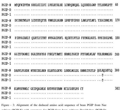
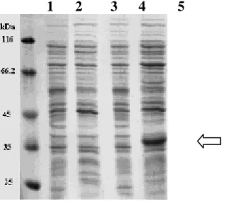
with that of Derakhshan cultivar previously reported (Hosseinzadeh Colagar et al., 2004 and accession number AY367002) and PGIP-1 from P.vulgaris cv. Saxa (accession numbers X64769 & A23205) indicates only a single nucleotide change from A to G at position 880 which alters the amino acid at position 294 from T (Threonine) to A (Alanine) (Fig 2, 3). The presence of G in this position in the DNA sequence creates a new HhaI (Hin6I) site. To confirm this nucleotide change from A to G in that position is not due to sequen-cing error,a restriction pattern was obtained by using HhaI digestion which shows the new HhaI (Hin6I) site in that position(Fig 4). PGIP was extracted from E. coli DH5α harboring pET21a(+) vector containing pgip gene (pETABRN). The extracted PGIP was assayed for Ascochyta rabie (IK04) and Fusarium oxysporum (F15) polyg-alacturonase activity inhibition. This pro-karyotic expressed IPTG inducible poly-peptide was unable to inhibit PG activity of A. rabie and F. oxysporum. This PGIP has a molecular mass of 37kDa when analyzed by SDS-PAGE (Fig 5), which corresponds to the deduced molecular weight of PGIP.
DISCUSSION
0 10 20 30 40 50 60 70
F. o xyspo rum A. rab iei
Is o l ate s
%
I
n
h
ib
it
io
n
Naz Derak hs han
hydrolyze the pectic fraction of the plant cell wall is reduced and the generation of elicitor active oligogalact-uronides is favored (Ridley et al., 2001). The overall effect of this interaction on the host plant is the limitation of fungal invasion. PGIPs exhibit different recognition specificities against the many PGs secreted by fungal pathogens (Wubben et al., 1999; Leckie et al., 1999; Hammond- Kosack and Jones, 1997). This functional redundancy likely ensures a higher level of protection against fungal pathogens and a selective advantage. All these proteins (PGIPs) share leucine-rich repeats (LRRs) of the extra-cellular or extracytoplasmic type, charac-terized by the presence of 9-10 repeats, each derived from modifications of 24 amino acid leucine-rich peptide (Leckie et al., 1999). The LRR element matches the consensus sequence LXXLX LXXNXLT/SGXIPXXL XXLXX (Leckie et al., 1999; Kajava 1998). PGIPs not only show similarity in the LRR region but also in the regions outside the LRR domain (Benntt et. al., 1996;DeLorenzo and Cervone 1997).
In our previous studies it was shown that polygalacturonase enzyme of highly virulent isolates of A. rabiei (IK04) and F. oxysporum (F15) is an important factor in pathogenicity (Zamani et al., 2000; Zamani et al., 2001; Motallebi et al.,
2003; Alani et al., 2004). In this work inhibitory activity of PGIP from P. vulgaris cv. Naz and cv. Derakhshan on PGs fromA. rabiei (IK04) and F. oxysporum (F15) is compared. The inhibitory activity of bean protein extract containing PGIP from two bean cultivars (Naz and Derakhshan) on PG of F. oxysporum
(F15) and A. rabiei (IK04) showed that Derakhshan PGIP was able to inhibit PG activity by 55% and 40% in F. oxysporum
Figure- 1: Polygalacturonase activity inhibition in Fusarium oxysporum (F15) and
Ascochyta rabiei (IK04) by PGIP extracted from Phaseolus vulgaris cv. Naz and cv. Derakhshan hypocotyle.
Figure -2: The nucleotide and the deduced amino acid sequences of the Phaseolus vulgaris cv. Naz pgip. Nucleotides are numbered on the left and amino acids are numberd on the right. Bold and underlined are the position of oligonucleotide primers SSRf & SSRr. Start codon (atg) and stop codon (taa) are shown in bold. The bold and boxed nucleotide and amino acid indicate the differences in Naz sequence compared with Derakhshan (AY367002) and Saxa (X64769 and A23205) cultivars.
1 atgactcaattcaatatcccagtaaccatgtcttcaagcttaagcataattttggtcatt M T Q F N I P V T M S S S L S I I L V I 61 cttgtatctttgagaactgcactctcagagctatgcaacccacaagataagcaagccctt L V S L R T A L S E L C N P Q D K Q A L 121 ctccaaatcaagaaagaccttggcaacccaaccactctctcttcatggcttccaaccacc L Q I K K D L G N P T T L S S W L P T T 60aa 181 gactgttgtaacagaacctggctaggtgttttatgcgacaccgacacccaaacatatcgc D C C N R T W L G V L C D T D T Q T Y R 241 gtcaacaacctcgacctctccggccataacctcccaaaaccctaccctatcccttcctcc V N N L D L S G H N L P K P Y P I P S S 301 ctcgccaacctcccctacctcaattttctatacattggcggcatcaataacctcgtcggt L A N L P Y L N F L Y I G G I N N L V G 120aa 361 ccaatcccccccgccatcgctaaactcacccaactccactatctctatatcactcacacc P I P P A I A K L T Q L H Y L Y I T H T 421 aatgtctccggcgcaatacccgatttcttgtcacagatcaaaaccctcgtcaccctcgac N V S G A I P D F L S Q I K T L V T L D 481 ttctcctacaacgccctctccggcaccctccctccctccatctcttctctccccaacctc F S Y N A L S G T L P P S I S S L P N L 180aa 541 ggaggaatcacattcgacggcaaccgaatctccggcgccatccccgactcctacggctcg G G I T F D G N R I S G A I P D S Y G S 601 ttttcgaagctgtttacggcgatgaccatctcccgcaaccgcctcaccgggaagattcca F S K L F T A M T I S R N R L T G K I P 661 ccgacgtttgcgaatctgaacctggcgttcgttgacttgtctcggaacatgctggagggt P T F A N L N L A F V D L S R N M L E G 240aa 721 gacgcgtcggtgttgttcgggtcagataagaacacgaagaagatacatctggcgaagaac D A S V L F G S D K N T K K I H L A K N 781 tctcttgcttttgatttggggaaagtggggttgtcaaagaacttgaacgggttggatctg S L A F D L G K V G L S K N L N G L D L 841 aggaacaaccgtatctatgggacgctacctcagggactagcgcagctaaagtttctgcaa R N N R I Y G T L P Q G L A Q L K F L Q 300aa 901 agtttaaatgtgagcttcaacaatctgtgcggtgagattcctcaaggtgggaacttgaaa
S L N V S F N N L C G E I P Q G G N L K 961 aggtttgacgtttcttcttatgccaacaacaagtgcttgtgtggttctcctcttccttcc R F D V S S Y A N N K C L C G S P L P S 1021 tgcacttaa
Figure - 3: Alignment of the deduced amino acid sequence of bean PGIP from Naz cultivar (PGIP-N) with sequences for PGIP from bean cultivars Derakhshan (PGIP-D) (AY367002) and Saxa (PGIP-1) (X64769 and A23205). Amino acids of the Naz sequence are numbered on the right. The arrow indicates the difference in amino acid between Naz and Derakhshan & Saxa PGIPs.
430 bp 144 bp 455 bp
150 bp
Figure - 4: A) Confirmatiom of Pfu PCR product by restriction pattern using
HhaI(Hin6I) restriction enzyme on 2% agarose.
The presence of G in position 880 in DNA sequence of Naz pgip creates a new HhaI (Hin6I) site. The restriction pattern was obtained using HhaI (Hin6I) digestion, which shows creation of a new site in that position. M= Marker; N= Naz; D= Derakhshan.
B) scheme of the 1029 bp PCR fragment containing the pgip gene from Phaseolus vulgaris cv. Naz and cv. Derakhshan showing HhaI sites. HhaI site marked by open triangle present in Naz and Derakhshan cultivars. Closed triangle show HhaI site present in Naz, absent in Derakhshan cultivars.
1 2 3 4 5
Fig -5: SDS – PAGE and molecular weight determination of PGIP extracted from E. coli
harboring pgip gene. The gel was stained with Coomassie blue.
A
B
Line 1- Protein molecular weight marker Line 2- E. coli BL21 (DE3) strain.
Line 3- E. coli BL21 (DE3)- harboring pET21(a) induced with 0.5 mM IPTG. Line 4- E. coli BL21 (DE3)- harboring pETABRN, noninduced.
Line 5- E. coli BL21 (DE3)- harboring pETABRN induced with 0.5 mM IPTG.
REFERENCES:
Alani,B., M. Motallebi and M.R. Zamani, Purification and partial characterization of polygalacturonase from highly virulent (HV) isolate of
Fusarium oxysporum (F23). Iranian J. Biol.16: 1-11 (2004)
Bennett, A., J. M. Labavitch, A. Powell and H. Stotz, Plant inhibitors of fungal polygalacturonases and their use to control fungal disease. Regents of the University of California, Oakland, Calif., United states (1996) Berger,D.K., D.Oelofse, M.S.Arendse,
E. Du Plessis and A. Dubery, Bean polygalacturonase inhibitor protein-1 (PGIP-1) inhibits polygalacturonases from Stenocarpella maydis . Physiol. Mol. Plant Pathol.57: 5-14 (2000) Brown, A.E. and N.K. Adikaram, A
role for pectinase and protease inhibitors in fungal rot development in tomato fruits. Phytopathol. 106:
249-251 (1983).
Buchanan,S.G.S. and N.J.Gay, Struc-tural and functional diversity in the leucine-rich repeat family of proteins.
Prog. Biophys. Mol. Biol. 65: 1–44 (1996).
Cervone, F., R. Castoria, F. Lechie and G. De Lorenzo, Perception of fungal elicitors and signal transduction. In:
Aducci, P. eds. Signal Transduction in Plants. Birkauser Verlag, Basel, Switzerland, Pp. 153-177 (1997) Collmer,A., J.L.Ried and M.S.Mount,
Assay methods for pectic enzymes.
Meth. Enzymol. 161: 329-335 (1988)
Cook BJ, R.P. Clay, C.W. Bergmann, P. Albersheim and A.G.Darvill, Fungal polygalacturonases exhibit different substrate degradation patterns and differ in their susceptibilities to polygalact- uronase inhibiting proteins. Molecular Plant Microbe Interact.12:703-711 (1999)
De Lorenzo,G. and F.Cervone, Poly-galacturonase- inhibiting proteins (PGIPs): their role in specificity and defense against pathogenic fungi.
Chapman & Hall, New York (1997) Degra, L., G. Salvi, D. Mariotti, G. De
Lorenzo and F.Cervone,A polygalact- uronase inhibiting protein in alfalfa callus cultures. J. Plant Physiol. 133:
364-366 (1988)
Desiderio,A., B.Aracri, F.Leckie, B.Mattei and G.Salvi, Polygalacturonase -inhibiting proteins (PGIPs) with different specificities are expressed in
Phaseolus vulgaris. Mol. Plant-Microbe Interact.10: 852-60 (1997) Devoto, A., A.J. Clark, L. Nuss, F.
Cervone and G.De Lorenzo, Develop-mental and pathogen-induced accumulation of transcripts of polygalacturonase-inhibiting protein in Phaseolus vulgaris L. Planta202: 284–292 (1997)
Doyle, J.J. and J.L. Doyle, Isolation of plant DNA from fresh tissue. Focus
12: 13–15 (1991).
Favaron, F., C. Castiglioni, R. D’Ovidio and P.Alghisi, Polygalacturonase inhibiting proteins from Allium porrum L. and their role in plant tissue against fungal endo-polygalacturonases. Physiol. Mol. Plant Pathol.50: 403–417 (1997) Hahn, M.G., A.G.Darvill, and P.
Albersheim, Host pathogen interaction XIX. The endogenous elicitor, a fragment of a plant cell wall polysaccharide that elicits phytoalexin accumulation in soybeans. Plant Physiol. 68: 1161-1169 (1981)
Hahn, M.G., P. Buchell, F. Cervone, S.H. Doares, R.A. O’Neill, A. Darvill and P. Albersheim, Roles of cell wall constituents in plant- pathogen interactions. In: Nester, E., T. Kosuge, eds. Plant- Microbe Interactions. New York: McGraw – Hill, 131-181 (1989)
Hammond-Kosack, K. E. and J.D.G. Jones, Plant disease resistance genes. Annu. Rev. Plant Physiol. Plant Mol. Biol.48: 575–607 (1997)
Henrik, U., A.L. T. Powell, S.E. Damon, L.C. greve, A.B.Bennett and J.M. Labavitch, Molecular characterization of polygalact- uronase inhibitor protein from Pyrus communis L. cv. Bartlett. Plant Physiol. 102: 133-138 (1993)
Hosseinzadeh Colagar A., M.R. Zamani and M. Motallebi, PGIP-PG interaction, cloning and partial characterization of pgip gene from
Phaseolus vulgaris cv. Derakhshan.
Iranian J. Biol. 17: 89-100 (2004) Huang,Q. and C.Allen,
Polygalact-uronase are required for rapid
colonization and full virulence of
Ralstonia solanacearum on tomato plants. Physiol. Mol. Plant Pathol.
57: 77-83 (2000).
Isshiki, A., M. Akimitsu and H. Yamamoto, Endopolygalacturonase is essential for citrus black rot caused by Alternaria citri but not brown spot caused by Alternaria alternate. Mol. Plant Microb.Interact. 14: 749-757 (2001)
Kajava, A.V., Structural diversity of leucine- rich repeat proteins. J. Mol. Biol. 277: 519-527 (1998)
Leckie, F., B. Mattei, C. Capodicasa, A. Hemmings, L. Nuss, B. Aracri, G. De Lorenzo and F. Cervone, The specificity of polygalacturonase-inhibiting protein (PGIP): a single amino acid substitution in the solvent-exposed b-strand/b-turn region of the leucine-rich repeats (LRRs) confers a new recognition capability. European Molecular Biology Organization (EMBO) 18: 2352–2363 (1999)
Machinandiarena, M.F., F.P. Olivieri, G.R. Dalco, C. R. Oliva, Isolation and
characterization of a
polygalacturonase- inhibiting protein from potato leaves. Accumulation in response to salicylic acid, wounding and infection. Plant Physiol. 39:
129-136. (2001)
Motallebi M., M.R. Zamani and A. Hosseinzadeh Colagar, Correlation of polygalacturonase activity and patho-genicity of Iranian isolates of
Ascochyta rabiei. J. Science and Technol. Agri. Natu. Resources 6: 159-169 (2003)
interaction. Fungal Genet. Biol. 36:
176-186 (2002)
Ridly,B.L., M.A.O’Neill and D.Mohnen, Pectin, structure, biosynthesis, and oligogalacturonide– related signaling.
Phytochem.57: 929-967 (2001) Rodriguez- Palenzuela, P. and T. J.
Burr, Polygalacturonase is a factor in
Agro-bacterium tumefaciens biovar 3.
J. Bacteriol. 173: 6547-6552 (1991) Salvi, G., F. Giarrizzo, G. De Lorenzo
and F.Cervone, A polygalacturonase inhibiting protein in the flowers of
Phaseolus vulgaris L. J.Plant Physiol.136: 513-518 (1990)
Sambrook, J., D.W. Russell, Molecular cloning: a laboratory manual. Cold Spring Harbor Press, NewYork, NY (2000).
Sweetingham,M.W., R.H. Cruickshank, D.H. Wong, Pectic zymograms and taxonomy and pathogenicity of the ceratobasidiaceae. Trans. Br. Mycol. Soc., 86: 305-311 (1986)
Ten Have, A., W. Mulder, J. Visser and J.A.van Kan, The endopolygalact-uronase gene Bepg1 is required for full virulence of Botrytis cinerea.
Mol. Plant Microb. Interact. 11:
1009-1016 (1998)
Wubben, W.M., A. ten Have, J.A. van Kan and J.Visser, Cloning and partial characterization of endopolygalact-uronase genes from Botrytis cinerea. Applied Env. Microbiol. 65: 1596-1602 (1999)
Yu, K.P., S.J. Poysa and P. Gepts, Integration of simple sequence repeat (SSR) markers into a molecular linkage map of common bean (Phaseolus vulgaris). The American Genetic Association 91: 429-434 (2000)
Zamani M.R., M. Motallebi and M.A. Arefpour, Comparative study of polygalacturonase activity from different Iranian isolates of Fusarium oxysporum. Iranian J. Agri. Sci. 31: 293-302 (2000)
Zamani M.R., M. Motallebi and M.J. Harighi, Pectic enzyme patterns of