_____________________________________________________________________________________________________
*Corresponding author: E-mail: [email protected];
Evaluation of Impact of Some Selected Plants on
Haematological Parameters of Testosterone and
Estradiol Induced Benign Prostatic Hyperplasia
Adult Male Rats
Melvin Nnaemeka Ugwu
1*, Atamgba Agbor Asuk
1, Mbeh Ubana Eteng
2and Edward Odey Emuru
11
Department of Medical Biochemistry, Faculty of Basic Medical Sciences, Cross River University of Technology, Calabar, Nigeria. 2Department of Biochemistry, Faculty of Basic Medical Sciences, University of Calabar, Calabar, Nigeria.
Authors’ contributions
This work was carried out in collaboration author all authors. Authors MNU and AAA designed the study, performed the statistical analysis, wrote the protocol and wrote the first draft of the manuscript. Authors MNU and MUE managed the analyses of the study. Author EOE managed the literature searches. All authors read and approved the final manuscript.
Article Information
DOI: 10.9734/IBRR/2019/v10i130113 Editor(s): (1) Dr. Dharmesh Chandra Sharma, Associate Blood Transfusion Officer ( ABTO), In-charge Blood Component & Aphaeresis Unit Blood Bank, Department of Pathology, J. A. Groups of Hospital and G. R. Medical College, India. Reviewers: (1) Awofadeju Stephen Olajide, Wesley Guild Hospital Ilesha, Obafemi Awolowo University Teaching Hospital, Nigeria. (2)Juliano Augusto Brum Scheffer, Brazil. (3)Erdal Benli, Ordu University, Turkey. Complete Peer review History:http://www.sdiarticle4.com/review-history/53052
Received 24 September 2019 Accepted 28 November 2019 Published 04 December 2019
ABSTRACT
Background: Benign prostate hyperplasia (BPH) is an age-related disease of unknown etiology, characterized by prostate enlargement. The effect of Prosopis africana (PA), Vernonia amydalina
(VA) and Ocimum gratissmum (OG), plant extracts on haematological parameters of BPH animal model was investigated.
Methods: BPH was induced in 45 male Wistar rats (250-350 g) by exogenous injection of testosterone and estradiol in staggered doses for 3 weeks. To confirm BPH induction, some animals
were sacrificed; histological inspection of prostate gland and PSA was carried out. Forty BPH induced rats were divided into 8 groups. Group 1 and 2, 3 and 4, 5 and 6 were treated with 50 mg/kg bw and 100 mg/kg bw doses of PA, VA and OG extracts respectively. Group 7 received finasteride (0.1 mg/kg bw). Group 8 BPH control and five rats without induction constitute group 9, the normal control and both received distilled water. After 45 days, the rats were anaesthetised by a brief exposure to trichloromethane vapour and 5 ml of blood was collected from the rats through cardiac puncture and dispensed into well-labelled EDTA containers to avoid coagulation. All analyses were completed within 24 h of sample collection.
Results: Results showed that induction of BPH caused a significant (P< 0.05) enlargement of prostate gland when compared to normal control. All extracts produced significant (P<0.05) reduction in the weight of the enlarged prostate when compared to the BPH control. There were significant (P ˂0.05) decline in RBC, PCV and Hb of BPH control when compared to the normal control and treated groups. In the treated groups the administration of the extracts and standard drug exhibited an increase in RBC, PCV and Hb concentration when compared with the BPH control. Also there was significant (P < 0.05) increase in the WBC, neutrophils, platelets, monocytes, lymphocytes and eosinophils levels in BPH control when compared to normal control and treated groups. In all treated groups there was significant decrease in WBC, neutrophils, platelets, monocytes, lymphocytes and eosinophils concentration levels when compared with the BPH control group.
Conclusion: The result of this study indicates that the extracts have the potential to reverse the inflammation caused by BPH and also have the capacity to boost the numbers of red blood cells probably by inhibiting the hemolysis caused by inflammatory factors or by enhancing the production of red blood cell from the bone marrow.
Keywords: Prosopis africana; Vernonia amygdalina and Ocimum gratissimum; prostate disorder; haematology.
1. INTRODUCTION
The prostate gland is a major secondary endocrine organ of males whose development and growth depends on androgen stimulation especially by dihydrotestosterone (DHT). It is shown that androgen and estrogens constitute the primary factors responsible for prostate diseases [1,2,3,4]. Aging is associated with increased accumulation of DHT which results in increased cell growth and hyperplasia [5]. Benign prostatic hyperplasia (BPH) is a common non-malignant urologic condition among older man which is characterized by prostate enlargement, bladder outlet obstruction (BOO), and lower uri-nary tract symptoms (LUTS).
In orthodox medical practice, various medications such as alpha-adrenergic blockers and 5 alpha-reductase inhibitors have been used successfully to manage the lower urinary tract symptoms associated with prostate hyperplasia [6]. In addition to refractory retention of urine (repeated urine retention) other complications from benign prostate hyperplasia such as haematuria, recurrent urinary tract infections, bladder calculi and renal insufficiency are managed with surgical interventions such as open prostatectomy and transurethral resection of the prostate [7].
Currently alternate medicine has been incorporated into the main stream medical services. As a result, more patients are resorting to Traditional Medicine to address their ailments including prostate conditions [6]. Other reasons for the increase in herbal medicine use in underdeveloped countries has been attributed to such factors as the high cost of pharmaceutical medicines and overcrowding of patients in clinics and hospitals [8]. Herbal medicine, botanical medicine or phytomedicine refers to using plant components such as seeds, berries, roots, leaves, bark, or flowers for medicinal purposes [9]. It has been noted that Traditional Medical practice has long been used outside of conventional medicine but mainly for primary health care [6].
that treatment of BPH animal model with
Prosopis africana seed extract for 45 days significantly inhibited the development of induced prostatic hyperplasia, which was evident in the reduction in the elevated prostate weight, reduced prostatic specific antigen (PSA), testosterone, estradiol and prolactin levels in the serum. The increased prostate weight is used as one of crucial markers of BPH according to previous study [17,18]. Also, Ugwu, et al. [19] has proven that administration of the aqueous extract of Vernonia amygdalina to rats induced with BPH has protective effects against the development of BPH as seen in the reduction in PSA levels, prostate weight, improved prostate histological patterns. It has been established that serum PSA levels increase abnormally in patients with benign prostatic hyperplasia and prostatitis [20]. Therefore reduction of the serum PSA level can show protective effects on benign prostatic hyperplasia.
The practice of traditional medicine is as old as the origin of man [21]. The use of plants in traditional medicine referred to as herbalism or botanical medicine [22] falls outside the mainstream of the Western or Orthodox medicine. It has been estimated that about two third of the world’s population (mainly in the developing countries) rely on traditional medicine as their primary form of health care. The use of traditional medicine in the treatment and management of diseases in the African continent cannot fade away and this could be attributed to the socio-cultural, socio-economic, lack of basic
health care and qualified personnel [23]. Plants contain active components such as
anthraquinones, flavonoids, glycosides, saponins, tannins, etc., which possess medicinal properties that are harnessed for the treatment of different diseases. Many studies reported that oral ingestion of medicinal drugs can alter the haematological parameters ranges to either positive or negative [24,25]. There is paucity of data on relationship between haematological parameters and BPH. Therefore it is paramount to study the effect of these three indigenous plants on haematological parameters in BPH condition.
Haematology refers to the study of the number and morphology of the cellular elements of blood particularly; the red blood cells (erythrocytes), white blood cells (leucocytes) and the platelets (thrombocytes) in addition to the use of these results in the diagnosis and monitoring of diseases [26]. Haematological studies are of
ecological and physiological interest in helping to understand the relationship of blood characteristics to the environment [27]. Evaluation of blood parameters can be used to determine the level of negative effect of foreign compounds, including medicinal plants [28,29, 30]. This method can also be used to explain haematological relating functions of plant products [31]. In addition, such investigation is necessary to risk examination as changes in the blood system have higher predictive value on human toxicity when data are explained from animal studies [24].
Blood transports nutrients and oxygen to body cells and removes waste products away from the cells [32]. It is a composite of erythrocytes, leucocytes, thrombocytes, plasma and accounts for 7% of human body weight [33]. Oxygen distribution to the periphery from the lungs through the pulmonary capillaries, removal of carbon dioxide from the tissues back to the lungs through the systemic capillaries and maintenance of acidic and basic values of the body are the three main functions of erythrocytes [34].
Leucocytes are the main cells of the immune system that provide innate and specific adaptive immunity. They are further divided into five different classes which include; basophils, neutrophils, eosinophils, lymphocytes and monocytes [35]. Platelets play a major role in hemostasis, thrombosis, clot retraction, vessel constriction and repair, inflammation including promotion of atherosclerosis, host defense and
even tumor growth/metastasis [36].
Haematological complications consist mainly of abnormalities in the function, morphology and metabolism of erythrocytes, leukocytes and platelets [37,38]. Hence the need to study the effect of these three plants on heamatological parameters in benign prostate hyperplasia condition.
2. MATERIALS AND METHODS
2.1 Plant Material I and II: Ocimum
gratissimumandVernoniaamygdalina
Fresh leaves of Ocimum gratissimum and
Calabar. Their fresh leaves were washed with clean water and dried under the shade for six days. Their dried leaves were milled using pestle and mortar to get powders that were used for extraction.
2.1.1 Preparation of extracts of Ocimum gratissimum and Vernonia amygdalina
The powered sample of Ocimum gratissimum and Vernonia amygdalina 100 grams were soaked into 100 ml of distilled water separately. These were filtered after 48 hours and filtrate were concentrated in hot air oven. The solutions were diluted with corn oil, to produce a solution 100 mg/ml respectively. Administrations of extracts were totally by gavage. Proper concentrations were administered by the use of oropharyngeal canula and calibrated hypodermic syringe.
2.2 Plant Material III: Prosopis africana
Prosopis seeds were purchased from Shibori market in Ogoja Local Government of Cross River State, Nigeria. The seeds (500 g) was sorted, cleaned and was boiled for 5 h using a gas cooker and allowed to cool to room temperature. The boiling helps to soften the hulls for easy removal and separation of the cotyledons. After it was dehulled and decorticated. The dehulled and boiled seeds were washed again with clean water. The processing of decortications was done by hand squeezing the seeds and washing with clean water. The wet decorticated seeds were kept in a large polythene sacks to exclude air and was fermented for three days according to the method described by Achi [39]. The fermentation was done at room temperature (≈25ºC) for 72 h. The fermented seeds were then sun-dried to a constant weight and milled using hammer mill to produce prosopis seed flour [40]. The flour was kept in a refrigerator at 4ºC prior to use.
2.3 Hormones
Testosterone propionate Brand name:
Ricostrone; a product of Greenfield pharma, Jiangsu Co Ltd., China. Estradiol valerate (by Medipharm Ltd., 108-Kotlakhpat industrial Est; Lahore, India. Testosterone propionate (T) and estradiol valerate E2 (puregynon depot) were used for the induction of prostate gland enlargement at a dose of 400 μg T and 80 μg E2 [41]. This was administered to the rats for three
weeks subcutaneously in the inguinal region after which a few rats were sacrificed and inspected for gross examination of prostate gland enlargement. All Chemicals used in this study were of analytical grade and were obtained from reputable companies.
2.4 Animals
A total of fifty (50) Wistar rats weighing between 250-350 g were obtained from the animal house of the Faculty of Basic Medical Sciences, University of Calabar, Nigeria. The rats were used for the experiment. The rats were acclimatized for two weeks before the experiment commences. The rats were exposed to approximately 12-hour light/dark cycles under humid tropical conditions, given tap water and feed ad libitum, and were housed in standard plastic cages (five per cage) throughout the 45-day duration of the study. The animal room was well ventilated with a temperature range of 27-29ºC.
2.5 Induction of BPH
BPH was induced by exogenous administration of testosterone and estradiol in staggered doses (three times a week respectively) for three weeks according to Bernoulli, [41] with modification by Mbaka, et al. [42].
2.6 Animal Grouping and Treatment
The rats were separated into nine (9) groups each made up of five (5) male rats. Forty-five (45) rats were induced with BPH which were grouped as group 1 to group 8 and remaining 5 which were randomly selected and used to confirm the induction of BPH. Groups 1 and 2 received 50 and 100 mg kg–1 body weight (bw) of
2.7 Sample Collection
After 45 days, the rats were anaesthetised by a brief exposure to trichloromethane vapour and 5ml of blood was collected from the rats through cardiac puncture and dispensed into well-labeled EDTA containers to avoid coagulation. All analyses were completed within 24 h of sample collection.
2.8 Haematological Assays (Full Blood Count)
Whole blood was used to determine the full blood count parameters and all the analysis were performed using standard diagnostic Diatro reagents (Diatro-Lyse-Diff, Diatro Cleaner, Diatro Diluent reagents) using Abacus junior haematology analyzer (Diatron. GmbH. Wein Austria). These were done according to manufacturer’s instructions.
2.9 Statistical Analysis
The experimental data were analysed for statistical significance by one-way analysis of variance and post hoc comparison using the SPSS version. The Independent Samples t-test was used to compare the means of two independent groups. All data were reported as mean ± SD and statistical significance was accepted at P ˂ 0.05.
3. RESULTS
3.1 Haematological Parameters
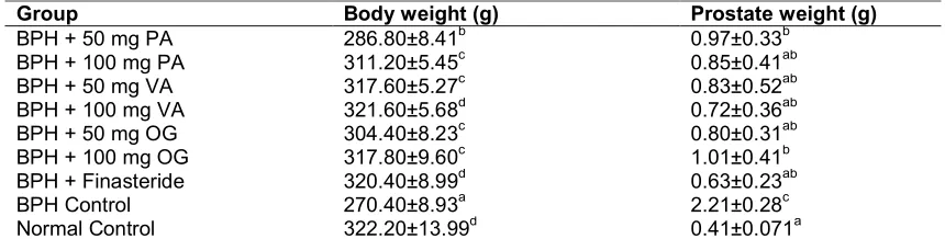
Results showed that induction of BPH caused a significant (P< 0.05) enlargement of prostate gland when compared to normal control. All
extracts produced significant (P<0.05) reduction in the weight of the enlarged prostate when compared to the BPH control (Table 1).
Results of red blood cells (RBC) count, packed
cell volume (PCV), haemoglobin (Hb)
concentration, white blood cells (WBC) count, neutrophils, platelets, monocytes, lymphocyte, eosinophils and basophils concentration for the treated and control groups are shown in Tables 2, 3 and 4.
There were significant (P ˂0.05) decline in RBC, PCV and Hb of BPH control when compared to the normal control and treated groups. In the treated groups the administration of the extracts and standard drug exhibited an increase in RBC, PCV and Hb concentration when compared with the BPH control.
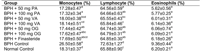
There were significant (P < 0.05) increases in the WBC, neutrophils, platelets, monocytes, lymphocytes and eosinophils levels in BPH control when compared to normal control and treated groups. In all treated groups there were significant decreases in WBC, neutrophils, platelets, monocytes, lymphocytes and eosinophils concentration levels when compared with the BPH control group.
4. DISCUSSION
Benign prostatic hyperplasia is caused by changes in hormone balance and consequently in cell growth, but molecular pathways leading to this condition are still largely unknown. Inflammatory component is believed to have an important role while presence and degree of inflammation corresponds to prostate volume and weight [43,44]. The origin of inflammation in
the prostate remains a subject of
Table 1. Effect of extract of PA, VA, OG and finasteride body weight and prostate weight
Group Body weight (g) Prostate weight (g)
BPH + 50 mg PA 286.80±8.41b 0.97±0.33b
BPH + 100 mg PA 311.20±5.45c 0.85±0.41ab
BPH + 50 mg VA 317.60±5.27c 0.83±0.52ab
BPH + 100 mg VA 321.60±5.68d 0.72±0.36ab
BPH + 50 mg OG 304.40±8.23c 0.80±0.31ab
BPH + 100 mg OG 317.80±9.60c 1.01±0.41b
BPH + Finasteride 320.40±8.99d 0.63±0.23ab
BPH Control 270.40±8.93a 2.21±0.28c
Normal Control 322.20±13.99d 0.41±0.071a
Values are expressed as Mean ± SD. Benign prostate hyperplasia (BPH), Prosopis africana (PA),Vernonia amygdalina (VA) and Ocimum gratissium (OG). Identical superscript (i.e. a) means there is no significant difference between the comparing group P>0.05. Non- identical superscripts (i.e. a, b, c, d) means there is
Table 2. Effects of extracts PA, VA, OG and finasteride on haematological parameters Group Rbcs (1012/l) Pcv (%) Hb (g/dl)
BPH + 50 mg PA 8.47±0.38ab 43.35±0.70a 14.23±0.36a
BPH + 100 mg PA 8.50±0.49ab 43.38±0.73a 14.27±0.37a
BPH + 50 mg VA 8.45±0.26ab 43.44±0.67a 14.46±0.37a
BPH + 100 mg VA 8.80±0.22b 43.57±1.05a 14.52±0.27a
BPH + 50 mg OG 8.59±0.26ab 43.37±0.74a 14.28±0.44a
BPH + 100 mg OG 8.58±0.47ab 43.40±0.66a 14.28±0.54a
BPH + Finasteride 8.61±0.36ab 43.58±1.16a 14.29±0.39a
BPH Control 8.24±0.44a 38.71±1.57b 12.98±0.21b
Normal Control 8.86±0.06b 43.68±0.50a 14.31±0.38a
Values are expressed as Mean ± SD. ‘Red blood cells (RBC), Packed cell volume (PCV), Haemaglobin concentration (Hb), Benign prostate hyperplasia (BPH), Prosopis africana (PA), Vernonia amygdalina (VA) and
Ocimum gratissium (OG). Identical superscript (i.e. a) means there is no significant difference between the
comparing group P > 0.05. Non- identical superscripts (i.e. a, b)means there is significance between the
comparing groups at P ˂ 0.05
Table 3. Effects of extract PA, VA, OG and finasteride on haematological parameters Group wbc (x109/l) Neutrophils (%) Platelets (x109/l)
BPH + 50 mg PA 17.21±0.72b 26.24±0.63b 902.52±23.49c
BPH + 100 mg PA 17.31±0.83b 26.27±0.62b 907.02±19.46c
BPH + 50 mg VA 17.27±0.40b 26.75±0.44b 925.34±67.49 c
BPH + 100 mg VA 17.35±0.23b 26.82±0.37b 929.81±40.01c
BPH + 50 mg OG 17.25±0.60b 26.39±0.70b 911.01±42.35c
BPH + 100 mg OG 17.31±0.69b 26.41±0.28b 914.81±54.87c
BPH + Finasteride 17.27±0.62b 26.50±0.48b 926.71±42.56c
BPH Control 24.87±0.89a 34.63±0.46a 1241.69±32.07a
Normal Control 17.48±0.5b 26.84±0.59b 943.98±27.00b
Values are expressed as Mean ± SD. White blood cell (WBC), Benign prostate hyperplasia (BPH), Prosopis africana (PA), Vernonia amygdalina (VA) and Ocimum gratissium (OG).Identical superscript (i.e. a) means there is no significant difference between the comparing group P>0.05. Non- identical superscripts (i.e. a, b, c) means
there is significance between the comparing groups at P ˂ 0.05
Table 4. Effects of extract PA, VA, OG and finasteride on haematological parameters Group Monocytes (%) Lymphocyte (%) Eosinophils (%)
BPH + 50 mg PA 17.28±0.47a 64.56±0.59a 5.62±0.56a
BPH + 100 mg PA 17.32±0.34a 64.66±0.63ab 5.77±0.25a
BPH + 50 mg VA 18.00±0.38bcd 65.55±0.43bc 6.01±0.31a
BPH + 100 mg VA 18.14±0.51cd 65.84±0.46c 6.14±0.36a
BPH + 50 mg OG 17.44±0.42ab 64.74±0.30ab 6.06±0.74a
BPH + 100 mg OG 17.62±0.47abc 64.79±0.31ab 6.09±0.21a
BPH + Finasteride 17.69±0.50abcd 64.85±0.30ab 6.18±0.26a
BPH Control 26.50±0.58e 72.63±1.27d 9.36±0.44b
Normal Control 18.31±0.37d 65.88±0.90c 6.20±0.21a
Values are expressed as Mean ± SD. Benign prostate hyperplasia (BPH), Prosopis africana (PA), Vernonia amygdalina (VA) and Ocimum gratissium (OG).Identical superscript (i.e. a) means there is no significant difference between the comparing group P > 0.05. Non- identical superscripts (i.e.a, b, c, d, e) means there is
significance between the comparing groups at P ˂ 0.05
argument and is likely to be multifactorial. It represents a chronic process of wound healing, which activates hyperproliferative programmes resulting in the formation of BPH nodules [43,44]. Acute and chronic inflammation leads to accumulation of immunocompetent cells in the
prostate, mainly T lymphocytes and
macrophages. However, many other cell types
may be observed, including neutrophils, eosinophils, and mast cells, depending on the type of offending agent [45]. Most of the lymphocyte populations around prostate gland are CD8+ T-lymphocytes while stroma mostly contains clusters of B-lymphocytes enclosed with CD4+ T-lymphocytes [46]. Not only
cells have cytokine receptors on membrane surface, participating in the local immune response [45, 47,48]. In a study by Cihan, et al. [49] patients with BPH had a higher level of neutrophils and lower level of mean lymphocyte count when compared to healthy individuals.
Inflammatory pathways are triggered by viral or bacterial antigens, as well as different chemical irritations and metabolic disorders. Both prostate epithelial and stromal cells and inflammatory cells produce cytokines (CCL-5, CCL-2), ILs (IL-1α, IL-1β, IL-6, IL-18), and hypoxia-inducible factor-1α (HIF-1α), creating local inflammatory microenvironment [47,50,51]. Abundant lymphoid infiltrates with a massive increase in CD4+ T-lymphocytes, as well as macrophages and mast cells, are noted in chronic prostate inflammation [52]. These cells participate in pathological changes characteristic for both BPH and prostate carcinoma. There are several studies demonstrating that ILs, which has a pro-inflammatory role, may lead to initiation and progression of BPH [45,50,51,53,54,55].
McDowell and associates showed how
inflammatory cells can be attracted to the prostate tissue microenvironment and can selectively promote the proliferation of prostate epithelial cells [56]. Further studies have confirmed IL-17 to be the initiator of BPH progression via activation of the nuclear-factor-kappa-B (NF-κB) pathway, which leads to secretion of other pro-inflammatory cytokines, such as IL-1, IL-6, and IL-8. Steiner, et al. [57] demonstrated that healthy prostates do not express IL-17, whereas prostates with inflammation and BPH do. Wang, et al. [58] (also found cyclooxygenase 2 (COX-2) expressed in macrophages and epithelial prostate cells within significant inflammation. Under certain conditions, if high level of T-lymphocytes is reached, surrounding cells are killed by CD8+ cytotoxic T cells and prostatic tissue is replaced
by fibromuscular nodules [48,52]. Local hypoxia and inflammation also promote fibroblast to myofibroblast transformation, which
leads to extracellular changes forming suitable microenvironment for continuous inflammation [47,52,59]. Inflammation is also continuously stimulated by androgens and changes within metabolic syndromes, but exact pathways are still mostly unknown [48,60, 61].
Chronic inflammation has been documented for years in benign prostatic hyperplasia (BPH). BPH is an immune inflammatory disease. Unravelling the specific nature of immune dysregulation may
help design novel drugs with these specific targets in mind [52]. Clinical evidence reported that chronic inflammation presented a key condition leading to prostate enlargement and to an increased symptoms score as well as a major risk of complications [62]. Previous study by Nickel, et al. [63] showed a positive association between inflammation and prostate volume, indeed patients with chronic inflammation at biopsy had higher volumes than those without inflammation. Robert, et al. [54] assessed the degree of prostatic inflammation using cytological and immunohistochemical parameters in 282 patients treated with surgery for complicated or symptomatic BPH.
Interestingly, the grade of prostatic inflammation was strongly associated with LUTS severity, and patients with chronic inflammation had higher, International Prostate Symptom Score (IPSS) than those without inflammation. Moreover, prostate volume was significantly higher in patients with high-grade inflammatory pattern. Burris, et al. [64] showed that LUTS improvement after radical prostatectomy correlated with degree of prostatic inflammation. The International Prostate Symptom Score (IPSS) is a short validated questionnaire which can document the baseline severity of lower urinary tract symptoms and can be used to monitor the impact of therapy. The score gives an idea of the severity of LUTS and the most bothersome symptoms. On follow-up, the score can give accurate documentation of patients’ progression and deterioration [65].
Assessment of haematological parameters can be used to determine the extent of deleterious effect of foreign compounds including plant extracts on the blood constituents of an animal. Such toxicity testing is relevant to risk evaluation as changes in the haematological system have higher predictive value for human toxicity, when data are translated from animal studies [24,25]. It can also be used to explain blood relating functions of chemical compounds/plant extract [31]. Haematological analysis was carried out for
Prosopis africana, Vernonia amygdalina and Ocimum gratissimum treated.
and children) or liver (children and adults). Packed cell volume (PCV) is used to measure red blood cell mass and an increase in red blood cell mass is equivalent to erythrocytosis and a decrease indicates an anemia. Some medicinal plants are known to cause destruction of red blood cells leading to anaemia [66].
In this present study, RBC decreased significantly in the BPH control when compared to the treated groups. A possible explanation for the observed decrease in RBC can be linked to red cell distribution width (RDW). The red cell distribution width blood test measures the amount of red blood cell variation in volume and size. It can also help to determine the underlying cause of anemia. In previous study Patel, et al. [67] reported that BPH patients have high RDW values which reflect an underlying inflammatory state that can impairs erythrocyte maturation and consequent inadequate production of the hormone erythropoietin. They further stated that undernutrition or oxidative damage can also result to this condition. Among these variables, oxidative stress and inflammation have been hypothesized as the important determinants of RDW [67]. Inflammatory states are strongly related to ineffective erythropoiesis, and it has been demonstrated that inflammatory cytokines, such as tumor necrosis factor (TNF)-α, IL-1β and IL-6, desensitize bone marrow erythroid progenitors to erythropoiesis, inhibit red blood cell (RBC) maturation and thereby promote anisocytosis.
A decrease in the number of red cells in the blood as observed in is often associated with the development of anaemia [68]. Red blood cells (RBC) or erythrocytes, are the most common type of blood cells and the vertebrate organism's principal means of delivering oxygen (O2) to the
body tissues through the circulatory system [69]. This could be due to the stimulation of lipid peroxidative system by toxins or disease resulting in the production of lipid peroxides which haemolyse the red blood cells [70]. The decrease in haematocrit (PCV) of rats observed in the BPH control, suggests that they may induce anaemia and inability of the cells to deliver oxygen to body tissues that require them. Packed Cell Volume (PCV) or Haematocrit is used clinically to signal to known or suspected anaemia [71]. Since the purpose of red blood cells is to transfer oxygen from the lungs to body tissues; a blood sample's haematocrit can become a point of reference of its ability to deliver oxygen [71]. Factors influencing RBCs will affect the haematocrit because RBCs
comprise 99% of the total cells of the blood [71].
A lower than normal haematocrit is
representative of anaemia especially the aplastic anaemia and some thalassemia syndromes [72].
In extract-treated groups, there was also a significant increase in these parameters when compared with the BPH groups. This could be due to the phytochemical constituents in the extract and also presence of minerals and vitamins. Some of these constituents are well known haemopoietic factors that have direct influence on the production of blood in the bone marrow [73,74]. The findings of this study seem to support this claim and restoration of these functions. Prosopis africana, Vernonia amygdalina and Ocimum gratissimum extracts seem to facilitate iron absorption, as adequate amount of this element is necessary for haemoglobin synthesis and for the animal tissues such as the kidneys and bones to take part in manufacture of RBCs.
Furthermore the increase in PCV as observed by the extracts is an indication that the extracts contain phytochemicals that can stimulate the secretion of erythropoietin—a glycoprotein hormone that stimulates stem cells in the bone marrow to produce RBCs [75]. This may be attributed to the presence of some compounds [76] and its richness in iron and vitamins [77,78, 79,80].
stromal cells and stimulates the proliferation of prostatic epithelial cells in vitro [84]. Monocyte chemotactic protein-1 might be another possible explanation of the correlation between WBC and prostate volume [84].
An increased number of WBC can occur abnormally as a result of an infection, cancer, or toxic chemical [85]. There have been reports that inflammation is a cause of prostatic enlargement [86]. So a possible explanation is that asymptomatic inflammation of the prostate may increase the WBC and the neutrophil count. Serum WBC was reportedly associated with serum monocyte chemotactic protein-1 levels [87]. Reduced WBC showed that the immune system was not compromised in animals treated with Prosopis africana, Vernonia amygdalina and
Ocimum gratissimum which may be due to its content of immune-boosting phytochemicals. Previous studies showed similar trend [88,89,90, 91]. This signifies that these plants can help preserve the body’s adaptive response to stress caused by BPH.
Eosinophils are pleiotropic multifunctional leukocytes involved in initiation and propagation of diverse inflammatory responses, as well as modulators of innate and adaptive immunity [81]. They are multifunctional leukocytes implicated in the pathogenesis of numerous inflammatory processes including parasitic helminth, bacterial and viral infections, tissue injury, tumor immunity, and allergic diseases [92,93,94]. In response to the diverse stimuli, eosinophils are recruited from the circulation into inflammatory foci where they modulate immune responses through an array of mechanisms. Triggering of eosinophils by engagement of receptors for cytokines, immunoglobulins, and complement can lead to the secretion of an array of proinflammatory cytokines, such as interleukin (IL)-2, IL-4, IL-5, IL-10, IL-12, IL-13, IL-16, IL-18, and transforming growth factor (TGF)-a/b, chemokines such as CCL5/RANTES and CCL11/eotaxin-1, and lipid mediators such as platelet-activating factor (PAF) and leukotriene (LT)C4 [95]. These molecules have proinflammatory effects that include upregulation of adhesion systems, modulation of cellular trafficking and activation and regulation of vascular permeability, mucus secretion, and smooth muscle constriction. This could explain the observed increase in eosinophils in the BPH control group. But the extracts were able to reduce this observed increase.
Platelet is implicated in blood clotting and plays a crucial role in reducing blood loss and repair of
vascular injury [96]. The significant increase in platelet concentration in the BPH control indicates that the disease condition can stimulate the biosynthesis of blood clotting factors [97, 98]. Mean platelet volume (MPV) is a simple indicator of platelet size and has been known to be a marker of platelet activity. Some platelet markers, such as MPV, have been reported to be related to inflammation [99]. MPV is correlated with inflammation in inflammatory bowel diseases, rheumatoid arthritis, and ankylosing spondylitis, as reported in previous studies [100]. In the present study, it was identified that platelets levels increased in asymptomatic BPH rats compared with other groups. It might be possible that the elevation of platelets in asymptomatic BPH rats result from the effect of inflammation on thrombopoiesis.
The relationship between MPV values and BPH and prostatitis remain unclear. MPV is a widely used laboratory indicator associated with platelet function based on inflammatory conditions [100]. MPV also represents platelet function, which is central to processes that are involved in coronary heart disease pathophysiology and endothelial dysfunction. Platelet parameters might be influenced by coronary risk factors including age,
obesity, smoking, diabetes mellitus,
hypertension, hyperlipidemia, metabolic syndrome, stroke, peripheral artery disease, and deep vein thrombosis [101]. MPV can also be affected by thyroid diseases, bullous pemphigoid,
malignancy, and medications such as
anticoagulant therapy and statins [102,103]. Karaman, et al. [104] initially reported that MPV values significantly decreased after treatment in all grades of prostatitis and BPH.
oxygen-carrying capacity of the blood and the amount of oxygen delivered to the tissues since red blood cells and haemoglobin (Hb) are very important in transferring respiratory gases [108, 109]. The reduction might be due to the presence of saponin, which has been reported to reduce haematological parameters probably due to lysis of blood cells or suppression of blood synthesis [110]. This report is contrary to the findings from this present study, which demonstrated increase in blood levels of erythrocyte. The possible explanation to this observed difference might be the duration of the administration and dose administered. It is therefore possible that the excessive and long time consumption of
Vernonia amygdalina by humans can lead to anaemic state.
Ofem
,
et al. [111] observed that extract O. gratissimum caused an increase in the erythrocyte count. This was confirmed by the increased haematocrit (PCV) and percentage Hb. In normal circumstances, local tissue anoxia apparently leads to the formation of a glycoprotein called erythropoietin, which stimulates increased production of erythrocytes [112]. It is very likely that O. gratissimum leaves extract contains erythropoietin-like agent(s) which is/are responsible for the increased production of erythrocytes. In Ofem’s study, examination of the differential counts revealed that high doses of O. gratissimum led to reduction in lymphocyte count [111]. The reduction in lymphocyte count could probably be due to cell margination rather than destruction. It is also possible that the inflammation due to BPH stimulate the bone marrow to produce neutrophils and release them into the blood. Neutrophils are the major granulocytes to be activated when the body is invaded by bacteria and they provide the first line of defense against invading microorganisms [113]. The granules of the neutrophil contains many enzymes, which makes it a powerful and effective killer machine.Obianime, et al. [114] reported that aqueous O. gratissimum leaf extract decreased PCV and Hb levels in the first week, whereas WBC and lymphocyte counts were increased during the first and second weeks, respectively, in male mice. This is contrary to our present study which demonstrated that O. gratissimum increased RBC, PCV and Hb. They attributed the reduced haematological effects of O. gratissimum to stimulation of adaptive mechanisms within the body against O gratissimum–induced toxicity on the blood cells. They suggested that O.
gratissimum may have two different effects— oxidative or antioxidative—depending on the tissue/organ system under investigation and the duration of administration [114].
5. CONCLUSION
This study indicates that the three extracts have the potential to reverse the inflammation caused by BPH and also have the capacity to boost the numbers of red blood cells probably by inhibiting the hemolysis caused by inflammatory factors or by enhancing the production of red blood cell from the bone marrow. Indicating that the plants can lead to the formation of a glycoprotein called erythropoietin, which stimulates increased production of erythrocytes.
CONSENT
It is not applicable.
ETHICAL APPROVAL
The Cross River University of Technology, Calabar, Nigeria, Animal Ethics Committee approved the study before the experiment and certified all experimental protocols.
COMPETING INTERESTS
Authors have declared that no competing interests exist.
REFERENCES
1. De Nunzio C, Tubaro A. BPH: Unmet needs in managing LUTS-a European perspective. Nat Rev Urol. 2011;9:9-10. 2. Farley SJ. BPH: Lift and separate to
relieve LUTS. Nat Rev Urol. 2011;8:352-352.
3. Shin IS, Lee MY, Jung DY, Seo CS, Ha HK. Ursolic acid reduces prostate size and dihydrotestosterone level in a rat model of benign prostatic hyperplasia. Food Chem Toxicol. 2012;50:884-888.
4. Obeagu EI, Amilo GI, Obeagu GU, Ugwuja SE, Agbo E.A. Evaluation of impact of level of prostate specific antigen on haematological parameters of men in Owerri, Nigeria. J Biomed Sci Appl. 2017; 1(3).
5. Carson C, Rittmaster R. The role of dihydrotestosterone in benign prostatic hyperplasia. Urology. 2003;61:2-7.
6. Kyei MY, Klufio GO, Ayamba A,
alternative practice in the management of
prostate diseases in southern Ghana. Ghana Med J.
2017;51(3):128-137.
7. Kyei MY, Mensah JE, Morton B, Gepi-Attee S, Klufio GO, Yeboah ED. Surgical management of BPH in Ghana: A need to improve access to transurethral resection of the prostate. East African Medical Journal.2012;89(8):25-29.
8. Ampofo JA, Andoh A, Tetteh W, Bello M. Microbiological profile of some Ghanaian herbal preparations—Safety issues and implications for the health professions. Open Journal of Medical Microbiology. 2012;2:121-130.
9. Izzo A, Ernst E. Interactions between herbal medicines and prescribed drugs: An updated systematic review. Drugs. 2009; 69(13):1777-1798.
10. Furnharm. Why do people choose and use complementary therapies? In: E. Ernst,
Ed., Complementary Medicine an
Objective Appraisal, Butterworth-Heine- mann, Oxford. 1996;170.
11. Ernst E, White A. The BBC survey of complementary medicine use in the UK. Complementary Therapies in Medicine.
2000;8(2000):32-36.
12. Farnsworth NR, Akerele O, Bingel AS, Soejart DD, Guo ZG. Medicinal plants in therapy. Bulletion of the World Health Organization. 1985;63(6):965-981.
13. Eisenberg DM, Davis RB, Ettner SL. Trends in alternative medicine use in the United States, 1990- 1997-Results of a Follow-Up National Survey. Journal of the American Medical Directors Association. 1998;280(18):1569-1575.
14. World Health Organization. Traditional Medicine; 2007.
Available:http://www.who.int/mediacentre/f actsheets/fs134/en/
15. Ugwu MN, Ogueche PN, Eteng MU, Eno MA. Protective effects of aqueous extract of Ocimum gratissimum on prostate functions in hormonal induced enlarged prostate in adult rats. Asian Journal of Research in Biochemistry. 2018;2(2):1-12. 16. Ugwu MN, Eteng MU, Ogueche PN,
Amaku EE. Effect of Prosopis africana
seed extract on histology and biochemical indices of prostate functions in testo-sterone and estradiol induced enlarged prostate in adult rats. The Pharmaceutical and Chemical Journal. 2018;5(1):1-9.
17. Arruzazabala ML, Mas R, Molina V, Noa M, Carbajal D. Effect of D-004, a lipid extract from the cubal royal palm fruit, on atypical prostate hyperplasia induced by phynylephrine. Drugs in R & D. 2006;7: 233–41.
18. Bisson JF, Hidalgo S, Rozan P, Messaoudi M. Therapeutic effect of ACTICOA powder, a cocoa polyphenolic extract on experi-mentally induced prostate hyperplasia in Wistar-Unilever rats. Journal of Medicinal Food.2007;10:628-635.
19. Ugwu MN, Mgbekem MA, Eteng MU. Effect of Aqueous extract of Vernonia amygdalina on biochemical indices of prostate functions in hormonal induced enlarged prostate in rats. Journal of Complementary and Alternative Medical Research.2018;6(1):1-12.
20. Akinyemi R, Huthman I, Adesanya O, Akpan H, Adefule A. Effect of the methanolic extract of Trichosanthes cucumerina seed (Snakegourd/Tomatoe) on experimentally enlarged prostate gland in adult wistar rats. Research & Reviews. 2012;1:10-20.
21. Doughari JH, Human SI, Bennade S,
Ndakidemi PA. Phytochemicals as
chemotherapeutic agents and antioxidants: Possible solution to the control of antibiotic resistant verocytotoxin producing bacteria. J Med Plant Res. 2009;3:839-848.
22. Evans WC. Pharmacognosy. (15th edtn). 2007; W.B Saunders, Edinburgh, Scotland. 23. Elujoba AA, Odeleye OM, Ogunyemi CM. Traditional medicine development for medical and dental primary health care delivery system in Africa. Afr J Tradit Complement Altern Med. 2005;2:46-61. 24. Olson H, Betton G, Robinson D, Thomas
K, Monro A, Kolaja G, Lilly P, Sanders J Sipes G, Bracken W, Dorato M, Deun KV, Smith P, Berger B, Heller A. Concordance of the toxicity of pharmaceuticals in humans and in animals. Regul. Toxicol. Pharmacol. 2000;32:56–67.
25. Daradka HM. Evaluation of haematological and biochemical activity of ethanolic extract of Zygophyllum simplex Linn. in Wistar Rats. Pakistan Journal of Biological Sciences.2016;19:179-184.
26. Merck M. Haematologic reference ranges. Mareck Veterinary Manual; 2012.
Available:http://www.merckmanuals.com/. 27. Ovuru SS, Ekweozor IKE. Haematological
ingestion in experimental rabbits. African Journal of Biotechnology. 2004;3:346-348. 28. Agbaje EO, Adeneye AA, Daramola AO.
Biochemical and toxicological studies of aqueous extract of Syzigium aromaticum
(L.) Merr. & Perry (Myrtaceae) in rodents. Afri J. Tradit Complemen Altern Med. 2009;6(3):241–254.
29. Ashafa AOT, Yakubu MT, Grierson DS, Afolayan AJ. Effects of aqueous leaf extract from the leaves of Chrysocoma ciliate (L) on some biochemical parameters of Wistar rats. Africa Journal of Biotechnology. 2009;8:1425-1430.
30. Ibrahim MB, Sowemimo AA, Sofidiya MO, Badmos KB, Fageyinbo MS, Abdulkareem FB, Odukoya OA. Sub-acute and chronic toxicity profiles of Markhamia tomentosa
ethanolic leaf extract in rats. Journal of Ethnopharmacology. 2016;193:68-75. 31. Yakubu MT, Akanji MA, Oladiji AT.
Haematological evaluation in male albino rats following chronic administration of aqueous extract of Fadogia agrestis stem. Pharmacognosy Magazine. 2007;3:34-38. 32. Cheeke PR, Shull LR. Natural toxicants in
feeds and poisonous plants. New York: AVI Publishing; 1999.
33. Austin CC, Perkins SL. Parasites in a biodiversity hotspot: A survey of haematozoa and a molecular phylogenetic analysis of Plasmodium in New Guinea skinks. J Parasitol. 2006;92:770-777. 34. Jagger JE, Bateman RM, Ellsworth ML,
Ellis CG. Role of erythrocyte in regulating local O2 delivery mediated by hemoglobin oxygenation. Am J Physiol Heart Circ Physiol. 2001;280:H2833-2839.
35. Mosmann TR, Coffman RL. TH1 and TH2 cells: Different patterns of lymphokine secretion lead to different functional properties. Annu Rev Immunol. 1989;7: 145-173.
36. Harrison P. Platelet function analysis. Blood Rev. 2005;19:111-123.
37. Comazzi S, Spagnolo V, Bonfanti U. Erythrocyte changes in canine diabetes mellitus: in vitro effects of hyperglycaemia and ketoacidosis. Journal on Comparative Clinical Pathology. 2004;12:199-205. 38. Jorum OH, Piero NM, Machocho AK.
Haematological effects of dichlorome-thane-methanolic leaf extracts of Carissa edulis (Forssk.) Vahl in Normal Rat Models, Journal of Haematology & Thromboembolic Diseases. 2016;4:1.
39. Achi OK. Microorganisms associated with natural fermentation of Prosopis africana
seeds for the production of okpiye. Plant Foods for Human Nutrition. 1992;42(4): 297–304.
40. Yusuf ND, Ogah DM, Hassan DI, Musa MM, Doma UO. Effect of Decorticated fermented Prosopis seed meal (Prosopis africana) on growth performance of broiler chicken. International Journal of Poultry Science.2008;7(11):1054-1057.
41. Bernoulli J. An experimental model of prostatic inflammation for drug discovery. Finland: University of Turku. 2008;139. 42. Mbaka GO, Ogbonnia SO, Olarewaju OT,
Duru FI. The effects of ethanol seed extract of Raphia hookeri (Palmaceae) on exogenous testosterone and estradiol induced benign prostatic hyperplasia in adult male rats. Journal of Morphological Science. 2013;30(4):235-243.
43. Bushman W. Etiology, epidemiology, and natural history of benign prostatic hyperplasia. Urol Clin North Am. 2009; 36:403–15.
44. Bostanci Y, Kazzazi A, Momtahen S, Laze J, Djavan B. Correlation between benign prostatic hyperplasia and inflammation. Curr Opin Urol. 2013;23:5–10.
45. Mrakovčić-Šutić I, Sotošek Tokmadžić V, Ilić Tomaš M, Sotošek S, Tulić V, Šutić I. Cross talk between NKT and regulatory T cells (Tregs) in prostatic tissue of patients with benign prostatic hyperplasia and prostate cancer. Period Biol. 2014;116: 409–15.
46. De Nunzio C, Kramer G, Marberger M, Montironi R, Nelson W, Schröder F. The controversial relationship between benign prostatic hyperplasia and prostate cancer: the role of inflammation. Eur Urol. 2011; 60:106–17.
47. Sfanos KS, Isaacs WB, De Marzo AM. Infections and inflammation in prostate cancer. Am J Clin Exp Urol. 2013;1:3– 11.
48. De Nunzio C, Presicce F, Tubaro A. Inflammatory mediators in the development and progression of benign prostatic hyperplasia. Nat Rev Urol. 2016; 13:613–26.
50. Nickel JC. Prostatic inflammation in benign prostatic hyperplasia–the third component? Can J Urol. 1994;1:1–4.
51. Mishra VC, Allen DJ, Nicolaou C, Sharif H, Hudd C, Karim OM. Does intraprostatic inflammation have a role in the patho-genesisandprogressionofbenignprostatic hyperplasia? BJU Int. 2007;100:327–31. 52. Kramer G, Mitteregger D, Marberger M. Is
benign prostatic hyperplasia (BPH) an immune inflammatory disease? Eur Urol. 2007;51(5):1202-16.
53. Nickel JC. Inflammation and benign prostatic hyperplasia. Urol Clin North Am. 2008;35:109–15.
54. Robert G, Descazeaud A, Nicolaiew N, Terry S, Sirab N, Vacherot F, Maille P, Allory Y, de la Taille A. Inflammation in benign prostatic hyperplasia: A 282 patients’ immunohistochemical analysis. Prostate. 2009;69:1774-1780.
55. Elkahwaji JE. The role of inflammatory mediators in the development of prostatic hyperplasia and prostate cancer. Res Rep Urol. 2012;31:1–10.
56. McDowell ME, Occhipinti S, Gardiner RA, Baade PD, Steginga SK. A review of prostate-specific antigen screening prevalence and risk perceptions for first-degree relatives of men with prostate cancer. Eur J Cancer Care (Engl). 2009; 18:545–55.
57. Steiner GE, Newman ME, Paikl D, Stix U, Memaran-Dagda N, Lee C. Expression and function of pro-inflammatory interleukin IL-17 and IL-17 receptor in normal, benign, hyperplastic and malignant prostate. Prostate. 2003;56:171–82. 58. Wang W, Bergh A, Damber JE. Chronic
inflammationinbenignprostatehyperplasia is associated with focal upregulation of cyclooxygenase-2, Bcl-2, and cell proliferation in the glandular epithelium. Prostate. 2004;61:60–72.
59. Krušlin B, Ulamec M, Tomas D. Prostate cancer stroma: An important factor in cancer growth and progression. Bosn J Basic Med Sci. 2015;15:1–8.
60. Alcaraz A, Hammerer P, Tubaro A, Schröder FH, Castro R. Is there evidence of a relationship between benign prostatic hyperplasia and prostate cancer? Findings of a literature review. Eur Urol. 2009;55: 864–73.
61. Vignozzi L, Gacci M, Maggi M. Lower urinary tract symptoms, benign prostatic
hyperplasia and metabolic syndrome. Nat Rev Urol. 2016;13:108–19.
62. Dong X, Liao Y, Chen K, Fang Y, Li W, Chen J, You L, Li Shuiping. Elevated red blood cell distribution width in benign prostatic hyperplasia patients with metabolic syndrome. Int J Clin Exp Med. 2015;8(1):1213-1219.
63. Nickel JC, Roehrborn CG, O’Leary MP, Bostwick DG, Somerville MC, Rittmaster RS. The relationship between prostate inflammation and lower urinary tract symptoms: Examination of baseline data from the REDUCE trial. Eur Urol. 2008;54: 1379-1384.
64. Burris MB, Cathro HP, Kowalik CG, Jensen D, Culp SH, Steers WD, Krupski TL. Lower urinary tract symptom improvement after radical prostatectomy correlates with degree of prostatic inflammation. Urology. 2014;83:186-190. 65. Vasanwala FF, Wong MYC, Ho H.S.S,
Foo, KT. Benign prostatic hyperplasia and male lower urinary symptoms: A guide for family physicians. Asian Journal of Urology. 2017;4:181e184.
66. Adedapo AA, Abatan MO, Olorunsogo OO. Toxic effects of some plants in the genus
Euphorbia on haematological and
biochemical parameters of rats.
Veterinarski Arhiv. 2004;74:53-62.
67. Patel KV, Semba RD, Ferrucci L, Newman AB, Fried LP, Wallace RB, Bandinelli S, Phillips CS, Yu B, Connelly S, Shlipak MG, Chaves PH, Launer LJ, Ershler WB, Harris TB, Longo DL, Guralnik JM. Red cell distribution width and mortality in older adults: A meta-analysis. J Gerontol A Biol Sci Med Sci. 2010;65:258-265.
68. Junqueira LC, Carneiro J, KRO. Basic Histology. A Lange Medical Book, seventh ed. Appleton and Lange. 2006; 72–125.
69. Erich S. In: Lipowsky, R., Sackmann, E. (Eds.), Biological Membranes Architecture and Function, Handbook of Biological Physics. 1995;1.
70. Kolanjiappan K, Manoharan S, Kayalvizhi M. Measurement of erythrocyte lipids, lipid peroxidation, antioxidants and osmotic fragility in cervical cancer patients. Clinica Chimica Acta. 2002;326:143–149.
72. Tamariz LJ, Young JH, Pankow JS, Yeh HC, Schmidt MI, Astor B, Brancati FL. Blood viscosity and haematocrit as risk factors for type 2 diabetes mellitus: the atherosclerosis risk in communities (ARIC) study. Am. J. Epidemiol. 2008;168:1153– 1160.
73. Sembulingam K, Sembulingam P.
Essentials of medical physiology. (5th edtn). Jaypee Brothers Medical Limited, India; 2010.
74. Ajugwo, AO, Mounbegna PE, Kemajou TS. Ofokansi VC. Effects of Moringa oleifera
leaves extract on haematological parameters of phenylhydrazine anaemia induced wistar rats. International Journal of Public Health & Safety.2017;2:4.
75. Ohlsson A, Aher SH. Early erythropoietin for preventing red blood cell transfusion in preterm and/or low birth weight infants. Cochrane Database Syst Rev. 2006;3. 76. Ufelle S, Neboh E, Ghasi S. Potential
haematopoietic properties of crude metha-nolic seed extract of Pentaclethra macrophylla in Wistar rats. Biokemistri. 2015;27(27):22–25.
77. Ayodele JT, Alao OA, Olagbemiro TO. The chemical composition of Sterculia setigera. Niger J Anim Sci. 2000;4(5).
78. Idu M, Uzoekwe S, Onyigbe HI. Nutritional evaluation of Sterculia setigera seeds and pod. Pak J Biol Sci. 2008;11(1):139–141. 79. Lohar PS, Lohar MS, Roychoudhury S.
Erythropoitic effect of some medicinal plants of India on experimental rat model. Slovak J Anim Sci. 2009;42:95–98.
80. Adelakun KM, Mustapha MK, Ogundiwin DI, Ihidero AA. Nutritional and anti-nutrient compostion of karaya gum tree (Sterculia setigera seed): A potential fish feed ingredient. J Fish. 2014;2(3):151–156. 81. Kruger P, Saffarzadeh M, Weber ANR,
Rieber N, Radsak M, von Bernuth H. Neutrophils: Between host defence, immune modulation and tissue injury. PLoS Pathog. 2015;11(3):e1004651. 82. LaRosa DF, Orange JS. Lymphocytes. J
Allergy Clin Immunol. 2008;121(2):S364-S369.
83. Auffray C, Sieweke MH, Geissmann F.
Blood monocytes: Development,
heterogeneity, and relationship with dendritic cells. The Annual Review of Immunology. 2009;27:669–92.
84. Fujita K, Imamura R, Tanigawa G, Nakagawa M, Hayashi T, Kishimoto N,
Hosomi M, Yamaguchi S. Low serum neutrophil count predicts a positive prostate biopsy. Prostate Cancer and Prostatic Diseases. 2012;15:386–390. 85. Mansour SA, Mossa AH, Heikal TM.
Haematoxicity of a new natural insecticide Spinosad on male Albino rats. Int. J. Agric. Biol. 2007;9:342-346.
86. Kramer G, Marberger M. Could
inflammation be a key component in the
progression of benign prostatic
hyperplasia? Curr Opin Urol. 2006;16(1): 25-29.
87. Fukami A, Yamagishi S, Adachi H, Matsui T, Yoshikawa K, Ogata K. High white blood cell count and low estimated glomerular filtration rate are independently associated with serum level of monocyte chemo-attractant protein-1 in a general population. Clin Cardiol. 2011;34(3):189-194.
88. Bub A, Watzl B, Blockhaus M, Briviba K, Liegibel U. Fruit juice consumption modulates antioxidative status, immune status and DNA damage. J. Nutr. Biochem. 2003;14:90-98.
89. Romieu I, Castro-Giner F, Kunzli N, Sunyer J. Air pollution, oxidative stress and dietary supplementation: A review. Eur. Respir. J. 2008;31:179-197.
90. Olajire AA, Azeez L. Total antioxidant activity, phenolic, flavonoid and ascorbic acid contents of Nigerian vegetables. Afr. J. Food Sci. Technol. 2011;2:22-29. 91. IwealaEEJ, OgidigoJO. Prostate specific
antigen, antioxidant and haematological parameters in prostatic rats fed Solanum macrocarpon L. leaves. Asian Journal of Biological Sciences. 2015;8(1):30-41. 92. Gleich GJ, Loegering DA. Immunobiology
of eosinophils. Annu Rev. Immunol. 1984; 2:429–59.
93. Weller PF. Eosinophils: Structure and functions. Curr Opin Immunol. 1994;6:85– 90
94. Rothenberg ME. Eosinophilia. N. Engl J Med. 1998;338:1592–1600.
95. Kita H. The eosinophil: A cytokine producing cell? J Allergy Clin Immunol. 1996;97:889–92.
96. Oyedemi SO, Yakubu MT, Griesson DS, Afolayan AJ. Toxicological effects of the aqueous extract of Strychnos henningsii
gilg in Wistar rats. J Nat Pharm. 2010;1:1. 97. Adebayo OJ, Adesokan AA, Olatunji LA,
leaves on haematological and serum lipids variables in rats. Biokemistri. 2005;17:45– 50.
98. Zaruwa MZ, Ibok NI, Ibok IU, Onyenonachi EC, Danchal C, Ahmed AG, Ahmed MU. & Sudi IY. Effects of Sterculia setigera Del. Stem Bark Extract on Haematological and
Biochemical Parameters of Wistar Rats. Biochemistry Insights. 2016;9:19-22.
99. Murat MR, Onur D, Hasan G, Mursel D. Mean platelet volume—A predictive factor for the diagnosis of non-symptomatic prostatitis: Results of Univariate and Multivariate Models American Journal of Men’s Health. 2017;11(1):35–40.
100. Balbaloglu O, Korkmaz M, Yolcu S, Karaaslan F, Beceren NG. Evaluation of mean platelet volume (MPV) levels in patients with synovitis associated with knee osteoarthritis. Platelets. 2014;25:81-85.
101. Gasparyan AY, Ayvazyan L, Mikhailidis DP, Kitas GD. Mean platelet volume: A link between thrombosis and inflammation? Current Pharmaceutical Design. 2011;17: 47-58.
102. Thomopoulos KC, Arvaniti V, Tsamantas AC, Dimitropoulou D, Gogos CA, Siagris D, Labropoulou-Karatza C. Prevalence of liver steatosis in patients with chronic hepatitis B: A study of associated factors and of relationship with fibrosis. European Journal of Gastroenterology & Hepatology. 2006;18:233-237.
103. Rifaioglu EN, Sen BB, Ekiz O. Mean platelet volume and eosinophilia relationship in patients with bullous pemphigoid. Platelets. 2013;25:264- 267.
104. Karaman H, Karakukcu C, Kocer D. Can mean platelet volume serve as a marker for prostatitis? International Journal of Medical Sciences. 2013;10:1387-1391. 105. Ajagbonna OP, Onifade KI, Suleiman U.
Haematological and biochemical changes in rats given extract of Calotropis procera.
Sokoto J. Vet.Sci. 1999;1(1):36-42.
106. Hassan D, Yusuf N, Musa M, Musa-Azara I, Barde R, Ogah D, Yakubu A, Ari M. Haematology and some serum parameters of broilers fed decorticated fermented
Prosopis africana seed meal. Agricultural science and technology. 2014;6(2):170-174.
107. Chike CPR, Njoku B, Green K, Akpojotor PI, Onyebuenyi MO, Numbara D. Effect of ethanolic leaf extract of Vernonia amygdalina (bitter leaf) extract on some of the haematological parameters in wistar rats. Journal of Complementary and Alternative Medical Research. 2018;5(1): 1-7.
108. Polenakovic M, Sikole A. Is erythropoietin a survival factor for red blood cells? J. Am. Soc. Nephrol. 1996;7(8):1178-1182. 109. Oyedeji KO. Bolarinwa AF, Akintola AM.
Effect of methanolic extract of Vernonia amygdalina on haematological and plasma biochemical parameters in male albino rats. J. Dental Med. Sci. 2013;3(5):64-67. 110. Schneider CR, Sheidt K, Brietmaier E.
Four new pregnant glycosides from
Gongrenema latifolium (Asclepidaceous). J. Parkische Chem Chenisker – Zutung. 2003;353:532–536.
111. Ofem, OE, Ani EJ, Eno AE. Effect of aqueous leaves extract of Ocimum gratissimumon haematological parameters in rats. Int J Appl Basic Med Res. 2012; 2(1):38–42.
112. Bowman WC, Rand MT. Textbook of phar-macology. Oxford: Blackwell Publishers. Drugs affecting coagulation, fibrinolysis, haematopoiesis and the functioning of blood cells. 1980;21.1–21.53.
113. Ganong WF. 22nd ed. Singapore: McGraw Hill. Review of Medical Physiology. 2005; 515–517.
114. Obianime AW, Aprioku JS, Esomonu C.
The effects of aqueous Ocimum
gratissimum leaf extract on some bio-chemical and haematological parameters in male mice. Asian J Biol Sci. 2011;4:44– 52.
_________________________________________________________________________________
© 2019 Ugwu et al.; This is an Open Access article distributed under the terms of the Creative Commons Attribution License (http://creativecommons.org/licenses/by/4.0), which permits unrestricted use, distribution, and reproduction in any medium, provided the original work is properly cited.
Peer-review history: