University of Pennsylvania University of Pennsylvania
ScholarlyCommons
ScholarlyCommons
Publicly Accessible Penn Dissertations
2019
Diverse Colloidal Crystals And Clusters Formed By Dna-Grafted
Diverse Colloidal Crystals And Clusters Formed By Dna-Grafted
Spheres Via Self-Assembly
Spheres Via Self-Assembly
Yifan Wang
University of Pennsylvania, [email protected]
Follow this and additional works at: https://repository.upenn.edu/edissertations
Part of the Chemical Engineering Commons, Chemistry Commons, and the Mechanics of Materials Commons
Recommended Citation Recommended Citation
Wang, Yifan, "Diverse Colloidal Crystals And Clusters Formed By Dna-Grafted Spheres Via Self-Assembly" (2019). Publicly Accessible Penn Dissertations. 3501.
https://repository.upenn.edu/edissertations/3501
Diverse Colloidal Crystals And Clusters Formed By Dna-Grafted Spheres Via Diverse Colloidal Crystals And Clusters Formed By Dna-Grafted Spheres Via Self-Assembly
Assembly
Abstract Abstract
Meta-materials with novel photonic, phononic, thermal, and mechanical properties are considered as future materials for new applications. Colloidal crystals are meta-materials made from assemblies of building blocks that are not found in naturally occurring materials. Unlike atoms and molecules, such building blocks could be obtained from DNA-coated colloids which have feasible and adjustable
interaction strengths as well as facilitating a further DNA-induced self-assembly into different geometries and structures. This DNA colloidal self-assembly prototyping process could be utilized as a fast and effective way for fabricating different structures, which shed light on the industrial production of the meta-materials.
In this work, an extensive number of colloidal building blocks and crystal structures were obtained by our lab. Specifically, first, a swelling/de-swelling based method invented by our lab allowed us to attach F-108 tri-block polymers with DNA strands on ends onto polystyrene (PS) colloids with a high DNA
concentration. This method was later updated and combined with a SPAAC based method so that a PS-PEO bi-block polymer with DNA strands on PS-PEO end could also be attached onto PS colloids, yet with a lower cost and higher efficiency. Second, these DNA-coated colloids were mixed and crystallized through a slowly quenching system, which ultimately yielded colloidal crystals with high crystallinity. In total ten types of crystal structures were discovered, and six of them have never been reported before in the field. What more intriguing is we also observed the transformations from a mother phase into multiple child phases with the conclusion that ten structures are basically from two origins. It is worthwhile to mention that a double diamond structure was discovered. We believe its intersected two diamond packing unit made it advantageous in optical properties over other structures. Third, colloidal clusters with octahedral, tetrahedral, icosahedral, and cubic symmetries were also obtained by applying a co-crystallization method and ligating impurity particles within the crystals. The yielded clusters will be used to build up second-order structures. Another important discovery but without DNA involved was we were able to produce anisotropic tetrahedral and cubic 3-methacryloxypropyl-trimethoxysilane (TPM) particles, which could be used in gravity-induced self-assembly.
Degree Type Degree Type Dissertation
Degree Name Degree Name
Doctor of Philosophy (PhD)
Graduate Group Graduate Group
Chemical and Biomolecular Engineering
First Advisor First Advisor John C. Crocker
Keywords Keywords
Crystallization, Crystal Transformtion, DNA Grafted Clusters, DNA Grafted Colloids, Particle Technology, Self Assembly
Subject Categories Subject Categories
Chemical Engineering | Chemistry | Mechanics of Materials
DIVERSE COLLOIDAL CRYSTALS AND CLUSTERS FORMED BY
DNA-GRAFTED SPHERES VIA SELF-ASSEMBLY
Yifan Wang A DISSERTATION
in
Chemical and Biomolecular Engineering
Presented to the Faculties of the University of Pennsylvania in
Partial Fulfillment of the Requirements for the Degree of Doctor of Philosophy
2019
Supervisor of Dissertation ______________________ Dr. John C. Crocker
Professor, Chemical and Biomolecular Engineering
Graduate Group Chairperson ______________________ Dr. John C. Crocker
Professor, Chemical and Biomolecular Engineering
Dissertation Committee
ii
ACKNOWLEDGEMENT
I would like to express my greatest gratitude to my advisor, Professor John Crocker, for
his guidance, support, encouragement, attention, and considerations through my entire 5
years PhD study and 6 years at lab since 2013. I started as a Master student at Penn and
suffered from several issues in my first year’s Master study. At the time I was very
frustrated and almost giving up hope, John kindly agreed my request to enter his lab and
do experiments with him. Later on, he even kindly offered me a chance to continue as his
PhD student which changed my whole life and made me what I am today. Through all of
our interactions, John is always intelligent, encouraging, and optimistic. In research, he
always has a ton of ideas to help me solve the problems, and is always very resourceful
and helpful for any of the request I made. Moreover, he not only teaches me science and
how to do research, but also teaches me life experience and philosophy to make me more
prepared for post-graduation life. He often shares his stories with me, even failing ones,
in a very funny way, and most of the times I think I would never be as resilient as he was
in that situation. In terms of our personal relationship, I can always feel his trust, pride,
and hope on me. Even sometimes I made mistakes or was incapable of finishing some
tasks, he was always patient, supportive, and lenient. Other than the research skills and
PhD degree I obtained from him, the relationship I grew with him and personalities I
learned from him are much more appreciated and cherished. I will always be proud as a
iii
I would also like to thank my thesis committee members, Professor Talid Sinno,
Professor Daeyeon Lee, and Professor David Chenoweth, for being in my thesis
committee, your guidance on my thesis work, and your suggestions along my research. I
would like to show my appreciations to other faculty members at CBE and SEAS,
especially to Professor Raymond Gorte, Professor Amish Patel, Professor Robert
Riggleman, Professor Kathleen Stebe, and Professor Ravi Radhakrishnan, for your help
and attentions during my Master study. I would like to thank Professor Arjun Yodh, for
letting me use your confocal microscopy. Also thank Dr. Jamie Ford, who trained me on
SEM and ESEM.
I am also thankful to my collaborators and other students who helped me with my
experiments. To Ian Jenkins, who simulated my systems with beautiful results, to Max
Ma, who helped me with confocal trouble shooting, to students in Daeyeon Lee’s lab, for
lending DLS to me to use, and to Mingxin He who worked with David Pine in NYU, for
his kind sharings and discussions.
I would like to thank my lab mates, who stayed with me every day and made my
research experience meaningful, colorful, and cheerful. I would like to thank Dr. James
McGinley, who was my mentor and to go person when I first joined the lab. James
trained me with every skill he knows and helped me wholeheartedly to learn. He also
kindly shared his connections when I was looking for jobs. I would like to thank Dr. Tae
Soup Shim, who is now a professor in Korea. He was like a big brother to me that not
only helped me with experiments, but also hung out with me as friends. We did late night
iv
would like to thank Chris Porter and Dr. Mehdi Molaei, who have been tolerating me for
4 and 5 years. Our office areas are connected, therefore we always discuss about
experiments and share funny stories. They are very smart and always give me ideas when
I get stuck in research. Talking and meeting with them are one of my most important
motives to get up and go to lab every day. I would also like to thank Dr. Zaki Estephan,
Yuchen Chenliu, Jennifer Hwang, Ehsan Atefi, Tianyu Liu, Charlie, Andrew, Greg,
Steven Henry, and Amruthesh for being a kind lab mate and friend to me.
I would like to thank my friends in the CBE Department, who made up a very
impressive part of my PhD life, for your kindness, support, and help. To Tianyi Yao,
Tianqi Chen, Yichen Ji, Tianren Zhang, and Entao Yang, thank you for embracing me as
part of the group, feeding me up, and helping me grow. I will always remember the times
we have lunch and dinner together, grab a coffee together, go to gym together, and play
board games together. You make me feel younger than I am and I really enjoy this
feeling. To my graduated CBE and SEAS friends, Zhe Sun, Gang Duan, Fuquan Tu,
Jiahan Zhuang, Sheng Mao, and Jingye Yu, thank you for sharing your experience and
tips with me.
I would like to thank different student organizations, including Chinese Students and
Scholars Association at Penn (CSSAP), Penn Chinese Soccer Club (PCSC), Graduate and
Professional Students Assembly (GAPSA), Penn Graduate Consulting Club (PGCC), and
Penn Wharton China Summit (PWCS), which endowed me with experiences to be a
student leader, chances to organize events, and educated me with knowledge that I cannot
v
Henry Han in PCSC, Paul Welfer in GAPSA, Caiyue Xu in PGCC, and George Chen in
PWCS, thank you for taking me as part of the group and trusting me in leading the events
and mapping the group future.
Then I would like to thank some of my very best friends, who are my mental reliance
and “saviors” throughout these years. To Daming Shi, who is also a CBE PhD alumni
form Penn. I can still remember when I first became a PhD student and didn’t have any
connection in the circle as my class were all gone. It was Daming that took me to
different occasions to help me know people and introduce me to others, which triggered
my virtuous cycle. Later on during our 5 years’ relationship at Penn, we often hang out
together and all of the unhappiness dissipated after a conversation and several drinks.
Thank you for being very resourceful and supportive and I’m really happy for you
settling down as a consultant to pursue your own life goal. To Jiahao Rong, who is a CBE
Master Alumni from Penn. Thank you for being considerate as my roommate and caring
for my life, also thank you for warm host every time when I go to Shanghai. To Haidong
Wang, who is not from Penn but has been my best friend for 13 years, thank you for
standing beside me every time in every occasion. I learn a lot from you, as always.
I would also like to thank another very important person, my girlfriend, Zoe Zou, for
her understandings, encouragements, and continuing considerations. PhD life is tough,
experiments are sometimes tedious, but I’m always rejuvenated and energetic even after a
short talk with her. Thank you for letting me pursue my career goal and providing endless
supports, and I think all of the efforts will be paid off when we finally find a perfect way
vi
Last but most importantly, I would like to thank my parents and family members for
your ultimate support, wholehearted cares, and unselfish love. Thank you my mother Hui
Han, and my father Sheguang Wang, you are the best parents in the world with
resourceful support, continuous encouragements, and open minded opinions. Every day,
you are making me as the most confident and the happiest person in the world trying to
tell me that I can pursue my own goals without any pressure from family. Whenever there
is a dilemma, you always stand by my side. Whenever there is a choice to make, you
always help me analyze but respect my final decision. My proudest thing is always you
are my parents and I hope I can be your pride and feed you back in the future. Also thank
you to my four grandparents on both my Mom and Dad’s sides for your considerations
and love. Especially I feel really sorry and apologetic that I cannot go back when my two
grandmas on both my Mom and Dad’s sides passed away due to my Visa issues. I hope in
heaven you could see me graduate today and I will make you be proud of me ever since.
With all of these, thanks again to everyone I met and knew that helped me make this
vii
ABSTRACT
DIVERSE COLLOIDAL CRYSTALS AND CLUSTERS FORMED BY
DNA-GRAFTED SPHERES VIA SELF-ASSEMBLY
Yifan Wang
Dr. John Corcker
Meta-materials with novel photonic, phononic, thermal, and mechanical properties are
considered as future materials for new applications. Colloidal crystals are meta-materials
made from assemblies of building blocks that are not found in naturally occurring
materials. Unlike atoms and molecules, such building blocks could be obtained from
DNA-coated colloids which have feasible and adjustable interaction strengths as well as
facilitating a further DNA-induced self-assembly into different geometries and structures.
This DNA colloidal self-assembly prototyping process could be utilized as a fast and
effective way for fabricating different structures, which shed light on the industrial
production of the meta-materials.
In this work, an extensive number of colloidal building blocks and crystal structures
were obtained by our lab. Specifically, first, a swelling/de-swelling based method
invented by our lab allowed us to attach F-108 tri-block polymers with DNA strands on
ends onto polystyrene (PS) colloids with a high DNA concentration. This method was
later updated and combined with a SPAAC based method so that a PS-PEO bi-block
polymer with DNA strands on PEO end could also be attached onto PS colloids, yet with
viii
crystallized through a slowly quenching system, which ultimately yielded colloidal
crystals with high crystallinity. In total ten types of crystal structures were discovered,
and six of them have never been reported before in the field. What more intriguing is we
also observed the transformations from a mother phase into multiple child phases with
the conclusion that ten structures are basically from two origins. It is worthwhile to
mention that a double diamond structure was discovered. We believe its intersected two
diamond packing unit made it advantageous in optical properties over other structures.
Third, colloidal clusters with octahedral, tetrahedral, icosahedral, and cubic symmetries
were also obtained by applying a co-crystallization method and ligating impurity particles
within the crystals. The yielded clusters will be used to build up second-order structures.
Another important discovery but without DNA involved was we were able to produce
anisotropic tetrahedral and cubic 3-methacryloxypropyl-trimethoxysilane (TPM)
ix
TABLE OF CONTENTS
ACKNOWLEDGEMENT ... II
ABSTRACT ... VII
LIST OF TABLES ... XII
LIST OF ILLUSTRATIONS ... XIII
CHAPTER 1 INTRODUCTION ... 1
1.1 Field Landscape ... 1
1.2 Introducing Self-assembly Systems ... 2
1.3 DNA Hybridizations and Binding Interactions ... 5
1.4 DNA-coated Building Blocks and Self-assembling ... 8
1.5 Ligation for More Rigid Building Blocks ... 11
1.6 Thesis Overview ... 12
Reference: ... 14
CHAPTER 2 SYSTEM DESIGN, METHODS, AND EXPERIMENTAL OVERVIEW ... 18
2.1 DNA design ... 18
2.2 Interaction Matrix and Size Ratio ... 21
2.3 Synthesizing DNA-coated colloids ... 23
2.3.1 F108 polymer and DNA conjugation ... 24
2.3.2 Particle Preparation (SPAAC-based) ... 25
2.4 Melting Temperature (Tm) Measurement ... 27
2.5 Crystallizations ... 28
2.5.1 In situ crystallization ... 28
2.5.2 Cooler Crystallization ... 29
x
2.6 Optimization Operations ... 30
2.6.1 Fractionation ... 30
2.6.2 Ligation ... 31
2.7 Imaging Techniques ... 33
Confocal Microscopy ... 33
Scanning Electron Microscopy (SEM) ... 34
References: ... 34
CHAPTER 3 DIMPLED POLYHEDRAL COLLOIDS FORMED BY COLLOIDAL CRYSTAL TEMPLATING ... 36
3.1 Abstract ... 37
3.2 Introduction ... 37
3.3 Experimental Section ... 39
3.4 Results and Discussion ... 45
3.5 Conclusions ... 57
References: ... 57
CHAPTER 4 COLLOIDAL CRYSTALS WITH DIAMOND SYMMETRY ... 62
AT OPTICAL LENGTHSCALES ... 62
4.1 Abstract ... 63
4.2 Introduction ... 63
4.3 Results ... 66
4.4 Discussion ... 74
4.5 Methods ... 76
References: ... 84
CHAPTER 5 COHERENT TRANSFORMATIONS OF DNA-GRAFTED COLLOIDS YIELD A PANOPLY OF CRYSTAL STRUCTURES ... 88
5.1 Abstract ... 89
xi
5.3 Methods ... 98
References: ... 105
CHAPTER 6 COLLOIDAL CLUSTERS OF DIFFERENT SYMMETRIES OBTAINED FROM CRYSTAL TEMPLATING AND CO-CRYSTALLIZATION ... 107
6.1 Abstract ... 108
6.2 Introduction ... 109
6.3 Results and Discussions ... 110
6.3.1 Systems to yield clusters ... 110
6.3.2 Cluster purification ... 114
6.3.3 Increase cluster sturdiness and stability ... 116
6.4 Experimental Methods ... 120
References: ... 124
CHAPTER 7 CONCLUSIONS AND FUTURE WORK ... 126
7.1 Conclusions ... 126
7.2 Future Work ... 127
xii
LIST OF TABLES
TABLE 1.1: METHOD FOR DESIGNING DNA SEQUENCES DEVELOPED BY
NADRIAN SEEMAN. ... 7
TABLE 6.1: TABLE 6.1 SHOWS THE QUALITATIVE RELATIONSHIP OF CRYSTALLINITY RELATIVE TO DNA AMOUNT ADDED AND SALT
xiii
LIST OF ILLUSTRATIONS
FIGURE 1.1: DNA STRANDS HYBRIDIZATIONS OF TWO-STRAND SYSTEM AND THREE-STRAND SYSTEM WITH LINKER. ... 7
FIGURE 1.2: LIGATION PROCESS IS THE JOINING OF TWO NUCLEIC ACID
FRAGMENTS THROUGH THE ACTION OF AN ENZYME. ... 12
FIGURE 2.1: FIGURE 2.1 SHOWED DIFFERENT INTERACTION MATRIX AND THE CORRESPONDING ALPHA VALUES. ... 23
FIGURE 3.1: A SCHEMATIC OF THE PROCESS FOR FORMING DIMPLED
PARTICLES WITH POLYHEDRAL SYMMETRY. ... 40
FIGURE 3.2: TPM EMULSIONS YIELD HIGHLY MONODISPERSE SPHERES AFTER POLYMERIZATION, WITH 1.53 µM DIAMETER ... 42
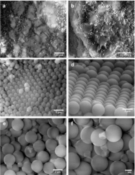
FIGURE 3.3: ESEM MICROGRAPHS REVEAL LARGE CRYSTALLINE DOMAINS OF PS PARTICLES ... 45
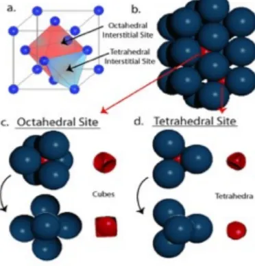
FIGURE 3.4: TWO DIFFERENT TYPES OF INTERSTITIAL SITES (TETRHDRAL AND OCTAHEDRAL SITES) IN A CLOSED-PACKED FCC CRYSTAL ... 47
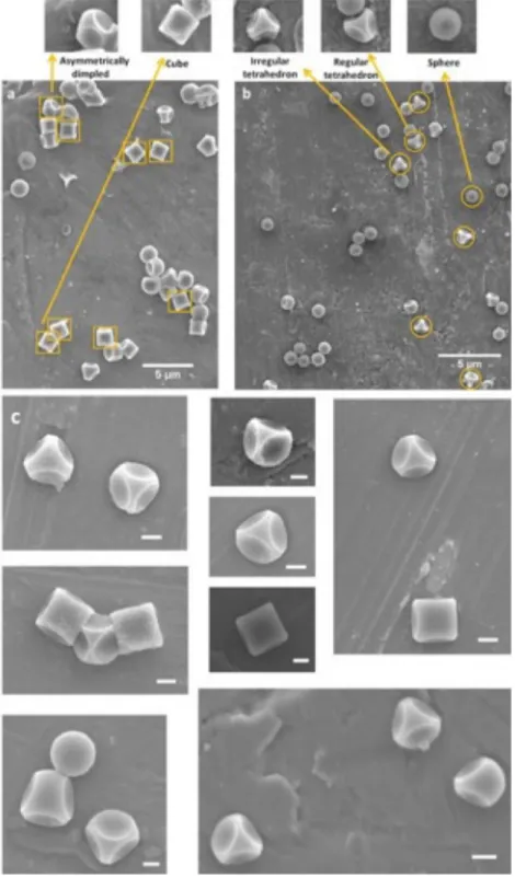
FIGURE 3.5: ELECTRON MICROGRAPHS OF DIMPLED PARTICLES REVEAL A MIXTURE OF SHAPES OF TPM PARTICLES. ... 48
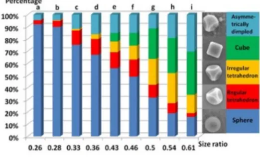
FIGURE 3.6: THE PERCENTAGE YIELDS (BY NUMBER) OF DIFFERENT SHAPED TPM PARTICLES DEPEND ON THE SIZE RATIO (RADIUS OF POLYMERIZED, AND THUS SHRUNKEN, SPHERICAL DROPLETS DIVIDED BY PS PARTICLE RADIUS). ... 49
FIGURE 3.7: ANALYSIS OF MINIMUM PARTICLE EDGE THICKNESSES, l PROVIDES INFORMATION REGARDING LATTICE EXPANSION FOR BOTH
xiv
FIGURE 3.8: AN SEM MICROGRAPH OF A SAMPLE ENRICHED IN TETRAHEDRAL PARTICLES USING GRADIENT FRACTIONATION. ... 56
FIGURE 4.1: PROPOSED APPROACHES FOR MAKING DIAMOND-LIKE
COLLOIDAL CRYSTALS. ... 65
FIGURE 4.2: POLYHEDRAL CRYSTALLITES HAVE A DOUBLE DIAMOND
STRUCTURE. ... 68
FIGURE 4.3: DOUBLE DIAMOND CRYSTALLITES DISPLAY DIAMOND (211) STRUCTURE. ... 69
FIGURE 4.4: DOUBLE DIAMOND CRYSTALS ARE STABLE IN SIMULATION. ... 72
FIGURE 4.5: CRYSTALLITES SURFACES DISPLAY LATTICE RECONSTRUCTION. ... 76
FIGURE 5.1: DNA INTERACTION STRENGTH IS MODULATED BY THE PRESENCE OF MAJOR AND MINOR STRANDS. ... 90
FIGURE 5.2: BINARY DNA COLLOIDS FORM A PANOPLY OF DISTINCT CRYSTAL STRUCTURES. ... 92
FIGURE 5.3: FOR MANY EXPERIMENTAL CONDITIONS (TOP ROW), SEVERAL DIFFERENT STRUCTURES (BOTTOM ROW) WILL APPEAR IN APPARENT
COEXISTENCE. ... 94
FIGURE 5.4: THE CRYSTAL STRUCTURES RESULTING FROM THE ‘TWO-STEP’ DIRECTED SELF-ASSEMBLY OF BINARY DNA-COLLOIDS CAN BE ARRANGED INTO A ‘FAMILY TREE’. ... 95
FIGURE 6.1: THE FIGURE IS A WHITE-LIGHT IMAGE OBSERVED UNDER OPTICAL MICROSCOPE WHICH SHOWS THE CRYSTALLINITY OF CSCL
STRUCTURES OUT OF THREE-COMPONENT CO-CRYSTALLIZATION. ... 112
FIGURE 6.3: DENSITY GRADIENT FRACTIONATION WAS USED TO PURIFY CLUSTER MIXTURE SUSPENSIONS. ... 113
xv
AND D ARE THE RED CHANNELS (SHOWING A PARTICLES). . ERROR! BOOKMARK NOT DEFINED.
FIGURE 6.4: THE FIGURE SHOWS THE FRACTIONATION OF A 245/445
OCTAHEDRAL CLUSTER SUSPENSIONS. ... 114
FIGURE 6.5: THE FIGURE SHOWS THE FRACTIONATION OF A 378/378 CUBIC CLUSTER SUSPENSIONS. ... 115
1
Chapter 1 Introduction
1.1 Field Landscape
Meta-materials are considered as future materials for many novel applications. Its unique
structures which are not made from atoms or molecules but from assemblies of building
blocks endowed it with superior photonic, phononic, thermal, and mechanical properties.
The mass production of such meta-materials will not only lower the cost of realizing
functional materials, but also will be beneficial for the development and progress of the
entire human beings.
Colloidal crystal is a typical type of meta-materials which is built from colloidal single
particles or colloidal clusters as building blocks. Hence, the acquisition of diverse types
of colloidal crystals as well as colloidal clusters and a further mass production of the
structures they form will drive the technology and knowledge into a more mature stage of
making meta-materials.
Our group, with Dr. John Crocker as the PI, has long been a pioneering group in the
colloidal science field across the world. Some of our achievements in this particular
colloidal crystal segment are we succeeded in making the colloidal crystal as the first
group in the world[1-3] and well understood the diffusionless transformation drove by the
structures[4, 5]. We also have a deep understanding in making colloidal clusters of
2
But we are far more ambitious than this. In order to make more types of colloidal
crystals and clusters, which will further expand the library of colloidal crystals, we still
face several challenges. Therefore, the open questions for the group and specifically for
me to answer in this thesis are, first, what is the system for colloidal crystals and clusters
to grow? Second, how to fast and effectively drive this grow in the system? Third, how to
make more efficient building blocks and what are the shapes of the final structures? And
forth, how to make building blocks sturdier and less fragile?
We’ll give a brief answer of the questions in the Introduction part, and will elaborate
through all the chapters below.
1.2 Introducing Self-assembly Systems
Self-assembly is defined as the spontaneous and reversible organization of small building
blocks into ordered structures by non-covalent interactions. Self-assembly systems are
everywhere. The most famous example of self-assembly system is the occurrence of the
life on Earth. It is confirmed that it happens because the sun generates a strong temperate
gradient in its environment so that the construction of cells and living organisms were
built through biologic macromolecular assemblies. That also indicates self-assembly only
happens when certain prerequisites are satisfied.
Self-assembly has been an important research focus and is still considered as one of
the most efficient methods for structure generation and growth due to its
“self-organized”. Self-assembly technique is widely used in multiple fields, including
3
fields[10]. Fabricating a crystal is also made possible by self-assembly as placing one
particle after one particle is not at all possible. Plus, the structure yielded out of this is at
equilibrium because of the systems reaches its lowest free energy. Many experimental
and theoretical work have been done to demonstrate the idea that multiple self-assembly
systems could be driven feasibly and efficiently with nanoparticles and colloids.
Our lab had a very nice review in 2017 categorizing assembly systems into four
groups[11], which are spherical particles, non-specific directional polyhedral particles,
specific homo directional polyhedral particles, and specific Janus directional particles.
First of all, a self-assembly system could be driven by spherical particles.
Self-assembly using gold nanoparticles were first introduced by Mirkin[12, 13] and
Alivisatos[14]. Later on, multiple types of crystals have been formed from DNA-coated
nanoparticles[15-17] and colloids[2, 5, 18-21]. Our lab is honorably to be the first group
to drive colloidal crystallization through self-assembly[1-3] and spearheaded in
understanding the system driving by spherical particles.
Second, polyhedral nanoparticles[22] and colloids [23, 24]with anisotropic
interactions could also be used in directed self-assembly. Glotzer group studied the
structure change of the assembled crystal with the variation of amount of faceting of the
building polyhedral blocks[25, 26]. Experimentally, Pine and Sacana’s lock and key
colloids[27-29] represent the mainstream of realizing such system. Our lab simplified the
method by innovating a crystal templating way and succeeded in making highly uniform
4
a nice review by Pradeep talking about different methods making such building blocks
[31].
In the third group, the interactions among building blocks are designed with certain
directions so that more complex structures could be assembled following these designed
interactions. Specifically, Pine’s group designed clusters with certain symmetries using
DNA patchy amidinated polystyrene microspheres[32] and later made interesting AB2
structures using colloidal clusters[33]. A DNA-origami method of building a polyhedral
cage was designed by Gang group[34] and was recently reported to form ordered
assemblies using nanoparticles.[35, 36]. Our group designed a fast and smart crystal
templating system to yield DNA coated clusters using sedimentation crystallization[6, 7],
and the potential assembled system has been simulated by Sinno group[37].
The last group building blocks are designed to be coated with different interactive
segments on the same particle species which made them look like patchy in different
regions. Though the system designed has been obtained[36], the further assembly with
the building blocks has not been explored. There have been studies on the gelling
properties of the DNA assemblies by Sciortino and Bellini group[38, 39], however the
followed up simulation work done by Sciotino [40]showed that the system never
crystallizes.
All in all, the self-assembly system is perfect for colloids and clusters to grow into
5
1.3 DNA Hybridizations and Binding Interactions
After understanding the self-assembly systems and its advantages over other systems, we
tried to find a fast and effective way the drive the growth in the system. Our lab used
synthetic single strands DNA as a means to drive colloids interactions per our tradition
and is updating the technology with a novel three strand system to apply for more
versatile purposes.
Deoxyribonucleic Acid (DNA), which is a molecule composed of two chains that coil
around each other to form a double helix structure, carries human being’s genetic code to
replicate and grow. Not like genetic DNAs, synthetic DNAs are less used in the function
of coding for proteins but more in driving directed interactions due to its “sticky
ends”[41]. DNA strands are comprised of combinations of four bases that interact with
each other thermodynamically via Watson-Crick base-pairings[42]. To be more specific,
adenine binds with thymine (A-T), and guanine binds to cytosine (G-C). The two types of
base pairs form different numbers of hydrogen bonds, A-T forms two hydrogen bonds,
and G-C forms three hydrogen bonds. In this case, DNA with high GC-content is more
stable than DNA with low GC-content.
The design of the strands determines the Gibbs free energy of the interaction which
could be calculated by nearest neighbor model[43, 44]. The interactions of the
complementary “sticky ends” would be dis-associated when the system temperature goes
to melting temperature (Tm), at which half of the interactions are “melted”[45].
A DNA designed method developed by Nadrian Seeman is used in the generation of
6
bases (table 1.1). The design starts by listing all possible 64 three-base combinations,
known as “codons” in a table. First, a codon is arbitrarily chosen from the list (like TCA
in this case), both TCA and its reverse-complement (TGA) are crossed of the list with the
red line. Second, the sequence is growing by a single arbitrary base (A) and the next
codon’s first two bases are the last two bases of the first codon, now the second codon
becomes CAA. Therefore, both CAA and its complementary codon TTG are crossed by
green line. Similarly, the strand grows again and AAC and GTT are crossed off by blue
lines. The strand could grow to either it reaches its desired length or all the available
codons are run out. The sequence grown in this way are avoiding hairpins, self-dimers,
and other unwanted interactions to the best effort which could be verified using
IDTDNA.com's OligoAnalyzer tool. Besides, The AAA, TTT, CCC, and GGG sequences
should be avoided if possible.
Other than hybridizing through base pairs of two complementary strands, a more
flexible and controllable system was introduced by our group that we introduced a DNA
linker as the third strand to the system so that the length of the sticky ends as well as the
interaction strength could be controlled and adjustable. In this case, other than a
self-complementary hybridization between strand 1 and strand 2, these two strands are both
binding with a linker strand which length and composition could be manipulated easily.
7
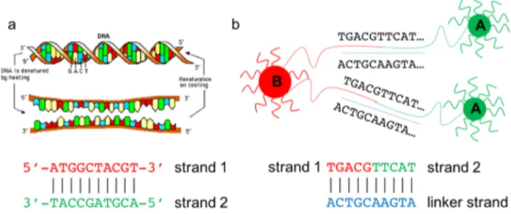
Figure 1.1: DNA strands hybridizations of two-strand system and three-strand system with linker.
Figure 1.1.a showed the two strands complementary hybridization system in which A binds to T, and C binds to G.
Figure 1.1.b on right showed the introduction of a linker strand, which consists of a three-strand system. In this
way, the length of the interacting base pairs and the interaction strengths could be controlled by linker design. Table 1.1: Method for designing DNA sequences developed by Nadrian Seeman.
DNA sequences designed using this method are avoiding hairpins, self-dimers, and other unwanted interactions to
the best effort as the strands will induce unwanted interactions are the complementary codons which are crossed off
8
DNA’s versatile hybridization and interaction properties as well as feasible strands
design made it a wonderful tool in generating particle-particle interactions and further
driving the growth of a self-assembly system into complex ordered colloidal structures.
1.4 DNA-coated Building Blocks and Self-assembling
After understanding self-assembly systems and applying DNA interactions to drive the
growth of the systems, our next challenge is to make building blocks with DNA
interactions so that the assembly will be smoothly driven. Out of four types of
self-assembly systems and their corresponding building blocks, our group is interested in
obtaining effective DNA-coated spherical colloids and clusters as building blocks,
respectively, and the structures they will assemble into. We’ll also take a glance at
non-specific directional polyhedral building blocks as a supplement.
Multiple research groups have reported on spherical colloidal particles and their
assembled structures. Mirkin[17, 46], Dijkstra[47], Gang[48], and Tkachenko[49]
discussed about the phase diagram of the crystals with the change of composition and
size ratio of the building blocks. Pine group made four types of crystal structures with
complementary DNA strands grafted on colloids[18, 50]. However, more types of the
colloidal crystals are still waiting to be discovered and the transformation phenomenon of
the floppy structures are still yet to be explored.
Our group is prestigious in the field as we succeeded in making the colloidal crystal as
9
drove by the structures[4, 5]. Recently we discovered ten types of colloidal crystals using
a slowly quenching method, in which six of them have never been discussed before. We
also found a very fascinating double diamond structure along the way[20] which shows a
very promising optical properties. Moreover, we observed the similar diffusionless
transformations within these ten crystals from a mother phase into multiple child phases
with the conclusion that ten structures are basically from two sources. This indicates that
diffusionless transformation is a nature for floppy colloidal crystals. (with more details to
be discussed in CHAPTER 5)
Colloidal clusters also have been obtained and used as building blocks to drive
self-assembly. Chaikin and Bausch made disordered clusters using polygamous particles and
by adding DNA linker [51, 52]. Capasso reported the assembly of DNA-functionalized
nanoparticles into hetero-pentamer clusters [53], and Kotov mixed different dimers
together and made chiral nanoparticle tetrahedral clusters[54].
Our group invented a fast and smart crystal templating system to yield DNA coated
clusters using sedimentation crystallization in a two component system[6, 7] by ligating
the impurity and host particles to stabilize the clusters. The cluster structures we got out
of the crystals are tetrahedral, octahedral, cuboctahedral, triangular orthobicupula, and
icosahedral clusters. Sinno group studied and proved the possibility of future
self-assembly by some of the clusters[37]. Recently, our group innovated a new method of
making cubic clusters using a three component co-crystallization system, which is more
10
Additionally, polyhedral non-spherical particles could be obtained as building blocks
for future DNA-induced self-assembly as well. The review written by Pradeep discussed
multiple ways of preparing such polyhedral non-spherical particles[31]. Moreover, Pine’s
lab made polyhedral lock-and-key particles with different polymeric materials [27, 55].
Our lab made tetrahedral and cubic polyhedral trimethoxysilyl propyl methacrylate
(TPM) particles without a DNA input, while we see a promising vista that the
DNA-induced self-assembly could also be applied in the system[30].
Another important thing we need to discuss is how to coat DNA strands onto the
building blocks with high efficiency and controllable concentration, yet remain an
affordable cost. Pine wrote a nice review about different methods of coating DNA onto
the colloidal particles. The methods including swelling/de-swelling method,
Azide-dibenzocyclooctyne (N3-DBCO) click chemistry (also known as SPAAC chemistry), and
other three methods were discussed [56].
Our group invented swelling/de-swelling method more than a decade ago using F-108
tri-block polymers [57] and obtained multiple types of colloidal crystals with very high
crystallinity. We recently updated swelling/de-swelling method and combined with
Pine’s SPACC chemistry [50, 58] to get a higher yield with an even lower cost and
controllable DNA concentration. Our group also studied into the variation of salt
concentration during the SPACC chemistry which will influence the homogeneity of the
system. The DNA homogeneity increase on the colloids were believed to later favor in
11
Both methods were proved to make crystals with high crystallinity which indicates a
high DNA concentration on the building blocks. Moreover, the two methods were both
applicable on carboxylate and sulfate PS particles, which give us space in designing
systems with different properties.
1.5 Ligation for More Rigid Building Blocks
So far, the only question we need to answer is unlike spherical building blocks, clusters
are more floppy and unstable. However, the stability of the building blocks must be
guaranteed so that they could survive from the microscopy measurement, purity
enrichment process, and the self-assembly experiments afterwards. Therefore, whether
we could make them sturdier or not is decisive for the future possibility of
self-assembling these clusters.
To stabilize the structures, ligation with T4 Ligase were found effective and fast to
stabilize DNA bridges. In other groups’ work, DNA gold nanoparticle dimer and
complexes were stabilized with T4 ligase[60-62].
Ligation is the joining of two nucleic acid fragments through the action of an enzyme.
The mechanism of DNA ligase is to form two covalent phosphodiester bonds between 3'
hydroxyl ends of one nucleotide with the 5' phosphate end of another. E. coli and T4 are
the most common ligase used. Also, ligase only functions on the phosphate group of the
5’ side, which requires a phosphorylation modification on 5’ side during DNA design.
12
A lot of factors effect ligation efficiency, including the overhang should be more than
4 bases and the control of a low salt concentration in the system[63]. Our grouped used
ligation technique in the work of double diamond preparation [7, 20]. (See CHAPTER 4
for details). We also studied and optimized ligation condition and process that ultimately
noticeably increased the sturdiness of the cluster building blocks. (See CHAPTER 6 for
details)
1.6 Thesis Overview
After answering all of the four questions in a short form, we are going to elaborate the
long answers in this thesis.
Figure 1.2: Ligation process is the joining of two nucleic acid fragments through the action of an enzyme.
Ligation needs DNA ligase, a specific type of enzyme, to happen. The mechanism of DNA ligase is to form two
covalent phosphodiester bonds between 3' hydroxyl ends of one nucleotide with the 5' phosphate end of another.
13
In Chapter Two, we will explain the DNA design, experimental system design,
crystallization methods (including in-situ crystallization and cooler crystallization),
optimization operations (ligation, and fractionation), and imaging tools (optical
microscope, confocal microscopy, and SEM) used in this thesis. Through which we’ll
give an overall description about the system set up and the logic about why we
implement. General protocols regarding above operations will be discussed with more
detailed description on experimental methods written in the materials and method section
in each following chapter. In Chapter Three, we’ll talk about a way to make highly
uniform dimpled polyhedral colloids from colloidal crystal templating. Tetrahedral and
cubic TPM particles were obtained through this crystal templating method and the purity
was enriched by density gradient fractionation. In Chapter Four, we’ll talk about a double
diamond colloidal crystal we discovered recently and the formation and transformation
mechanism behind that. The double diamond structure was produced by mixing two
component colloids out of cooler crystallization with different interaction matrix and size
ratios, and the double diamond structure was considered as a child phase of NaCl
structure. In Chapter Five, we generalize the crystallization method to make ten types of
DNA colloidal crystals in total in which five of them have never been discovered before.
The interaction matrix, size ratios, and stoichiometry were very well studied so that we
could predict the incidence of a certain type structure. Moreover, transformation
mechanisms from mother phase to child phases were very well studied and understood. In
Chapter Six, we use three components co-crystallization system to generate cubic and
octahedral clusters for future self-assembly. The three species particles were mixed,
14
gradient fractionation was used to increase the purity of the clusters. In Chapter Seven,
we’ll conclude what we have done and achieved in this thesis, and what’s the meaning to
DNA-directed self-assembly. We’ll finalize the thesis by suggesting some work and ideas
our lab could focus in the future.
Reference:
1. Kim, A.J., et al., Probing interfacial equilibration in microsphere crystals formed by DNA-directed assembly. Nature Materials, 2009. 8(1): p. 52-55.
2. Biancaniello, P.L., A.J. Kim, and J.C. Crocker, Colloidal interactions and self-assembly using DNA hybridization. Physical Review Letters, 2005. 94(5).
3. Kim, A.J., P.L. Biancaniello, and J.C. Crocker, Engineering DNA-mediated colloidal crystallization. Langmuir, 2006. 22(5): p. 1991-2001.
4. Scarlett, R.T., et al., A mechanistic view of binary colloidal superlattice formation using DNA-directed interactions. Soft Matter, 2011. 7(5): p. 1912-1925.
5. Casey, M.T., et al., Driving diffusionless transformations in colloidal crystals using DNA handshaking. Nature Communications, 2012. 3.
6. McGinley, J.T., et al., Assembling colloidal clusters using crystalline templates and reprogrammable DNA interactions. Soft Matter, 2013. 9(38): p. 9119-9128. 7. McGinley, J.T., et al., Crystal-Templated Colloidal Clusters Exhibit Directional DNA
Interactions. Acs Nano, 2015. 9(11): p. 10817-10825.
8. Lehn, J.M., Perspectives in Supramolecular Chemistry - from Molecular
Recognition Towards Molecular Information-Processing and Self-Organization.
Angewandte Chemie-International Edition in English, 1990. 29(11): p. 1304-1319. 9. Douglas, S.M., I. Bachelet, and G.M. Church, A Logic-Gated Nanorobot for
Targeted Transport of Molecular Payloads. Science, 2012. 335(6070): p. 831-834. 10. Bezryadin, A., R.M. Westervelt, and M. Tinkham, Self-assembled chains of
graphitized carbon nanoparticles. Applied Physics Letters, 1999. 74(18): p. 2699-2701.
11. Porter, C.L. and J.C. Crocker, Directed assembly of particles using directional DNA interactions. Current Opinion in Colloid & Interface Science, 2017. 30: p. 34-44. 12. Mirkin, C.A., et al., A DNA-based method for rationally assembling nanoparticles
into macroscopic materials. Nature, 1996. 382(6592): p. 607-609.
13. Mirkin, C.A., et al., DNA-induced assembly of gold nanoparticles: A method for rationally organizing colloidal particles into ordered macroscopic materials.
15
14. Alivisatos, A.P., et al., Organization of 'nanocrystal molecules' using DNA. Nature, 1996. 382(6592): p. 609-611.
15. Nykypanchuk, D., et al., DNA-guided crystallization of colloidal nanoparticles.
Nature, 2008. 451(7178): p. 549-552.
16. Park, S.Y., et al., DNA-programmable nanoparticle crystallization. Nature, 2008. 451(7178): p. 553-556.
17. Macfarlane, R.J., et al., Nanoparticle Superlattice Engineering with DNA. Science, 2011. 334(6053): p. 204-208.
18. Wang, Y., et al., Crystallization of DNA-coated colloids. Nature Communications, 2015. 6.
19. Rogers, W.B. and V.N. Manoharan, Programming colloidal phase transitions with DNA strand displacement. Science, 2015. 347(6222): p. 639-642.
20. Wang, Y.F., et al., Colloidal crystals with diamond symmetry at optical lengthscales. Nature Communications, 2017. 8.
21. Rogers, W.B., W.M. Shih, and V.N. Manoharan, Using DNA to program the self-assembly of colloidal nanoparticles and microparticles. Nature Reviews
Materials, 2016. 1(3).
22. Shevchenko, E.V., et al., Structural diversity in binary nanoparticle superlattices.
Nature, 2006. 439(7072): p. 55-59.
23. Damasceno, P.F., M. Engel, and S.C. Glotzer, Predictive Self-Assembly of Polyhedra into Complex Structures. Science, 2012. 337(6093): p. 453-457. 24. Rossi, L., et al., Cubic crystals from cubic colloids. Soft Matter, 2011. 7(9): p.
4139-4142.
25. van Anders, G., et al., Entropically Patchy Particles: Engineering Valence through Shape Entropy. Acs Nano, 2014. 8(1): p. 931-940.
26. Glotzer, S.C. and M.J. Solomon, Anisotropy of building blocks and their assembly into complex structures. Nature Materials, 2007. 6(8): p. 557-562.
27. Sacanna, S., et al., Lock and key colloids. Nature, 2010. 464(7288): p. 575-578. 28. Rossi, L., et al., Shape-sensitive crystallization in colloidal superball fluids.
Proceedings of the National Academy of Sciences of the United States of America, 2015. 112(17): p. 5286-5290.
29. Sacanna, S., et al., Shaping colloids for self-assembly. Nature Communications, 2013. 4.
30. Wang, Y.F., J.T. McGinley, and J.C. Crocker, Dimpled Polyhedral Colloids Formed by Colloidal Crystal Templating. Langmuir, 2017. 33(12): p. 3080-3087.
31. Lilian C.Hsiao, S.P., Experimental synthesis and characterization of rough particles for colloidal and granular rheology. Current Opinion in Colloid & Interface Science, 2019.
32. Wang, Y.F., et al., Colloids with valence and specific directional bonding. Nature, 2012. 491(7422): p. 51-U61.
16
34. Tian, Y., et al., Prescribed nanoparticle cluster architectures and low-dimensional arrays built using octahedral DNA origami frames. Nature Nanotechnology, 2015. 10(7): p. 637-+.
35. Tian, Y., et al., Lattice engineering through nanoparticle-DNA frameworks.
Nature Materials, 2016. 15(6): p. 654-+.
36. Liu, W.Y., et al., Diamond family of nanoparticle superlattices. Science, 2016. 351(6273): p. 582-586.
37. Zanjani, M.B., et al., Colloidal Cluster Assembly into Ordered Superstructures via Engineered Directional Binding. Acs Nano, 2016. 10(12): p. 11280-11289. 38. Fernandez-Castanon, J., et al., Small-angle neutron scattering and molecular
dynamics structural study of gelling DNA nanostars. Journal of Chemical Physics, 2016. 145(8).
39. Biffi, S., et al., Phase behavior and critical activated dynamics of limited-valence DNA nanostars. Proceedings of the National Academy of Sciences of the United States of America, 2013. 110(39): p. 15633-15637.
40. Rovigatti, L., et al., Gels of DNA Nanostars Never Crystallize. Acs Nano, 2014. 8(4): p. 3567-3574.
41. Seeman, N.C., Nucleic-Acid Junctions and Lattices. Journal of Theoretical Biology, 1982. 99(2): p. 237-247.
42. SantaLucia, J. and D. Hicks, The thermodynamics of DNA structural motifs. Annual Review of Biophysics and Biomolecular Structure, 2004. 33: p. 415-440.
43. SantaLucia, J., A unified view of polymer, dumbbell, and oligonucleotide DNA nearest-neighbor thermodynamics. Proceedings of the National Academy of Sciences of the United States of America, 1998. 95(4): p. 1460-1465.
44. SantaLucia, J., H.T. Allawi, and A. Seneviratne, Improved nearest-neighbor parameters for predicting DNA duplex stability. Biochemistry, 1996. 35(11): p. 3555-3562.
45. Rogers, W.B. and J.C. Crocker, Direct measurements of DNA-mediated colloidal interactions and their quantitative modeling. Proceedings of the National Academy of Sciences of the United States of America, 2011. 108(38): p. 15687-15692.
46. Li, T.I.N.G., et al., Modeling the Crystallization of Spherical Nucleic Acid
Nanoparticle Conjugates with Molecular Dynamics Simulations. Nano Letters, 2012. 12(5): p. 2509-2514.
47. Filion, L. and M. Dijkstra, Prediction of binary hard-sphere crystal structures.
Physical Review E, 2009. 79(4).
17
49. Tkachenko, A.V., Generic phase diagram of binary superlattices. Proceedings of the National Academy of Sciences of the United States of America, 2016. 113(37): p. 10269-10274.
50. Wang, Y.F., et al., Synthetic Strategies Toward DNA-Coated Colloids that Crystallize. Journal of the American Chemical Society, 2015. 137(33): p. 10760-10766.
51. Wu, K.T., et al., Polygamous particles. Proceedings of the National Academy of Sciences of the United States of America, 2012. 109(46): p. 18731-18736. 52. Hecht, F.M. and A.R. Bausch, Kinetically guided colloidal structure formation.
Proceedings of the National Academy of Sciences of the United States of America, 2016. 113(31): p. 8577-8582.
53. Fan, J.A., et al., DNA-Enabled Self-Assembly of Plasmonic Nanoclusters. Nano Letters, 2011. 11(11): p. 4859-4864.
54. Yan, W.J., et al., Self-Assembly of Chiral Nanoparticle Pyramids with Strong R/S Optical Activity. Journal of the American Chemical Society, 2012. 134(36): p. 15114-15121.
55. Kim, S.H., et al., Synthesis and Assembly of Colloidal Particles with Sticky Dimples.
Journal of the American Chemical Society, 2012. 134(39): p. 16115-16118. 56. Moon, J., et al., DNA-Coated Microspheres and Their Colloidal Superstructures.
Macromolecular Research, 2018. 26(12): p. 1085-1094.
57. Kim, A.J., V.N. Manoharan, and J.C. Crocker, Swelling-based method for preparing stable, functionalized polymer colloids. Journal of the American Chemical Society, 2005. 127(6): p. 1592-1593.
58. Oh, J.S., et al., High-Density PEO-b-DNA Brushes on Polymer Particles for Colloidal Superstructures. Chemistry of Materials, 2015. 27(24): p. 8337-8344.
59. Jenkins, I.C., J.C. Crocker, and T. Sinno, Interaction Heterogeneity can Favorably Impact Colloidal Crystal Nucleation. Physical Review Letters, 2017. 119(17). 60. Claridge, S.A., et al., Enzymatic ligation creates discrete multinanoparticle
building blocks for self-assembly. Journal of the American Chemical Society, 2008. 130(29): p. 9598-9605.
61. Kanaras, A.G., et al., Site-specific ligation of DNA-modified gold nanoparticles activated by the restriction enzyme StyI. Small, 2007. 3(1): p. 67-70.
62. Kanaras, A.G., et al., Towards multistep nanostructure synthesis: Programmed enzymatic self-assembly of DNA/gold systems. Angewandte
Chemie-International Edition, 2003. 42(2): p. 191-+.
18
CHAPTER 2 System Design, Methods, and Experimental Overview
2.1 DNA design
In this paper, we used DNA base pair hybridizations to generate interactions between
colloids. We grafted DNA strands on to colloids and colloids bind to each other as DNA
strands hybrid via base pairs. There are four types of bases found in DNA, which are
adenine (A), cytosine (C), guanine (G) and thymine (T). These four bases are attached to
the sugar-phosphate to form the complete nucleotide, as shown for adenosine
monophosphate. Adenine pairs with thymine and guanine pairs with cytosine, forming
A-T and G-C base pairs.
The design of the strands determines the Gibbs free energy of the interaction which
could be calculated by nearest neighbor model[1, 2]. The interactions of the
complementary “sticky ends” would be dis-associated when the system temperature goes
to melting temperature (Tm), at which half of the interactions are “melted”[3].
A DNA designed method developed by Nadrian Seeman is used in the generation of
DNA sequences and works well when the length of sticky ends is from five to twenty
bases. The detailed ways of designing the strands were introduced in CHAPTER 1.3.
After design of the sticky ends, a Poly-T bases spacer is added on the 5’ or 3’ side of the
designed strand and its non-self-complementary component. Usually, a Poly-T bases
spacer consists of 30-50 bases. The entire sequence is then analyzed using
IDTDNA.com's OligoAnalyzer tool, on which hairpins, self-dimers, and other unwanted
19
Other than hybridizing through base pairs of two complementary strands, a more
flexible and controllable system was introduced by our group that we introduced a DNA
linker as the third strand to the system so that the length of the sticky ends as well as the
interaction strength could be controlled and adjustable. In this case, other than a
self-complementary hybridization between strand 1 and strand 2, these two strands are both
binding with a linker strand which length and composition could be manipulated easily.
(See details in Figure 1.1)
The DNA strands designed and used in the thesis are:
1)
L1' (Ligatable, Phosphorylated):
5- /5Phos/TCA ACCTACTCC CAC ATT TTT TTT TTT TTT TTT TTT TTT TTT
TTT TTT TT/3AmMO/ -3
L2 (Complementary via linker to L1 & L1', Non-Phosphorylated):
5- /5AmMC6/TTT TTT TTT TTT TTT TTT TTT TTT TTT TTT TTT TTT TTT
TTT TTT TTT TTA CGC ATC T -3
L12_Linker_5 (5 base interaction region + nick site + 16 base region):
5- TGT GGG AGT AGG TTG AAG ATG -3
20
B1-nonphos: 5’-TCAACCTACTCCCACA TTT TTT TTT TTT TTT TTT TTT TTT
TTT-3’-DBCO
B1-phos: 5’-TCAACCTACTCTTTTT TTT TTT TTT TTT TTT TTT TTT TTT TTT
-3’-DBCO
B2: DBCO-5’-TTT TTT TTT TTT TTT TTT TTT TTT TTT TTT TTT TTT TTT
ACGCATCT-3’
B12 5 base Linker: 5’-GAGTAGGTTGAAGATG-3’
Kick-off: 5’-TGTGGGAGTAG-3’
Other designs that succeeded but with no data shown:
3)
NY1: (non phospho)
DBCO-5’-TTT TTT TTT TTT TTT TTT TTT TTT TTT TTT TTT TTA CAC CCT
CCA-3’
NY2ph: (Ligatable, phosphorylated)
5’-phospho-ACT TCA TTT TTT TTT TTT TTT TTT TTT TTT TTT TTT
TTT-3’-DBCO
NY2: (non phospho kickable)
21 NY linker:
5’-GAA GTT GGA GGG TGT AA-3’
NY Kick: (kick non phospho)
5’-AGG CAG ATG A-3’
2.2 Interaction Matrix and Size Ratio
We use DNA interactions to drive interactions between colloids. However, the
composition of the DNA strands on the colloids determines the interactions of the system,
and we call this interaction matrix.
For a two species particles system with ‘A’ and ‘B’, our four adjustable parameters are
two ‘like’ attraction strengths, UAA and UBB, one ‘unlike’ one, UAB= UBA, (defined as
positive-valued parameters) and the spheres’ diameter ratio. The interactions are realized
and modulated by grafting various amounts of complementary DNA strands to the two
particle species’ surfaces [4, 5]. We assume the green strands on A particles are
complementary with red strands on B particles, and the interaction matrix could be varied
as the composition of strands change. Also we define an alpha value between 0 and 1,
1>𝛂>0, if there is only one strand on the particles, we define this as 𝛂=0, if there is a
mixture of two complementary strands on the particles, 𝛂>0 and the value is decided by
the percentage of one strand over sum of both strands. Say if there are two green strands
22
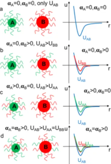
Figure 2.1 showed a detailed demonstration of different interaction matrix and the
corresponding alpha values. In figure 2.1.a, only green strands are on A particles and red
strands are on B particles. As green strands bind to red strands, in the system there are
only interactions between A and B particles. Therefore, only UAB exists in the system and
𝛂A=𝛂B=0. For figure 2.1.b, as there is a mixture of red and green strands on B particles,
𝛂B>0, therefore red particles not only bind to green particles, but also bind to red particles
themselves as well. In this case, 𝛂A=0, 𝛂B>0, and UAB>UBB. In figure 2.1.c, similarly,
green particles bind to both red and green particles. So that in the system 𝛂A>0, 𝛂B=0,
and UAB>UAA. 2.1.d showed when red and green particles both have a mixture of strands
on them, so that in system, 𝛂A=𝛂B>0, UAB>UAA=UBB.
Other than interaction matrix, we also change size ratios for colloids mixings. Size
ratio is defined as the diameter ratio of two species colloids. Our crystallization
experiments are valid starting the size ratio as small as 0.43, and as big as 1.
In this thesis, CHAPTER 4 and CHAPTER 5 will have a more detailed discussion on
23
2.3 Synthesizing DNA-coated colloids
As described in the Introduction Chapter, our group invented swelling/de-swelling
method more than a decade ago using F-108 tri-block polymers [5] and obtained multiple
types of colloidal crystals with very high crystallinity. We recently updated
swelling/de-swelling method and combined with Pine’s SPACC chemistry [6, 7] to get a higher yield Figure 2.1: Figure 2.1 showed different interaction matrix and the corresponding alpha values.
In figure 2.1.a, only green strands are on A particles and red strands on B particles. In the system there are only
interactions between A and B particles. Therefore, only UAB exists in the system and 𝛂A=𝛂B=0. For figure 2.1.b, as
there is a mixture of red and green strands on B particles, therefore red particles not only bind to green particles,
but also bind to red particles as well. In this case, 𝛂A=0, 𝛂B>0, and UAB>UBB. In figure 2.1.c, similarly, green
particles bind to both red and green particles. So that in the system 𝛂A>0, 𝛂B=0, UAB>UAA. 2.1.d showed when red
24
with an even lower cost and controllable DNA concentration. Here we will give a brief
description of the two methods, with more details to be discussed in the chapter 4, 5, and
6.
2.3.1 F108 polymer and DNA conjugation
500mg F108, 2ml dichloromethane, and 30µL TEA were added to the glass vial, allowed
to dissolve completely on a heat plate with stir bar mixing, and then 100mg of fresh
4-NPCF were added and dissolved. The glass vial was then wrapped with parafilm, put on
ice and allowed to react for 3-5 hours. Four washing solutions were prepared and frozen
at -20°C in clean 50ml centrifuge tubes, the first contained 14.6ml ethanol and 0.4ml
HCl, the other three were 14.9ml ethanol and 0.1ml HCl. After the reaction, the first
washing solution was added, F108 precipitated, the tube was then shaken and chilled at
-20°C for 30 minutes to complete precipitation, centrifuged at 4000rpm for 6 minutes at
2°C to form a pellet, and the supernatant discarded. This process was repeated three more
times with the remaining washing solutions. After the final wash, the supernatant was
discarded and the pellet was warmed by hand until fully redispersed. Activated F108 was
split into multiple tubes and dried overnight under vacuum. These samples remain
useable for >2 months when stored at -20°C.
15µL of DNA solution (2000µM, in this paper, DNA strand we used were L1’ and L2,
see above) was mixed with 1µL 1M pH=10 carbonate buffer. 15mg activated Pluronic
F108 (dried from -20°C storage) was dissolved in 1ml 10mM pH=4 citric acid buffer,
gently vortexed to full dissolution, settled by micro-centrifuge and used immediately.
25
volume 20µL), gently vortexed for 30 minutes (after which a yellow reaction product was
evident) and settled by micro-centrifuge, then incubated overnight at room temperature.
The F108-grafted DNA solution can be stored up to 2 months at 4°C.
Particle preparation and DNA grafting.
First, 80µL polystyrene (PS) microspheres/beads were washed three times by dilution
with 920µL bio-water, centrifugation at 8000rpm for 35 minutes, and discarding of
supernatant. Next, 20µL of F108-grafted DNA solution (either L1’ DNA, L2 DNA, or
any combination of L1’ and L2 with 20µL total volume), 35µL washed 10% solid
fraction colloids and 340µL 1xTE solution were combined. To swell the particles, 4µL
toluene was added in to the tube, followed by 0.1µL-1µL green or red BODIPY dye,
depending on the particle species, A or B. The tube was then tightly sealed and wrapped
with parafilm and slowly rotated overnight (not vortexed). To evaporate the toluene, the
sample was settled by micro-centrifuge, opened, and put into the pre-heated oven (80°C )
for 20 to 40 minutes, with periodic mixing. To remove toluene and unracted DNA, the
particles were washed 4-6 times in 1xTE solution to a total sample volume of 1 mL, as
before. After the last wash, the supernatant was removed and 350µL 1-xTE was added to
regain the solid fraction as 10%. DNA-grafted particles can be stored at 4°C for at least 2
months.
2.3.2 Particle Preparation (SPAAC-based) Polymer Activation
0.1g (6900g/mol, 14.5µmol) SEO polymer was dissolved into 2ml DCM in a 2 ml
26
the vial. The reaction was carried out for 2 hours at 0 degree and subsequently 22 hours at
room temperature with a stir bar stirred. After the reaction, the SEO polymer solution was
added into the pre-frozen plastic centrifuge tube containing 2ml methanol solution with
3% HCl for salt removal. 45ml diethyl ether was added subsequently and precipitates
were observed after slight shakes. The tube was put in freezer for 10 minutes to generate
more precipitates. Later on, the suspension was centrifuged in a swing-bucket rotor at
2500rpm for 3 minutes. After the centrifugation, salt removal washing process with 3%
HCl in 2 ml methanol solution and 45 ml diethyl ether were repeated for 3 times in total.
For the last wash, pure methanol solution was added instead of HCl in methanol solution.
The pellet was fully dried under vacuum for around 1 hour.
Polymer Azidation
Dried polymer pellet was fully dissolved in 2 ml N-N DMF solution with 0.01g
NaN3(0.15 mmol). More DMF was added if the pellet was not fully dissolved. The
reaction was carried out for a day at 65-70 °C in glycerol bath with a stir bar. After
glycerol bath, 10 ml methanol and 40 ml diethyl ether were added to the polymer
solution. Precipitates were observed with slight shakes. The solution was put in freezer
for 20 minutes to generate more precipitates. Later on, the suspension was centrifuged in
a swing-bucket rotor at 2500 rpm for 3 minutes. After the centrifugation, washing with 10
ml methanol and 40 ml diethyl ether were repeated to remove NaN3 for 3 times in total.
The final pellet was fully dried under vacuum for a day. The pellet could be stored in
freezer as powders or be stored in the fridge as a 1 mmolar solution in DI water. The
27 SPAAC Chemistry
50 µl 1m molar SEN (azidated di-block polymer), 50 µl DNA solution (L1’ or L2) in
bio water (90 µM to 180 µM), and 300 µl 200mM NaCl in 1-xTE solution were
combined followed with 1wt% ( or 1 mg/ml) F127 added into an Eppendorf tube. The
tube was shaken on vortex mixer for 2 days at a vortex level 5-8 to process SPAAC click
chemistry.
Physical DNA Grafting
25 µl PS particles were washed in bio-water for 3-4 times, and saved for use. 25 µl
washed PS particles, 50 µl clicked DNA solution, and 200 µl 1-xTE were combined into
an Eppendorf tube. 5 µl was added followed by 0.8 µl green dye or 0.4 µl red dye. The
tube was spin on a spinner for 4 hours. After the spinning, the tube was put in a
pre-heated 75°C oven for 10 minutes for toluene evaporation. The particles were
subsequently centrifuged and washed with 1-xTE in a fixed angle rotor at 8000 rpm for
30 minutes for a total of 4-5 times. The final PS volume fraction was 0.5%.
2.4 Melting Temperature (Tm) Measurement
Melting Temperature (Tm) of the system was analyzed and measured in order to testify
the stability of a certain binding system with different DNA strands. To be more specific,
particles with various interacting strands were mixed under a certain salt concentration
and the aggregates were observed under microscope with a sealed coverslip chamber.
28
objective heater controller. The temperatures of the condenser and objective were kept
the same and increased together from room temperature at the rate of ~1 degree per
minute. When the system reached a point at which half of the aggregates melted, but still
with some dynamic clusters remaining, we marked down the temperature as the Tm of
the system to indicate the binding strength of DNA strands of this condition. The Tm
could be tuned by DNA binding strength and changing the salt concentration. Tm
analysis and measurement could be reproduced with an error bar of +/- 0.5 degrees.
2.5 Crystallizations
We use three ways of crystallizations in this thesis, here we provide a representative
example protocol for each method, more detailed crystallization protocols will be
discussed in the main thesis.
2.5.1 In situ crystallization
To observe and understand crystal formation handy, we crystallized samples on a DIC
microscope (LEICA DMIRB) with a X100 oil immersion objective and condenser, both
of which were temperature controlled (BIOPTECHS). Two particle species were mixed
at a 1:1 number stoichiometry (we adjusted number stoichiometry for other than AB type
crystal) with a total volume of 6 µl-8 µl (specifically, we calculated volume of each
species from their diameters), 1 µl DNA linker (100 µM to 500 µM) were added and well
mixed, 1 µl-3 µl NaCl in 1-xTE solution (1M) were finally added to make the final
volume 10 µl. The sample was well mixed and mounted in a sample chamber formed by
two coverslips separated with a silicone vacuum grease sealant. After mounting on the
29
where particle aggregates broke apart and started to melt. To form crystals, the
temperature was quickly decreased to 0.5-2 °C below Tm and crystals were formed in a
few minutes and growth was completed in about 30 minutes. To obtain larger crystals,
after staying a few minutes of nucleation at the lower temperature, the temperature can be
increased by 0.3–0.6 °C reducing the rate of further nucleation, and slowing the rate of
crystal growth.
2.5.2 Cooler Crystallization
Cooler crystallization is a slowly quenching method, in which temperature goes down as
0.4 degree per hour. Specifically, two particle species were mixed at a 1:1 number
stoichiometry (we adjusted number stoichiometry for other than AB type crystal) with a
total volume of 200 µl (we calculated volume of each species from their diameters) were
mixed in a 0.2 ml PCR tube and was centrifuged at 8000 rpm for 30 minutes. 192-194 µl
supernatant was discarded leaving 6 µl-8 µl of suspension. 1 µl 5-base linker (100-500
µM, see detailed structure above) and 1 µl-3 µl NaCl solution in 1-xTE (1 M) were added
to make total volume 10 µl and volume fraction of particles around 20%. The pellet was
re-dispersed, and further settled by micro-centrifuge. A large insulated cooler was filled
with several liters of tap water heated to above 50°C. The PCR tube was first melted in a
small 50°C bath, mixed again, and was then settled again by micro-centrifuge, wrapped
tightly with parafilm and submerged completely in the larger hot water bath. The cooler
lid was tightly closed and the quenching continued for about 3 days. Once the cooler
temperature was several degrees below the estimated crystal melting temperature, the