University of Pennsylvania
ScholarlyCommons
Publicly Accessible Penn Dissertations
2019
Expanding Peptide Stapling With S-Tetrazine
Matthew Henry Bunner
University of Pennsylvania, mhbunner@gmail.com
Follow this and additional works at:https://repository.upenn.edu/edissertations Part of theOrganic Chemistry Commons
This paper is posted at ScholarlyCommons.https://repository.upenn.edu/edissertations/3374
For more information, please contactrepository@pobox.upenn.edu.
Recommended Citation
Bunner, Matthew Henry, "Expanding Peptide Stapling With S-Tetrazine" (2019).Publicly Accessible Penn Dissertations. 3374.
Expanding Peptide Stapling With S-Tetrazine
Abstract ABSTRACT
EXPANDING PEPTIDE STAPLING WITH s-TETRAZINE Matthew H. Bunner
Professor Amos B. Smith, III
This dissertation presents efforts toward the extension of synthetic methods for the creation of peptide macrocycles via the addition of dichloro-s-tetrazine to peptide sequences containing lysine, serine, threonine, and tyrosine. Chapter one reviews the work and interest of the Smith group in the s-tetrazine chromophore and the history of stapled peptides in the chemical literature. Chapter two describes (A) the development of a synthetic protocol for the creation of peptide macrocycles from peptide sequences containing a single cysteine and a single lysine residue using dichloro-s-tetrazine including discussion on amino acid tolerance and seven successful examples, (B) efforts toward the development of a synthetic protocol for the creation of peptide macrocycles from peptide sequences containing a single cysteine residue and a single serine /
threonine / tyrosine residue, and (C) efforts toward the investigation of the photochemical dissociation of the created cysteine / lysine peptide macrocycles. Chapter three describes our efforts to improve the synthesis of dichloro-s-tetrazine in a large-scale procedure.
Degree Type Dissertation
Degree Name
Doctor of Philosophy (PhD)
Graduate Group Chemistry
First Advisor Amos B. Smith, III
Subject Categories Organic Chemistry
EXPANDING PEPTIDE STAPLING WITH s-TETRAZINE
Matthew H. Bunner
A DISSERTATION
in
Chemistry
Presented to the Faculties of the University of Pennsylvania
In Partial Fulfillment of the Requirements for the
Degree of Doctor of Philosophy
2019
Supervisor of Dissertation
________________________________
Amos B. Smith, III
Rhodes-Thompson Professor of Chemistry
Graduate Group Chairperson
________________________________
David W. Christianson
Roy and Diana Vagelos Professor in Chemistry and Chemical Biology
Dissertation Committee:
Jeffrey D. Winkler, Merriam Professor of Chemistry
David M. Chenoweth, Associate Professor of Chemistry
ii
This thesis is dedicated to my family.
Every new height I’ve reached was thanks to the environment and foundation you
iii
ACKNOWLEDGMENT
S
First and foremost I would like to thank Professor Amos B. Smith, III. Your
support and encouragement over the last 5 years has been incredibly valuable. There were
many times that I felt lost in the woods and wanted to give up and scrap this project, then
you would remind me that “nothing worth doing is easy” and I’d decide to try one more
experiment. Your work ethic and your commitment to your students have displayed
values I will be trying to emulate for the rest of my life.
Next, I owe an enormous thanks to the members of my committee: Professor
Jeffery D. Winkler, Professor David M. Chenoweth, and Professor Feng Gai. Your
helpful suggestions and constructive criticism really helped me shape this project and
showed me options I had overlooked.
To the late Dr. George Furst and Dr. Jun Gu, this project would not have come as
far as it did without your help, both in instructing me as to which NMR experiments
would be most beneficial to my work and in making the necessary instruments available
to me. The trust you placed in me to run the Cryoprobe with no supervision, and to
acquire data for other research groups during off hours, opened a lot of doors when I
went looking for employment.
The past and current members of the Smith Group have all come together to
provide a wonderful environment for science. No one will every say what we do is easy,
but it is much easier with good comrades.
Finally, I would like to thank my parents. Even from 600 miles away, you were
iv
ABSTRACT
EXPANDING PEPTIDE STAPLING WITH s-TETRAZINE
Matthew H. Bunner
Professor Amos B. Smith, III
This dissertation presents efforts toward the extension of synthetic methods for
the creation of peptide macrocycles via the addition of dichloro-s-tetrazine to peptide
sequences containing lysine, serine, threonine, and tyrosine. Chapter one reviews the
work and interest of the Smith group in the s-tetrazine chromophore and the history of
stapled peptides in the chemical literature. Chapter two describes (A) the development of
a synthetic protocol for the creation of peptide macrocycles from peptide sequences
containing a single cysteine and a single lysine residue using dichloro-s-tetrazine
including discussion on amino acid tolerance and seven successful examples, (B) efforts
toward the development of a synthetic protocol for the creation of peptide macrocycles
from peptide sequences containing a single cysteine residue and a single serine /
threonine / tyrosine residue, and (C) efforts toward the investigation of the photochemical
dissociation of the created cysteine / lysine peptide macrocycles. Chapter three describes
v
TABLE OF CONTENTS
ABSTRACT
... iv
LIST OF TABLES
... vii
LIST OF FIGURES
... vii
CHAPTER 1. EVALUATION OF
S
-TETRAZINE IN
BIOPHYSICS: EARLY WORK
... 1
Importance of Peptides as Therapeutics ... 11
Challenges of Peptides as Therapeutics ... 11
Early Peptide Macrocycles ... 12
Synthetic Peptide Macrocycles ... 14
Modern Stapled Peptides ... 16
References – Chapter 1 ... 18
CHAPTER 2. EXPANDING PEPTIDE STAPLING WITH
DICHLORO-
S
-TETRAZINE
... 24
Modification of Cysteine/Lysine Peptides with Dichloro-s-tetrazine ... 25
Macrocyclization of Modified Peptides (3) Possessing a Lysine Side Chain ... 34
Scope and Tolerance of Cysteine/Lysine Stapling ... 38
Cys-Tet-Lys Macrocycles as Photochemical Substrates – Unexpected Difficulties! 49 Summary ... 55
References – Chapter 2 ... 57
vi
References – Chapter 3 ... 66
EXPERIMENTAL
... 69
vii
LIST OF TABLES
Table 1.1. Photochemical dissociation of substituted tetrazines………4
Table 1.2. Evaluation of spacer length on photocleavage of s-tetrazine………6
Table 2.1. Competing residue scope……….39
LIST OF FIGURES
Figure 1.1. Photodissociation of s-tetrazine………..…………..1Figure 1.2. Eliminated 1,4 bonding photodissociation pathway……….1
Figure 1.3. Selective photodissociation of 12C15N-s-tetrazine………2
Figure 1.4. Synthesis of [1-Cys, 6-Cys]-S,S-Tet-oxytocin……….5
Figure 1.5. On-resin synthesis of s-tetrazine trimer peptide macrocycles………..6
Figure 1.6. Liquid-phase peptide coupling of tetrazine macrocycles……….7
Figure 1.7. Fragment union strategy for the construction of α-helical peptides………….8
Figure 1.8. Peptide stapling and unstapling with dichloro-s-tetrazine………9
Figure 1.9. Dye-conjugation by inverse electron demand Diels Alder reaction………...10
Figure 1.10. Gramicidin S……….12
Figure 1.11. Four possible configurations of peptide macrocycles………...……...13
Figure 1.12. Protein secondary structure cartoon of human PCNA……….14
Figure 1.13. Early all-hydrocarbon peptide stapling………15
Figure 1.14. One-component and two-component peptide stapling……….16
Figure 2.1. Modification of cys-ala-lys trimer peptide with dichloro-s-tetrazine……….25
Figure 2.2. Proposed degradation pathway of hydroxy-tetrazine……….27
Figure 2.3. Attempts at two-step, one-pot macrocyclization...……….30
Figure 2.4. Revised plan………...30
viii
Figure 2.6. “Block” and “shell” freezing of a 50 mL reaction mixture in a 100 mL
vessel………..33
Figure 2.7. Mass spectrum of crude isolate of compound 3……….34
Figure 2.8. Investigating macrocyclization conditions……….34
Figure 2.9. HPLC purification of macrocycle 4 synthesis on small scale using sodium phosphate and sodium bicarbonate………35
Figure 2.10. HPLC purification of scaled up synthesis of 4 using sodium phosphate….36 Figure 2.11. Successful conditions for the synthesis of macrocycle 4……….37
Figure 2.12. LC chromatogram data for macrocycle 12a……….39
Figure 2.13. More cys-tet-lys macrocycles………...40
Figure 2.14. Differences in proton exchangeability between 2 and 4………...42
Figure 2.15. Solubility problems with macrocycles……….42
Figure 2.16. Attempts at making cys-ser/thr macrocycles with s-tetrazine………..44
Figure 2.17. Attempts at forming cys-tyr macrocycles with s-tetrazine………...45
Figure 2.18. Synthesis of 36 – reaction mixture at 60 minutes……….45
Figure 2.19. HPLC and LC/MS data from the first purification of 36……….46
Figure 2.20. Second HPLC purification of macrocycle 36 and 1H NMR spectrum of isolated material……….47
Figure 2.21. Third HPLC purification of 36 and LC/MS analysis of isolated material…48 Figure 2.22. Synthesis of small molecule alkyl-tetrazine ethers………...49
Figure 2.23. UV-vis absorption spectra of S,S-tetrazine and S,N-tetrazine peptides……50
Figure 2.24. Invetigation of S,N-tetrazine photochemistry, first attempt……….52
Figure 2.25. Net difference in IR spectrum after 5 hours of irradiation………...53
Figure 2.26. UV-Vis absorption during the photolysis of 4………..53
Figure 2.27. Degradation pathways of cyanamides………..54
Figure 2.28. Attempted trapping strategies………...55
ix
Figure 3.2. Chavez and Hiskey’s synthesis of dichloro-s-tetrazine (1) using Coburn and
coworker’s intermediate (39)……….62
Figure 3.3. Modified synthesis of dichloro-s-tetrazine (1) using trichloroisocyanuric acid (41)……….62
Figure 3.4. Use of sodium nitrite for the synthesis of 3,6-bis(3,5-dimethyl-1H-pyrazol-1-yl)-s-tetrazine (39)……….63
Figure 3.5. Full route for the synthesis of dichloro-s-tetrazine (1)………...64
Figure A.1. 500 MHz 1H-NMR Spectrum of Peptide 2 in d 6-DMSO………109
Figure A.2. 126 MHz 13C-NMR Spectrum of Peptide 2 in d6-DMSO…..………110
Figure A.3. LC/MS Chromatogram of Peptide 2………...………111
Figure A.4. 500 MHz 1H-NMR Spectrum of Peptide 10a in d6-DMSO………112
Figure A.5. 126 MHz 13C-NMR Spectrum of Peptide 10a in d6-DMSO..………113
Figure A.6. 126 MHz DEPT-135 Spectrum Peptide 2 in d6-DMSO………….………114
Figure A.7. HMBC Spectrum of Peptide 10a in d6-DMSO………...………115
Figure A.8. LC/MS Chromatogram of Peptide 10a………...………116
Figure A.9. 500 MHz 1H-NMR Spectrum of Peptide 10b in d6-DMSO……...………117
Figure A.10. 126 MHz 13C-NMR Spectrum of Peptide 10b in d6-DMSO..……..……118
Figure A.11. 126 MHz DEPT-135 Spectrum Peptide 10b in d6-DMSO..……….119
Figure A.12. HMBC Spectrum of Peptide 10b in d6-DMSO………...…….……120
Figure A.13. 500 MHz 1H-NMR Spectrum of Peptide 10c in d6-DMSO……...….…121
Figure A.14. 126 MHz 13C-NMR Spectrum of Peptide 10c in d 6-DMSO..……..….…122
Figure A.15. 126 MHz DEPT-135 Spectrum Peptide 10c in d6-DMSO..……….123
Figure A.16. COSY Spectrum of Peptide 10c in d6-DMSO………....….….……124
Figure A.17. HSQC Spectrum of Peptide 10c in d6-DMSO………...……...……125
Figure A.18. HMBC Spectrum of Peptide 10c in d6-DMSO…..………...………126
Figure A.19. 500 MHz 1H-NMR Spectrum of Peptide 10d in d6-DMSO……....….…127
x
Figure A.21. 126 MHz DEPT-135 Spectrum Peptide 10d in d6-DMSO..……….129
Figure A.22. COSY Spectrum of Peptide 10d in d6-DMSO………....….….……130
Figure A.23. HSQC Spectrum of Peptide 10d in d6-DMSO………...……...……131
Figure A.24. HMBC Spectrum of Peptide 10d in d6-DMSO…..………...…………132
Figure A.25. LC/MS Chromatogram of Peptide 10d………...………..………133
Figure A.26. 500 MHz 1H-NMR Spectrum of Peptide 19 in d6-DMSO………...….…134
Figure A.27. 126 MHz 13C-NMR Spectrum of Peptide 19 in d6-DMSO..…...….…135
Figure A.28. 126 MHz DEPT-135 Spectrum Peptide 19 in d6-DMSO..…………..….136
Figure A.29. HMBC Spectrum of Peptide 19 in d6-DMSO………...…....….…...……137
Figure A.30. 500 MHz 1H-NMR Spectrum of Peptide 32 in d6-DMSO………...….…138
Figure A.31. 126 MHz 13C-NMR Spectrum of Peptide 32 in d6-DMSO..…...….…139
Figure A.32. 500 MHz 1H-NMR Spectrum of Peptide SI-1 in d6-DMSO…...…….…140
Figure A.33. 126 MHz 13C-NMR Spectrum of Peptide SI-1 in d6-DMSO…...……....141
Figure A.34. 126 MHz DEPT-135 Spectrum of Peptide SI-1 in d6-DMSO..…...…142
Figure A.35. COSY Spectrum of Peptide SI-1 in d6-DMSO………....….………143
Figure A.36. HSQC Spectrum of Peptide SI-1 in d6-DMSO………...……..……144
Figure A.37. HMBC Spectrum of Peptide SI-1 in d6-DMSO…..………...…………145
Figure A.38. LC/MS Chromatogram of Peptide SI-1………...…….146
Figure A.39. 500 MHz 1H-NMR Spectrum of Peptide SI-2 in d6-DMSO…...…….…147
Figure A.40. 126 MHz 13C-NMR Spectrum of Peptide SI-2 in d6-DMSO…...……....148
Figure A.41. 126 MHz DEPT-135 Spectrum of Peptide SI-2 in d6-DMSO..…...…149
Figure A.42. COSY Spectrum of Peptide SI-2 in d6-DMSO………....….………150
Figure A.43. HSQC Spectrum of Peptide SI-2 in d6-DMSO………...……..……151
Figure A.44. HMBC Spectrum of Peptide SI-2 in d6-DMSO…..………...…………152
Figure A.45. LC/MS Chromatogram of Peptide SI-2………...…….153
xi
Figure A.47. 126 MHz 13C-NMR Spectrum of Peptide SI-3 in d6-DMSO…...……....155
Figure A.48. 126 MHz DEPT-135 Spectrum of Peptide SI-3 in d6-DMSO..…...…156
Figure A.49. LC/MS Chromatogram of Peptide SI-3………...…….157
Figure A.50. 500 MHz 1H-NMR Spectrum of Peptide SI-4 in d 6-DMSO…...…….…158
Figure A.51. 126 MHz 13C-NMR Spectrum of Peptide SI-4 in d 6-DMSO…...……....159
Figure A.52. 126 MHz DEPT-135 Spectrum of Peptide SI-4 in d6-DMSO..…...…160
Figure A.53. COSY Spectrum of Peptide SI-4 in d6-DMSO………....….………161
Figure A.54. HSQC Spectrum of Peptide SI-4 in d6-DMSO………...……..……162
Figure A.55. HMBC Spectrum of Peptide SI-4 in d6-DMSO…..………...…………163
Figure A.56. LC/MS Chromatogram of Peptide SI-4………...…….164
Figure A.57. 500 MHz 1H-NMR Spectrum of Peptide SI-5 in d6-DMSO…...…….…165
Figure A.58. 126 MHz 13C-NMR Spectrum of Peptide SI-5 in d6-DMSO…...……....166
Figure A.59. LC/MS Chromatogram of Peptide SI-5………...…….167
Figure A.60. 500 MHz 1H-NMR Spectrum of Peptide SI-6 in d6-DMSO…...…….…168
Figure A.61. 126 MHz 13C-NMR Spectrum of Peptide SI-6 in d6-DMSO…...……....169
Figure A.62. 126 MHz DEPT-135 Spectrum of Peptide SI-6 in d6-DMSO..…...…170
Figure A.63. 500 MHz 1H-NMR Spectrum of Peptide SI-7 in d6-DMSO…...…….…171
Figure A.64. 126 MHz 13C-NMR Spectrum of Peptide SI-7 in d 6-DMSO…...……....172
Figure A.65. 126 MHz DEPT-135 Spectrum of Peptide SI-7 in d6-DMSO..…...…173
Figure A.66. HMBC Spectrum of Peptide SI-7 in d6-DMSO…..………...…………174
Figure A.67. LC/MS Chromatogram of Peptide SI-7………...…….175
Figure A.68. 500 MHz 1H-NMR Spectrum of Peptide SI-8 in d6-DMSO…...…….…176
Figure A.69. 126 MHz 13C-NMR Spectrum of Peptide SI-8 in d6-DMSO…...……....177
Figure A.70. 126 MHz DEPT-135 Spectrum of Peptide SI-8 in d6-DMSO..…...…178
Figure A.71. LC/MS Chromatogram of Peptide SI-8………...…….179
xii
Figure A.73. 126 MHz 13C-NMR Spectrum of Peptide SI-9 in d6-DMSO…...……....181
Figure A.74. LC/MS Chromatogram of Peptide SI-9………...…….182
Figure A.75. 500 MHz 1H-NMR Spectrum of Macrocycle 4 in d6-DMSO…...…....…183
Figure A.76. 126 MHz 13C-NMR Spectrum of Macrocycle 4 in d 6-DMSO…..……....184
Figure A.77. 126 MHz DEPT-135 Spectrum of Macrocycle 4 in d6-DMSO…...…185
Figure A.78. HMBC Spectrum of Macrocycle 4 in d6-DMSO…………...…………186
Figure A.79. 500 MHz 1H-NMR Spectrum of Macrocycle 12c in d6-DMSO…...…187
Figure A.80. 126 MHz 13C-NMR Spectrum of Macrocycle 12c in d 6-DMSO……...188
Figure A.81. 126 MHz DEPT-135 Spectrum of Macrocycle 12c in d6-DMSO…....…189
Figure A.82. TOCSY Spectrum of Macrocycle 12c in d6-DMSO………..………..…190
Figure A.83. HSQC Spectrum of Macrocycle 12c in d6-DMSO…………...…….…191
Figure A.84. HMBC Spectrum of Macrocycle 12c in d6-DMSO………...…………192
Figure A.85. 500 MHz 1H-NMR Spectrum of Macrocycle 12d in d6-DMSO…...…193
Figure A.86. 126 MHz 13C-NMR Spectrum of Macrocycle 12d in d6-DMSO……...194
Figure A.87. 126 MHz DEPT-135 Spectrum of Macrocycle 12d in d6-DMSO…....…195
Figure A.88. COSY Spectrum of Macrocycle 12d in d6-DMSO………..…...……..…196
Figure A.89. HSQC Spectrum of Macrocycle 12d in d6-DMSO…………...…….…197
Figure A.90. HMBC Spectrum of Macrocycle 12d in d6-DMSO………...…………198
Figure A.91. 500 MHz 1H-NMR Spectrum of Macrocycle 13 in d6-DMSO…...…199
Figure A.92. 126 MHz 13C-NMR Spectrum of Macrocycle 13 in d6-DMSO……...200
Figure A.93. 126 MHz DEPT-135 Spectrum of Macrocycle 13 in d6-DMSO…...…201
Figure A.94. COSY Spectrum of Macrocycle 13 in d6-DMSO………..…...…………202
Figure A.95. HSQC Spectrum of Macrocycle 13 in d6-DMSO…………...……...…203
Figure A.96. HMBC Spectrum of Macrocycle 13 in d6-DMSO………...………..…204
Figure A.97. 500 MHz 1H-NMR Spectrum of Macrocycle 14 in d6-DMSO…...…205
xiii
Figure A.99. 126 MHz DEPT-135 Spectrum of Macrocycle 14 in d6-DMSO…...…207
Figure A.100. COSY Spectrum of Macrocycle 14 in d6-DMSO………..…....…….…208
Figure A.101. HSQC Spectrum of Macrocycle 14 in d6-DMSO…………...…….…209
Figure A.102. HMBC Spectrum of Macrocycle 14 in d6-DMSO………...……....…210
Figure A.103. 500 MHz 1H-NMR Spectrum of Macrocycle 15 in d 6-DMSO…...…211
Figure A.104. 126 MHz 13C-NMR Spectrum of Macrocycle 15 in d6-DMSO…...212
Figure A.105. 126 MHz DEPT-135 Spectrum of Macrocycle 15 in d6-DMSO…....…213
Figure A.106. COSY Spectrum of Macrocycle 15 in d6-DMSO………..…....…….…214
Figure A.107. HSQC Spectrum of Macrocycle 15 in d6-DMSO…………...…….…215
Figure A.108. HMBC Spectrum of Macrocycle 15 in d6-DMSO………...……....…216
Figure A.109. 500 MHz 1H-NMR Spectrum of Macrocycle 16 in d6-DMSO…...…217
Figure A.110. 126 MHz 13C-NMR Spectrum of Macrocycle 16 in d6-DMSO…...218
Figure A.111. 126 MHz DEPT-135 Spectrum of Macrocycle 16 in d6-DMSO…....…219
Figure A.112. COSY Spectrum of Macrocycle 16 in d6-DMSO………..…....…….…220
Figure A.113. HSQC Spectrum of Macrocycle 16 in d6-DMSO…………...…….…221
Figure A.114. 126 MHz 13C-NMR Spectrum of Compound 42 in D2O…….…...222
Figure A.115. 500 MHz 1H-NMR Spectrum of Compound 43 in CDCl3…...…223
Figure A.116. 126 MHz 13C-NMR Spectrum of Compound 43 in CDCl 3…...224
Figure A.117. 500 MHz 1H-NMR Spectrum of Compound 39 in CDCl3…...…225
Figure A.118. 126 MHz 13C-NMR Spectrum of Compound 39 in CDCl3…...226
Figure A.119. 126 MHz 13C-NMR Spectrum of Compound 38 in d 6-DMSO………..227
1
CHAPTER 1. Evaluation of
s
-Tetrazine in Biophysics: Early
Work
In 1975, King and Hochstrasser reported that, upon excitation at an appropriate
wavelength by a tunable dye laser, s-tetrazine would fragment to give two equivalents of
hydrogen cyanide and one equivalent of nitrogen gas (Figure 1.1).1 This discovery
was followed by a collaborative effort between King, Hochstrasser, and Smith to
investigate the reaction pathway of this photodissociation via the synthesis and
subsequent decomposition of 1,4-s-tetrazine-15N2. Through mass analysis of the products
of the photolysis, King et al. were able to show that it was highly unlikely a
Dewar-benzene type intermediate was involved because the required 1,4-bonding pattern would
yield nitrogen gas molecules registering m/z values of 28 or 30 (Figure 1.2). Instead, the
m/z value observed was 29, corresponding to exclusively 14N15N molecular nitrogen.2
Figure 1.1. Photodissociation of s-tetrazine1
Figure 1.2. Eliminated 1,4 bonding photodissociation pathway2 N N
N N
H
H !"
N N N N
H
H
*
2 eq.N H
+ N2
15N
N 15N
N
N
15N
15N
N
15N N
N 15N
N
15N
15N
15N
N N
15N
2
Dellinger and other members of this collaboration also reported a method for the
exclusive photodecomposition of 12C214N4-tetrazine in the presence of s-tetrazine
molecules containing the natural abundance of 13C and 15N, thereby proving that the
enrichment of a material via photo-ablation was possible (Figure 1.3).3,4 This constituted
laser-induced isotope separation.
3
A wealth of literature appeared over the next few years concerning the reaction
pathways of different s-tetrazine derivatives and how those pathways changed based on
irradiation in vapor phase, mixed crystal, or solution phase experiments.5-10 The
conclusions drawn from these experiments were that the photodissociation pathway of s
-tetrazines is highly dependent on substitution, solvent, photon energy, and light source
intensity.6,7 One trend that held throughout these investigations was that attaching
substituents to the parent ring lowered the yield of the photodissociation.7,8 This trend is
exemplified by 3,6-diphenyl-s-tetrazine,8 in that π-bond delocalization outside of the
tetrazine core permits the phenyl substituents to serve as “heat fins” for the dissipation of
vibrational energy (Table 1.1, Entry 1-3). However, Tucker and coworkers were able to
demonstrate that including sulfur disrupts the vibrational connection to the substituents,
thereby increasing the efficiency of the photochemical process. Two possible
explanations exist as to why the change to sulfur increases the yield of the photochemical
reaction. One is that the sulfur atoms are sufficiently massive to serve as a barrier to
energy transfer via vibration.11 This effect is referred to as “the heavy atom-effect.”12 The
other possible explanation is that the switch to sulfur has opened up new photochemical
pathways that did not exist in the carbon and hydrogen substituted s-tetrazines. There was
some experimental evidence presented for this, but further work was never carried out.11
Other work also highlight the huge difference in photochemical properties between s
-tetrazines substituted with heteroatoms.13-15 Not only could 3,6-bis(benzylthio)-s
-tetrazine undergo photolysis at an acceptable yield under irradiation by 355 nm light, but
the compound would also undergo photolysis upon irradiation with a 410 nm light source
4
biological investigation because peptides and proteins do not generally absorb at this
wavelength. This beneficial effect carried over to an s-tetrazine derivative substituted
with two cysteine residues (Table 1, Entry 5). Upon irradiation, fragmentation of the
tetrazine chromophore occurred within a few hundred picoseconds, making this a
possible molecule for studying folding interactions in proteins.
Table 1.1. Photochemical dissociation of substituted tetrazines.11
Entry R Yield (Light Source)
1 ~90% (532 nm)
2 ~17% (532 nm)
3 ~1% (532 nm)
4 24% (355 nm)
12% (410 nm)
5 11% (410 nm)
To verify the compatibility of the s-tetrazine chromophore with biological
systems and peptide synthesis, Tucker and coworkers developed a method to bridge two
proximal cysteine residues of a growing peptide chain during solid phase peptide
synthesis via dichloro-s-tetrazine (1) and an orthogonal protection strategy using highly
acid-labile monomethoxytrityl (Mmt) in place of the trityl groups standard to Fmoc solid
phase peptide synthesis (Figure 1.4).11 This resulted in the creation of an oxytocin H
Me
S
NH S
O O O N
N N N
R R
R R
N N
N N N
N N N
R R
* picosecond
5
analogue with s-tetrazine bridging the disulfide bond, proving that the tetrazine
chromophore was compatible with both Fmoc solid phase peptide synthesis and with
cyclic peptides.11 Photolysis rate and yield of this cyclic tetrazine construct was similar to
the acyclic versions (Table 1.1, Entry 4-5).11
Figure 1.4. Synthesis of [1-Cys, 6-Cys]-S,S-Tet-oxytocin11
C Y I Q N C P L G
O
HN AcHN
S Mmt
S Mmt O
tBu O
NH Trt
NH
O Trt
Rink Resin
1. TFA / TIPSH / CH2Cl2
(1 : 4 : 95), 2 min, repeat 5 x 2.
ClCH2CH2Cl / iPr2NEt
(9 : 1), 5 h
N N N N
Cl Cl
C Y I Q N C P L G
O
HN AcHN
O tBu
O NH
Trt
NH
O Trt
N N N N
S S
TFA/TIPSH (97.2 : 2.5) 4 hr, HPLC
C Y I Q N C P L G
O
NH2
AcHN
HO O NH2
NH2
O
N N N N
S S
6
This method for the creation of s-tetrazine cyclic peptides via incorporation of
dichloro-s-tetrazine during solid phase synthesis was then further investigated by Abdo
and coworkers.16 To test the amino acid compatibility of the synthetic protocol, Abdo et
al. synthesized a series of peptide trimers featuring two cysteine residues in an i, i+2
arrangement with a variable central amino acid (Figure 1.5).16 A variety of reactive or
sterically bulky amino acids where included at the variable position (R), with all tested
residues except arginine being well tolerated.16 Next, using this method, the effect of
amino acid spacer length (i, i+x) on the photochemical reaction of s-tetrazine was
evaluated (Table 1.2).16 The original i, i+2 configuration, corresponding to a half turn of
Table 1.2.Evaluation of spacer length on photocleavage of s-tetrazine16
Entry Cys Relationship Photochemical Yield
1 (i, i + 2) > 25 %
2 (i, i + 3) > 15 %
3 (i, i + 4) ~ 10 %
Figure 1.5. On-resin synthesis of s-tetrazine trimer peptide macrocycles16
S S
N N
N N
X
R = Ala, Lys X = OH, NHMe Y = H, Fmoc, Ac n = 1 - 3
N H
O R
H N O
N H O
Y
hν (355 nm)
n
X
N H
O R
H N O
N H O
Y
n
S S
N N
Y
O H
N S O
N H R
O H
N S X
N-methylindole
or Wang resin Mmt Mmt
X
1) DCM / TIPSH / TFA 2) dichloro-s-tetrazine (1) DCE, DIPEA
3) TFA / TIPSH / H2O
Y
O H
N O
N H R
O H
N X
X = OH, NHMe Y = H, Fmoc, Ac
N N
N N
S S
7
an α-helix, was found to give the best yield (Table 1.2, Entry 1). Finally, Abdo and
coworkers demonstrated that, while this on-resin macrocyclization method was not
amendable to long peptide sequences, these trimer peptide macrocycles could undergo
further liquid phase peptide couplings, so other peptide fragments could be added to build
up larger peptide structures of interest (Figure 1.6).16
These trimer peptide tetrazine macrocycles were then used as the cores of a
tri-component union strategy to make alanine-rich α-helical peptides with a constrained
photo-cleavable kink built into the center (Figure 1.7).17 By strategically placing 13C-18O
labeled carbonyl pairs at different points within the peptide, it was possible to measure
rotational changes in the peptide backbone using ultrafast 2D-IR spectroscopy.18 Upon
photochemical cleavage of the tetrazine chromophore, the kinked helix at the center of
the peptide was released, and progress of the formation of a single new helical turn could
be followed via the unique IR signal of the enriched carbonyl pair. These measurements
established that the kink does not propagate through the helix upon release. Rather, a S
S N N N N HO
N H O
H N O
NH2
O
1. Boc2O
1,4-dioxane : H2O (1 : 1)
2 h
2. BnNH-Phe-NH2
HBTU, DIPEA, DMF 1 h
S S
N N N N H
N
N H O
H N O
N H O
Boc N
H O Bn
1. 30% TFA / CH2Cl2
30 min
2. HO2C-Lys(Cbz)-Boc
HBTU, DIPEA, DMF 1 h
S S
N N N N H
N
N H O
H N O
N H O N
H O Bn
O H N
Boc
H N
8
simple rotation about the previously restricted backbone to reestablish a full turn of the
helix.18 According to Hochstrasser (private communication to Smith) there is currently no
theory to explain this folding process.
A
BocHN C C CO2H
N N N N S S K A K A F Y
AcHN A CO2H
6 HN NH G A K A K A
H2N CONHMe
4 A A NH 2 13 19 HNCbz Cbz Cbz Cbz
1) HBTU, DIPEA, oxyma, DMF, 2 h 2) TFA / MeCN (1:3), 2 h 3) HBTU, DIPEA, oxyma, DFM, 8 h 4) TFA, 24 h A
2
A
HN C C
N N N N S S O HN O K A K A F Y AcHN A 6 NH2 NH2 A 2 G A K A K A CONHMe 4 A A NH2 2 13 19 NH2 K K A F Y AcHN 6 NH2 NH2 A 2 G A K A K A CONHMe 4 A NH2 2 13 19 NH2 S,S-Tet-AKAA5*A6* O H
N 13C
N H 18O 13C H N 18O N H O S,S-Tet-AKAA11*A12* S,S-Tet-AKAA17*A18* N N N N S S A A A A B
9
With this background, Brown and Smith devised and reported a method to get
away from on-resin synthesis and instead to incorporate the s-tetrazine linker into fully
deprotected peptides in solution. That is, they developed a method for the creation of
stapled peptides, via the incorporation of dichloro-s-tetrazine between the thiol side-
chains of two proximal cysteine residues in unprotected peptides prepared by standard
Fmoc solid-phase peptide synthesis (SPPS).19 Following purification, these stapled
peptides could be subjected to steady-state irradiation under UV-A lamps, cleaving the
tetrazine chromophore and releasing the peptide macrocycle as the dithiocyanate. Unlike
the early introduction of staples like Grubbs and Verdine (vide infra),20,21 this was the
first example of a synthetic organic staple that could be successfully removed with little
perturbation to the parent peptide. These masked thiols could then be freed by exposing
Figure 1.8. Peptide stapling and unstapling with dichloro-s-tetrazine (1)19
O HO
O H N
HS O
N H
OH O H N
SH O
N H
NH2
OH O
H2N Phase Transfer
Reaction 50 mM NaH2PO4
(1) CHCl3, 1 min
N
N N N
Cl Cl
O
O OH
O H N
O N H
OH O H N
O N H NH2
OH H2N
N
N N N
S S
O
O OH
O H N
O N H
OH O H N
O N H NH2
OH H2N
UV-A Lamps MeOH
24 h
S S
N N
N N
O
H2N OH
HS H
O
OH N
S H
H2N
87 %
78 % 97 %
10
the dithiocyanate peptide to a solution of cysteine, removing the nitriles in a fashion
similar to native-chemical ligation (Figure 1.8).22 Brown was also able to perform an
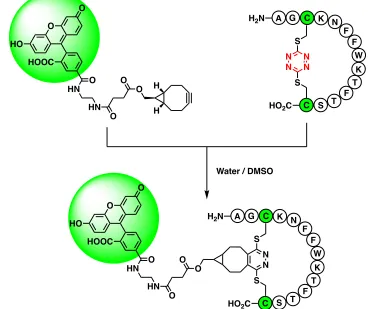
inverse electron-demand Diels-Alder reaction between the tetrazine chromophore and a
fluorescein dye conjugated to bicyclononynol (Figure 1.9).19 This reaction, commonly
called “tetrazine ligation,” is a bioorthogonal click reaction and the most common use of
tetrazine in the literature today.23,24
The question that remained at this point in the Hochstrasser-Smith collaboration
was: Are there other nucleophile side chains that might participate in cysteine cyclic
ligations to expand the utility of this cyclic peptide construct tactic? Before we discuss
our investigative effort to probe this question, we should provide some discussion on (a) N
K A
H2N
F G C
W K T
HO2C
F
F S T C N N N N
S
S O
O
HN O
H
H O
HN O
O
HO HOOC
Water / DMSO
N K A
H2N
F G C
W K T
HO2C
F
F S T C N N S
S O
HN O O HN O
O
HO HOOC
O
11
the history of peptide macrocycles, (b) their importance to the chemical community, (c)
prominent methods for their creation, and (d) where s-tetrazine fits within that history.
Importance of Peptides as Therapeutics
Peptides and proteins have long been thought to hold great therapeutic potential
by the medicinal chemistry community.25 Through the manipulation of their primary
structure and secondary conformations, it might be possible to unlock new peptide tools
to probe biological mysteries or treat illnesses. That is, peptides nicely bridge the
chemical space in between small molecules and large biologics,26 offering the potential
therapeutics to modulate targets previously considered “undruggable.”25 For example,
small molecules are typically considered ill-suited to target the shallow activation site and
large surface area of protein-protein interactions (PPIs).27 Small molecules are also more
likely to produce side effects because of the accumulation of metabolites in sensitive
tissues.28 When compared to larger biologics, peptides are often better suited to
modulating intracellular targets as their smaller size increases their relative ability to
diffuse across cell membranes.28 From a more practical standpoint, peptides also have a
much lower manufacturing cost, and much better room-temperature stability, than large
biological therapeutics.28
Challenges of Peptides as Therapeutics
This is not to say that peptides are free of challenges as therapeutic molecules.
Being made of amino acids, peptides are typically considered poor targets because of
their pharmacokinetic profile.28 Because many proteolytic enzymes recognize common
12
proteases.28 Additionally, the liver and kidneys both rapidly remove circulating peptides
from the blood, often resulting in biological half-lives in the range of minutes.28 The oral
availability of peptides is also rather poor due to low absorption by the impermeable
gastrointestinal epithelium, the activity of peptidases, and the acidic environment of the
stomach.29 Lastly, peptides tend not to be as conformationally rigid as small molecules.26
The time peptides spend sampling different local energy minima across conformational
space is time not spent providing a therapeutic benefit, lowering binding efficiency and
overall efficacy.
Early Peptide Macrocycles
Nature provided inspiration for a solution to these problems with the discovery of
antibacterial peptide gramicidin S (Figure 1.10) by Gause and Brazhnikova in 1944.30
Synge later identified that gramicidin S is a cyclic peptide.31 Used to treat septic gunshot
wounds in World War II, this therapeutically useful cyclic peptide paved the way for the
thousands of cyclic peptides known today.28 Many of these peptide macrocycles have
N HN HN
H N H N
N H
N H O
H2N O
NH NH
N O
O
O O
O H
H
O
O O
NH2
13
therapeutic properties, like oxytocin, cyclosporine a, colistin, and somatostatin.32 The
conformational constraint created by the cyclic configuration of these molecules imparts
many beneficial properties that are not found in their linear counterpart.33 Cyclic peptides
are often more potent, resistant to proteolytic degradation, and are better able to penetrate
cell membranes.32
Depending on the functional groups present within a peptide, macrocyclization
can occur in four ways: head-to-tail, head-to-side chain, side to-tail, or side
chain-to-side chain (Figure 1.11).32 The most common peptide macrocycle found in nature
involves a head-to-tail cyclization, forming a large lactam ring between the N- and
C-terminus.34 The second most common cyclic peptides are side chain-to-side chain
macrocycles formed via disulfide bridge between the thiols of two cysteine residues.33,34
Because both of these structural motifs are common in nature, proteins have evolved to
specifically target these bonds for degradation. So while these early macrocycles did
NH2
COOH R1
R2
N R2 H
R1 HO2C
Side chain-to-tail
C N O
R2
Head-to-tail H
R1
R2 R1 CO2H
Side chain-to-side chain H2N
C
R1
O
R2
NH2
Head-to-side chain
14
exhibit a conformational rigidity that caused them to spend more time in an active form,
they were still susceptible to the same degradative weaknesses discussed above for their
linear counterparts. To overcome this, scientists began developing methods of creating
peptide macrocycles using non-naturally observed bond linkages.
Synthetic Peptide Macrocycles
The rational design of bioactive peptide macrocycles ultimately seeks to replicate
the naturally occurring three-dimensional secondary structures of proteins, such as turns,
α-helices, and β-sheets (Figure 1.12). The largest class of protein secondary structure,
and therefore the most commonly featured structural motif in protein-protein interactions
(PPIs), is the α-helix.35 The most common way of creating stable α-helical peptides is
through the creation of side chain-to-side chain macrocycles, early on referred to as
15
“stapled” peptides, although the term has now come to include all side chain-to-side
chain peptide macrocycles.36 The first major breakthrough in the creation of stapled
peptides using non-native bonds was achieved by Blackwell and Grubbs via ring closing
metathesis using two allylic ether serine residues spaced in an I, i+4 fashion (Figure
1.13).20 This work was later expanded upon by Verdine and coworkers when they
introduced an all hydrocarbon staple, investigating stereochemical configuration, α
-substitution, linker length, and residue spacing for the optimal increase in overall helicity
and eventual biological activity.21 The current state of all-hydrocarbon peptide stapling
has been nicely reviewed by Walensky and Bird.38 The successful implementation of a
synthetic staple resulting in improved biological activity by Verdine and coworkers
ignited a veritable arms race to develop new methods for the creation of peptide
macrocycles.
O O O O
RCM
RCM
Grubbs
20Verdine
21HO Fmoc
H N O
16 Modern Stapled Peptides
From this beginning, the field of peptide stapling has exploded in the last 20 years
as more and more techniques emerge. These methods can be divided into two categories:
one-component and two-component stapling (Figure 1.14).37 One-component stapling is
an intramolecular strategy that uses side-chain functionality present within the peptide to
form the macrocycles. Prominent examples from this category include (a) lactam stapling
Peptide N H
O
Peptide
N N N
Peptide Peptide
S S
Peptide
F F
S
F F S
Peptide
NH HN
O O
Peptide N
N
N N
N N
One-component staples
Two-component staples
17
using native lysine and aspartic acid residues originally pioneered by Rosenblatt39 and
extensively studied by Fairlie,40 (b) the all-hydrocarbon work already discussed by
Grubbs20 and Verdine,21 (c) thiol-ene click photochemistry as pioneered by Anseth,41 and
(d) the copper catalyzed azide-alkyne cycloaddition, brought to solid phase peptide
synthesis by Meldal.42 Two-component stapling on the other hand, comprises an
intermolecular, bicomponent strategy that joins the side-chains of two amino acids with a
second component to form a macrocycle by bridging the two residues.43 The chemical
space of two component staples is incredibly diverse, with great strides made by the
following series of colleagues: Pentelute,44-49 Pentelute and Buchwald,50-51 Woolley,52-54
DeGrado and Greenbaum,55 Caddick and Chudasama,56 Wilson,57 Inouye,58 Baran,59
Derda,60 Rivera,61 and Spring.62-63 These strategies taken together come with unique
benefits and drawbacks. Many involve the use of poisonous metals, which can be hard to
remove to a satisfactory level for eventual biological medical application. Others involve
the use of non-natural amino acids, which increase the cost of production. These stapling
strategies also do not allow for the cleavage of the peptide macrocycle (i.e., unstapling)
18 References – Chapter 1
1. Hochstrasser, R.M.; King, D.S. J. Am. Chem. Soc.1975, 97(16), 4760-4762.
2. King, D.S.; Denny, C.T.; Hochstrasser, R.M.; Smith, A.B., III. J. Am. Chem. Soc.
1977, 99(1), 271-273.
3. Dellinger, B.; King, D.S.; Hochstrasser, R.M.; Smith, A.B., III. J. Am. Chem. Soc.
1977, 99(9), 3197-3198.
4. Dellinger, B.; King, D.S.; Hochstrasser, R.M.; Smith, A.B., III. J. Am. Chem. Soc.
1977, 99(22), 7138-7142.
5. Hochstrasser, R.M.; King, D.S.; Smith, A.B., III. J. Am. Chem. Soc.1977, 99(12),
3923-3933.
6. Paczkowski, M.; Pierce, R.; Smith, A.B., III; Hochstrasser, R.M. Chem. Phys.
Lett.1980, 72(1), 5-9.
7. Haynam, C.A.; Young, L.; Morter, C.; Levy, D.H. J. Chem. Phys. 1984, 81(11),
5216-5217.
8. Windisch, V.L.; Smith, A.B., III; Hochstrasser, R.M. J. Chem. Phys. 1988, 92,
5366-5370.
9. Scheiner, A.C.; Schaefer, H.F., III. J. Chem. Phys.1987, 87(6), 3539-3556.
10.Zhao, X.; Miller, W.B.; Hintsa, E.J.; Lee, Y.T. J. Chem. Phys. 1989, 90(10),
5527-5535.
11.Tucker, M.J.; Courter, J.R.; Chen, J.; Atasoylu, O.; Smith, A.B., III; Hochstrasser,
R.M. Angew. Chem. Int. Ed.2010, 49, 3612-3616.
19
13.Gong, Y.-H.; Miomandre, F.; Méallet-Renault, R.; Badré, S.; Galmiche, L.; Tang,
J.; Audebert, P.; Clavier, G. Eur. J. Org. Chem.2009, 6121-6128.
14.McGrane, S.D.; Bolme, C.A.; Greenfield, M.T.; Chavez, D.E.; Hanson, S.K.;
Scharff, R.J.; J. Phys. Chem. A.2016, 120, 895-902.
15.Allain, C.; Piard, J.; Brosseau, A.; Han, M.; Paquier, J.; Marchandier, T.;
Lequeux, M.; Boissière, C.; Audebert, P. ACS Appl. Mater. Interfaces. 2016, 8,
19843-19846.
16.Abdo, M.; Brown, S.P.; Courter, J.R.; Tucker, M.J.; Hochstrasser, R.M.; Smith,
A.B., III. Org. Lett.2012, 14(13), 3518-3521.
17.Courter, J.R.; Abdo, M.; Brown, S.P.; Tucker, M.J.; Hochstrasser, R.M.; Smith,
A.B., III. J. Org. Chem.2014, 79, 759-768.
18.Tucker, M.J.; Abdo, M.; Courter, J.R.; Chen, J.; Brown, S.P.; Smith, A.B., III;
Hochstrasser, R.M. Proc. Natl. Acad. Sci.2013, 110(43), 17314-17319.
19.Brown, S.P.; Smith, A.B., III. J. Am. Chem. Soc.2015, 137, 4034-4037.
20.Blackwell, H.E.; Grubbs, R.H. Angew. Chem. Int. Ed. 1998, 37(23), 3281-3284.
21.Schafmeister, C.E.; Po, J.; Verdine, G.L. J. Am. Chem. Soc. 2000, 122,
5891-5892.
22.Dawson, P.E.; Muir, T.W.; Clark-Lewis, I.; Kent, S.B.H. Science. 1994, 266,
776-779.
23.Blackman, M.L.; Royzen, M.; Fox, J.M.; J. Am. Chem. Soc. 2008, 130,
13518-13519.
24.Devaraj, N.K.; Weissleder, R.; Hilderbrand, S.A. Bioconjugate Chem. 2008, 19,
20
25.Klein, M. Expert Opin. Drug Discov.2017, 12(11), 1117-1125.
26.Morrison, C. Nat. Rev. Drug Discov.2018, 17, 531-533.
27.Craik, D.J.; Fairlie, D.P.; Liras, S.; Price, D. Chem. Biol. Drug Des. 2013, 81,
136-147.
28.Tsomaia, N. Eur. J. Med. Chem.2015, 94, 459-470.
29.Renukuntla, J.; Vadlapudi, A.D.; Patel, A.; Boddu, S.H.S.; Mitra, A.K. Int. J.
Pharm.2013, 447, 75-93.
30.Gause, G.F.; Brazhnikova, M.G. Nature. 1944, 3918, 703.
31.Synge, R.L.M. Biochem. J.1945, 39, 363-367.
32.White, C.J.; Yudin, A.K. Nat. Chem.2011, 3, 509-524.
33.Fairlie, D.P.; Dantas de Araujo, A. Biopolymers (Peptide Science). 2016, 106(6),
843-852.
34.Lawson, K.V.; Rose, T.E.; Harran, P.G. Proc. Natl. Acad. Sci. 2013,
E3753-E3760.
35.Pelay-Gimeno, M.; Glas, A.; Kock, O.; Grossmann, T.N. Angew. Chem. Int. Ed.
2015, 54, 8896-8927.
36.Azzarito, V.; Long, K.; Murphy, N.S.; Wilson, A.J. Nat. Chem.2013, 5, 161-173.
37.Lau, Y.H.; de Andrade, P.; Wu, Y.; Spring, D.R. Chem. Soc. Rev. 2015, 44,
91-102.
38.Walensky, L.D.; Bird, G.H. J. Med. Chem.2014, 57, 6275-6288.
39.Chorev, M.; Roubini, E.; McKee, R.L.; Gibbons, S.W.; Goldman, M.E.;
21
40.Shepherd, N.E.; Hoang, H.N.; Abbenante, G.; Fairlie, D.P. J. Am. Chem. Soc.
2005, 127, 2974-2983.
41.Aimetti, A.A.; Shoemaker, R.K.; Lin, C.-C.; Anseth, K.S. Chem. Commun. 2010,
46, 4061-4063.
42.Tornøe, C.W.; Christensen, C.; Meldal, M. J. Org. Chem.2002, 67, 3057-3064.
43.Iegre, J.; Gaynord, J.S.; Robertson, N.S.; Sore, H.F.; Hyvönen, M.; Spring, D.R.
Adv. Therap. 2018, 1, 1800052.
44.Spokoyny, A.M.; Zou, Y.; Ling, J.J.; Yu, H.; Lin, Y.-S.; Pentelute, B.L. J. Am.
Chem. Soc.2013, 135, 5936-5949.
45.Zhang, C.; Dai, P.; Spokoyny, A.M.; Pentelute, B.L. Org. Lett. 2014, 16,
3652-3655.
46.Zou, Y.; Spokoyny, A.M.; Zhang, C.; Simon, M.D.; Yu, H.; Lin, Y.-S.; Pentelute,
B.L. Org. Biomol. Chem.2014, 12, 566-573.
47.Vinogradov, A.A.; Choo, Z.-N.; totaro, K.A.; Pentelute, B.L.; Org. Lett.2016, 18,
1226-1229.
48.Lautrtte, G.; Touti, F.; Lee, H.G.; Dai, P.; Pentelute, B.L. J. Am. Chem. Soc.2016,
138, 8340-8343.
49.Wolfe, J.M.; Fadzen, C.M.; Holden, R.L.; Yao, M.; Hanson, G.J.; Pentelute, B.L.
Angew. Chem. Int. Ed.2018, 57, 4756-4759.
50.Rojas, A.J.; Zhang, C.; Vinogradova, E.V.; Buchwald, N.H.; Reilly, J.; Pentelute,
B.L.; Buchwald, S.L. Chem. Sci.2017, 8, 4257-4263.
22
52.Kumita, J.R.; Smart, O.S.; Woolley, G.A. Proc. Natl. Acad. Sci. 2000, 97(8),
3803-3808.
53.Flint, D.G.; Kumita, J.r.; Smart, O.S.; Woolley, G.A. Chem. Biol. 2002, 9,
391-397.
54.Woolley, G.A. Acc. Chem. Res. 2005, 38, 486-493.
55.Jo, H.; Meinhardt, N.; Wu, Y.; Kulkarni, S.; Hu, X.; Low, K.E.; Pavies, P.L.;
DeGrado, W.F.; Greenbaum, D.C. J. Am. Chem. Soc.2012. 134, 17704-17713.
56.Lee, M.T.W.; Maruani, A.; Baker, J.R.; Caddick, S.; Chudasama, V. Chem. Sci.
2016, 7, 799-802.
57.Grison, C.M.; Burslem, G.M.; Miles, J.A.; Pilsl, L.K.A.; Yeo, D.J.; Imani, Z.;
Warriner, S.L.; Webb, M.E.; Wilson, A.J. Chem. Sci.2017, 8, 5166-5171.
58.Fujimoto, K.; Majino, M.; Inouye, M. Chem. Eur. J.2008, 14, 857-863.
59.Malins, L.R.; deGruyter, J.N.; Robbins, K.J.; Scola, P.M.; Eastgate, M.D.;
Ghadiri, M.R.; Baran, P.S. J. Am. Chem. Soc.2017, 139, 5233-5241.
60.Kalhor-Monfared, S.; Jafari, M.R.; Patterson, J.T.; Kitov, P.I.; Dwyer, J.J; Nuss,
J.M; Derda, R. Chem. Sci.2016, 7, 3785-3790.
61.Vasco, A.V.; Pérez, C.S.; Moralex, F.E.; Garay, H.E.; Vasilev, D.; Gavin, J.A.;
Wessjohann, L.A.; Rivera, D.G.; J. Org. Chem.2015, 80, 6697-6707.
62.Lau, Y.H.; Wu, Y.; Rossmann, M.; Tan, B.X.; de Andrade, P.; Tan, Y.S.; Verma,
C.; McKenzie, G.J.; Venkitaramn, A.R.; Hyvönen, M.; Spring, D.R. Angew.
Chem. Int. Ed.2016, 54, 15410-15413.
63.Lau, Y.H.; de Andrade, P.; Quah, S.-T.; Rossmann, M.; Laraia, L.; Sköld, N.;
23
Venkitaraman, A.R.; Brown, C.J.; Lane, D.P.; Spring, D.R. Chem. Sci. 2014, 5,
24
CHAPTER 2. Expanding Peptide Stapling with Dichloro-
s
-Tetrazine
While the work of Brown and Smith1 nicely validated the use of dichloro-s
-tetrazine (1) as a peptide stapling and unstapling reagent in peptide sequences containing
two free cysteine residues and in turn highlighted the utility of this tactic, the use of two
cysteine residues can be problematic. Cysteine is one of the least commonly encoded
proteogenic amino acids.2-3 Hence, every inclusion of a cysteine in a natural amino acid
sequence is purposeful, but the inclusion of cysteine residues in sequences solely for use
of the stapling tactic can add unnecessary difficulty. For example, cysteine makes an
excellent nucleophilic scavenger,2 and in particular it tends to pick up cationic fragments
cleaved from other residues in the global deprotection and resin cleavage protocols
common to Fmoc-SPPS.4 The inclusion of other thiol scavengers in the cleavage cocktail
can of course help minimalize this byproduct formation, but as the intramolecular cation
transfer from another residue to cysteine proceeds faster than an intermolecular one,
some amount of undesired adduct is usually isolated. Cysteine is also sensitive to
oxidation.2-4 Therefore peptides containing free cysteine need to be handled in dilute,
acidic environments to suppress the formation of disulfides. The free thiol side-chains of
cysteine are also quite hydrophobic, making it necessary to include ion-forming residues
like arginine or lysine in the sequence to aid solubility. Finally, the presence of cysteine N N
N N
Cl Cl
25
is accompanied by a characteristic odor that many find unpleasant. Although workable,
more than a few scientists may avoid handling these compounds for that reason. For all
these reasons, the feasibility of utilizing other nucleophilic side chain-containing amino
acids, lysine in particular, to form stapled peptides from dichloro-s-tetrazine was
explored as the subject of this thesis.
Modification of Cysteine/Lysine Peptides with Dichloro-s-tetrazine
This research program was thus initiated by investigating the modification of a
cys-ala-lys trimer peptide, isolated as the trifluoroacetic acid (TFA) salt (2), with
dichloro-
s-tetrazine (1), expecting to arrive at the acyclic product 3 or the closed macrocycle 4
(Figure 2.1). Not wanting to repeat history, the first conditions explored involved the
exact protocol previously developed by Brown and Smith for the creation of
tetrazine-bridged disulfide macrocycles from peptides containing two proximal cysteine residues.1
Isolation and purification by HPLC and analysis by LCMS showed the isolated material
to be an approximate 2:1 mixture of the desired para-chloro product 3 to undesired para
-hydroxy product 5.
Figure 2.1. Modification of cya-ala-lys trimer peptide with dichloro-s-tetrazine
2 3 4
C A K
AcHN S
CONH2
NN
N N
H N
C A K
AcHN S
CONH2
N N N N
NH3
Cl
C A K
AcHN HS
CONH2
NH3
4. eq
PBS buffer, pH 5, 100 mM
(1:1) Buffer : MeCN 2 min
+ N N
N N
Cl Cl
26
Attempts at further purification of the isolated mixture resulted in the almost
complete loss of 3, with a small amount of closed macrocycle 4 detectable in the
chromatogram along with the major product 5. One might think that this still provides a
path forward, as alcohols can be converted to any number of electron-withdrawing
leaving groups that are useful in the SNAr reaction manifold, but this fails to take into
account the extremely electron deficient character of tetrazines and their potential to
undergo cyclo-elimination. If one were to draw the resonance contributor of 5 wherein
one of the lone pairs of the phenolic alcohol is delocalized into the s-tetrazine ring, the
subsequent movement of electrons would result in the dissociation of the s-tetrazine
moiety, yielding an equivalent of nitrogen gas, isocyanic acid (6), and thiocyanate
peptide 7(Figure 2.2). This proposed degradation pathway of 3 to 5 to 7 is supported by
mass spectrometry evidence. After five minutes, the major component of the reaction
mixture is still product 3 with little sign of degradation (Figure 2.2C). However, after
stirring 24 hours, new mass spectrometry analysis (Figure 2.2D) clearly shows that,
while some compound 3 still remains, the major components of the sample are now
compounds 5 and 7. The results of this experiment made it abundantly clear that the
competing reactivity of water while handling any compounds such as 3 would be a
complicating factor not observed in previous studies by Brown.1 Because of this
5 C A K AcHN
S
CONH2 N
N N N
NH3
27
150 200 250 300 350 400 450 500 550 600 650 700 750 800 850 900 950 1000
m/z (Da) 458.267
100.00% 387.244 63.79%
401.345 33.71%474.23433.08% 328.212
22.06% 460.291 11.70% 200.100
10.97% 329.301 801.5227.90%
3.87% 620.8590.63% 721.4181.41% 915.5711.40% Mass spectrum - one day
Figure 2.2. A) Proposed degradation pathway of hydroxy-tetrazine; B) Degradation of 3
to 5 to 7 with molecular weights; C) Formation of compound 3 – mass spectrum of the reaction mixture after 5 minutes; D) Formation of comound 3 – mass spectrum of the reaction mixture after one day
150 200 250 300 350 400 450 500 550 600 650 700 750 800 850 900 950 1000
m/z (Da) 476.206
100.00%
478.185 46.29%
477.249 15.60% 479.332
6.96% 155.037
3.85% 243.1511.21% 328.2541.59% 584.3250.76% 951.4630.61%
Mass spectrum - 5 minutes
A
B
C
D
5
C A K
AcHN S
CONH2
N N N N
NH3
OH S
N N N N
OH
S N
N • N
N
HO
S N N N
7
C A K
AcHN S
CONH2
N NH3
Exact Mass: 387.1809 Exact Mass: 458.1929
NH • O
6
3
C A K
AcHN S
CONH2
N N N N
NH3
Cl


![Figure 1.4. Synthesis of [1-Cys, 6-Cys]-S,S-Tet-oxytocin11](https://thumb-us.123doks.com/thumbv2/123dok_us/9213020.1456849/20.612.216.435.191.654/figure-synthesis-of-cys-cys-s-tet-oxytocin.webp)