Molecular Genetic Studies on Disorders
of Calcium and Phosphate Homeostasis.
David Bryan Parkinson
MRC Molecular Medicine Group, Clinical Sciences Centre,
Hammersmith Hospital, DuCane Road, London.
Submitted for the degree of PhD
ProQuest Number: 10016734
All rights reserved
INFORMATION TO ALL USERS
The quality of this reproduction is dependent upon the quality of the copy submitted.
In the unlikely event that the author did not send a complete manuscript and there are missing pages, these will be noted. Also, if material had to be removed,
a note will indicate the deletion.
uest.
ProQuest 10016734
Published by ProQuest LLC(2016). Copyright of the Dissertation is held by the Author.
All rights reserved.
This work is protected against unauthorized copying under Title 17, United States Code. Microform Edition © ProQuest LLC.
ProQuest LLC
789 East Eisenhower Parkway P.O. Box 1346
Abstract of Thesis
Molecular Genetic Studies on Disorders of Calcium and Phosphate Homeostasis.
The aim of my project has been to investigate the molecular basis for the mineral disorders of familial hypoparathyroidism and X-linked hypophosphataemic rickets. For familial hypoparathyroidism, autosomal dominant, autosomal recessive and X-linked recessive forms of inheritance have been established. For four pedigrees which show an autosomal mode of inheritance (two recessive and two dominant), I have investigated the parathyroid hormone (PTH) gene on chromosome lip for abnormalities which may be associated with the pathology of the disease. In order to facilitate segregation analysis in these families, I have characterised a novel tetranucleotide polymorphism at the PTH locus. By segregation analysis and direct DNA sequencing of the PTH gene and its associated promoter, I have identified a donor splice site mutation at the exon 2/intron 2 boundary of the PTH gene in one pedigree with autosomal recessive hypoparathyroidism and have demonstrated that this mutation cosegregates with hypoparathyroidism in this family. In order to characterise the effect of this mutation upon PTH mRNA processing, as parathyroid tissue was not available, I have used the sensitivity of the polymerase chain reaction to detect the illegitimate or non-tissue specific transcription of the PTH gene in total RNA isolated from cultured lymphocytes from both unaffected and affected individuals from this family. Analysis of the PTH transcript from affected individuals demonstrated that this mutation caused exon skipping to occur and that the
PTH mRNA lacked exon 2 of the coding sequence, thereby causing parathyroid hormone deficiency. Analysis in the other three remaining pedigrees allowed the exclusion of the PTH gene as the cause of hypoparathyroidism.
chromosome (Xq26-27), I have used the flanking markers, 4D.8 (locus DXS98) and pCDRl (locus CDR) from this region for pulsed field gel electrophoresis studies to generate physical map data around this locus. The markers 4D.8 and pCDRl have been used to screen the ICI yeast artificial chromosome (YAC) library, and I have investigated and characterised the YAC clones obtained from this region containing the gene.
Acknowledgements.
I would like to thank all my colleagues in the MRC Molecular Medicine Group, especially Carol Wooding, for their help and support during my time in the group. Thanks also to Dr Sue Povey, my external supervisor, and to Professor James Scott. Particular thanks are also offered to my supervisor. Dr Raj Thakker, for his help and enthusiasm.
TABLE OF CONTENTS.
Abstract of T h e s i s ... 2
Aclcnowledegemexits... 4
Table of C o n t e n t s ... 5
List of F i g u r e s ... 8
List of T a b l e s ... 12
List of A p p e n d i c e s ... 12
1.INTRODUCTION ... 14
1.1 PARATHYROID HORMONE ... 14
1.1.1 The Parathyroid Hormone G e n e ... 17
1.1.2 Transcriptional Control of the Parathyroid Hormone Gene ... 19
1.1.3 Biosynthesis of Parathyroid Hormone . . 20
1.1.4 Parathyroid Hormone Related Peptide . . 21
1.1.5 The PTH/PTHrp R e c e p t o r ... 23
1.2 VITAMIN D ... 26
1.2.1 The Vitamin D Receptor (VDR) ... 29
1.3 DISORDERS OF CALCIUM AND PHOSPHATE HOMEOSTASIS 31 1.3.1 Familial Idiopathic Hypoparathyroidism 31 1.3.2 Inherited Forms of Rickets ... 40
2. MATERIALS AND M E T H O D S ... 54
2.1 Patients and Families Studied ... 54
2.1.1 Autosomal Idiopathic Hypoparathyroidism 54 2.1.2 X-Linked Recessive Idiopathic Hypoparathyroidism ... 5 6 2.2 Molecular Biology Protocols ... 57
2.2.1 Extraction of Nucleic Acids ... 57
2.2.2 Restriction Enzyme Digestion ... 67
2.2.3 Agarose Gel Electrophoresis ... 67
2.2.4 Transfer of DNA from Agarose Gels onto Nylon Filters (Southern Blotting) . . . 69
2.2.7 Microbiological/Cloning Protocols . . . 73
2.2.8 Sequencing of D N A ... 7 6 2.2.9 Polymerase Chain Reaction (PGR) Amplification of D N A ... 77
2.2.10 cDNA S y n t h e s i s ... 7 9 2.2.11 Isolation and Characterisation of Yeast Artificial Chromosome Clone DNA . . . . 80
2.3 Linkage Disequilibrium Analysis ... 85
2.4 Buffers and stock solutions ... 8 6 3. AUTOSOMAL IDIOPATHIC H Y P O P A R A T H Y R O I D I S M ... 93
3.1 Pedigree A / 8 9 ... 94
3.2 Pedigree 7 / 9 0 ... 107
3.3 Pedigree 2 / 8 9 ... 109
3.4 Pedigree 23/92 ... 112
3.5 D i s c u s s i o n ... 115
4. A NOVEL TETRANUCLEOTIDE (AAAT)^ POLYMORPHISM IN THE PARATHYROID HORMONE G E N E ... 123
4.1 (AAAT)n Tetranucleotide Polymorphism ... 123
4.2 Segregation Studies in Families with Idiopathic Hypoparathyroidism ... 127
4.3 Linkage Disequilibrium Analysis for the Tetranucleotide (AAAT)^, PstI and TaqI Polymorphisms at the PTH L o c u s ... 12 9 4.4 D i s c u s s i o n ... 133
5. X-LINKED RECESSIVE IDIOPATHIC HYPOPARATHYROIDISM. 135 5.1 Physical Mapping Studies Using Pulsed Field Gel Electrophoresis (PFGE) ... 136
5.2 Isolation of Yeast Artificial Chromosome (YAC) Clones for CDR and D X S 9 8 ... 139
5.3 Alu-PCR Fingerprinting of CDR and DXS98 Positive YAC C l o n e s ... 147
5.5 D i s c u s s i o n ... 157
6. MOLECULAR MAPPING OF THE MOUSE CALBINDIN D9K GENE USING A N INTERSPECIFIC B A C K CROSS... 168
6.1 Mapping of the Human Calbindin D9K Gene on the Human X Chromosome by Somatic Cell Hybrid A n a l y s i s ... 170
6.2 Isolation of the Rat Calbindin D9K cDNA . . . 172
6.3 Localisation of Calbindin D9K on the Mouse X-Chromosome using an Interspecific Backcross Segregating for h y p... 173
6.4 Isolation of the Normal Mouse Calbindin D9K cDNA S e q u e n c e ... 178
6.5 Investigation of the Calbindin D9K Gene in the hyp M o u s e ... 181
6 . 6 D i s c u s s i o n ... 188
7. S U M M A R Y ... 190
LIST OF FIGURES.
1.1: The physiological actions of parathyroid hormone
(PTH) in calcium h o m e o s t a s i s ... 16 1.2: Schematic representation of the genomic organisation
of the parathyroid hormone (PTH) g e n e ... 18 1.3: Schematic representation of the PTH/PTHrp receptor 25
1.4: Photochemical and metabolic pathways of Vitamin D3 28 1.5: Family W\81 (key individuals) segregating for
X-linked recessive idiopathic hypoparathyroidism (HPT) with the distal X-chromosome loci DXS294, Factor IX,
DXS105, CDR, DXS98 and D X S 5 2 ... 36 1,6: Schematic representation of deletion mapping data
for the patients Manchester 1 and Manchester 2 with
haemophilia B due to a Factor IX deletion . . . . 39 1.7: Diagram of the human X-chromosome showing the
position of the X-linked hypophosphataemic rickets
(HYP) gene at Xp22 . 3 1 - p 2 1 . 3 ... 47 1.8: Details of the interspecific M. m. domesticus/ M.
spretus cross ... 50 1.9: Diagram of the mouse X chromosome indicating gene
order and genetic distance, in centiMorgans ±
standard error, between the loci ... 51 2.1: Schematic representation of the vectorette
procedure for the isolation of YAC clone terminal
s e q u e n c e s ... 82 3.1: Segregation analysis at the PTH locus in pedigree
A/89 with autosomal recessive idiopathic hypoparathyroidism using PstI and TaqI restriction
fragment length polymorphisms ... 9 5 3.2: Schematic representation of the human parathyroid
hormone (PTH) g e n e ... 98 3.3: Autoradiograph showing the nucleotide sequences of
the PTH gene exon 2-intron 2 boundary obtained from
a normal individual and patient IV. 1 from pedigree
3.4: Ddel restriction enzyme analysis in members of
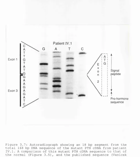
family A / 8 9 ... 100 3.5: Autoradiograph showing a 123 bp segment from the
total 258 bp DNA sequence of the normal PTH cDNA synthesized from illegitimately transcribed PTH m R N A ... 102 3.6: The illegitimate transcription of the PTH gene in
EBV- transformed lymphocytes ... 104 3.7: Autoradiograph showing an 18 bp segment from the
total 168 bp DNA sequence of the mutant PTH cDNA from patient IV. 1 ... 106 3.8: Segregation analysis at the PTH locus in pedigree
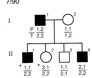
7/90 with autosomal dominant idiopathic hypoparathyroidism using PstI (P) and TaqI (T)
restriction fragment length polymorphisms . . . . 108 3.9: Segregation analysis at the PTH locus in pedigree
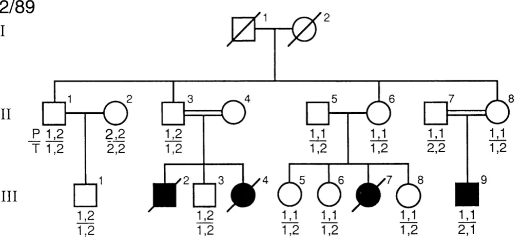
2/89 with autosomal recessive idiopathic hypoparathyroidism using PstI (P) and TaqI (T) R F L P s ... Ill 3.10: Pedigree 23/92 with hypoparathyroidism,
sensorineural deafness, and renal dysplasia . . . 113 4.1: Tetranucleotide (AAAT) „ polymorphism associated with
the PTH g e n e ... 125 4.2: Mendelian inheritance of the tetranucleotide (AAAT)^
polymorphism associated with the PTH gene . . . . 126 4.3: Segregation studies in families A/89, 2/89 and 7/90
with idiopathic hypoparathyroidism ... 128 4.4: Schematic representation of the PTH gene ... 132 5.1: Autoradiographs of pulse field blot hybridised with
the probes pCDRl (locus CDR) and 4D.8 (locus D X S 9 8 ) ... 138 5.2: Nucleotide sequence of 392 base pairs from the
5.4: Autoradiograph of pulsed field blot of CDR and DSX98 positive yeast artificial chromosome clones
hybridised with the probes pCDRl and 4D.8 . . . . 145 5.5: Autoradiograph of pulsed field blot of duplicate
samples of CDRYACl yeast artificial chromosome clone
DNA hybridised with pBR322 plasmid D N A ... 14 6 5.6: Alu-PCR fingerprint analysis of CDR and DXS98
positive YAC c l o n e s ... 148 5.7: Polymerase chain reaction amplification of
vectorette libraries constructed from CDRYACl using the restriction enzymes Alul, EcoRV, PvuII and
R s a l ... 152 5.8: Localisation by PCR of sequence tagged site derived
from right arm terminal sequence of CDRYACl clone
using oligonucleotide primers CDRVRl and CDRVR2 . 153 5.9: Genomic localisation by PCR of sequence tagged site
derived from left arm terminal sequence from DXS98YAC1 clone using oligonucleotide primers D98VL1
and D 9 8 V L 2 ... 155 6.1: Localisation of the human calbindin D9K gene by
analysis of the somatic cell hybrids 1W1LA4.9, 835,
Sinl76 and C F 3 7 ... 171 6.2: Autoradiograph of Southern blot of TaqI digested
male backcross progeny DNA hybridised with rat calbindin cDNA to reveal Mus musculus domesticus and
Mus spretus RFLV a l l e l e s ... 174 6.3: Molecular mapping of the calbindin D9K gene on the
mouse X chromosome in male progeny of the
interspecific backcross segregating for hyp . . . 176 6.4: Molecular mapping of the calbindin D9K gene on the
mouse X chromosome in female progeny of the
interspecific backcross ... 177 6.5: Nucleotide sequence of the 428 base pair mouse
calbindin D9K c D N A ... 180
6.6 : Identification of two forms of calbindin D9K cDNA in kidney RNA from normal and hyp Mus musculus
6.7: Autoradiograph of sequencing gel showing the region of nucleotide sequence difference between the two
forms of mouse calbindin D9K c D N A ... 185
6.8 : Intron 2/exon 3 nucleotide sequence of mouse and rat
LIST OF TABLES.
Table 1 : LOD scores for X-linked markers and HPT . . . 37 Table 2: Clinical and Biochemical Findings in Members of
Family A / 8 9 ... 55 Table 3: Linkage disequilibrium for PTH polymorphisms . 131
LIST OF APPENDICES.
APPENDIX I: Oligonucleotide Sequences ... 203 APPENDIX II: Abbreviations U s e d ... 205
1.INTRODUCTION
The homeostatic control of calcium and phosphate in man and all terrestrial vertebrates is principally regulated by parathyroid hormone and the active forms of vitamin D. The actions of parathyroid hormone and vitamin D are both
cooperative and coordinated in order to regulate the total body content of the two ions, and to control the equilibrium between the insoluble mineral phase of bone and the soluble fractions of calcium and phosphate in the extracellular fluids of the body.
In addition to parathyroid hormone and vitamin D, the recently identified parathyroid hormone-related peptide (PTHrp), which shares N-terminal homology with parathyroid hormone, has been postulated to have a role in foetal mineral homeostasis and the regulation of maternal bone resorption and intestinal calcium absorption during lactation.
1.1 PARATHYROID HORMONE
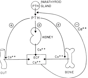
The physiological role of parathyroid hormone (PTH), through concerted actions on its target organs of the kidney, bone and intestine, is to increase the flow of calcium into the extracellular fluid and thereby regulate the level of the ion in the plasma. In accordance with this, the production of parathyroid hormone is regulated by the concentration of calcium in the plasma. A decrease in the level of plasma calcium rapidly stimulates the production of PTH and conversely the secretion of the hormone is inhibited by an increase in plasma calcium (Sherwood et al 1966, Habener et al
1976, Mayer et al 1976) .
acts in a divergent manner upon the kidney to increase the renal reabsorption of calcium and to directly inhibit the tubular reabsorption of phosphate, thereby ensuring the desired rise in plasma calcium levels whilst preventing hyperphosphataemia. The third action of parathyroid hormone is the indirect enhancement of the intestinal absorption of calcium. This effect is achieved by the hormone functioning as an activator of the 2 5 (OH) vitamin D 1-alpha-hydroxylase enzyme in the proximal tubular cells of the kidney, thereby
PARATHYROID p i n)g l a n d
PT
KIDNEY
Co + + EOF
BONE GUT
1.1.1 The Parathyroid Hormone Gene
Human parathyroid hormone is encoded by a single gene on the short arm of chromosome 11, band llpl5 (Mayer et al 1983, Naylor et al 1983) . The nucleotide sequence of the human preproparathyroid hormone complementary DNA (cDNA) was
described by Hendy et al (1981) , and demonstrates 90% nucleotide and 85% amino acid homology in coding sequence with the bovine PTH cDNA sequence (Kronenberg et al 197 9) . The parathyroid hormone gene is approximately 4 Kilobases in size, and consists of three exons (or coding sequences) and two
introns (Vasicek et al 1983) . The complete genomic sequence of the PTH gene, together with DNA sequence upstream from the
transcription start site containing elements of the PTH promoter was published by Reis et al (1990) . Exon 1 of the PTH gene is 85 base pairs in size (Figure 1.2) and contains 5' untranslated sequence. Exon 2, which is 90 base pairs in size, contains the initiation methionine codon (ATG) and encodes the 25 amino acid signal sequence together with the first three amino acids of the pro-sequence of the prepro-PTH molecule. Exon 3 is 612 base pairs in size and encodes the remaining 3 amino acids of the 6 amino acid pro-sequence and the 84 amino acid PTH peptide. The exon-intron boundary consensus sequences required for the post-transcriptional splicing of the PTH gene messenger RNA conform to the "gt-ag" rule proposed by Breathnach et al (1978) .
PTH
g e n e
Un
translated
Signal
Pro
peptide
sequence
IH---PTH
Un
peptide
translated
%Exon 1%
^ 85bp^
Intron 1
# #
301
%
t Z E x o n
P
^
Intron 2)::
: lOSbp ÿ
Exon 3</
612bp
3
'~K
Figure 1.2: Schematic representation of the genomic organisation of the parathyroid hormone (PTH) gene. The PTH gene consists of 3 exons and 2 introns. Exon 1 contains 5' untranslated sequence, exon 2 contains the initiation codon (ATG) and encodes the 25 amino acid signal peptide together with the first three amino acids of the pro-sequence, and exon 3 encodes the remainder of the pro sequence and the 84 amino acid PTH peptide. Two restriction fragment length polymorphisms are associated with the PTH gene with the enzymes TaqI and PstI (Schmidtke et al 1984). The TaqI polymorphic site (denoted by a closed triangle) is within intron 2 of the PTH gene, and the PstI polymorphic site (open triangle) is situated approximately 1.7 Kilobase pairs downstream of the g e n e .
1.1.2 Transcriptional Control of the Parathyroid Hormone Gene
In addition to the control of secretion of PTH from the parathyroid glands, the level of extracellular calcium exerts
an effect upon the rate of biosynthesis of parathyroid hormone. Studies performed by Russell et al (1983) have demonstrated that increases in the level of extracellular calcium cause a decrease (which is reversible) in the rate of PTH gene expression and decreased PTH mRNA levels in dispersed bovine parathyroid cells. More specific molecular studies on the control of parathyroid hormone gene expression by calcium have been performed by Okazaki et al (1992) and have identified a 15 base pair palindromic sequence (TGAGACAGGGTCTCA) approximately 3.5 Kilobase pairs upstream form the PTH gene, the negative calcium response element (nCaRE), which is required for the negative regulation of gene expression by extracellular calcium.
The effects of 1 , 2 5 (OH) 2 vitamin D3 levels upon PTH mRNA transcription have also been examined by Silver at al (1985) and Russell et al (1986), using the technique of "nuclear run off" in order study the effect of the active vitamin D metabolite upon PTH mRNA transcription. Both studies demonstrated a specific effect of 1 , 2 5 (OH) 2 vitamin D3 in decreasing the rate of PTH mRNA transcription. Furthermore, experiments performed by Okazaki et al (1988), using plasmid constructs containing fragments of the PTH promoter fused to
1.1.3 Biosynthesis of Parathyroid Hormone
The parathyroid hormone gene, which consists of 3 exons and 2 introns, is transcribed in the nucleus as a large precursor mRNA molecule from which the intronic sequences are removed prior to translation of the mRNA. This process known as mRNA splicing requires the presence of consensus sequences (Breathnach et al 1978, Mount 1982) at the exon-intron boundaries, which are recognised by the splicing machinery of the cell. The mature spliced PTH mRNA is then transported to the cytoplasm and is translated to give the 115 amino acid preproPTH molecule. During translation, the 25 amino acid hydrophobic signal sequence of the preproPTH molecule binds with a particle known as the signal recognition particle (Walter at al 1981), which mediates entry of the nascent preproPTH peptide into the lumen of the rough endoplasmic reticulum. The 25 amino acid signal sequence is then removed by a signal peptidase to yield the intermediate pro-PTH precursor, which is transported to the Golgi apparatus of the cell. In the Golgi, the six amino acid prohormone sequence is enzymatically removed to yield the mature 84 amino acid PTH peptide. The parathyroid hormone is then packaged into secretory granules that fuse with the plasma membrane of the cell and release PTH into the bloodstream.
1.1.4 Parathyroid Hormone Related Peptide
The possibility that malignant tumours could cause hypercalcaemia by producing a substance resembling parathyroid hormone was first suggested by Albright at al (1941), based upon the coexistence of hypophosphataemia and hypercalcaemia in a patient with renal carcinoma. This humoral hypercalcaemia of malignancy (HHM), which often occurs in patients with
squamous cell carcinoma of the lung without bony métastasés, appeared to be caused by tumour products acting on bone to promote resorption and on the kidney to restrict calcium excretion (Stewart et al 1983) . Moseley et al (1987) purified a protein with similar biological activities to parathyroid hormone from the culture medium of a human lung cancer cell line, and demonstrated that the cancer cells contained no PTH mRNA indicating the presence of a protein distinct from PTH.
The complementary DNA (cDNA) for this parathyroid hormone-related peptide (PTHrp) has been cloned (Suva et al
1987, Mangin et al 1988) and localised (Mangin et al 1988) to the short arm of chromosome 12 (12pl2.1 - p l l . 2) . The cDNA encodes a protein of 177 amino acids, containing a prepro sequence of 36 amino acids followed by the mature peptide of 141 residues in length. Of the first 13 amino acids of the mature peptide, 8 are identical to parathyroid hormone, although the sequences of the two peptides diverge completely after this point. The genomic organisation of parathyroid hormone related peptide gene has been reported by Yasuda et al
(1989) ; the gene consists of 7 exons and spans approximately 13 Kilobases of genomic DNA. Hammonds et al (1989) reported the presence of two forms of PTHrp, one of which is the full length 141 amino acids, and another form truncated at its
are derived from a common ancestral sequence.
The function of parathyroid hormone-related peptide in
human physiology is under intense study at the present time. It has been postulated that the peptide plays a role in foetal and neonatal calcium metabolism (Burton et al 1990, Moniz et
al 1990) . In addition, it has been demonstrated that PTHrp is expressed in lactating mammary tissue (Thiede et al 1988), and that PTHrp may act to regulate maternal bone resorption and intestinal calcium absorption during lactation (Budayr et al
1.1.5 The PTH/PTHrp Receptor
Parathyroid hormone regulates calcium and phosphate metabolism by binding to specific receptors located on the plasma membrane in kidney and bone. Pharmacological and physicochemical evidence (Juppner et al 1988), from experiments performed with rat osteosarcoma cells, strongly indicates that both parathyroid hormone and parathyroid
hormone related peptide bind to the same receptor.
The physiological effects of PTH on its target tissues are principally mediated by two distinct receptor-linked effector systems, adenylate cyclase and phospholipase C, which initiate a cascade of intracellular events upon binding of PTH. Binding of PTH to its receptor activates the stimulatory guanyl nucleotide regulatory subunit, Gg, (Nissenson at al
1981), leading to activation of adenylate cyclase (Chase at al
1967, Chase at al 1968, Keutmann at al 1985), accumulation of cyclic AMP (cAMP), and activation of cAMP-dependent protein kinases (Partridge at al 1981, Livesay at al 1982). Mutation of the a subunit of the stimulatory G protein (Ggtt) of adenyl cyclase has been demonstrated (Patten at al 1990) to be associated with the condition of pseudohypoparathyroidism type lA (Albright's hereditary osteodystrophy), in which there is a resistance of target organs to parathyroid hormone and other hormones that act by stimulating adenyl cyclase activity.
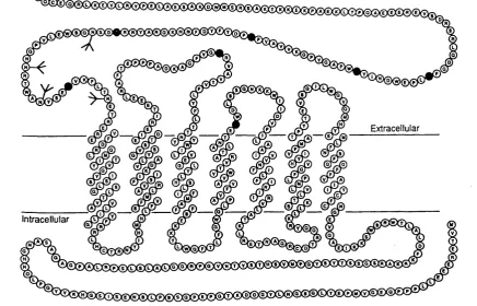
The complementary DNA (cDNA) encoding the PTH / PTHrp receptor has recently been isolated by Juppner et al (1991) from an opossum kidney cDNA library using an expression cloning strategy. Nucleotide sequencing of the cDNA revealed an open reading frame encoding a 585 amino acid protein, which when expressed in COS 7 cells, binds PTH and PTHrp with equal affinity leading to activation of intracellular adenylate
E xtracellular
intracellular
1.2 VITAMIN D
The primary physiological function of vitamin D, as for parathyroid hormone, is the homeostatic control of calcium in
the extracellular fluid. Whereas it is parathyroid hormone that is the principal regulator for the minute-to-minute homeostatic control of calcium, it is the physiological role of vitamin D to maintain the day-to-day overall balance of calcium in the body. The classification of vitamin D as a vitamin, an essential dietary requirement required only in trace amounts, is somewhat of an historical misnomer. Vitamin D conforms to a much greater extent to the definition of a hormone, a substance produced in one part of an organism which has an effect on another part of the organism.
The control of extracellular calcium levels by vitamin D is achieved through the concerted action of the hormone upon its target tissues of the intestine and bone, although the principal physiological effects of vitamin D in calcium homeostasis are mediated through the action of the hormone on the small intestine. The active form of vitamin D, 1,25 (OH) 2 vitamin D3, functions to enhance the absorption of dietary calcium and phosphorus in the small intestine. For the transport of calcium in the duodenum, the major calcium transport protein, is the calbindin D9K (Wasserman et al
1983), or calbindin. It has been demonstrated in the rat that 1 , 2 5 (OH) 2 vitamin D3 specifically induces an increase in the rate of calbindin gene transcription (Leonard et al 1984) and that the expression of calbindin correlates with calcium transport activity in the small intestine (Bruns et al 1987) .
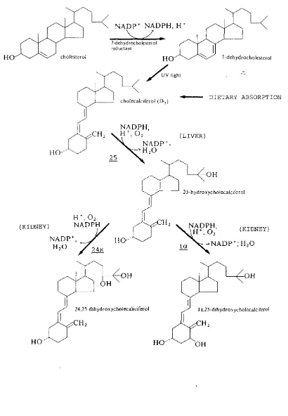
vitamin D3 (cholecalciferol) can be produced in sufficient quantities in man provided that the individual is adequately exposed to sunlight. In primitive man, living with very few clothes and exposed to sunlight for considerable periods of time, sufficient vitamin D3 would have been synthesized in the skin, only when man began to live in colder
climes and covered most of his body in clothes has he become dependent upon a dietary source of the vitamin. In the skin. Vitamin D3 is synthesized from its precursor molecule,
cholesterol, via the intermediate of 7-dehydrocholesterol, utilising ultraviolet light in the epidermis for the conversion of 7-dehydrocholesterol to vitamin D3
(cholecalciferol; Figure 1.4).
Whether vitamin D3 is produced in the skin, or is provided in the diet, it must be modified by two hydroxylations (Figure 1.4) which occur sequentially in the liver and in the kidney before it may function in the homeostatic control of calcium.
The first hydroxylation reaction occurs in the liver to produce 25 (OH) vitamin D3, which is released into the bloodstream and transported to the kidney, where the second hydroxylation reaction takes place to form the active vitamin D metabolite, 1 , 2 5 (OH) 2 vitamin D3. The activity of the renal mitochondrial 2 5 (OH)D-la-hydroxylase enzyme has been demonstrated to be strongly regulated by plasma inorganic phosphorus concentration, hypophosphataemia activating the enzyme (Tanaka et al 1973, Gray et al 1979) . In addition, hypocalcaemia, by the stimulation of parathyroid hormone
secretion, acts to increase the activity of the la-hydroxylase enzyme (Fukase et al 1982).
Another hydroxylation of 25 (OH) vitamin D3 may also occur in the kidney; a 24-hydroxylation (Figure 1.4), which produces
24,25 (OH) 2 vitamin D3. Although it is generally accepted that the 1,25 (OH) 2 vitamin D3 is the active form of the hormone
NADP+ NADPH, H
7-dehydrocholesterol
reductase
cholesterol
H H O dehydrocholesterol
( K I D N E Y )
U V light
D IE T A R Y A B S O R P T IO N
c h o lec a lc ife ro l ( D j )
NADPH,
:CH ( L I V E R
N A D P + ,
H2O
H O
25
O H
25-h yd ro x y c h o le c a ic ife ro l
:CH NADPH
(K ID N E Y )
N A D P \
H2O 2 4 R H O la
O H O H
2 4.2 5 -d ih y d ro x y c h o le c a iic ife ro l
:CH
H O
OH
1 x,25-dihydroxycholecalciferol
H O O H
1.2.1 The Vitamin D Receptor (VDR)
1 , 2 5 (OH) 2 vitamin D3 mediates its biological effects by binding to intracellular receptors (DeLuca et al 1990) which belong to the steroid receptor superfamily (Baker et al 1988). Binding of 1 , 2 5 (OH) 2 vitamin D3 to its receptor induces an allosteric change that allows the receptor-hormone complex to bind to its DNA response element in the promoter region of a
target gene. This interaction results in the transcription of specific genes the products of which affect the biological response to the hormone. Isolation of the complementary DNA for the vitamin D receptor (VDR) by Baker et al (1988) , indicated that the VDR protein was highly homologous to other steroid receptors and the thyroid hormone receptor. The area of strongest homology is at the N-terminus of the VDR protein, a hydrophilic region which is rich in cysteine, lysine, and arginine residues, and constitute the two zinc finger DNA binding domain of the VDR molecule (Evans 1988) .
The investigation of the promoter regions of the rat calbindin D9K (Darwish et al 1992) , rat (Demay et al 1990) and human (Kerner et al 1989) osteocalcin genes, which have been demonstrated to be transcriptionally controlled by 1,25 (OH) 2 vitamin D3, has allowed the characterisation of the vitamin D responsive element, or VDRE, for these genes. The vitamin D receptor-ligand complex, as for the thyroid hormone and retinoic acid receptors (Umesono et al 1991), interacts with a direct repeat consensus sequence in the promoter of the gene. For example, the characterisation of the VDRE for the rat osteocalcin promoter (Demay et al 1990) revealed the presence of three AGGTCA-related sequences in close proximity
5^-TGGGTGAATGAGGACATTACTGACCG-3^ , which confer responsiveness to 1 , 2 5 (OH) 2 vitamin D3.
1.3 DISORDERS OF CALCIUM AND PHOSPHATE HOMEOSTASIS
1.3.1 Familial Idiopathic Hypoparathyroidism
Hypoparathyroidism is an endocrine disorder which is
characterised by hypocalcaemia and hyperphosphataemia due to a lack of active circulating parathyroid hormone. Hypoparathyroidism may occur as an isolated endocrinopathy, termed idiopathic hypoparathyroidism, or may also arise due to a number of other causes, for instance as part of a polyglandular autoimmune disorder, or as a congenital defect,
for example DiGeorge syndrome.
Studies performed by Fattorossi et al (1988) demonstrated that antibodies in sera from patients with autoimmune hypoparathyroidism were cytotoxic to cultured bovine parathyroid cells, as well as to endothelial cells from bovine adrenal medulla and pulmonary artery. The mechanism by which such auto-antibodies arise is, as yet, unclear, but the observation of their cytotoxicity to endothelial cells from several different tissue types indicates an explanation for the involvement of several organs in autoimmune hypoparathyroidism. Hypoparathyroidism may also occur as part of a congenital defect, for example the DiGeorge syndrome, where there is an abnormality in the development of the derivatives of the third and fourth pharyngeal pouches. Affected individuals are athymic, aparathyroid, have aortic arch abnormalities and facial dysplasia. De la Chapelle (1981) first mapped the gene causing DiGeorge syndrome to chromosome
2 2qll, and postulated that deletion or an abnormality at this locus caused the observed phenotype, but positional cloning strategies have yet to identify the gene or genes involved in this developmental disorder.
have been established.
Autosomal Idiopathic Hypoparathyroidism.
For the autosomal inherited forms of idiopathic hypoparathyroidism, the parathyroid hormone gene located on chromosome llplS (Mayer et al 1983, Naylor et al 1983) has been investigated for abnormalities that may be associated with the endocrinopathy. Ahn et al (198 6) performed segregation studies using the PstI and TaqI restriction fragment length polymorphisms (RFLP's) at the PTH locus (Schmidtke et al 1984) in eight pedigrees with familial isolated hypoparathyroidism.
Linkage analysis in two of these families with autosomal dominant hypoparathyroidism (pedigrees C and D, Ahn et al
1986) demonstrated segregation of PTH alleles with hypoparathyroidism. The results of segregation analysis in the remaining 6 pedigrees excluded an association between hypoparathyroidism and the PTH gene in four of these families, whilst the other two pedigrees were uninformative. Arnold et
X-Linked Recessive Idiopathic Hypoparathyroidism.
Idiopathic hypoparathyroidism has been reported to occur as an X-linked recessive disorder in two multi-generation kindreds designated P/60 and W/81 (Peden 1960, Whyte et al
1981) from Missouri, USA. Affected individuals, who are all males, suffer from infantile onset of epilepsy and hypocalcaemia due to parathyroid hormone deficiency. In addition, the autopsy of an affected male from pedigree P/60 (Whyte et al 1986) revealed the absence of parathyroid tissue, thereby indicating that there is an isolated congenital defect in parathyroid gland development.
Linkage Studies.
the best estimate of the genetic distance between the two l o c i .
Previous family linkage analysis using cloned human X-chromosome sequences identifying restriction fragment length polymorphisms (RFLPs) have mapped the gene causing X-linked recessive hypoparathyroidism {HPT) to the long arm of the X-chromosome at Xq26-27 (Thakker et al 1990). This analysis established linkage between the X-linked recessive hypoparathyroid gene {HPT) and the locus DXS98 (4D.8) with a peak LOD score of 3.82 (0=0.05). Multipoint analysis indicated that the HPT locus is proximal to the DXS98 locus but distal to the F9 (Factor IX) locus on the distal long arm of the X-chromosome establishing the locus order Xcen-F9-HPT-DXS98-Xqter. Additional markers are required however as the recombination rate between HPT and F9 is 4% and that between HPT and DXS98 is 3%. Further linkage studies were therefore undertaken using the DNA probes pCDR and CX55.7, defining the loci CDR and DXS105 respectively, which have been localised to Xq26-27 (Anson et al 1988) in order to define a more detailed genetic map of the region containing the HPT gene. The probe pCDR corresponds to the cDNA for the cerebellar degeneration-related antigen, a target molecule recognized by autoantibodies in patients with paraneoplastic cerebellar degeneration (Dropcho et al 1987, Chen et al 1990). The probe CX55.7 consists of a single copy 2.4 Kbp EcoRI fragment (Willard et al 1985) . The probe pCDR reveals a restriction fragment length polymorphism (RFLP) with the enzyme H i n d i
(Siniscalco et al 1989), and the probe CX55.7 reveals RFLP's with the enzymes TaqI (Hofker et al 1987) , and StuI (Rekila et
al 1988) . The TaqI polymorphism for the probe CX55.7 was found to be uninformative in pedigrees P/60 and W/81; the data shown for linkage analysis for the probe CX55.7 (locus DXS105) is that obtained using the StuI polymorphism.
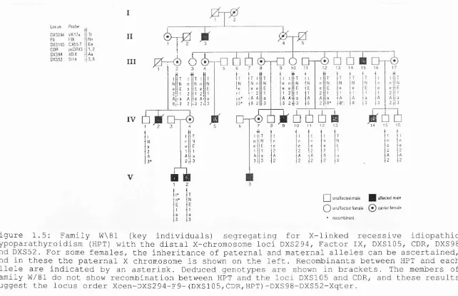
the two loci in this kindred. The pedigree is also informative (Thakker at al 1990) for the X-linked RFLP loci DXS294, F9, DXS98 and DXS52 (Figure 1.5). Individual IV. 4 is a carrier mother who is informative and phase known for DXS294, F9, DXS105 and DXS52, her affected son V.l is recombinant for HPT
and the proximal loci DXS2 94 and F9 but non-recombinant for DXS105, DXS98 and D X S 5 2 . Individual II.4 is a deceased carrier mother whose genotype was deduced from her eleven children. Her unaffected son I I I. 6 is recombinant for HPT and the distal loci DXS98 and DXS52 but is non-recombinant for the proximal locus DXS105. Thus the members of pedigree W/81 do not show recombination between HPT and the loci DXS105 and CDR, and these results suggest the locus order Xcen-DXS294-F9-(DXS105, CDR, HPT)-DXS98-DXS52-Xqter. The combined results of two point linkage analysis from families W/81 and P/60 are shown in Table 1.
L o c u s P ro b e
D XS294 V K 1 7 a Tt
F9 FIX Nn
D X S 105 0 X 5 5 .7 Ee C D R p cD R 1 3 1 ,2
D X S 98 4 D .8 Aa D X S 52 S t14 3 ,8
II
III
è > - r 0
2 3
■e 6 6 - H
T t N n
E e
1 2
a A
3 2 T I N n
E e
1 2
a A
3 2
■O
à 6 è-H
T I N n
e e
1 2
A A
8 2
a O Q
1 0 11
u
n n e e
1 2
A A
3 2
1 2 1 3 1 4 1 5 1 6 1 7
IV 6
A
o t^
o A ù ù 6 è
A i ù
1 ' a 3 4 ' s 6 7 8 ' 9 1 0 11 1 2 1 3 1 4 1 5 1 6
V
■ H
1 1 T t T t t 1 t T t
N n N n N n n n ■ n N n
e e E e E e e e e E e
2 1 1 1 ■ 1 2 2 2 2 1 2
A A a A a A A A A a A
3 * 2 3 3 3 2 2 2 2 8 2
I I u n a ffe c te d m a le a ffected m ale
( ^ 2 ) u n a ffe c te d fe m a le ca rrie r fem ale
* re c o m b in a n t
Figure 1.5: Family W\81 (key individuals) segregating for X-linked recessive idiopathic hypoparathyroidism (HPT) with the distal X-chromosome loci DXS294, Factor IX, DXS105, CDR, DXS98 and DXS52. For some females, the inheritance of paternal and maternal alleles can be ascertained, and in these the paternal X chromosome is shown on the left. Recombinants between HPT and each allele are indicated by an asterisk. Deduced genotypes are shown in brackets. The members of family W/81 do not show recombination between HPT and the loci DXS105 and CDR, and these results suggest the locus order Xcen-DXS294-F9-(DXS105,CDR,HPT)-DXS98-DXS52-Xqter.
Table 1: LOD scores for X-linked markers and HPT.
Locus LOD Scores Z(6)
0 Z (0) Z (0.0 0 1) Z (0.05) Z (0.10) Z (0.2 0) Z (0.30) Z (0.40)
DXS294 0.107 2.48 -0.74 2.28 2.48 2.24 1 . 6 8 0.93
F9 0.088 1 . 8 8 0.35 1.82 1.87 1.64 1.23 0 . 6 8
DXS105 0.000 7.62 7.61 6.94 6.22 4.21 3.08 1.36
CDR 0.000 3.58 3.57 3.25 2.91 2.21 1.50 0.78
DXS98 0.046 4.16 2.90 4.16 3.97 3.21 2.19 0.99
DXS52 0.133 2.70 -4.11 2.07 2.63 2.53 1.87 0.98
Deletion Mapping Studies
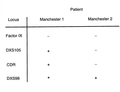
In order to further characterise the region containing the X-linked hypoparathyroid gene, deletion mapping studies were examined by Dr Derek Blake and Professor Kay Davies at the Institute of Molecular Medicine, Oxford, in two male patients (Manchester 1 and Manchester 2) with haemophilia B due to deletions involving the Factor IX gene locus, but who did not have the manifestations of hypoparathyroidism.
The results obtained for the deletion mapping are shown in
Figure 1.6.
For the patient Manchester 1 (Anson et al 1988) , DNA hybridisation analysis revealed the presence of the three loci DXS105, CDR and DXS98. However, for the haemophilia patient Manchester 2, hybridisation signals were detected with the probe DXS98, but were absent with the probes DXS105 and CDR. Assuming that the deletion in this patient is continuous, these results would therefore indicate that the X-linked recessive hypoparathyroid gene locus is not included in the deletion and is thus distal to the two loci CDR and DXS105, suggesting the locus order:
Patient
Locus
Manchester 1
Manchester 2
Factor IX
— —DXS105
+
—CDR
+
—DXS98
+
+
Figure 1.6: Schematic representation of deletion mapping data for the patients Manchester 1 and Manchester 2 with haemophilia B due to a Factor IX deletion (Anson et al 1988). The results for Southern blot hybridisation analysis with probes for the Factor IX, DXS105, CDR and DXS98 loci are shown. The presence of a hybridisation signal for a probe is denoted by a plus sign ( + ) , and absence of a hybridisation signal is denoted by a minus (-) sign. For the patient Manchester 1, hybridisation signals were obtained for the loci DXS105, CDR and DXS98. For the patient Manchester 2, hybridisation signals were obtained only with the DXS98 locus, and were absent for the DXS105 and CDR loci. These results therefore indicate that HPT is distal to CDR and DXS105.
1.3.2 Inherited Forms of Rickets
There are a variety of causes of hereditary rickets, which may be classified according to the predominant metabolic abnormality into two main groups. For the first group, the observed hypophosphataemia is due to a renal tubular defect,
which may consist of a single phosphate transport defect as occurs for X-linked hypophosphataemic rickets, or multiple transport defects in the handling of phosphate, amino acids, glucose, bicarbonate and potassium as occurs in the Fanconi syndrome. Such disorders are termed Vitamin D resistant r i c k e t s .
X-LINKED (VITAMIN D RESISTANT) HYPOPHOSPHATAEMIC RICKETS.
Vitamin D-resistant hypophosphataemic rickets was first described by Albright et al (1937) who recognized that some patients did not respond to therapy with normal doses of vitamin D, but would respond to large doses of vitamin D. Clinically, X-linked hypophosphataemic rickets (HYP) is
characterised by short stature, hypophosphataemia, and inadequate mineralisation of both the cartilagenous growth plate and bone. The X-linked dominant mode of inheritance was first delineated by Winters et al (1958), who noted that in a large pedigree from North Carolina there were no instances of male to male transmission of either bone disease or hypophosphataemia, the bone disease was much less severe in females, and that all daughters of hypophosphataemic males were themselves hypophosphataemic.
The biochemical and physiological features of HYP are due to a decrease in the renal tubular reabsorption of phosphate in the kidney, although the primary defect causing this has yet to be elucidated.
Hypophosphataemic Mouse Models:
Hyp and
Gyro
The investigation of the etiology of X-linked hypophosphataemic rickets has been greatly facilitated by the characterisation of two hypophosphataemic mouse models: hyp
and gyro. The hyp mouse was first described by Eicher et al
(1976), with mutant male mice manifesting with
described by Lyon et al (1986) . The mutation in the gyro
mouse, as with hyp, causes hypophosphataemia due to a defect in renal phosphate transport, but in addition the gyro
mutation causes circling behaviour, inner ear abnormalities, and sterility in males. The human counterpart to the phenotype caused by the gyro mutation has not yet been identified. Davies et al (1984) reported individuals with X-linked hypophosphataemic rickets who in addition presented with sensorineural deafness and tinnitus, but it is not known whether this results from bony overgrowth or calcification of the ligaments in the inner ear, or as an independent inherited
defect. The gyro gene (symbol gy) has been mapped close to hyp
Phosphate Transport Studies in Patients with X-linked
Hypophosphataemic Rickets and in the
Hyp Mouse
Dennis et al (1977) described the presence of two separate phosphate transport systems in mammalian kidney, one of which is PTH sensitive, located in the late proximal convoluted tubule and controlled by an X-linked gene, and another which is PTH insensitive, located in the early proximal tubule and controlled by an autosomal gene p r o d u c t . This finding is in agreement with the earlier studies of Glorieux and Scriver (1972), who described evidence that the hypophosphataemia in patients with X-linked hypophosphataemic rickets was due to a defect in the PTH sensitive component of phosphate transport, which is responsible for approximately two-thirds of the total net reabsorption of phosphate in the human kidney. Physiological experiments in the hyp mouse have defined that there is a defect in a sodium dependent phosphate transport molecule at the brush border (Tenenhouse et al 1978) of cells of the proximal convoluted tubule, and have demonstrated that in the hyp mouse, the defect is also independent of PTH (Cowgill et al 1979) .
Abnormalities of Vitamin D Metabolism
et al 1973) and in healthy adults (Gray et al 1979) . In addition, studies of the effects of parathyroid hormone upon synthesis of 1,25 (OH) 2 vitamin D3 in man (Lyles et al 1982) and the hyp mouse (Nesbitt et al 1986) have demonstrated that raising serum PTH levels fails to increase the 1-alpha-hydroxylase enzyme activity. Experiments performed by Nesbitt et al (1989) demonstrated that the lack of modulation of the 1-alpha-hydroxylase enzyme by parathyroid hormone in hyp mice was not due to an abnormality of intracellular cAMP production by the PTH receptor-linked adenyl cyclase enzyme. Nesbitt et
al therefore proposed three possible explanations for the abnormal control of the 1-alpha-hydroxylase enzyme in the hyp mouse kidney: Firstly, a defect in the cAMP-dependent activation of intracellular protein kinases. Secondly abnormal protein phosphorylation by cAMP activated protein kinases. Thirdly, since the aberrant renal 1-alpha-hydroxylase activity is confined to the segment of the kidney tubule in which abnormal phosphate transport occurs (Tenenhouse et al 1978, Cowgill et al 1979), the defective modulation of the enzyme may result from a c AMP-independent alteration of the intracellular milieu (diminished phosphate flux) that may directly impair mitochondrial function or inhibit new protein synthesis.
Studies performed in the gyro mouse, however, have demonstrated (Davidai et al 1990) that there is normal control of the renal 1-alpha-hydroxylase enzyme and production of 1 , 2 5 (OH) 2 vitamin D3 in response to hypophosphataemia and parathyroid hormone. This finding therefore indicates that the defect of phosphate transport in the proximal tubule of gyro
There is considerable controversy over whether the abnormalities observed in patients with X-linked hypophosphataemic rickets and in the hyp mouse occur due to an intrinsic renal defect or are due to the presence of a circulating humoral factor that alters proximal tubule brush border function and vitamin D metabolism. Bell et al (1988), using established primary cultures of renal epithelial cells from normal and hyp male mouse kidneys, observed that the cultured cells exhibited abnormal phosphate uptake and vitamin D metabolism after 5-6 days in culture, and therefore concluded that the defect was intrinsic to the hyp mouse kidney cells. In contrast, cross-transplantation experiments performed by Nesbitt et al (1992) obtained results that appeared to indicate unequivocally that the kidney is not the target organ for the genetic abnormality that underlies the
hyp phenotype. The hyp mouse phenotype was neither transferred nor corrected by renal transplantation, and Nesbitt et al
therefore postulated that the disorder in the hyp mouse, and by extrapolation in patients with HYP, is the result of the action of a humoral factor (of unknown source) rather than an intrinsic renal abnormality. Nesbitt et al suggested that a possible explanation for the disparate findings between the cross-transplantation and cell culture experiments may be that whilst the cultured renal cells from the hyp mice may have been removed from the influence of the humoral factor, they may not have been exposed to "essential factors" necessary to reinitiate the correct expression of the hyp gene product; thus the abnormalities of these cells in vivo would be perpetuated in culture.
Mapping of the Human X-Linked Hypophosphataemic Rickets Gene
From the study of comparative mapping data of the human and mouse X chromosomes, and the known location of the hyp
gene. Buckle et al (1985) predicted that the human X-linked hypophosphataemic rickets (HYP) gene may be either between the alpha-galactosidase gene and the hypoxanthine-guanine phosphoribosyl transferase (HPRT) gene or in the distal part
of the short arm of the human X chromosome. Studies using X chromosome probes detecting restriction fragment length polymorphisms (Read et al 1986, Machler et al 1986) established linkage with the marker 99.6 (locus DXS41, Figure 1.7) with a peak LOD score of 4.82 at 10% recombination. Multilocus linkage analysis indicated that the X-linked hypophosphataemic rickets (HYP) gene mapped distal to the DXS41 locus on the short arm of the X chromosome at Xp22.31-p21.3. Thakker et al (1987), by a study of 16 families, established that the HYP gene lay distal to the DXS41 locus
(probe 99.6) and proximal to the DXS43 locus (probe pD2), thereby defining bridging markers for the disease (Figure
A
^ # # 0
#####:#
. S O S ' / S ^
D X S 8 5 D X S 4 3
HYP
D X S 4 1 D X S 2 8 D X S 8 4 D X S 7
7 8 2 D 2 9 9 . 6
Cl
7 5 4 L I . 2 8
D X V S 1 p D p 3 4
D X S 1 7 5 9
D X S 5 1 5 2 A
Figure 1.7: Diagram of the human X-chromosome showing the position of the X-linked hypophosphataemic rickets (HYP) gene at X p 2 2 .31-p21.3. Reproduced from Thakker and O'Riordan
Econs et al (1992) performed further analysis in five families with hypophosphatémie rickets and demonstrated linkage between the markers DXS365 (peak LOD score 13.65 at 0% recombination) and DXS257 (peak LOD score 8.09 at 6%
recombination) with the HYP locus. Multipoint analysis indicated the locus order Xtel-DXS43-DXS257-HYP-DXS41-Xcen. The marker DXS365 was localised to the DXS43 to DXS41 interval but could not be located with respect to HYP and DXS257 (Econs
et al 1992) . Rowe et al (1992) further investigated the genetic map around the HYP locus, and defined DXS274 as the closest proximal marker, giving the locus order:
Xtel-DXS43-(DXS197,DXS257)-HYP-DXS274-DXS41-Xcen.
The identification of a microsatellite polymorphism at the DXS365 and DXS443 loci (Brown at al 1992) and use of the Genethon markers AFM234yfl2 and AFM163yh2, which map to the HYP region, for linkage analysis in 14 families with hypophosphataemic rickets (Rowe et al 1993) allowed the further refinement of the genetic map around the HYP locus. The results of this linkage analysis, together with analysis of a yeast artificial chromosome clone isolated using the marker AFM163yh2 has allowed the proposal of the locus order:
<--- H Y P --- >
Mapping of the Murine Hypophosphataemic Rickets
(hyp)
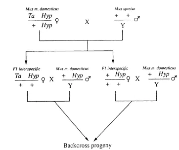
LocusLinkage studies performed by Eicher et al (1976) using the murine X-linked mutant genes Tabby (symbol Ta) and Bent-Tail (symbol Bn) localised the hyp gene to the distal end of the mouse X-chromosome. In order to facilitate the more precise localisation of the hyp gene, Kay et al (1991) utilised the approach of an interspecific backcross, segregating for hyp and Ta (the putative homologue of the human genetic disease hypohidrotic ectodermal dysplasia), in order to map the hyp locus within a framework of markers on the mouse X-chromosome. For this approach, crosses between the two evolutionary divergent mouse species. Mus spretus and Mus
musculus domesticus, were established (Figure 1.8),
facilitating the detection of restriction fragment length variants or RFLV's for DNA probes in the backcross progeny.
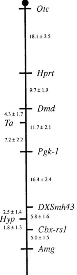
Multipoint analysis of recombination events between loci in the backcross progeny was performed indicating the gene order Xcen-Pgkl-DXSmh43-hyp-Cbxrsl-Amg-Xter. The distance between DXSmh4 3 and hyp is 2.5 ± 1.4 centiMorgans, and the distance between Cbx-rsl and hyp is 1.8 ± 1.3 centiMorgans
M u s m. domeslicus
Ta Hyp
+ Hyp
M u s sprclu s
+ +
X (f
Y
F I interspecific
Ta Hyp
+ +
M u s m. domeslicus
+ Hyp
(f
F I interspecific
+ Hyp
Y + +
9 X
M u s m. domeslicus
+ Hyp
Cf Y
Backcross progeny
Figure 1.8: Details of the interspecific M. m. domesticus/ M. spretus cross. Ova from female M. m. domesticus mice carrying the mutations Ta and hyp were fertilised with sperm from a M. spretus male. Of the resultant FI female progeny, two carried both the Ta and hyp mutations and two carried only the hyp