Kabell S, Handberg KJ, Li Y, Kusk M, Bisgaard M: Detection of vvIBDV in vacci-nated SPF chickens. Acta vet. scand. 2005, 46, 219-227.– The purpose of our exper-iment was to investigate, if apparently healthy, vaccinated chickens may be involved in maintaining and spreading infectious bursal disease virus (IBDV) in poultry environ-ments. We aimed at simultaneous detection and identification of very virulent field strain IBDV (vvIBDV) as well as vaccine strain IBDV in experimentally infected chick-ens. Two groups of specific pathogen free (SPF) chickens were vaccinated using the in-termediate infectious bursal disease (IBD) vaccine D78. Group 1 was vaccinated at the age of one week and group 2 at the age of three weeks. Both groups were challenged with vvIBDV at the age of four weeks. A third, vaccinated, non-challenged group served as negative control. No clinical symptoms were observed in any of these groups. The chickens were euthanised and submitted to autopsy and sample preparation in groups of three at fixed intervals from the age of 28 to 44 days. Gross pathological lesions were not observed. Lymphoid tissues from the bursa of Fabricius, bone marrow, spleen and thymus in addition to cloacal- and bursal swaps were analysed by one-step reverse tran-scription polymerase chain reaction (RT-PCR). Positive results were confirmed by two-step strain specific duplex (DPX) RT-PCR. The vaccine strain was detected in bursa tis-sues from all groups, while the challenge strain was detected in few bursal as well as non-bursal tissue samples.
The results indicate a possibility of replication of vvIBDV in vaccinated chickens. IBD; field virus; vaccination; D78; vvIBDV; Duplex RT-PCR.
Detection of vvIBDV in Vaccinated SPF Chickens
By S. Kabell1, K. J. Handberg1, Y. Li1, M. Kusk1andM. Bisgaard2
1Danish Institute for Food and Veterinary Research, Hangøvej 2, DK-8200 Aarhus N. 2Department of Veterinary
Pathobiology, The Royal Veterinary and Agricultural University, 4 Stigbøjlen, DK-1870 Frederiksberg C, Den-mark.
Introduction
Gumboro disease or infectious bursal disease (IBD) is associated with reduced production parameters, increased mortality and immuno-suppression in young chickens (Jorgensen et al. 1995; Lasher & Davis1997; Saif1998). The etiological agent, infectious bursal disease virus (IBDV) is a small and very stable RNA-virus that may survive even thorough cleaning and disinfection of poultry houses (Chettle et al.1989). Two serotypes, serotypes 1 and 2 are recognised (McFerran et al. 1980), of which only serotype 1 is pathogenic to the domesti-cated chicken. Extensive virus replication takes place in the bursa of Fabricius of the infected chicken, resulting in lesions in the bursa tissue
The disease is mainly controlled by vaccination (van den Berg2000, Muller et al.2003). Most of the Danish commercial breeders are vacci-nated and serologically tested to ensure high and uniform levels of maternally derived anti-bodies (MDA) in their offspring. MDA and high biosecurity levels in all parts of the pro-duction intentionally prevent intropro-duction and spread of diseases, including IBD. However, clinical outbreaks of IBD, defined as reported cases with typical clinical signs, increased mor-tality and pathological lesions including one or more chicken flocks in the same farm were re-ported from 1998 (Flensburg2001) until 2003, interrupted by a 14 months break including 2001. Based upon previous experiences, vacci-nation of broilers in and around affected farms was recommended for up to three broods suc-ceeding an outbreak. The vaccine strains were live, intermediate, attenuated IBDV strains. From March 2002 to February 2003, a total of 43 cases of clinical outbreaks of IBD were re-ported, involving approximately 10% of broiler producing farms in Denmark. Approximately one third of the flocks involved in these out-breaks had been vaccinated, 60% twice and 40% once, giving rise to questions concerning presence of virulent field virus in vaccinated flocks. For this reason, our experiment was aimed at investigating, if vaccinated chickens could contain virulent virus.
High levels of maternally derived antibodies (MDAs) inhibit an active immune response to-wards vaccination (Lucio & Hitchner 1979; Naqi et al.1983). The level of antibody that a vaccine can break through depends on the vac-cine strain (Winterfield et al.1980). Mild vac-cine strains are efficient only when chickens have no or very low levels of MDA, while the intermediate strains including D78, and the "hot" strains can break through higher levels of MDA titres (van den Berg et al.1991). Chick-ens from more than one parent flock may be
mixed in the same broiler house, adding to the variation in the level of MDA. Even anticipat-ing optimal timanticipat-ing of a santicipat-ingle vaccination, op-timal vaccine quality and opop-timal handling of vaccines, some chickens in the flock may not respond optimally to the vaccination due to MDA. Consequently, broiler farmers are often advised to vaccinate a flock twice. The first vac-cination is carried out early to benefit chickens with the lowest levels of MDA, while the sec-ond vaccination is carried out at a later age, when the highest initial levels of MDA have waned, often at the age of three weeks. Groups 1 and 2 in the present experiment represent early and late vaccination respectively. Consid-ering that SPF chickens have no MDA against IBDV, we vaccinated group 1 at seven days, the earliest vaccination time recommended, to al-low ample time for antibody production prior to challenge with field virus. Group 2 was only al-lowed one week to develop immunity before challenge, relying on the chickens' ability to produce virus-neutralizing antibodies three to four days post infection (Skeeles et al.1979). This group had the advantage of a more mature age as to immuno competence (Mast and God-deeris1999) before vaccination.
The objective of the present study was detection of virulent IBDV in lymphoid tissues and swab samples from vaccinated chickens indicating virus replication and excretion respectively. We used a commercial RT-PCR assay for detection of virus RNA, succeeded by a modification of a strain-specific RT-PCR assay recently devel-oped at our laboratory (Kusk et al.2005).
Materials and methods
Chickens
1500, Andersen BV, The Netherlands) and pro-vided with unlimited access to water and a commercial chicken feed. A light programme ensured 18 hours of daylight interrupted by six hours of darkness after the age of one week.
IBDV strains and inoculation protocol The vaccine strain Nobilis®Gumboro D78 Vet
(Intervet International B.V. Boxmeer, The Netherlands) (D78) with a titre of 105
ELD50/ml was used according to the manufac-turer's instructions; one dose was given orally to each chicken by syringe. A vvIBDV strain isolated from a Danish outbreak in 1998 (Handberg et al.2001), Gen Bank Accession No. AY850693 (DK01) was extracted from bursa tissues of previously infected chickens. The bursa tissues were homogenised in a mor-tar using a pestle and sterile sand. The ho-mogenate was suspended in 10% phosphate buffered saline (PBS) containing 10,000 U/ml penicillin, 10,000 µg/ml streptomycin, 250 µg/ml gentamycin, 500 U/ml nystatin and 5% FBS (Invitrogen). After centrifugation for 15 minutes at 3000 rpm, the supernatant was col-lected and diluted in Hank's buffered saline solution (HBSS) (GibcoTM, Scotland) to 10-1,
10-2, 10-3, 10-4, 10-5 and inoculated on the
chorio-allantoic membrane (CAM) of embry-onated hen's eggs to determine virus titres (Busby et al.1964). The titre value was 104.2
ELD50/ml. After a 1,000 fold dilution in HBSS, 0.2 ml per chicken was inoculated as drops in the eyes, nostrils and beak.
Experimental design
Twenty-four chickens from group 1 were inoc-ulated with D78 at the age of seven days, and 24 chickens from group 2 were similarly inocu-lated with D78 at the age of 21 days. Both groups were challenged with DK01 at the age of 28 days. A group of nine chickens vaccinated at the age of 21 days and not challenged served
as negative control. All three groups were ob-served twice daily for clinical symptoms. The chickens in groups 1 and 2 were bled and eu-thanised three at a time at the ages of 28, 29, 30, 31, 36, 38, 42 and 44 days. These chickens were numbered 1 to 24 in both groups. Chickens in the negative control group were sampled only on days 21, 30 and 38 and numbered 1 to 9. Eu-thanisation was performed in accordance with Article 2 (1) in Directive 86/609/EEC of 24 November 1986. Necropsy was performed im-mediately after euthanisation. Tissue samples from the bursa of Fabricius, bone marrow, spleen and thymus were removed and frozen at -80°C. Cloacal swabs were collected and frozen at -80°C. As RT-PCR results of cloacal swabs from group 1 and the negative control group turned out to be inconsistent (not shown), we decided to collect bursa swabs in addition to cloacal swabs from group 2.
In order to estimate the analytical sensitivity of the two RT-PCR assays, the assays were applied to 0, 10, 100, 1,000 and 10,000 fold dilutions of RNA extractions from each of the virus strains D78 and DK01. Qiagen RT-PCR could detect RNA from D78 in 0, 10 and 100 fold dilutions, and from DK01 in 0, 10, 100 and 1,000 fold di-lutions, whereas DPX RT-PCR could detect RNA from D78 in 0 and 10 fold dilutions and from DK01 in 0, 10 and 100 fold dilutions. As the results indicated that apparently the Qiagen RT-PCR performed with better sensitivity than the DPX RT-PCR, the Qiagen RT-PCR was used for the initial analyses. In case of at least one positive result within one type of samples, all similar samples from the same group of chickens were subjected to duplex (DPX) RT-PCR analysis to allow confirmation of the virus strain(s) involved.
Serology
per-formed according to the OIE manual, 2000 (OIE2000).
Extraction of RNA
Frozen tissues were homogenized and sus-pended as previously described. Each swab sample was shaken in 2 ml of the previously de-scribed PBS solution for one hour before the swab was discarded. The suspensions were cen-trifuged for 15 minutes at 3000 rpm. RNA was extracted from the supernatants using Qiagen Rneasy® Mini Kit (Qiagen GmbH, Hilden,
Germany). The protocol was modified as fol-lows: initially 400 µl of sample supernatant was mixed with 300 µl of lysis buffer containing 6 µl ß-mercapto ethanol (Sigma-Aldrich Co., Ger-many). This mixture was incubated at room temperature for one hour. From this point on the manufacturer's instructions were followed.
Qiagen RT-PCR
The QIAGEN OneStep RT-PCR Kit (Qiagen GmbH, Hilden, Germany) was used according to the manufacturer's instructions. Briefly, 10 µl of a 5 ×reaction buffer, 2 µl dNTP mix, 2 µl en-zyme mixture, 100 pmol of each oligonucleo-tide and 5 µl of RNA were mixed with RNase-free water to a final volume of 50 µl. All ingredients were kept on ice during handling. The PCR reaction was performed in a Biometra T3 Thermocycler (Biometra GmbH, Germany) as follows: 30 minutes at 50°C (RT reaction); 94°C for 15 minutes (initial PCR activation); 39 three-step cycles of 94°C for 30 sec, 58°C for 1 minute and 68°C for 2 minutes; 68°C for 7 min-utes (final extension).
Reverse transcription (RT) reaction
The iScriptTMcDNA Synthesis Kit (BioRad
Laboratories, Hercules, USA) was used accord-ing to the manufacturer's instructions. Briefly, 5 µl of RNA extraction was mixed with 1 µM of RT primers (Kusk et al. 2005), denatured for
five minutes at 94°C, chilled on ice, annealed at 55°C for five minutes and chilled on ice. Four-teen µl of RT mixture containing 1 ×RT buffer with 4 units of iScript Reverse Transcriptase were added, and after five minutes at room tem-perature the RNA samples were reverse tran-scribed for 30 minutes at 42°C, inactivated at 85°C for 5 minutes and stored at -20°C.
Duplex RT-PCR
Duplex RT-PCR (DPX RT-PCR) represents a modification of the use of primers in the multi-plex RT- PCR (iScript) previously described by Kusk et al. (2005). The primers included in the PCR mixture were only the four specific primers for D78 and DK01, fp776, D78-rp955, DK01-fp775, DK01-rp1028, and the concentration was increased to 1µM of each primer.
Results
Clinical, pathological and serological results No clinical symptoms were observed in any chicken involved in the experiment. Gross pathological lesions were not observed during the experiment. Serological analyses showed that all the chickens had seroconverted the first time blood was collected after vaccination (data not shown).
Qiagen RT-PCR
ana-lytical sensitivity analyses indicated better sen-sitivity using the Qiagen RT-PCR assay than the DPX RT-PCR assay. Consequently, series of samples showing negative results when sub-jected to Qiagen RT-PCR were regarded as be-ing truly negative and exempted from further analyses. As RT-PCR results of cloacal swab analyses were inconsistent after the samples had been stored, all these results were excluded.
DPX RT-PCR
RNA from D78 was detected in bursa tissue samples from 10 of the 24 chickens in group 1
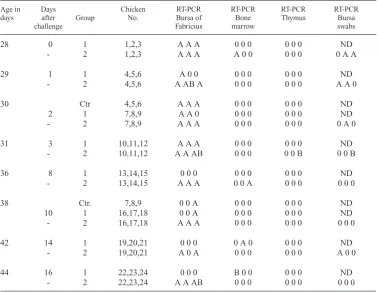
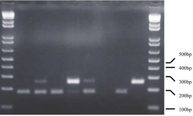
at the ages of 28, 29, 30, 31 and 38 days, and in a bone marrow sample from a 42 days old chicken. RNA from the challenge virus DK01 was not detected in bursa tissues from this group, but in a single bone marrow sample from a 44 days old chicken (Table 1). In group 2, RNA from D78 was found in all bursa tissue samples with one exception from day 42, while RNA from DK01 was detected in three chick-ens at the ages of 29, 31 and 44 days, respec-tively, as shown in Fig. 1 and Table 1. Analyses of non-bursal samples from group 2 revealed RNA from D78 in two bone marrow samples at Ta bl e 1 . DPX RT-PCR results of lymphoid tissue analyses from groups 1, 2 and the negative control group (Ctr), vaccinated at one, three and three week of age, respectively, and challenged with field virus at four weeks. The chickens were sampled in groups of three.
Age in Days Chicken RT-PCR RT-PCR RT-PCR RT-PCR
days after Group No. Bursa of Bone Thymus Bursa
challenge Fabricius marrow swabs
28 0 1 1,2,3 A A A 0 0 0 0 0 0 ND
- 2 1,2,3 A A A A 0 0 0 0 0 0 A A
29 1 1 4,5,6 A 0 0 0 0 0 0 0 0 ND
- 2 4,5,6 A AB A 0 0 0 0 0 0 A A 0
30 Ctr 4,5,6 A A A 0 0 0 0 0 0 ND
2 1 7,8,9 A A 0 0 0 0 0 0 0 ND
- 2 7,8,9 A A A 0 0 0 0 0 0 0 A 0
31 3 1 10,11,12 A A A 0 0 0 0 0 0 ND
- 2 10,11,12 A A AB 0 0 0 0 0 B 0 0 B
36 8 1 13,14,15 0 0 0 0 0 0 0 0 0 ND
- 2 13,14,15 A A A 0 0 A 0 0 0 0 0 0
38 Ctr. 7,8,9 0 0 A 0 0 0 0 0 0 ND
10 1 16,17,18 0 0 A 0 0 0 0 0 0 ND
- 2 16,17,18 A A A 0 0 0 0 0 0 0 0 0
42 14 1 19,20,21 0 0 0 0 A 0 0 0 0 ND
- 2 19,20,21 A 0 A 0 0 0 0 0 0 A 0 0
44 16 1 22,23,24 0 0 0 B 0 0 0 0 0 ND
- 2 22,23,24 A A AB 0 0 0 0 0 0 0 0 0
the ages of 28 and 36 days, respectively. DK01 was identified in a single thymus sample of a 31 days old chicken. In the bursal swab samples, RNA from D78 was detected in six chickens at the ages of 28, 29, 30 and 42 days. The chal-lenge strain was detected in a single bursal swab sample from a 31 days old chicken. In the negative control group, RNA from D78 was identified in four of six bursas sampled at the ages of 30 and 38 days.
Discussion
Clinical outbreaks of IBD in vaccinated broiler flocks have caused concern in the Danish poul-try induspoul-try. The reasons for these outbreaks could be high levels of MDA at the time of vac-cination that would block responses towards the vaccine, or it could be improper practical han-dling of the vaccine, decreasing its effect or dis-tribution, or new antigenic variant strains could be emerging. While these factors are most often investigated in connection with each outbreak, a potential reversion of the vaccine strain to
vir-ulence or survival of virulent strains in vacci-nated chickens remains to be investigated. Continuous vaccinations contain a risk that vac-cine strains may survive in the environment and regain virulence after passage in chickens ( Ya-maguchi et al.2000). As vaccination of broilers has not been routinely used in Denmark, we speculated that in our case it was more likely, that surviving field virus might infect, replicate and persist in vaccinated chickens.
Serum samples from the experimental chick-ens documented seroconversion before chal-lenge, and as might be anticipated from previ-ous studies, vaccine virus was detected in most bursa samples, contrary to the other lymphoid tissues (Kabell et al.2005). The detection of vaccine virus in bursa swabs from group 2 is concordant with the bursa tissue results and could be interpreted as excretion of virus. One bone marrow sample from group 1 and two from group 2 contained vaccine virus RNA, which cannot be explained by a mechanism connected to the challenge, as one sample was
100bp 200bp 300bp 400bp 500bp
positive before inoculation with DK01. How-ever, vaccine strain IBDV has been detected in non-bursal lymphoid organs before ( Barlic-Maganja et al. 2002), and even though virus strain and lymphoid tissues were different, a difference in sensitivity between the MPX and DPX RT-PCR assays might explain this diver-gence with previous results (Kabell et al.2005). The load of D78 seemed less in group 1 than in group 2, probably illustrating that the vaccine strain gradually disappears from the tissues with time (Table 1). In the negative control group, vaccine virus seemed to be disappearing earlier than in the similarly vaccinated group 2, but the number of samples is too small for fur-ther interpretations. The virulent strain DK01 was detected in bursa tissues from three chick-ens from group 2, one of which also contained DK01 in thymus tissue and the bursal swab. Presence of DK01 in the bursa and thymus from group 2 and in a bone marrow sample from group 1 indicates virus replication in the vaccinated chickens in spite of successful vac-cination.
The number of chickens found positive for DK01 was small, however the number of exper-imental chickens was also of limited size. Our observations do not exclude that surviving field virus in a broiler house may infect, replicate and be excreted from vaccinated chickens. An observation on the same aspect but regarding Newcastle disease virus (Alexander et al.1999) concluded that vaccination does not prevent replication and excretion of challenge virus. Assuming that viable IBDV may be excreted under predisposing conditions including sus-ceptible chickens, the final result may be a dis-ease outbreak.
Less field virus replication was observed in chickens challenged three weeks after vaccina-tion compared with chickens challenged one week after vaccination, adding to the general recommendations of protecting vaccinated
chickens from field virus as long as possible, al-lowing time for development of an optimal im-mune response.
In conclusion, the simultaneous identification of RNA from D78 and DK01 in bursa tissues, and DK01 in lymphoid tissues and bursal swabs indicated replication and excretion of very vir-ulent field virus from vaccinated chickens, sug-gesting an explanation to previous field out-breaks in vaccinated flocks. Further research into the impact of stress factors influencing dual IBDV infections under field conditions is in progress.
References
Alexander DJ, Manvell RJ, Banks J, Collins MS, Par-sons G, Cox B, Frost KM, Speidel EC, Ashman S, Aldous EW: Experimental assessment of the pathogenicity of the Newcastle disease viruses from outbreaks in Great Britain in 1997 for chick-ens and turkeys, and the protection afforded by vaccination. Avian Pathol. 1999, 28,501-511. Allan WH, Cullen GA, Faragher JT:
Immunosup-pression by Infectious Bursal Agent in Chickens Immunized Against Newcastle Disease. Vet. Rec. 1972, 90,511-512.
Barlic-Maganja D, Zorman-Rojs O, Grom J: Detec-tion of infectious bursal disease virus in different lymphoid organs by single-step reverse transcrip-tion polymerase chain reactranscrip-tion and microplate hy-bridization assay. J. Vet. Diagn. Investig. 2002, 14,243-246.
Busby DWG, House W, MacDonald JR: Neutralisa-tion. In D.W.G. Busby, W. House & J.R. MacDon-ald, Virological Technique 1st ed. 1964, 143-146. J. & A. Churchill, London, United Kingdom. Chettle NJ, Stuart JC, Wyeth PJ:Outbreak of virulent
infectious bursal disease in East Anglia. Vet. Rec. 1989, 125,271-272.
Cheville ONF:Studies on the pathogenesis of Gum-boro disease in the bursa of Fabricius, spleen and thymus of the chicken. Am. J. Pathol. 51: 527-551. 1967.
Handberg K, Flensburg MF, Ahrens P, P.H.Jorgensen PH:Characterisation of Danish isolates of IBDV: a preliminary study. II. International Symposium on Infectious Bursal Disease and Chicken Infec-tious Anaemia 2001, 157-162. Giessen, Germany, Institut für Geflügelkrankheiten, Justus Liebig University. European Commission, COST Action 839. 16-06-2001.
Jorgensen PH, Otte L, Nielsen OL, Bisgaard M: In-fluence of Subclinical Virus-Infections and Other Factors on Broiler Flock Performance. British Poult. Sci. 1995, 36,455-463.
Kabell S, Handberg KJ, Kusk M, Bisgaard M: Detec-tion of infectious bursal disease virus (IBDV) in various lymphoid tissues of experimentally in-fected SPF chickens by different RT-PCR assays. Avian Diseases 2005. In press.
Kusk M, Kabell S, Jorgensen PH, Handberg KJ: Dif-ferentiation of five strains of infectious bursal dis-ease virus: development of a strain specific multi-plex PCR. Vet. Microbiol. 2005, 109,159-167. Lasher HN, Davis VS:History of infectious bursal
disease in the USA - The first two decades. Avian Dis. 1997, 41,11-19.
Ley DH, Yamamoto R, Bickford AA:The pathogene-sis of infectious bursal disease – serologic, histopathologic and clinical chemical observa-tions. Avian dis. 1983, 27,1060-1085.
Lucio B, Hitchner SB: Infectious Bursal Disease Emulsified Vaccine – Effect Upon Neutralizing-Antibody Levels in the Dam and Subsequent Pro-tection of the Progeny. Avian Dis. 1979, 23, 466-478.
Mast J, Goddeeris BM:Development of immuno-competence of broiler chickens. Vet. Immunol. Immunopathol. 1999, 70,245-256.
McFerran JB, Mcnulty MS, Mckillop FR, Connor TJ, Mccracken RM, Collins DS, Allan GM:Isolation and serological studies with infectious bursal dis-ease viruses from fowl, turkeys and ducks – demonstration of a second serotype. Avian Pathol. 1980, 9,395-404.
Muller H, Islam MR, Raue R:Research on infectious bursal disease – the past, the present and the fu-ture. Vet. Microbiol. 2003, 97,153-165. Naqi SA, Marquez B, Sahin N:Maternal Antibody
and Its Effect on Infectious Bursal Disease Im-munization. Avian Dis. 1983, 27,623-631. Nielsen OL, Sorensen P, Hedemand JE, Laursen SB,
Jorgensen PH:Inflammatory response of differ-ent chicken lines and B haplotypes to infection with infectious bursal disease virus. Avian Pathol. 1998, 27,181-189.
OIE: OIE Manual of standards for diagnostic tests and vaccines. 2000, 4th edition, Chapter 2.7.1. Saif YM:Immunosuppression induced by infectious
bursal disease virus. Vet. Immunol. Immun-opathol. 1991, 30,45-50.
Saif YM:Infectious bursal disease and hemorrhagic enteritis. Poult. Sci. 1998, 77,1186-1189. Skeeles JK, Lukert PD, Debuysscher EV, Fletcher OJ,
Brown J:Infectious Bursal Disease Viral-Infec-tions.1. Complement and Virus-Neutralizing An-tibody-Response Following Infection of Suscepti-ble Chickens. Avian Dis. 1979, 23,95-106. van den Berg TP, Meulemans G:Acute Infectious
Bursal Disease in Poultry – Protection Afforded by Maternally Derived Antibodies and Interfer-ence with Live Vaccination. Avian Path. 1991, 20, 409-421.
van den Berg TP, Gonze M, Meulemans G: Acute in-fectious bursal disease in poultry: isolation and characterization of a highly virulent strain. Avian Pathol. 1991, 20,133-143.
van den Berg TP:Acute infectious bursal disease in poultry: A review. Avian Pathol. 2000, 29, 175-194.
Winterfield RW, Dhillon AS, Thacker HL, Alby LJ: Immune-Response of White Leghorn Chicks from Vaccination with Different Strains of Infec-tious Bursal Disease Virus and in the Presence of Maternal Antibodies. Avian Dis. 1980, 24, 179-188.
Yamaguchi T, Setiyono A, Kobayashi M, Takigami S, Fukushi H, Hirai K:Infectious bursal disease live vaccines: Changes in the virus population during serial passage in chickens and chicken embryo fi-broblast cells. Avian Dis. 2000, 44,284-290.
Sammendrag
Fund af meget virulent IBD-virus hos vaccinerede kyllinger.
gan-gen med fastsatte intervaller fra dag 28 til 44. Der kunne ikke påvises hverken kliniske symptomer eller patologiske forandringer hos nogen af grupperne. Lymfoidt væv fra bursa Fabricius, knoglemarv, milt og thymus, samt bursa- og kloaksvabere blev under-søgt ved one-step reverse transcription polymerase chain reaction (RT-PCR). Positive resultater blev
bekræftet ved two-step duplex (DPX) RT-PCR. Vac-cinestammen blev fundet i bursavæv fra alle grupper. RNA fra DK01 blev fundet i en knoglemarvsprøve fra gruppe 1 samt i tre bursa-prøver, en thymus og en bursa svaber fra gruppe 2. Resultaterne tyder på, at meget virulent IBD virus kan opformeres i og ud-skilles fra vaccinerede kyllinger.
(Received May 17, 2005; accepted July 31, 2005).