R E S E A R C H A R T I C L E
Open Access
Risk factors for complications and mortality
of percutaneous endoscopic gastrostomy
insertion
Gyu Young Pih, Hee Kyong Na
*, Ji Yong Ahn
*, Kee Wook Jung, Do Hoon Kim, Jeong Hoon Lee, Kee Don Choi,
Ho June Song, Gin Hyug Lee and Hwoon-Yong Jung
Abstract
Background:Percutaneous endoscopic gastrostomy (PEG) is a relatively safe procedure; however, acute and
chronic complications of PEG have been reported. We aimed to determine risk factors associated with complications and 30-day mortality after PEG, based on 11 years of experience at a single tertiary hospital. Methods:In total, 401 patients who underwent first PEG insertion at the Asan Medical Center, Seoul, Korea, between January 2005 and December 2015 were eligible. Medical records were retrospectively reviewed to determine clinical characteristics and outcomes of 139 and 262 patients who underwent pull-type and introducer-type PEG, respectively. Results:The median age of the overall population was 68 years, and the median body mass index was 19.5 kg/m2. Acute and chronic complications developed in 96 (23.9%) and 105 (26.2%) patients. Acute ileus and chronic tube obstruction were significantly more frequent in the introducer-type PEG group (p= 0.033 and 0.001, respectively). The 30-day mortality rate was 5.0% (median survival: 10.5 days). Multivariate analysis revealed that underlying malignancy was a predictor of acute complications; age≥70 years and diabetes mellitus were predictors of chronic complications. The median follow-up was 354 days. Neurologic disease and malignancy were the most common indications for PEG. Neurologic diseases were classified into two groups: stroke and the other neurologic disease group (including dementia, Parkinson’s disease, neuromuscular disease, and hypoxic brain damage). Multivariate analysis showed that 30-day mortality was significantly lower in the other neurologic disease group and higher in patients with platelet count < 100,000/μL, and C-reactive protein (CRP)≥5 mg/dL.
Conclusions:PEG is a relatively safe and feasible procedure, but it was associated with significantly higher early mortality rate in patients with platelet count < 100,000/μL or CPR≥5mg/dL, and lower early mortality rate in neurologic disease group including dementia, Parkinson's disase, neuromuscular disease, and hypoxic brain damage. In addition, acute complications in patients with underlying malignancy, and chronic complications in patients aged ≥70 and those with diabetes mellitus should be considered during and after PEG.
Keywords:Stomach, Endoscopy, Gastrostomy, Complication, Mortality
* Correspondence:hkna77@naver.com;ji110@hanmail.net
Department of Gastroenterology, Asan Medical Center, University of Ulsan College of Medicine, 88, Olympic-ro 43-gil, Songpa-gu, Seoul 05505, Korea
Background
Percutaneous endoscopic gastrostomy (PEG) is the most common method of enteral nutrition in patients who are expected to require supplementary enteral feeding for longer than 2–3 weeks [1]. Since its introduction in the 1980s, PEG has been associated with superior outcomes with respect to complication and mortality rates com-pared with radiological or surgical gastrostomy [2, 3]. Although PEG is a relatively safe and minimally invasive procedure, acute and chronic complications have been reported. In addition, the 30-day mortality after PEG has been reported to be 3.3–23.9% [4–9]. Therefore, the identification of risk factors associated with complica-tions and early mortality is crucial when evaluating which patients will benefit most from PEG procedures. We aimed to identify potential risk factors associated with acute complications, chronic complications and 30-day mortality after PEG using data from a large num-ber of patients who underwent tube insertion using the pull- or introducer-type PEG technique during an 11-year period at a single center in Korea.
Methods Patients
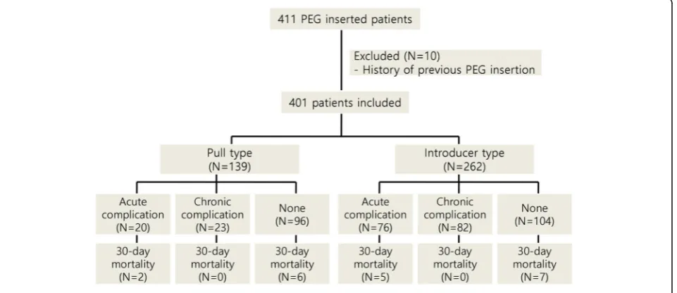
A total of 411 patients who underwent PEG at the Asan Medical Center in Seoul, Korea, between January 2005 and December 2015 were included in this retrospective study. Medical records of all patients were retrospect-ively reviewed for investigating clinical characteristics of patients. Demographic data, including underlying dis-ease, body mass index (BMI), indications for PEG, com-plications after PEG, comorbidities, and baseline laboratory values, were assessed. After exclusion of ten patients with a history of previous PEG insertion, 401
patients were included in the final analysis. A flow chart of the study population is shown in Fig 1. The study protocol was reviewed and approved by the institutional review board of the Asan Medical Center, Ulsan College of Medicine.
PEG methods
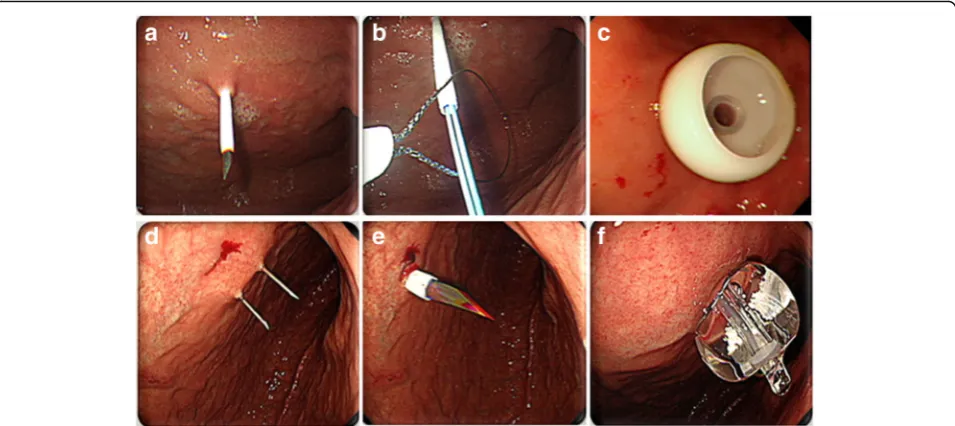
PEG was performed by either the pull (i.e., Ponsky-Gauderer) technique using a 20- or 24-Fr PEG tube (US Endoscopy®, Mentor, OH, US), or the intro-ducer (Russell) technique using a 15-Fr Cliny® PEG tube (Create Medic Co., Ltd., Yokohama, Japan) (Fig.2). PEG insertion was performed by gastroenterologists in all pa-tients. Among the 401 patients, the pull type technique was performed in 139 (34.7%) patients and the intro-ducer type was administered in 262 (65.3%) patients.
At the Asan Medical Center, all patients underwent the pull-type PEG until November 2010; from December 2010 to October 2011, both techniques were used, but from November 2011, all but three patients received the introducer-type PEG.
PEG insertion by the pull technique was initiated with insertion of a transilluminated endoscope to determine the puncture site on the abdominal wall, with endo-scopic monitoring of the anterior gastric wall. A guide-wire was introduced through the puncture site and drawn out through the esophagus and oral cavity using biopsy forceps. The PEG tube was then connected to the guidewire and reinserted through the oral cavity, pulled down, and extracted through the puncture site on the abdominal wall. An immediate endoscopic examination was performed to confirm the appropriate placement of the PEG tube.
Fig. 1Study flow chart
PEG insertion by the introducer technique was also initiated using a transilluminated endoscope; however, this technique used a gastropexy device and trocar to penetrate the abdominal wall and did not require the PEG tube to be pulled through the oral cavity. Following puncture of the gastric wall, the PEG tube was directly inserted through the abdominal wall and fixed by filling the tube balloon with 5 cc of saline.
Three hundred seventy seven (94.0%) patients received prophylactic antibiotic 30 min before the procedure. Most of the prophylactic antibiotic regimen was 2 g of cefazolin. However, patients who were already receiving antibiotic treatment were not required to change the antibiotic regimen unless the current regimen was not effective against gram-positive cocci. Before the proced-ure, all patients were sedated with intravenous midazo-lam (0.05 mg/kg) and pethidine (25 or 50 mg).
Definitions
Post-procedural complications were classified into two groups according to the time of occurrence. Acute com-plications were defined as those developing≤7 days after PEG, whereas chronic complications included those that occurred > 7 days after the procedure. Bleeding, pneu-moperitoneum, aspiration pneumonia, ileus, wound in-fection, and Mallory-Weiss tear were classified as acute complications. Chronic complications included tube leakage, tube obstruction, spontaneous tube removal, wound infection, buried bumper syndrome, and recur-rent aspiration pneumonia.
Bleeding was defined as an event requiring interven-tion (such as hemoclipping, embolizainterven-tion, or epineph-rine injection) to control bleeding and follow-up endoscopic examination to check lesion healing. In addition, bleeding complications included mucosal tear bleeding and bleeding requiring compression during and after the procedure. Aspiration pneumonia was defined as the presence of a newly developed symptom (such as cough, purulent sputum, and fever) with indicative chest X-ray image change after the PEG procedure. Ileus in-cluded symptomatic and non-symptomatic cases with indicative X-ray or computed tomography (CT) image of ileus and decreased bowel sounds.
Post-PEG management
On the day of the PEG procedure, all patients were kept NPO with natural tube drainage. On post-PEG day 1100 mL of clear water was flushed via the gastrostomy tube in patients whose abdomen was soft and in those who showed no abdominal discomfort. All patients underwent routine abdominal and chest X-ray on the same day as the procedure or the day after the proced-ure to check the position of the tube, as well as to detect any complications, such as pneumoperitoneum, aspir-ation pneumonia, and ileus. If any complicaspir-ations were detected, further imaging study or follow-up X-ray was performed. Patients who tolerated the water flush with-out abdominal pain or vomiting and had no evidence of leakage at the site of PEG were able to receive tube feed-ing with 100–200 mL of fluid.
Statistical analysis
Descriptive data are expressed as mean ± standard devi-ation or median and interquartile range (IQR). Statistical analysis was performed using SPSS version 21.0. Fisher’s exact test and the chi-square test were used to determine differences between categorical variables. Logistic regres-sion analysis was used to evaluate predictors of complica-tions and 30-day mortality. A p value < 0.05 was considered to be statistically significant for all analyses.
Results
Baseline characteristics
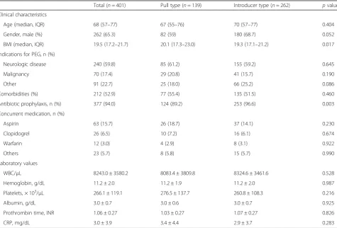
Age, gender, BMI, indications for PEG, comorbidities, and baseline laboratory values for the study subjects are sum-marized in Table1. The median age was 68 (IQR, 57–77) years, and median BMI was 19.5 (IQR, 17.2–21.7) kg/m2. There were 262 (65.3%) male patients in the study group. Indications for PEG were classified into three categories: patients undergoing PEG for neurologic disease (n= 240, 59.9%) included patients with stroke (n= 85), Parkinson’s disease (n= 61), neuromuscular disease (n= 53), dementia (n= 24), and hypoxic brain damage (n= 17); patients who underwent PEG for malignancy (n= 70, 17.4%) in-cluded patients with esophageal and head and neck
cancer (n= 21 and 49, respectively). In addition, there were 14 patients with lung cancer, three with stomach cancer, and one with hematologic cancer, colon can-cer, pancreatic cancan-cer, spine tumor, and breast cancan-cer, respectively.
In the overall study group, 186 (46.4%) patients had co-morbidities, including hypertension (n= 173), diabetes mel-litus (n= 90), and chronic kidney disease (CKD, n= 21). Antibiotic prophylaxis was used in 377 (94.0%) patients. Analysis of medication history for antiplatelet agents and anticoagulants revealed that 101 (25.2%) patients were treated with aspirin, clopidogrel, or warfarin; 12 patients (3.0%) were treated with dual aspirin/clopidogrel medica-tion; and 11 patients had discontinued aspirin and clopido-grel at least 5 days before the procedure. None of the cases of bleeding were reported in patients treated with dual an-tiplatelet agents. Among 51 patients who were taking as-pirin without clopidogrel, 32 patients had discontinued aspirin at least 7 days before the PEG procedure and 19 pa-tients discontinued treatment less than 7 days before the procedure. However, bleeding was reported in only one pa-tient who discontinued aspirin less than 7 days before the procedure. In 14 patients treated with clopidogrel without aspirin, 11 patients had discontinued medication at least
Table 1Baseline patient characteristics
Total (n= 401) Pull type (n= 139) Introducer type (n= 262) pvalue
Clinical characteristics
Age (median, IQR) 68 (57–77) 67 (55–76) 70 (57–77) 0.404
Gender, male (%) 262 (65.3) 82 (59) 180 (68.7) 0.052
BMI (median, IQR) 19.5 (17.2–21.7) 20.1 (17.3–23.0) 19.3 (17.1–21.2) 0.017
Indications for PEG, n (%)
Neurologic disease 240 (59.8) 85 (61.2) 155 (59.2) 0.645
Malignancy 70 (17.4) 29 (20.8) 41 (15.7) 0.190
Other 91 (22.7) 25 (18.0) 66 (25.2) 0.086
Comorbidities (%) 212 (52.9) 77 (55.4) 135 (51.5) 0.460
Antibiotic prophylaxis, n (%) 377 (94.0) 124 (89.2) 253 (96.6) 0.003
Concurrent medication, n (%)
Aspirin 63 (15.7) 26 (18.7) 37 (14.1) 0.230
Clopidogrel 26 (6.5) 10 (7.2) 16 (6.1) 0.674
Warfarin 12 (3.0) 4 (2.9) 8 (3.1) 0.922
Others 23 (5.7) 8 (5.8) 15 (5.7) 0.990
Laboratory values
WBC/μL 8243.0 ± 3580.2 8083.4 ± 3809.8 8324.6 ± 3461.6 0.528
Hemoglobin, g/dL 11.2 ± 2.0 11.2 ± 1.9 11.2 ± 2.0 0.987
Platelets, × 103/μL 266.1 ± 119.1 276.5 ± 137.7 260.8 ± 108.3 0.216
Albumin, g/dL 3.0 ± 0.7 3.0 ± 0.6 3.0 ± 0.7 0.925
Prothrombin time, INR 1.06 ± 0.27 1.03 ± 0.27 1.07 ± 0.27 0.826
CRP, mg/dL 3.0 ± 3.9 3.4 ± 4.4 2.9 ± 3.7 0.283
5 days before the procedure. Although three patients dis-continued treatment less than 5 days before the procedure, no bleeding events were reported.
Clinical outcomes
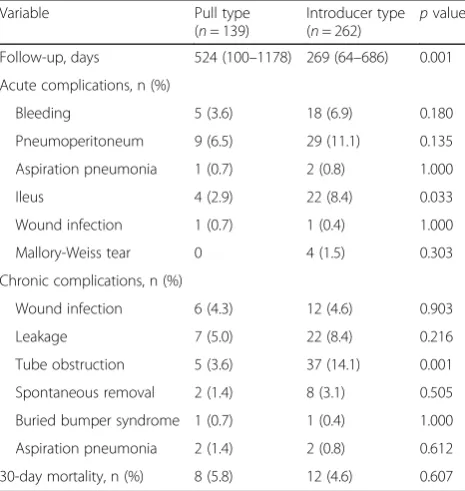
The median follow-up time was 354 (IQR, 79–787) days. Post-PEG complications are summarized in Table 2. With the exception of ileus and chronic tube obstruc-tion, there were no significant differences in the inci-dence of acute or chronic complications between the pull- and introducer-type PEG groups (Table2).
Acute post-PEG bleeding was observed in 23 (5.7%) patients. Although no additional intervention was re-quired in the majority of cases, two patients received epinephrine injection, three required hemoclipping, and one underwent embolization to manage bleeding.
Pneumoperitoneum developed in 38 (9.5%) patients within the first week of PEG. Of eight patients with symptomatic pneumoperitoneum who presented with the signs of peritoneal irritation as well as imaging ab-normalities on chest X-ray or CT, none required surgical intervention; all patients showed improvement with con-servative management.
Ileus was observed in 26 (6.5%) patients, but none re-quired surgical management. Wound infection was de-tected in 18 (4.5%) patients, among which three patients required not only antibiotic treatment but also removal of the PEG tube. Leakage was present in 29 (7.2%) pa-tients, 26 of whom underwent PEG tube change.
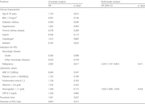
Risk factors for complications
Multivariate analysis revealed that underlying malig-nancy was a predictor of acute complications (odds ratio [OR], 2.205; 95% confidence interval [CI], 1.197–4.061;
p= 0.011) and that age≥70 years (OR, 1.022; 95% CI, 1.003–1.042;p= 0.021) and diabetes mellitus (OR, 1.877; 95% CI, 1.058–3.331; p= 0.031) were predictors of chronic complications (Tables3and4).
Subgroup analysis was conducted to compare the ma-lignancy group and neurologic disease groups. No sig-nificant differences in procedure time, PEG type, and acute complication rate were seen between the two groups, with the exception of the incidence of pneumo-peritoneum (p= 0.016; Table 5). However, all pneumo-peritoneum events were benign and improved with conservative care.
Prognosis
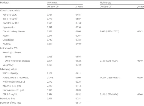
The 30-day mortality rate was 5.0%, and 20 patients died within 30 days of PEG insertion. On multivariate ana-lysis, platelet count < 100,000/μL (OR, 14.294; 95% CI, 3.358–60.851; p= 0.000), C-reactive protein (CRP)≥ 5 mg/dL (OR, 3.101; 95% CI, 1.021–9.414; p= 0.046), and the other neurologic disease group were found to be significant factors associated with 30-day mortality (OR, 0.125; 95% CI, 0.016–0.994; p= 0.049) (Table 6). The other neurologic disease group (including dementia, Par-kinson’s disease, neuromuscular disease, and hypoxic brain damage) was a significant factor that showed a lower incidence of 30-day mortality than stroke and ma-lignancy groups.
Among the 20 patients who died within 30 days of PEG, the cause of death was identified in 12 patients. The most common cause of death was pneumonia (n= 7). Mortality in two patients was considered to be associated with the PEG insertion procedure. One patient was admitted to the hospital to undergo chemotherapy for breast cancer with brain metastasis and leptomeningeal seeding; the patient presented with radiologic changes indicative of aspiration pneumonia and desaturation the day after PEG insertion and died 12 days after PEG as a result of aggravation of pneumonia. The other patient suffered from liver cirrhosis due to hepatitis B and C combined autoimmune hepatitis and was admitted to the hospital because of hepatic en-cephalopathy. The patient presented with fever on the day of PEG insertion and showed signs and symptoms of peri-tonitis with supporting diagnostic paracentesis result, which was normal the day before PEG. However, abdomen-pelvis CT showed no signs of peritonitis and the patient died 5 days after PEG as a result of uncontrolled infection. In addition, three patients presented with aggra-vation of previous pneumonia after PEG without evidence of a correlation with PEG; three patients developed aspir-ation pneumonia at least 10 days after PEG insertion.
Table 2Acute and chronic complications of percutaneous endoscopic gastrostomy according to the insertion type
Variable Pull type
(n= 139)
Introducer type
(n= 262) p
value
Follow-up, days 524 (100–1178) 269 (64–686) 0.001
Acute complications, n (%)
Bleeding 5 (3.6) 18 (6.9) 0.180
Pneumoperitoneum 9 (6.5) 29 (11.1) 0.135
Aspiration pneumonia 1 (0.7) 2 (0.8) 1.000
Ileus 4 (2.9) 22 (8.4) 0.033
Wound infection 1 (0.7) 1 (0.4) 1.000
Mallory-Weiss tear 0 4 (1.5) 0.303
Chronic complications, n (%)
Wound infection 6 (4.3) 12 (4.6) 0.903
Leakage 7 (5.0) 22 (8.4) 0.216
Tube obstruction 5 (3.6) 37 (14.1) 0.001
Spontaneous removal 2 (1.4) 8 (3.1) 0.505
Buried bumper syndrome 1 (0.7) 1 (0.4) 1.000
Aspiration pneumonia 2 (1.4) 2 (0.8) 0.612
Other causes of mortality unrelated to the PEG procedure included peritonitis, uncontrolled bleeding, liver failure, kidney failure, and traumatic brain injury. The cause of death was unknown in eight patients.
Discussion
PEG is a common procedure indicated for patients with normal gastrointestinal function who are expected to re-quire prolonged enteral feeding. However, patients who require PEG usually have chronic underlying diseases and vulnerable general condition. Although there are currently no established standard criteria for patients re-quiring PEG, guidelines published by the American Gastroenterological Association recommend that PEG is performed only in patients who are expected to survive for more than 30 days after the procedure [10]. Despite continuing efforts to evaluate risk factors associated with PEG-related complications and mortality, several studies have reported different risk factors [8, 11–14]. In addition, although PEG has been shown to be a safer ap-proach than radiological or surgical insertion, the com-plication rate with PEG is reported to be 13.2–42.9%
[12, 15, 16]. Complications including bleeding, wound infection, tube blockage, tube leakage, aspiration pneu-monia, perforation, and buried bumper syndrome are as-sociated with PEG [17].
In the present study, the technique of tube insertion was not associated with the rate of acute or chronic complications, with the exception of tube obstruction and ileus. The rate of tube obstruction was significantly higher with introducer- than pull-type PEG (3.6% vs. 14.1%, p= 0.001). As the introducer-type PEG uses a thinner 15-Fr tube than the pull-type PEG (20- or 24-Fr tube), our finding suggests that the use of a thinner tube is associated with a higher obstruction rate. The Euro-pean Society for Clinical Nutrition and Metabolism guidelines recommend that PEG tubes should be at least 15 Fr to minimize the risk of tube obstruction [1].
In addition, the rate of ileus was significantly higher in the introducer-type PEG group than the pull-type PEG group (2.9% vs. 8.4%, p= 0.033). Transient ileus rarely occurs after PEG, with a reported rate of 1–2% [18]. In the present study, all patients with ileus showed im-provement after conservative management. Diabetes
Table 3Univariate and multivariate logistic regression analysis of predictors of acute complications associated with percutaneous endoscopic gastrostomy
Predictor Univariate analysis Multivariate analysis
OR pvalue OR (95% CI) pvalue
Clinical characteristic
Age≥70 years 1.129 0.615
BMI < 19 kg/m2 0.955 0.196
Diabetes mellitus 0.590 0.099
Hypertension 1.002 0.993
Chronic kidney disease 0.578 0.389
Aspirin 0.546 0.114
Clopidogrel 1.072 0.885
Warfarin 0.705 0.655
Indication for PEG
Neurologic disease
Stroke 0.580 0.098
Other neurologic diseases 0.920 0.744
Malignancy 2.003 0.017 2.205 (1.197–4.061) 0.011
Laboratory values
WBC≥12,000/μL 0.694 0.347
Platelet count < 100,000/μL 1.163 0.798
Prothrombin time≥1.3 1.158 0.783
Albumin < 3.0 g/dL 1.333 0.243
Hemoglobin < 11 g/dL 1.390 0.175 1.029 (1.000–1.059) 0.054
CRP≥5 mg/dL 1.240 0.484
Procedure time 1.031 0.037
Diameter of PEG tube 0.843 0.412
mellitus may contribute to an increase in gastroparesis or ileus, but no significant difference in the rate of dia-betes mellitus was seen between the two groups (25.2% vs. 21.0%, p= 0.339). Conversely, analysis for underlying diseases revealed that the percentage of patients with neuromuscular disease was significantly higher in the introducer-type group (7.2% vs. 16.4%, p= 0.009). Neuromuscular diseases, such as amyotrophic lateral sclerosis, are associated with delayed gastric emptying and prolonged colonic transit time, which may contrib-ute to the development of post-PEG ileus [19,20].
In the present study, older age and diabetes mellitus were risk factors associated with chronic complications of PEG. Old age with debility and senility is a well-known factor affecting various medical conditions, and a previous study showed old age to be a risk factor for early mortality after PEG [11]. Because aging is
Table 4Univariate and multivariate logistic regression analysis of the predictors of chronic complications associated with percutaneous endoscopic gastrostomy
Predictor Univariate analysis Multivariate analysis
OR (95% CI) pvalue OR (95% CI) pvalue
Clinical characteristic
Age≥70 years 2.366 0.001 1.974 (1.186–3.285) 0.009
BMI < 19 kg/m2 1.161 0.542
Diabetes mellitus 1.851 0.022 1.731 (1.007–2.977) 0.047
Hypertension 1.857 0.011
Chronic kidney disease 0.570 0.376
Aspirin 1.985 0.022
Clopidogrel 1.316 0.550
Warfarin 1.174 0.812
Indication for PEG
Neurologic disease
Stroke 1.012 0.968
Other neurologic diseases 1.580 0.063
Malignancy 0.337 0.009 0.473 (0.199–1.082) 0.075
Laboratory values
WBC≥12,000/μL 0.419 0.054 0.434 (0.176–1.070) 0.070
Platelet count < 100,000/μL 0.226 0.152
Prothrombin time≥1.3 0.869 0.806
Albumin < 3.0 g/dL 1.048 0.849
Hemoglobin < 11 g/dL 0.837 0.470
CRP≥5 mg/dL 0.643 0.154
Procedure time 0.611 0.153
Diameter of PEG tube
15-Fr 0.031
20-Fr 0.143
(0.034–0.607)
0.008
24-Fr 0.976
(0.434–2.194)
0.952
Table 5Subgroup analysis between malignancy group and neurologic disease group
Variable Malignancy Neurologic disease pvalue
Acute complication, n (%)
Bleeding 3 (4.3) 12 (5.0) 1.000
Pneumoperitoneum 12 (17.1) 18 (7.5) 0.016
Aspiration pneumonia 2 (2.9) 1 (0.4) 0.129
Ileus 5 (7.1) 15 (6.3) 0.784
Wound infection 0 1 (0.4) 1.000
Mallory-Weiss tear 0 3 (1.3) 1.000
Pull-type PEG, n (%) 29 (41.4) 85 (35.4) 0.359
associated with immune system imbalance, infection-related complications may increase in older pa-tients [21, 22]. In the present study, infection-related chronic complications (including wound infection and recurrent pneumonia) comprised 21% of all chronic complications after PEG.
Diabetes mellitus is considered to be a significant risk factor associated with wound infection after invasive procedures and surgery. Furthermore, diabetes mellitus alters immunity by suppressing polymorphonuclear leukocyte function and cutaneous responses to antigen challenges [23]. In the present study, wound infection in the chronic period following PEG occurred in 18 (4.5%) patients. Given that PEG is an invasive procedure with penetration from the skin to the stomach, wound heal-ing is crucial for the prevention of infection and tube leakage. In this study, significant increases in chronic complications associated with wound healing were seen in patients with diabetes mellitus underwent PEG. In addition, tube leakage was the second most common chronic complication, developing in 29 (7.2%) patients. Diabetes mellitus may also have contributed to the tube
leakage observed in nine patients who presented with signs of inflammation, such as redness, pain, swelling, and pus-like discharge at the insertion site.
Several risk factors, such as elevated CRP level, low al-bumin level, older age, low BMI, and diabetes mellitus, have been reported to be associated with early mortality after PEG [8, 11–14]. In the present study, 30-day mor-tality was associated with high CRP levels, low platelet count, and neurologic diseases. In a prospective cohort study, Blomberg et al. reported that the combination of low albumin and high CRP was a predictor of 30-day mortality after PEG [14]. Additional studies reported ele-vated CRP level as an independent risk factor for 30-day mortality after PEG [13,16].
In the current study, CRP which is an acute phase reactant was not significantly associated with acute com-plications. The most common acute complications after PEG were pneumoperitoneum, bleeding, and ileus, which are not associated with active inflammation or in-fection. Pneumoperitoneum following PEG occurs fre-quently and is usually a benign finding without peritoneal inflammation [24]. Also, in the present study,
Table 6Univariate and multivariate analysis for determining factors associated with 30-day mortality after percutaneous endoscopic gastrostomy
Predictor Univariate Multivariate
OR (95% CI) pvalue OR (95% CI) pvalue
Clinical characteristic
Age≥70 years 0.721 0.485
BMI < 19 kg/m2 0.775 0.607
Diabetes mellitus 0.596 0.418
Hypertension 0.549 0.230
Chronic kidney disease 5.353 0.006 3.990 (0.993–17.072) 0.062
Aspirin 0.271 0.207
Clopidogrel 0.749 0.783
Warfarin 0.000 0.999
Indication for PEG
Neurologic disease
Stroke 0.926 0.893
Other neurologic diseases 0.094 0.022 0.125 (0.016–0.994) 0.049
Malignancy 1.193 0.759
Laboratory values
WBC≥12,000/μL 1.167 0.811
Platelet count < 100,000/μL 21.778 0.000 14.294 (3.358–60.851) 0.000
Prothrombin time≥1.3 2.170 0.324
Albumin < 3.0 g/dL 2.227 0.113
Hemoglobin < 11 g/dL 3.950 0.009
CRP≥5 mg/dL 2.994 0.032 3.101 (1.021–9.414) 0.046
Procedure time 0.991 0.751
Diameter of PEG tube 0.813
all of the patients with pneumoperitoneum improved with conservative management. Furthermore, infection-related complications (such as aspiration pneu-monia and wound infection) were seen in only 0.7% and 0.5% of patients, respectively.
By contrast, CRP was a significant factor for 30-day mortality after PEG. CRP is a marker of inflammation and severity of the underlying disease and has been reported as a prognostic factor for various different disease entities, including cardiovascular disease, renal failure, ischemic stroke, chronic obstructive pulmonary disease, acute pan-creatitis, and postoperative complications [25–30]. Con-sidering the fact that, in 8 of the 20 patients who died within 30 days of PEG, death was associated with infec-tion, pneumonia, or peritonitis, elevated CRP levels may be significantly associated with post-PEG prognosis.
In this study, stroke (21.2%), malignancy (17.5%), and Parkinson's disease (15.2%) were the most common under-lying diseases. In stroke patients, CRP was reported as a predictor not only of early mortality but also long-term mortality [31, 32]. In addition, an association between baseline CRP and early death after diagnosis of any cancer has been reported [33]. Moreover, baseline CRP has also been reported as a predictor of mortality in patients with Parkinson’s disease [34]. In this study, patients had chronic underlying diseases, including stroke, cancer, and Parkin-son’s disease, and CRP was a significant factor predicting early mortality, as seen in previous studies.
In addition, platelet count was also associated with early mortality after PEG in the present study. Multivari-ate analysis showed that low plMultivari-atelet count significantly increased the risk of 30-day mortality. Platelet is a known local host defense factor against endovascular in-fections, as demonstrated in an animal model of induced thrombocytopenia, which was associated with disease progression [35]. Also, platelet count was reported as an independent prognostic factor of mortality in patients with sepsis as well as in critically ill patients [36,37].
The present study has several limitations. First, because of the retrospective design of the study, some data were missing from the medical records. In addition, the rate of complications and mortality was determined only by chart review. Therefore, the potential for incorrect assessment of complications and cause of death cannot be excluded. Secondly, the median follow-up period was relatively shorter than total research periods. As a tertiary hospital, we usually transfer patients to a local hospital or nursing hospital after an active phase of disease. In this study, the median number of tube changes after PEG was only 1.07. Third, the present study was conducted at a tertiary refer-ral hospital where the disease severity is higher than that seen in general hospitals. Inevitable ethical problems and the challenges associated with designing large, randomized controlled studies mean that trials of gastrostomy are rare.
Conclusions
In conclusion, this study of data collected over a long time frame shows that PEG is a relatively safe and feas-ible procedure. Patients in neurologic diseases group in-cluding dementia, Parkinson’s disease, neuromuscular disease, and hypoxic brain damage showed significantly lower early mortality rate than other diseases group (in-cluding stroke and malignancy). However, platelet count < 100,000/μL, or CRP≥5 mg/dL were risk factors associ-ated with early mortality after PEG. In addition, acute complications in patients with underlying malignancy, and chronic complications in patients aged ≥70 and those with diabetes mellitus should be considered during and after PEG.
Availability of data and materials
The datasets used and analyzed during the current study are available from the corresponding author on reasonable request.
Authors’contributions
HKN and JYA designed the study. GYP collected the data. GYP, HKN, and JYA analyzed the data and interpreted the results. GYP drafted the manuscript. HKN and JYA revised the manuscript. KWJ, DHK, JHL, KDC, HJS, GHL, and HYJ interpreted the data and reviewed the manuscript. All authors read and approved the final manuscript.
Ethics approval and consent to participate
The study was approved and authorized by the hospital’s ethical committee (Asan Medical Center). Ethical approval number: 2017–1399. The consent of patients was waived by the hospital’s ethical committee.
Consent for publication Not applicable.
Competing interests
The authors declare that they have no competing interests.
Publisher’s Note
Springer Nature remains neutral with regard to jurisdictional claims in published maps and institutional affiliations.
Received: 28 February 2018 Accepted: 15 June 2018
References
1. Loser C, Aschl G, Hebuterne X, Mathus-Vliegen EM, Muscaritoli M, Niv Y, Rollins H, Singer P, Skelly RH. ESPEN guidelines on artificial enteral nutrition– percutaneous endoscopic gastrostomy (PEG). Clin Nutr. 2005;24(5):848–61. 2. Rustom IK, Jebreel A, Tayyab M, England RJ, Stafford ND. Percutaneous
endoscopic, radiological and surgical gastrostomy tubes: a comparison study in head and neck cancer patients. J Laryngol Otol. 2006;120(6):463–6. 3. Gauderer MW, Ponsky JL, Izant RJ, Jr.: Gastrostomy without laparotomy: a
percutaneous endoscopic technique. J Pediatr Surg 1980, 15(6):872–875. 4. Lim JH, Choi SH, Lee C, Seo JY, Kang HY, Yang JI, Chung SJ, Kim JS.
Thirty-day mortality after percutaneous gastrostomy by endoscopic versus radiologic placement: a systematic review and meta-analysis. Intest Res. 2016;14(4):333–42.
5. Grant MD, Rudberg MA, Brody JA. Gastrostomy placement and mortality among hospitalized Medicare beneficiaries. JAMA. 1998;279(24):1973–6. 6. Wollman B, D'Agostino HB, Walus-Wigle JR, Easter DW, Beale A. Radiologic,
endoscopic, and surgical gastrostomy: an institutional evaluation and meta-analysis of the literature. Radiology. 1995;197(3):699–704.
8. Lang A, Bardan E, Chowers Y, Sakhnini E, Fidder HH, Bar-Meir S, Avidan B. Risk factors for mortality in patients undergoing percutaneous endoscopic gastrostomy. Endoscopy. 2004;36(6):522–6.
9. Lee TH, Shih LN, Lin JT. Clinical experience of percutaneous endoscopic gastrostomy in Taiwanese patients–310 cases in 8 years. J Formos Med Assoc. 2007;106(8):685–9.
10. Kirby DF, Delegge MH, Fleming CR. American Gastroenterological Association technical review on tube feeding for enteral nutrition. Gastroenterology. 1995;108(4):1282–301.
11. Smith BM, Perring P, Engoren M, Sferra JJ. Hospital and long-term outcome after percutaneous endoscopic gastrostomy. Surg Endosc. 2008;22(1):74–80. 12. Loser C, Wolters S, Folsch UR. Enteral long-term nutrition via percutaneous
endoscopic gastrostomy (PEG) in 210 patients: a four-year prospective study. Dig Dis Sci. 1998;43(11):2549–57.
13. Figueiredo FA, da Costa MC, Pelosi AD, Martins RN, Machado L, Francioni E. Predicting outcomes and complications of percutaneous endoscopic gastrostomy. Endoscopy. 2007;39(4):333–8.
14. Blomberg J, Lagergren P, Martin L, Mattsson F, Lagergren J. Albumin and C-reactive protein levels predict short-term mortality after percutaneous endoscopic gastrostomy in a prospective cohort study. Gastrointest Endosc. 2011;73(1):29–36.
15. Ljungdahl M, Sundbom M. Complication rate lower after percutaneous endoscopic gastrostomy than after surgical gastrostomy: a prospective, randomized trial. Surg Endosc. 2006;20(8):1248–51.
16. Lee C, Im JP, Kim JW, Kim SE, Ryu DY, Cha JM, Kim EY, Kim ER, Chang DK, Small Intestine Research Group of the Korean Association for the Study of Intestinal Disease. Risk factors for complications and mortality of percutaneous endoscopic gastrostomy: a multicenter, retrospective study. Surg Endosc. 2013;27(10):3806–15.
17. Rahnemai-Azar AA, Rahnemaiazar AA, Naghshizadian R, Kurtz A, Farkas DT. Percutaneous endoscopic gastrostomy: indications, technique, complications and management. World J Gastroenterol. 2014;20(24): 7739–51.
18. Larson DE, Burton DD, Schroeder KW, DiMagno EP. Percutaneous endoscopic gastrostomy. Indications, success, complications, and mortality in 314 consecutive patients. Gastroenterology. 1987;93(1):48–52. 19. Hillemand B. Gastric emptying and amyotrophic lateral sclerosis
(proceedings). Lille Med. 1979;24(1):13.
20. Toepfer M, Schroeder M, Klauser A, Lochmuller H, Hirschmann M, Riepl RL, Pongratz D, Muller-Felber W. Delayed colonic transit times in amyotrophic lateral sclerosis assessed with radio-opaque markers. Eur J Med Res. 1997; 2(11):473–6.
21. Tortorella C, Piazzolla G, Antonaci S. Neutrophil oxidative metabolism in aged humans: a perspective. Immunopharmacol Immunotoxicol. 2001;23(4): 565–72.
22. Lord JM, Butcher S, Killampali V, Lascelles D, Salmon M. Neutrophil ageing and immunesenescence. Mech Ageing Dev. 2001;122(14):1521–35. 23. Joshi N, Caputo GM, Weitekamp MR, Karchmer AW. Infections in patients
with diabetes mellitus. N Engl J Med. 1999;341(25):1906–12. 24. Gottfried EB, Plumser AB, Clair MR. Pneumoperitoneum following
percutaneous endoscopic gastrostomy. A prospective study. Gastrointest Endosc. 1986;32(6):397–9.
25. Muir KW, Weir CJ, Alwan W, Squire IB, Lees KR. C-reactive protein and outcome after ischemic stroke. Stroke. 1999;30(5):981–5.
26. de Beaux AC, Goldie AS, Ross JA, Carter DC, Fearon KC. Serum concentrations of inflammatory mediators related to organ failure in patients with acute pancreatitis. Br J Surg. 1996;83(3):349–53.
27. Panichi V, Migliori M, De Pietro S, Taccola D, Metelli MR, Palla R. Plasma C-reactive protein in haemodialysis. Blood Purif. 1999;17(2–3):142–8. 28. Westhuyzen J, Healy H. Review: biology and relevance of C-reactive protein
in cardiovascular and renal disease. Ann Clin Lab Sci. 2000;30(2):133–43. 29. Nikfardjam M, Mullner M, Schreiber W, Oschatz E, Exner M, Domanovits H,
Laggner AN, Huber K. The association between C-reactive protein on admission and mortality in patients with acute myocardial infarction. J Intern Med. 2000;247(3):341–5.
30. Man SF, Connett JE, Anthonisen NR, Wise RA, Tashkin DP, Sin DD. C-reactive protein and mortality in mild to moderate chronic obstructive pulmonary disease. Thorax. 2006;61(10):849–53.
31. Naess H, Nyland H, Idicula T, Waje-Andreassen U. C-reactive protein and homocysteine predict long-term mortality in young ischemic stroke patients. J Stroke Cerebrovasc Dis. 2013;22(8):e435–40.
32. Dewan KR, Rana PV. C-reactive protein and early mortality in acute ischemic stroke. Kathmandu Univ Med J (KUMJ). 2011;9(36):252–5.
33. Allin KH, Bojesen SE, Nordestgaard BG. Baseline C-reactive protein is associated with incident cancer and survival in patients with cancer. J Clin Oncol. 2009;27(13):2217–24.
34. Sawada H, Oeda T, Umemura A, Tomita S, Kohsaka M, Park K, Yamamoto K, Sugiyama H. Baseline C-reactive protein levels and life prognosis in Parkinson disease. PLoS One. 2015;10(7):e0134118.
35. Sullam PM, Frank U, Yeaman MR, Tauber MG, Bayer AS, Chambers HF. Effect of thrombocytopenia on the early course of streptococcal endocarditis. J Infect Dis. 1993;168(4):910–4.
36. Lee KH, Hui KP, Tan WC. Thrombocytopenia in sepsis: a predictor of mortality in the intensive care unit. Singap Med J. 1993;34(3):245–6. 37. Williamson DR, Lesur O, Tetrault JP, Nault V, Pilon D. Thrombocytopenia in
the critically ill: prevalence, incidence, risk factors, and clinical outcomes. Can J Anaesth. 2013;60(7):641–51.