R E S E A R C H
Open Access
Proteomic analysis of adipose tissue during
the last weeks of gestation in pure and
crossbred Large White or Meishan fetuses
gestated by sows of either breed
F. Gondret
1*, B. Guével
2, M. C. Père
1, H. Quesnel
1, Y. Billon
3, E. Com
2, L. Canario
4, I. Louveau
1and L. Liaubet
4Abstract
Background:The degree of adipose tissue development at birth may influence neonatal survival and subsequent health outcomes. Despite their lower birth weights, piglets from Meishan sows (a fat breed with excellent maternal ability) have a higher survival rate than piglets from Large White sows (a lean breed). To identify the main pathways involved in subcutaneous adipose tissue maturation during the last month of gestation, we compared the proteome and the expression levels of some genes at d 90 and d 110 of gestation in purebred and crossbred Large White or Meishan fetuses gestated by sows of either breed.
Results:A total of 52 proteins in fetal subcutaneous adipose tissue were identified as differentially expressed over the course of gestation. Many proteins involved in energy metabolism were more abundant, whereas some proteins participating in cytoskeleton organization were reduced in abundance on d 110 compared with d 90. Irrespective of age, 24 proteins differed in abundance between fetal genotypes, and an interaction effect between fetal age and genotype was observed for 13 proteins. The abundance levels of proteins known to be responsive to nutrient levels such as aldolase and fatty acid binding proteins, as well as the expression levels ofFASN,a key lipogenic enzyme, andMLXIPL, a pivotal transcriptional mediator of glucose-related stimulation of lipogenic genes, were elevated in the adipose tissue of pure and crossbred fetuses from Meishan sows. These data suggested that the adipose tissue of these fetuses had superior metabolic functionality, whatever their paternal genes. Conversely, proteins participating in redox homeostasis and apoptotic cell clearance had a lower abundance in Meishan than in Large White fetuses. Time-course differences in adipose tissue protein abundance were revealed between fetal genotypes for a few secreted proteins participating in responses to organic substances, such as alpha-2-HS-glycoprotein, transferrin and albumin.
Conclusions:These results underline the importance of not only fetal age but also maternal intrauterine environment in the regulation of several proteins in subcutaneous adipose tissue. These proteins may be used to estimate the maturity grade of piglet neonates.
Keywords:Adipose tissue, Fetus, Genetics, Maturity grade, Proteome
* Correspondence:Florence.gondret@inra.fr
1PEGASE, Agrocampus Ouest, INRA, 35590, Saint-Gilles, France
Full list of author information is available at the end of the article
Background
Development and growth are ongoing processes that begin at conception and continue throughout life. Physiological maturity at birth is recognized as a major determinant of neonatal survival, postnatal growth and subsequent health outcomes [1–3]. For instance, preterm newborns with a low maturity grade have difficulty adapting to extra-uterine life [4]. In pigs, among which mortality affects a significant proportion of neonates [5], lower maturity at birth is also associated with lower survival [6,7]. Physiological maturity at birth is defined as the state of full development allowing neonatal survival; thus, it results from the sufficient devel-opment of organs, tissues and body systems during gesta-tion. Inter-individual differences in physiological maturity may be detected by measuring body size, organ weight, en-ergy reserves and/or cellular characteristics and functional-ity. Various findings support the view that the degree of adipose tissue development at birth is important for, or at least indicative of, the physiological maturity of neonates. In particular, piglets with low birth weight had lower body fat content than their appropriately grown littermates [8]. Reduced body fat content in piglets was also observed when birth weight was reduced in response to high- or low-protein gestation diets [9]. Conversely, neonates from fat-breeds sows had better survival than neonates from modern-breeds sows that have been intensively selected for lean growth rate [10, 11]. Piglets from dams selected for higher subcutaneous fat thickness [12,13] also had higher postnatal survival. Finally, piglets with higher breeding values for birth-to-weaning survival exhibited a greater percentage of body fat at full term [6]. Taken together, these data indicated that fetal development of adipose tissue should be considered in predicting piglet sur-vival. Fetal development of adipose tissue could also be important in predetermining postnatal growth of adipose tissue and predisposing animals to metabolic disorders in later life [14].
This study was undertaken to achieve a better un-derstanding of the biological pathways involved in adipose tissue maturation by exploring adipose tissue features in four genotypes of pig fetuses during the last three weeks of intrauterine life. Two gestational time points (d 90 and d 110) were chosen to collect subcutaneous adipose tissue in pure and reciprocal crossbred fetuses gestated by either fat (Meishan) or lean (Large White) maternal breeds. The Meishan is a Chinese breed that produces small piglets with extremely low mortality at birth, whereas the Large White is a modern European breed exhibiting high piglet mortality [15]. Proteome profiling has been used as an effective approach to assess different biological processes in adi-pose tissue [16–18] and as a valuable tool for identify-ing traits related to metabolic adaptation and adipose dysfunction [19].
Methods
Animals and sample collection
A total of 48 piglets of four fetal genotypes and two ges-tational ages were considered, which led to eight experi-mental groups (n= 6 fetuses per experimental group) as previously described [20]. In brief, 12 Meishan (MeiS) and 12 Large White (LW) sows were inseminated with mixed semen from MeiS and LW boars, so that each lit-ter was composed of pure (MeiS or LW) and crossbred (F1_MeiS from MeiS sows and F1_LW from LW sows) fetuses. Semen was collected from 3 LW boars and 3 MeiS boars and combined into 3 different mixtures, each derived from one boar per breed. Both parental genes and uterine environment differed between the fetuses. All sows were anesthetized at d 90 or d 110 after con-ception (average gestation term: 114 d). Their fetuses were quickly obtained by caesarean section. Blood (ap-proximately 5 mL) was immediately collected from the umbilical artery via a 21-gauge needle and a 5-mL syr-inge and placed in heparinized tubes. Plasma was pre-pared by low-speed centrifugation (2,000×g for 10 min at 4 °C) and stored at−20 °C until further analysis. Fe-tuses were euthanized by an intra-cardiac injection of 5 mg of KCl. The number, weights and sexes of the fe-tuses were recorded. In each litter, two male fefe-tuses, one purebred and one crossbred, were then selected. They were chosen so that their weight was representative of the mean weight of the fetuses of the same genotype in the lit-ter. For each of the selected fetuses, dorsal subcutaneous adipose tissue was then rapidly collected from the third lumbar vertebra to the last rib level by an incision made on the dorsal side of the body. Any residual skin frag-ments were carefully trimmed off the adipose tissue sam-ples, and the adipose tissue was cut into small pieces, snap frozen in liquid nitrogen, placed into 2-mL ster-ile tubes and then stored at −75 °C until analyses.
Lipid content in adipose tissue
Triglycerides content in subcutaneous fetal adipose tissue was determined using the method described by Xu et al. [21]. Briefly, 100 mg of subcutaneous adipose tissue sample was homogenized in 5 mL of ethanol at room temperature, followed by centrifugation at 15,000 ×g for 10 min. Triglycerides content was determined in super-natant using an enzymatic kit (Triglycérides Enzymatique PAP 150 #61236; Biomérieux, Marcy l’Etoile, France) and a clinical chemistry analyzer Konelab 20i (Thermo Fischer Scientific, Courtaboeuf, France).
Soluble proteins extracted from adipose tissue
Mini EDTA-free Protease Inhibitor Tablet, Sigma Aldrich, France). The mixture was stirred for 1 h on ice using glass bead agitators (Heidolph, Schwabach, Germany) and then centrifuged at 10,000×gfor 15 min at 4 °C. The resulting supernatant contained soluble proteins, this protein sub-fraction allowing the majority of enzymes and some of the less-expressed proteins to be more easily studied by 2D gel electrophoresis [22]. Before proteomics analyses, ex-tracts were concentrated using Amicon Ultra-4 10 K centrifugal filter device (Millipore, Molsheim, France) to ensure a minimal protein concentration above 2 mg/mL [17, 18]. The total protein concentration of the extracts was assessed by Bradford reagent (BioRad, Hercules, CA, USA) using bovine serum albumin as a standard. Protein extracts were stored at−75 °C until use.
High-resolution two-dimensional gel-based differential analysis of adipose proteins
The two dimensional-differential gel electrophoresis (2D– DIGE) procedure was performed as previously described [18]. Briefly, soluble adipose proteins (50μg) were minim-ally labelled with 400 pmol of amine-reactive Cy3 or Cy5 fluorescent cyanine dyes (GE Healthcare, Saclay, France) and an internal standard comprising equal amounts of each protein extract was labelled with Cy2 dye. For each gel, two samples were run with the pooled standard (Additional file1) in 24 cm precast immobilized pH 4–7 gradient strips and using an IPGphor system (GE Health-care). Isoelectric conditions were optimized to obtain well-resolved protein spots in 2D–gels at both ages: step-wise voltage increase with 120 V for 1 h, 200 V for 1 h, 500 V for 1 h, 1,000 V for 6 h, linear gradient up to 8,000 V within 1.5 h, and 8,000 V for 7.5 h for a total fo-cusing of 74,300 V-h. Proteins were then separated on 12.5% SDS polyacrylamide gels using a vertical Ettan Dalt Six device (GE Healthcare). Two preparative gels (one for d 90 and one for d 110) made with 600 μg of proteins from the corresponding pooled extracts were run in the same conditions and stained with LavaPurple (Serva Elec-trophoresis, CliniSciences, Nanterre, France). Representa-tive 2D–gels of fetal adipose tissue obtained at d 90 or d 110 of gestation were shown in Additional file2.
Gel imaging
After SDS-PAGE, the gels were scanned with a Typhoon 9400 Imager (GE Healthcare) at 200μm resolution (pixel size) using appropriated excitation and emission wave-lengths, filter and photomultiplier (PMT) sensitivity for each dye (PMT values: 415, 400 and 430 for Cy2, Cy3 and Cy5, respectively). Gel images were processed with the DeCyder software (v5.00.08; GE Healthcare), which allows quantification, gel matching and statistical analyses. First, the Differential In-gel Analysis (DIA) module co-detected the 3 images from a single gel (i.e., the internal standard
as the primary image and two samples as secondary and tertiary images, respectively), measured spot volume in each image, and expressed these values as a spot ratio of the internal standard. Second, the Biological Variation Analysis (BVA) module used those images to match pro-tein spots across comparable gels, using the internal standard for gel-to-gel matching.
Protein identification
Preparative gels were matched with the DIGE images to edit the picking list. Spots with a very weak intensity or bad-defined in preparative gels were excluded from the list. Tentative identification was thus performed on 182 spots. These spots were excised, digested, and peptides were processed as previously described [18]. Mass fin-gerprints were acquired using a matrix-assisted laser desorption ionization-time (MALDI) combined with a time-of-flight (TOF)/TOF mass spectrometer (Ultraflex, Bruker Daltonics). When applicable, precursor ions in the MALDI-MS mass spectrum were subjected to fragment ion analysis in the tandem (MS/MS) mode. Lists of mono-isotopic peaks were submitted to SwissProt sequence data-base restricted to Mammalia taxonomy (65,132 sequences) and NCBI non-redundant sequences databases of mamma-lian proteins (845,127 sequences) using the MASCOT search engine (version 2.2.07; http://www.matrixscience. com) and stored in ProteinScape bioinformatics platform (v1.3 SR2; Burker Daltonics). Search conditions were as follows: initial rather open mass window of 70 ppm for an internal calibration, one missed cleavage allowed, fixed modifications of cysteines by iodoacetamide, and methio-nine oxidation as variable modifications. Results were scored using the probability-based Mowse score. A score greater than 61 for the SwissProt database and 72 for the NCBInr database, respectively, was retained for identifica-tion. For the MS/MS experiment, the mass tolerance was set at 100 ppm for the parent ion and at 0.5 Da for the frag-ment ions. A score greater than 27 indicated peptides with significant homology, and scores greater than 32 (for the SwissProt database) or 43 (for the NCBInr database) indi-cated a significant identification. The rate of success in pro-tein identification averaged 72% in this experiment.
Gene-ontology based pathway analysis
appreciated by the modification of Fisher’s exact test (P< 0.05) and the enrichment calculated for individual specific terms in each functional group. To order the relative im-portance of the gene groups, the E score was calculated by minus log transformation of the geometric mean of the modified Fisher Exact EASE Scores of all the annotation terms that belong to the cluster.
Expression levels of selected genes by qPCR in adipose tissue
Expression levels of encoded proteins that were identified with differential abundance due to age and(or)genotype by proteomics were investigated by qPCR. Other selected genes encode proteins that cannot be visualized on 2D gels: the fatty acid synthase (FASN), a whole enzymatic system composed of two identical 272 kDa multifunctional poly-peptides acting in lipogenesis, and well-known transcrip-tional regulators of adipogenesis and lipid metabolism. Gene primers (Additional file 3) were designed using PrimerExpress software (version 3.0, Applied Biosystems, Foster City, CA, USA) from pig sequences, taking into ac-count intron-exon organization. Samples of subcutaneous adipose tissue were homogenized in Trizol reagent (Invitro-gen, Cergy-Pontoise, France) using a TissueLyser (Qia(Invitro-gen, Courtaboeuf, Paris) and treated as described previously [23]. Reverse transcription was performed (High capacity cDNA reverse transcription kit, Applied Biosystems, Foster City, CA) from total RNA. Gene expression was measured in triplicates on a StepOnePlus real-time PCR system (Applied Biosystems) using the Fast SYBR® Green Master Mix reagent in standard thermal cycling conditions and specific primers. For each gene, the normalized expression level N was calculated according to the following formula: N = E-ΔCq (sample-calibrator)/NF where the calibrator is a pool of samples, E the PCR efficiency and NF, a normalization factor. This NF factor was calculated as the geometric mean of two housekeeping genes (HPRT1andPPIA) declared as the most stables among 5 tested genes [24] by using the geNorm algorithm (https://genorm.cmgg.be/).
Plasma concentrations of nutrients and IGF-I
Glucose, fructose, lactate were chosen as indicators of carbohydrate metabolism, and albumin was used to indi-cate protein metabolism. Blood parameters were analyzed using commercial kits (the Glucose RTU, Lactate PAP and Albumin kits from bioMérieux, Marcy l’Etoile, France, and the Fructose kit from Thermo Electron, Cergy-Pontoise, France) on a Konelab analyzer. In addition, the plasma con-centration of insulin-like growth factor 1 (IGF-I) was deter-mined after an acid-ethanol extraction using the IRMA IGF-I kit (Immunotech, Marseille, France). The intra-assay coefficient of variance was 7.4%, and the lower limit of de-tection was 7.5 ng/mL.
Statistical analysis
Protein spots abundance, triglycerides content, expres-sion levels of target genes in adipose tissue, and plasma concentrations of nutrients and IGF-I were compared by ANOVA for the effects of developmental age, fetal geno-type and the interaction between age and genogeno-type (A × G), by using the GLM procedure of the SAS software (SAS Institute, Cary NC, New-York). Protein spot abun-dance values were analyzed after logarithmic transform-ation to fit normal distribution. AP-value of less than 0.05 was used as the cut-off. Lists of spots identified with a dif-ferential abundance due to age, fetal genotype and(or) an interaction effect between age and genotype were edited in picking lists. Benjamini-Hochberg (BH) multiplicity correction of the P-values for a control of the FDR was also calculated. Correlation was calculated between the mean abundance of different spots identified as albumin in adipose tissue and the albumin concentration measured in plasma, by using the CORR procedure of SAS. Both correlation coefficient (r) andP-value were shown.
Results
Fetal weight and lipid concentration in fetal adipose tissue
The body weight of the fetuses increased almost twofold (597 g to 1,114 g on average; P= 0.001) during the last three weeks of gestation (n= 48; Fig.1a). Fetal weight also tended to differ (P= 0.08) between genotypes, owing to the lighter weight (P< 0.05) of MeiS fetuses at d 110. The triglycerides content of the subcutaneous adipose tissue in-creased between d 90 (2.03% on average) and d 110 (3.73% on average). This content was greater in pure MeiS fetuses than in pure LW fetuses at both ages (Fig.1b), and the dif-ference was more pronounced at d 110 than at d 90 (A × G interaction: P< 0.001). The triglycerides content of the adipose tissue was similar in F1_LW and F1_MeiS fetuses at d 90, whereas it was higher in crossbred F1 fetuses hav-ing MeiS paternal genes (F1_LW) than in F1 fetuses havhav-ing LW paternal genes at d 110.
A powerful experimental design to test for changes in adipose tissue proteins during gestation
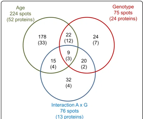
A total of 1,944 to 2,233 spots were detected on the 2D gels, and 75% were successfully matched across the differ-ent gels. A total of 224 protein spots had differdiffer-ential abun-dance (P< 0.05) in the subcutaneous adipose tissue of 110-day-old fetuses and 90-day-old fetuses (Fig.2), and 52 unique proteins were identified by MS/MS (Additional file
Adipose proteins affected by advancing gestation
The adipose tissue proteins whose abundance was modi-fied with advancing gestation could be clustered into 10 biological processes and 3 main functions (Table1). The abundance ratios of the protein spots (d 110 vs. d 90) are detailed in Additional file5. The most apparent effect of advancing gestation was observed on proteins associated with energy-related pathways, lipid binding and the cyto-skeleton. Among them, several proteins participating in hexose metabolic process [aldolase (ALDOC), lactate de-hydrogenase (LDHB), aldo-keto reductase (AKR1B1), gal-actose mutarotase (GALM), galactokinase (GALK1)], pyruvate metabolism [pyruvate dehydrogenase (PDHB), malate dehydrogenase (MDH1)] and oxidation reduc-tion [acyl-CoA dehydrogenase (ACADS), prostaglandin
reductase (PTGR2), aldehyde dehydrogenase (ALDH9A1), periredoxins (PRDX1, PRDX6)] were more abundant in the subcutaneous adipose tissue of 110-day-old fetuses than in that of 90-day-old fetuses. The ATP synthetase subunit ATP5B was the only protein in these energy-related processes with a decreased abundance between d 90 and d 110. With advancing gestation, there was an increase in the abundance of fatty acid binding proteins (FABP3, FABP4), annexin A4 (ANXA4), chaperonin heat-shock proteins (HSPD1, HSPA1B), non-sarcomeric my-osin regulatory light chains (MYL12A, MYL12B) and gel-solin (GSN). Conversely, the abundance of proteins involved in cytoskeleton organization such as cofilin (CFL1), fascin-1 (FSCN1), vinculin (VCL), tropomyosin-3 (TPM3) and actin itself (ACTB) declined with advancing gestation. Other proteins corresponding to microtubule-based processes such as stathmin (STMN1), tubulins (TUBA1B, TUBB) and desmin (DES) also had reduced abundance at d 110. The abundance of Rho GDP-dissociation inhibitor alpha (ARHGDIA) was reduced for 110-day-old fetuses compared with 90-day-old fetuses. Fi-nally, apolipoprotein A1 (APOA1) and vimentin (VIM) were two proteins identified to have proteoforms that var-ied according to age of development. Additionally, 8 proteins that were not clustered in significantly enriched functional pathways were also found to be differentially expressed between d 90 and d 110; de-tailed information can be found in Additional file 5.
a
b
Fig. 1Body weight and lipid concentration in subcutaneous adipose tissue of pure and crossbred Large White or Meishan fetuses gestated by sows of either breed. Purebred Large White (LW) or Meishan (MeiS) sows were inseminated with mixed semen from LW and MeiS boars. Pairs of purebred and crossbred (F1) fetuses were excised at d 90 or d 110 of gestation (n= 6 per age and per genotype). LW and F1_LW were from LW sows, whereas MeiS and F1_MeiS were from MeiS sows. A total of 48 fetuses were weighed (a). The triglyceride concentration was measured in the dorsal subcutaneous adipose tissue (b). Analysis of variance was used to determine the effects of gestational age, fetal genotype and the interaction between age and genotype (A × G). Least squares means sharing a common superscript letter did not significantly differ (P> 0.05)
Table 1Main biological processes and functional categories of proteins in subcutaneous adipose tissue of pig fetuses as affected by age of development
Functional annotationa P-value Enrich. Identified proteinsb
Cluster 1 (E score = 3.70) d 110≥d 90:
Pyruvate metabolism < 0.001 20.5 LDHB, MDH1, PDHB, AKR1B1,
Oxidation reduction < 0.001 6.2 ACADS, PTGR2, ALDH9A1, PRDX1, PRDX6
Cluster 2 (E score = 2.55) d 110≥d 90:
Glycolysis < 0.001 13.7 LDHB, MDH1, PDHB, ALDOC,
Hexose process < 0.001 9.0 GALM, GALK1, ALDH9A1
Generation of precursor metabolites and energy 0.02 4.6 d 90≥d 110:
ATP5B
Cluster 3 (E score = 2.46) d 110≥d 90:
Actin binding 0.003 7.8 GSN
d 90≥d 110: CFL1, VCL, FSCN1, TPM3
Cluster 4 (E score = 2.11) d 110≥d 90:
FABP3, FABP4, APOA1 d 90≥d 110: VCL, APOA1
Lipid binding < 0.001 85.4
Cluster 5 (E score = 2.01) d 110≥d 90:
HSPD1, HSPA1B d 90≥d 110: HSPA8, CLIC1
Response to protein stimulus 0.005 10.8
Molecular chaperone < 0.001 88.8
Cluster 6 (E score = 1.67) d 110≥d 90:
GSN, MYL12A, MYL12B, CRKL d 90≥d 110:
CFL1, ACTB, VCL
Regulation of actin cytoskeleton 0.001 5.3
Cluster 7 (E score = 1.59) d 110≥d 90:
HSPD1, HSPA1B, PHB, PRDX1, ANXA4 d 90≥d 110:
CFL1, LGALS1, ARHGDIA
Regulation of apoptosis 0.020 2.9
Cluster 8 (E score = 1.47) d 110≥d 90:
GSN
Cytoskeleton organization 0.015 4.0
Actin filament organization 0.02 12.0 d 90≥d 110:
TUBA1B, TUBB, CFL1, FSCN1, TPM3, DES, STMN1
Cluster 9 (E score = 1.46) d 110≥d 90:
CRKL, APOA1 d 90≥d 110: ARHGDIA, CFL1, APOA1
Ras protein signal transduction 0.005 10.9
Rho protein signal transduction 0.007 22.8
Cluster 10 (E score = 1.45) d 110≥d 90:
ALDOC, ACADS, FABP3, FABP4 d 90≥d 110:
AHSG, CFL1, SERPINA1
Response to endogenous stimulus 0.002 5.0
Response to hormone stimulus 0.03 3.9
Cluster 11 (E score = 1.41) d 110≥d 90:
PRDX1, PRDX6 d 90≥d 110: PDIA6
Cell redox homeostasis 0.020 13.7
Cluster 12 (E score = 1.18) d 110≥d 90:
TPM3, APOA1, VIM d 90≥d 110:
ATP5B, CFL1, ACTB, VCL, ARHGDIA, APOA1, VIM
Cell motion 0.001 4.8
Cluster 13 (E score = 1.10) d 110≥d 90:
CLEC3B, APOA1 d 90≥d 110:
AFP, AHSG, SERPINA1, APOA1
Plasma < 0.001 20.6
a
The E score of the cluster was measured by minus log transformation of the geometric mean of the modified Fisher Exact EASE Scores of all annotation terms that belong to this cluster, and it was intended to order the relative importance of the different clusters. The fold enrichments (enrich.) of specific individual annotation terms were also indicated for the most biologically-informative terms within each cluster
bLists of regulated proteins participating to each cluster were indicated. d 110 > d 90: proteins having a higher abundance in subcutaneous adipose
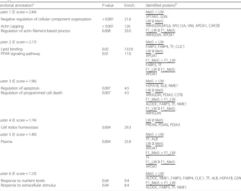
Adipose proteins affected by fetal genotype
Most of the proteins whose abundance was affected by fetal genotype (Table 2) participated in biological path-ways that were shown to differ between the two gesta-tional stages. Supporting information is provided in Additional file 6. In particular, proteins responding to nutrient levels such as aldolase (ALDOC), nucleoside di-phosphate kinase (NME1) and fatty acid binding pro-teins (FABP3, FABP4) were more abundant in MeiS fetuses than in LW fetuses. Similarly, proteins involved in the response to oxidative stress such as the heat shock protein HSPA1B, serum albumin (ALB) and the chloride intracellular channel CLIC1 were more abundant in MeiS fetuses than in LW fetuses. Other than CLIC1, the abundance levels of these proteins in F1_MeiS fetuses
were close to those measured in MeiS littermates, whereas in F1_LW fetuses, they were generally intermediate be-tween those of pure LW and pure MeiS fetuses. As a consequence, FABP3, ALDOC, NME1 and TF in adipose tissue were more abundant in crossbred fetuses gestated in a MeiS uterus than in crossbred fetuses gestated in an LW uterus. In the category of proteins regulating cytoskel-eton organization, gelsolin (GSN) and spectrin (SPTAN1) were more highly expressed in MeiS fetuses than in LW fetuses. By contrast, the abundance of other proteins par-ticipating in cytoskeleton organization, such as capping actin protein of muscle Z-line beta subunit (CAPZB) and myosin light chains 6 (MYL6) and 12A (MYL12A) were lower in MeiS fetuses than in LW fetuses; the abundance of these proteins in F1 fetuses was close to the levels
Table 2Main biological processes and functional categories of proteins in subcutaneous adipose tissue of pig fetuses as affected by genotype
Functional annotationa P-value Enrich. Identified proteinsb
Cluster 1 (E score = 2.44) MeiS > LW:
SPTAN1, GSN LW≥MeiS:
ARHGDIA,MYL6, MYL12A, VIM, APOA1, CAPZB F1_LW≥F1_MeiS:
ARHGDIA, APOA1 Negative regulation of cellular component organization < 0.001 21.6
Actin capping
Regulation of actin filament-based process
< 0.001 0.008
126 20.0
Cluster 2 (E score = 2.17) MeiS > LW:
FABP3, FABP4, TF, CLIC1 LW≥MeiS:
APOA1
F1_MeiS > F1_LW: FABP3, TF F1_LW≥F1_MeiS: APOA1
Lipid binding
PPAR signaling pathway
0.02 0.01
133.0 17.0
Cluster 3 (E score = 1.96) MeiS > LW:
HSPA1B, ALB, NME1 LW≥MeiS:
ARHGDIA, PDIA3, CSTB F1_MeiS > F1_LW: ALDOC, FABP3, TF, NME1 F1_LW≥F1_MeiS: ARHGDIA Regulation of apoptosis
Regulation of programmed cell death
0.007 0.007
4.5 4.5
Cluster 4 (E score = 1.74) LW≥MeiS:
PRDX6, PDIA6, PDIA3
Cell redox homeostasis 0.004 29.3
Cluster 5 (E score = 1.40) MeiS > LW:
TF, ALB LW≥MeiS: APOA1
F1_MeiS > F1_LW: TF
F1_LW≥F1_MeiS: APOA1
Plasma 0.004 25.8
Cluster 6 (E score = 1.23) MeiS > LW:
ALDOC, NME1, FABP3, FABP4, CLIC1, TF, ALB, HSPA1B, GSN F1_MeiS > F1_LW:
ALDOC, FABP3, TF, NME1 Response to nutrient levels
Response to extracellular stimulus
0.04 0.04
9.4 8.4
a
The E score of the cluster was measured by minus log transformation of the geometric mean of the modified Fisher Exact EASE Scores of all annotation terms that belong to this cluster, and it was intended to order the relative importance of the different clusters. The fold enrichments (enrich.) of specific individual annotation terms were also indicated for the most biologically-informative terms within each cluster
b
measured in LW fetuses. In addition, ARHGDIA was less abundant when fetuses grew in a MeiS uterus (i.e., MeiS and F1_MeiS) than when fetuses grew in an LW uterus (i.e., LW and F1_LW). Similarly, proteins with an anti-oxidative capacity (PRDX6) and participating in apoptotic cell clearance such as protein disulfide isomerases (PDIA3 and PDIA6) had a lower abundance in MeiS fetuses than in LW fetuses. An intracellular thiol protease inhibitor identified as type 1 cystatin (CSTB) was also less highly expressed in MeiS than in LW fetuses. The abundance levels of these redox proteins in F1 fetuses were inter-mediate between those found in the adipose tissue of MeiS and LW fetuses.
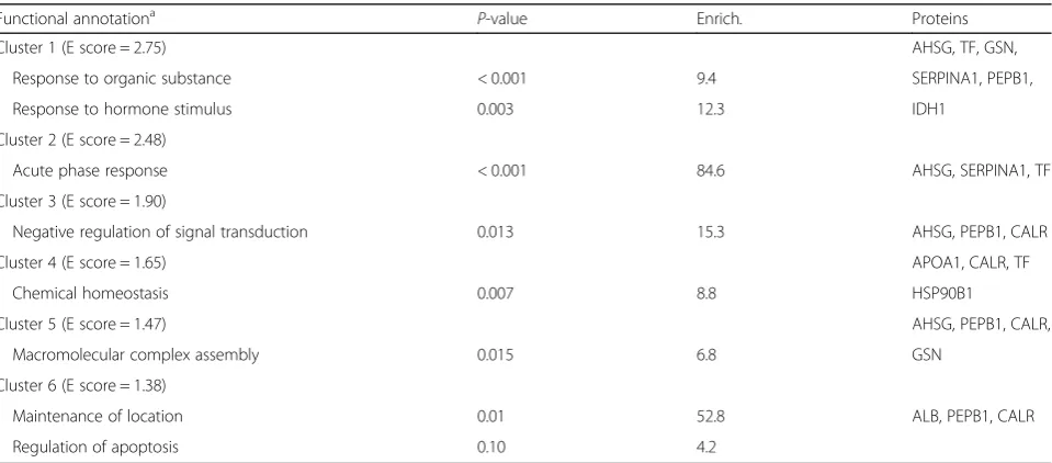
Interaction effects between developmental age and fetus genotype
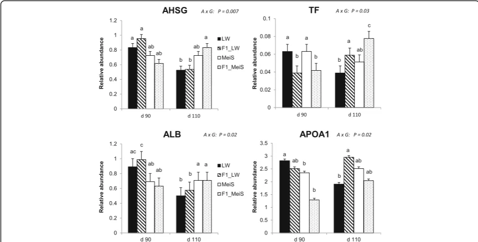
Proteins affected by an interaction (P< 0.05) between de-velopmental age and fetal genotype were involved in two main pathways: one is associated with responses to or-ganic substances, and the other is related to the acute-phase response (Table 3). The complete list of proteins is given in Additional file7. Various adipose proteins known to be secreted were identified. In particular, fragments of alpha-2-HS-glycoprotein (AHSG) as well as albumin (ALB) exhibited reduced abundance with advancing gestation in LW and F1_LW fetuses, whereas the abundance of these proteins remained almost constant between the two ages in the adipose tissue of MeiS and F1_MeiS fetuses (Fig. 3). Apolipoprotein A1 (APO1) was another protein with a lower abundance at d 110 than at d 90 in LW fetuses,
whereas its abundance was stable (F1_LW and MeiS) or in-creased (F1_MeiS) with advancing gestation in the three other genotypes. The abundance of transferrin (TF) was significantly lower in F1 fetuses than in their purebred littermates at d 90 but was the highest in the crossbred genotypes at d 110. Among other identified proteins, isoci-trate dehydrogenase (IDH1) was also oppositely regulated in MeiS and LW fetuses during the last month of gestation. Indeed, this protein was less abundant in 110-d-old fetuses than in 90-day-old fetuses of the LW genotype, but it was higher in 110-day-old than in 90-day-old MeiS fetuses (Additional file 7: Table S7). As a consequence, IDH1 was approximately twofold more abundant in adi-pose tissue of pure MeiS fetuses than in that of pure LW fetuses at d 110 of gestation.
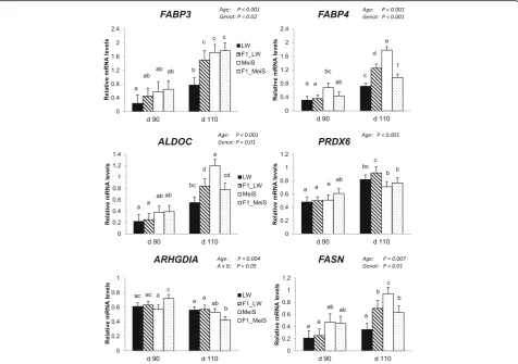
Expression levels of target genes
The expression levels of different genes were examined by qPCR to support and extend the proteomics data. As ex-pected, both developmental age and fetal genotype affected the expression levels of the adipocyte-type (FABP4) and heart/muscle-type (FAPB3) fatty acid binding proteins as well as that of aldolase (ALDOC). Differences between pure LW and pure MeiS fetuses were clearly observed at d 110, with the lowest expression level in LW fe-tuses (Fig. 4). Similarly, the mRNA levels ofPRDX6 in-creased with advancing gestation; however, there was no significant difference among the four fetal genotypes. Con-versely, a decrease in ARHGDIA mRNA level was ob-served with advancing age (P< 0.001), and this decrease
Table 3Main functional categories of proteins affected by an interaction between genotype and developmental age effects in
subcutaneous adipose tissue of pig fetuses
Functional annotationa P-value Enrich. Proteins
Cluster 1 (E score = 2.75) AHSG, TF, GSN,
Response to organic substance < 0.001 9.4 SERPINA1, PEPB1,
Response to hormone stimulus 0.003 12.3 IDH1
Cluster 2 (E score = 2.48)
Acute phase response < 0.001 84.6 AHSG, SERPINA1, TF
Cluster 3 (E score = 1.90)
Negative regulation of signal transduction 0.013 15.3 AHSG, PEPB1, CALR
Cluster 4 (E score = 1.65) APOA1, CALR, TF
Chemical homeostasis 0.007 8.8 HSP90B1
Cluster 5 (E score = 1.47) AHSG, PEPB1, CALR,
Macromolecular complex assembly 0.015 6.8 GSN
Cluster 6 (E score = 1.38)
Maintenance of location 0.01 52.8 ALB, PEPB1, CALR
Regulation of apoptosis 0.10 4.2
a
was much more accentuated in F1_MeiS than in the 3 other genotypes.
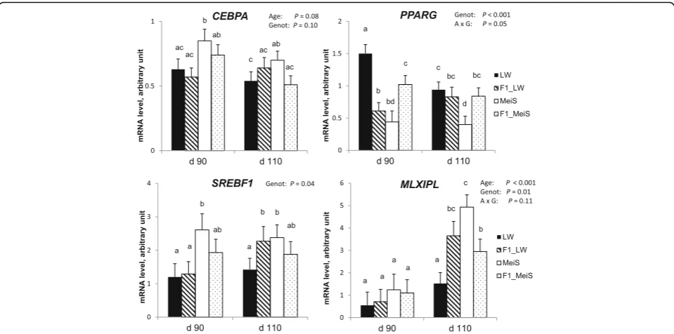
To extend this analysis, we also studied genes known to play a significant role in adipogenesis and lipogenesis. The expression level of FASN increased (P< 0.01) with gestational age (Fig.4). At d 110 of gestation,FASN ex-pression was higher in MeiS fetuses than in LW fetuses and did not differ between F1 crossbred genotypes. Simi-larly, MLXIPL was up-regulated (P< 0.001) by advancing gestation (Fig. 5). At d 110, its expression was higher in the adipose tissue of pure MeiS fetuses than in that of pure LW fetuses (P< 0.05) and intermediate in the two types of F1 fetuses.SREBF1expression was not affected by age during the studied period, but it exhibited a greater level of expression in pure MeiS fetuses than in pure LW fetuses at both time points (P< 0.05); its expression level was almost similar in F1_LW and F1_MeiS fetuses. The expression level of CEBPA, a gene involved in cell cycle regulation and homeostasis, tended to decrease (P= 0.08) between d 90 and d 110 of gestation; the highest levels were found at d 90 in the adipose tissue of pure MeiS fe-tuses and, to a lesser extent, of crossbred F1_MeiS. Finally,
PPARG was significantly down-regulated during the
last month of gestation in the adipose tissue of pure LW
fetuses only. At both time points, it was more highly expressed in pure LW fetuses and less highly expressed in pure MeiS fetuses.
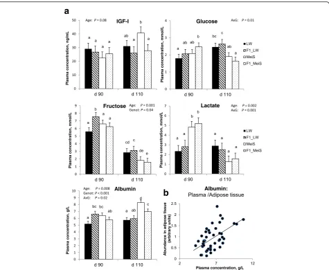
Plasma concentrations of nutrients and IGF-I
Plasma IGF-I concentration increased during late gesta-tion in pure MeiS fetuses, whereas it remained almost stable in the three other genotypes (Fig. 6a); it was the highest in pure MeiS fetuses at d 110. There was an interaction effect of developmental age and fetal geno-type on the circulating concentrations of the studied nu-trients (except fructose). Plasma glucose concentration increased from d 90 to d 110 in pure and crossbred fe-tuses gestated by the LW sows but remained stable in pure MeiS and decreased during this period in F1_MeiS fetuses. At d 110, plasma glucose tended to be lower (P= 0.10) in fetuses gestated by MeiS sow than in fetuses from LW sows. Plasma fructose decreased (P< 0.001) from d 90 to d 110 in all genotypes. The circulating concentra-tion of fructose was the highest in F1 fetuses gestated by LW sows at both ages, and it was the lowest in pure and crossbred fetuses gestated by MeiS sows at d 110. Blood fructose was about threefold higher than glucose in fetuses at d 90, whereas differences were no longer observed at
d 110. The circulating concentration of lactate remained al-most stable in fetuses gestated by LW sows but dramatic-ally decreased (P< 0.05) from d 90 to d 110 in fetuses gestated by MeiS sows. The plasma concentration of al-bumin increased (P< 0.01) from d 90 to d 110 in MeiS and F1_MeiS but remained almost stable during this period in LW and F1_LW fetuses. The plasma albumin concentration was greater in MeiS fetuses than in LW fe-tuses at both ages. At d 110, it was also greater in cross-bred fetuses that grew in MeiS uteri (F1_MeiS) than those developing in LW uteri (F1_LW). There was a positive cor-relation (r= 0.55) between albumin concentration in plasma and albumin abundance in fetal adipose tissue (Fig.6b).
Discussion
Approximately 60% of uterine energy deposition in sows occurs during the last 30 d of gestation, [25] and the body weight of pig fetuses almost doubles (present results; [26])
during this period. Previous studies have proposed d 90 of gestation as the midway point of adipose tissue develop-ment in pig fetuses [27]. The present study brings new clues that allow a better understanding of the maturation of pig adipose tissue during the last month of gestation.
Identification of proteomic changes in subcutaneous adipose tissue of pig fetuses due to advancing gestation
Proteins involved in 10 biological pathways underwent pronounced changes from d 90 to d 110 after concep-tion. Specifically, they were associated with metabolism, the cytoskeleton, redox, chaperones, Rho protein signal transduction regulation, apoptosis and others; these pathways correspond to the categories of proteins found to be regulated during adipocyte differentiation in cell culture models [28–30]. Most of these proteins that in-crease in abundance with advancing gestation are known to play important roles in energy metabolic processes.
This suggests an increased capacity for carbohydrate handling and oxidation in the intact adipose tissue of pig fetuses from d 90 to d 110, which is in agreement with previous histochemical studies [31–33] and molecular analyses [23–34] showing an increase in the activity of lipogenic enzymes and a decrease in ATPase activity in pig adipose tissue with advancing gestation. During the last month of gestation, ANXA4, a protein playing roles in carbohydrate recognition [35], and gelsolin (GSN), a calcium-regulated protein having specific roles in pro-moting lipogenic genes in 3 T3-L1 adipocyte cells [36], increase in abundance, which also supports this view. These changes associated with the marked increase in expression (both at the mRNA and protein levels) of FASN, the key enzyme catalyzing fatty acid synthesis, and FABP4, the adipocyte-type fatty acid binding protein, may explain the doubling in adipose tissue triglyceride content during this period. Conversely, we showed a decreased abundance with advancing gestation in different fragments of cytoskeletal proteins, notably, several related to tubulin (such as α- and β-tubulins and stathmin) and actin (such as TPM3, a protein that provides stability to actin filaments, and β-actin itself ). These changes are likely associated with the programmed morphological changes that occurr during adipogenic differentiation [28–30,37,38].
Genotype-associated differences in the abundance of several metabolic proteins suggest better anabolic function of adipose tissue in pure MeiS fetuses
When analyzed a few days before birth, the pure MeiS fetuses exhibited the highest triglycerides content in sub-cutaneous adipose tissue, although they have lower body weight than pure LW or crossbred F1 fetuses. This find-ing contrasts with the positive relationship usually re-ported between body fat mass and birth weight when these traits were analyzed within European breeds [8]. Various proteins involved in lipid metabolism (ALDOC, FABP3, FABP4, and GSN) had a greater abundance in the adipose tissue of MeiS fetuses than in that of LW fe-tuses. A greater abundance in IDH1, an enzyme involved in cytosolic NADPH production as an essential cofactor for lipogenesis, was also found in the MeiS fetuses at d 110 of gestation. At the mRNA level, we showed that
FASN, the key lipogenic enzyme; MLXIPL, a pivotal transcriptional mediator of glucose-related stimulation of lipogenic genes in porcine primary adipocytes [39]; andSREBF1, another classically recognized gene regulator controlling the expression of genes participating in the use of glucose for anabolic purposes [40], were more highly expressed in the adipose tissue of pure MeiS than in that of pure LW fetuses. Altogether, these data strongly argue that the adipose tissue of pure MeiS fetuses has superior
metabolic function. This may explain why Meishan neo-nates exhibited a greater number of enlarged adipose cells than did the Large White neonates [11]. Because contrast-ing time-course changes between pure MeiS and LW fe-tuses were observed in the expression levels of PPARG and CEBPA,two transcriptional regulators of adipogene-sis, time points earlier in gestation may be considered in further studies to clarify breed-associated differences in adipose cell development. Finally, three redox proteins (PRDX6, PDIA3, and PDIA6) were less abundant in MeiS than in LW fetuses at the two gestational ages. Up-regulation of redox proteins has been reported to occur during adipocyte development, when cells must cope with increased production of reactive oxygen species (ROS) [41], and two proteins of the peroxiredoxin family
(PRDX1, PRDX6) were also found to be more abundant in pig adipose tissue at d 110 than at d 90 of gestation in the present study. Taken together, these data sug-gest that ROS generation was lower and/or that ROS scavenging was more effective in MeiS adipose tissue. The increased abundance of CLIC1, a chloride intracellu-lar channel whose knockdown in cells led to increased sensitivity to hydrogen peroxide [42], was another element supporting this assumption.
Differences in the abundance of adipose proteins with regulatory roles in cellular growth were revealed between fetal genotypes
At the two examined time points of gestation, proteins par-ticipating in the regulation of cytoskeleton organization and
a
b
filament dynamics (MYL6, MYL12A, VIM and CAPZB) were decreased in abundance in the subcutaneous adipose tissue of pure MeiS fetuses. These differences may reflect the lower number of connective tissue fibers reported to exist in the interstitial space of the adipose tissue in neo-nates from obese breeds compared with lean breeds [43]. Because proteins included in this pathway exhibited de-creased abundance with advancing gestation, these data further argue that the adipose tissue of MeiS fetuses just before birth is more mature than that of LW fetuses or hy-brids. During stem cell lineage commitment, several pro-teins are known to activate a number of downstream effectors regulating cytoskeletal organization, thereby exert-ing effects on cellular growth. Among them, Rho proteins including Rho GDP-dissociation inhibitor alpha (ARHG-DIA) are important actors in the regulation of cell growth [44, 45]. In particular, the down-regulation of ARHGDIA may promote the proliferation, cell cycle progression and migration of different cell types [46]. Moreover, Rho GTPases are known to regulate the phosphorylation of myosin regulatory light chains [47]. In the present study, ARHGDIA protein and the myosin light chain elements MYL12A and MYL6 were less abundant in the adipose tissue of MeiS fetuses. In addition, we showed that cysta-tin B (CSTB), a protein encoded by a gene whose down-regulation increased cell viability and decreased apoptosis [48], had a lower abundance, whereas spectrin (SPTAN1), a gene involved in tumorigenesis [49], was more abundant in the adipose tissue of pure MeiS fetuses than in that of pure LW fetuses at both gestational time points. Altogether, these protein changes suggested that the regulation of the cell cycle in adipose tissue may be different during the last month of gestation in purebred fetuses of fat vs. lean breeds. In support of this interpretation, preadipocytes have been reported to have an elevated proliferation rate in MeiS compared with LW primary cell cultures [50]. In MeiS fe-tuses, the sharp increase from d 90 to d 110 in the circulat-ing concentration of IGF-I, a member of the larger family of insulin-related growth factors, may have promoted prea-dipocyte proliferation [51] in this genotype.
Maternal influences were important factors explaining differences in the maturity grade of adipose tissue between pig fetuses
Comparing the relative abundance of adipose proteins be-tween pure and crossbred fetuses may reveal new, relevant information regarding the main effects of fetal genetics and uterine environment on the mechanisms involved in adipose tissue maturation. In crossbred fetuses gestated by MeiS sows, the abundance levels of metabolically active proteins known to be responsive to nutrient levels were close to those found in their pure MeiS littermates, whereas in F1_LW fetuses, they were generally intermedi-ate between the levels in pure LW and pure MeiS fetuses.
In particular, FABP3, ALDOC, NME1 and TF in adipose tissue were more abundant in crossbred fetuses that devel-oped in a MeiS uterus than in those that develdevel-oped in an LW uterus. This suggests that uterine influences are im-portant for the metabolic function of adipose tissue during gestation. Differences in placental and endometrial vascu-larity between MeiS and LW sows during the late stages of gestation may be involved. Indeed, the vascular density of the MeiS placenta increases by about one-third between d 90 and d 110 of gestation while remaining almost con-stant in LW dams [52]. It is also recognized that uterine type determines conceptus size, whereas conceptus geno-type controls placental efficiency [52]. In the current study, circulating concentrations of glucose and fructose, two energy-yielding nutrients, were lower in the arterial blood of 110-day-old pure and crossbred fetuses gestated by MeiS sows than in that of fetuses gestated by LW sows. Time-dependent decreases in circulating glucose and lac-tate concentrations were also observed in fetuses geslac-tated by MeiS sows, whereas plasma glucose concentration in-creased with advancing gestation in fetuses gestated by LW sows (present results) and LW × Landrace sows [23]. This suggests that circulating nutrient concentrations might be less important than blood flow for adipose tissue metabolism. Conversely, the abundance of proteins par-ticipating in cytoskeleton organization (except ARHGDIA) in the adipose tissue of F1 fetuses was close to that mea-sured in pure LW fetuses. The lack of differences in the abundance of these proteins between the two types of crossbred fetuses suggests that parental genes are more in-fluential than the uterine environment in the regulation of adipose cell growth.
Adipose secreted proteins may be indicative of accelerated maturation during gestation
Possible indicators of accelerated maturation of adipose tissue in fetuses should be sought among the few identi-fied proteins that show different time courses depending on the fetal genotype. This interaction between age and genotype especially affected certain secreted proteins, namely, albumin, transferrin and fetuin-A, that were previously identified in culture media conditioned by porcine stroma-vascular preadipose cells [16] and serum from fetal and neonatal pigs [53]. In the adipose tissue of pure and crossbred fetuses gestated by LW sows, the abundance of fetuin-A (the protein encoded by the gene
accelerated adipose tissue maturation in the offspring of MeiS sows during late gestation. Fetuin-A mediates the cross-talk between liver and adipose tissue, and changes in fetuin-A abundance are associated with deficient fetal growth and complications in later life [55]. Its precise role in adipose tissue, however, remains to be clarified. Another adipose tissue protein showing an opposite time-course be-tween pure LW and MeiS fetuses was albumin. In mice, plasma albumin is taken up by white adipose tissue at an al-most constant rate from fetal to early postnatal life [56]. Moreover, the albumin concentration in the plasma of pig fetuses increases during late gestation (present results; [23]) but is lower in LW fetuses than in MeiS fetuses at d 110. Similar to the present findings, the serum concentration of albumin was found to be lower in pig neonates from lean breeds than in those from obese breeds [12,13] and lower in neonates from highly selected white-type sows than in neonates from less selected sows [7]. Finally, high plasma albumin concentration has been suggested to be indicative of advanced development and increased physiological maturity in pig fetuses [57]. Importantly, albumin may regulate fatty acid uptake by the tissue [58], such that inter-individual variations in adipose tissue albumin content could be important for early survival when piglets feed on colostrum and milk.
Conclusion
During the last three weeks of gestation, increased expres-sion of metabolic proteins with roles in energy generation and lipid binding together with reduced expression of pro-teins involved in cytoskeleton organization contributed to the rapid development of pig adipose tissue. During this period, differences between fetus genotypes in the abun-dance of adipose proteins involved in energy metabolism likely contributed to the higher maturity grade of adipose tissue of fetuses gestated by Meishan sows. These proteins may be regulated by differences in maternal environment, such as placental efficiency. Altogether, however, the num-ber of proteins that were differentially regulated by fetus genotype was one-third the number regulated by gesta-tional age. This confirmed that although some cellular and metabolic differences can be observed between obese and lean pig breeds at d 110 of gestation [32], maternal obesity has less visible influence on adipose tissue during gestation than during the postnatal period [59]. Acceler-ated developmental changes in adipose tissue during the fetal period may program the greater fat accretion ob-served in the Meishan breed during postnatal life.
Additional files
Additional file 1:Running plan for the 48 samples in Differential Gel Electrophoresis. (DOCX 29 kb)
Additional file 2:Representative two dimensional gels of fetal adipose proteins stained by silver nitrate. The first image corresponds to subcutaneous adipose tissue d 90, the second one at d 110 of gestation for pure Large White fetuses. (DOCX 1257 kb)
Additional file 3:Primers for target gene expression by qPCR. (DOCX 30 kb)
Additional file 4:Mass parameters for protein identities. (XLS 474 kb)
Additional file 5:Proteins showing a differential abundance in adipose tissue with developmental age. (DOCX 42 kb)
Additional file 6:Proteins showing a differential abundance in adipose tissue according to fetus genotype. (DOCX 96 kb)
Additional file 7:Proteins in adipose tissue affected by age in a different manner according to fetus genotype. (DOCX 32 kb)
Abbreviations
2D–DIGE:Two-dimension differential gel electrophoresis; ACADS: Acyl-CoA dehydrogenase; ACTB: Actin beta; AHSG: Alpha2-HS glycoprotein; AKR1B1: Aldo-keto reductase family 1, member B1; ALB: Albumin; ALDH9A1: Aldehyde dehydrogenase 9 family member A1; ALDOC: Aldolase-C; APOA1: Apolipoprotein-A1; ARHGDIA: Rho GDP-dissociation inhibitor 1; ATP5B: ATP synthase subunit beta; CAPZB: F-actin-capping protein subunit beta; CFL1: Cofilin; CLIC1: Chloride intracellular channel 1; CTSB: Cystatin; d 110: 110 days of gestation; d 90: 90 days of gestation; DES: Desmin; FABP3: Fatty acid binding protein heart/muscle type; FABP4: Fatty acid binding protein adipocyte type; FSCN1: Fascin actin-bundling protein 1; GSN: Gelsolin; HSPA1B: Heat shock protein family A (Hsp70) member 1B; HSPD1: Heat shock protein family D (Hsp60); IDH1: Isocitrate dehydrogenase; LDHB: Lactate dehydrogenase B; LW: Large White; MDH1: Malate
dehydrogenase 1; MeiS: Meishan; MS: Mass spectrometry; MYL12A: Myosin light chain 12A; MYL6: Myosin light chain 6; PDHB: Pyruvate dehydrogenase; PDIA3: Protein disulfide-isomerase A3; PDIA6: Protein disulfide-isomerase A3; PRDX1: Peroxiredoxin-1; PRDX6: Peroxiredoxin-6; PTGR2: Prostaglandin reductase 2; ROS: Reactive oxygen species; SPTAN1: Spectrin alpha-chain; STMN1: Stathmin 1; TF: Transferrin; TPM3: Tropomyosin 3; TUBA1B: Tubulin alpha-1B; TUBB: Tubulin beta class 1; VCL: Vinculin; VIM: Vimentin
Acknowledgements
The authors are very grateful to all the staff of experimental pig facilities (INRA, GenESI, Saint-Pierre d’Amilly, France) for expert assistance in surgeries and Sandrine Tacher, Christine Tréfeu, Sophie Daré-Michelot and Nathalie Bonhomme (INRA, PEGASE, Saint-Gilles, France) for laboratory technical analyses.
Funding
The project received financial support from the French National Agency for Research (PORCINET project, ANR-09-GENMOO5). This work was also supported by grants from Biogenouest, IBiSA and Conseil Regional de Bretagne (France).
Availability of data and materials
Mean values of all data generated or analyzed during this study, mass spectrometry parameters and detailed identification of proteins are included in this published article and its additional information files. Individual data are available from the corresponding author on reasonable request.
Authors’contributions
LL, LC, HQ and YB: conceived the study and its animal design. LL, LC, YB, IL, MCP, FG and HQ participated to the animal design and sample collection; FG: coordinated the study on adipose tissue; EC and BG: generated proteomics data; MCP and HQ: generated plasma nutrient data and other phenotypic traits on fetuses; FG: generated lipid concentration data; FG and IL: generated qPCR data; FG: performed statistics, analyzed the data and interpreted the results; FG and IL: drafted the manuscript. All authors helped to draft the manuscript and read and approved the final version.
Ethics approval
Consent for publication
Not applicable
Competing interests
The authors declare that they have no competing interests.
Author details
1PEGASE, Agrocampus Ouest, INRA, 35590, Saint-Gilles, France.2Protim,
Inserm U1085, Irset, Université Rennes 1, Campus de Beaulieu, 35042 Rennes Cedex, France.3GenESI, INRA, Le Magneraud, 17700, Saint-Pierre-d’Amilly, France.4GenPhyse, INRA, INPT, INPT-ENV, Université de Toulouse, 31320 Castanet-Tolosan, France.
Received: 7 September 2017 Accepted: 9 February 2018
References
1. Gondret F, Lefaucheur L, Juin H, Louveau I, Lebret B. Low birth weight is associated with enlarged muscle fiber area and impaired meat tenderness of the longissimus muscle in pigs. J Anim Sci. 2006;84:93–103.
2. Hales CN, Barker DJ, Clark PM, Cox LJ, Fall C, Osmond C, et al. Fetal and infant growth and impaired glucose tolerance at age 64. BMJ. 1991;303:1019–22. 3. Morrison JL, Duffield JA, Muhlhausler BS, Gentili S, McMillen IC. Fetal growth
restriction, catch-up growth and the early origins of insulin resistance and visceral obesity. Pediatr Nephrol. 2010;25:669–77.
4. National Research Council. Preterm Birth: Causes, Consequences, and Prevention. Washington, DC: The National Academies Press; 2007. Richard E. Behrman, Adrienne Stith Butler, Editors, Committee on Understanding Premature Birth and Assuring Healthy Outcomes. 792 pages. 978–0–309-10159-2
5. GTTT. 2015. [Résultats Porcs Bretagne]. Agricultures&Territoires. Chambres d’Agriculture Bretagne.
6. Leenhouwers JI, Knol EF, de Groot PN, Vos H, van der Lende T. Fetal development in the pig in relation to genetic merit for piglet survival. J Anim Sci. 2002;80:1759–70.
7. Canario L, Père MC, Tribout T, Thomas F, David C, Gogué J, et al. Estimation of genetic trends from 1977 to 1998 of body composition and physiological state of large white pigs at birth. Animal. 2007;1:1409–13.
8. Morise A, Sève B, Macé K, Magliola C, Le Huërou-luron I, Louveau I. Impact of intrauterine growth retardation and early protein intake on growth, adipose tissue, and the insulin-like growth factor system in piglets. Pediatr Res. 2009;65:45–50.
9. Rehfeldt C, Lefaucheur L, Block J, Stabenow B, Pfuhl R, Otten W, et al. Limited and excess protein intake of pregnant gilts differently affects body composition and cellularity of skeletal muscle and subcutaneous adipose tissue of newborn and weanling piglets. Eur J Nutr. 2012;51:151–65. 10. Le Dividich J, Mormède P, Catheline M, Caritez JC. Body composition and
cold resistance of the neonatal pig from European (large white) and Chinese (Meishan) breeds. Biol Neonate. 1991;59:268–77.
11. Herpin P, Le Dividich J, Amaral N. Effect of selection for lean tissue growth on body composition and physiological state of the pig at birth. J Anim Sci. 1993;71:2645–53.
12. Mersmann HJ, Pond WG, Stone RT, Yen JT, Lindvall RN. Factors affecting growth and survival of neonatal genetically obese and lean swine: cross fostering experiments. Growth. 1984;48:209–20.
13. Stone RT, Campion DR, Klindt J, Martin RJ. Blood parameters and body composition in fetuses from reciprocal crosses of genetically lean and obese swine. Proc Soc Exp Biol Med. 1985;180:191–5.
14. Louveau I, Perruchot MH, Bonnet M, Gondret F. Invited review: Pre- and postnatal adipose tissue development in farm animals: from stem cells to adipocyte physiology. Animal. 2016 Nov;10:1839–47.
15. Canario L, Cantoni E, Le Bihan E, Caritez JC, Billon Y, Bidanel JP, et al. Between-breed variability of stillbirth and its relationship with sow and piglet characteristics. J Anim Sci. 2006;84:3185–96.
16. Hausman GJ, Poulos SP, Richardson RL, Barb CR, Andacht T, Kirk HC, et al. Secreted proteins and genes in fetal and neonatal pig adipose tissue and stromal-vascular cells. J Anim Sci. 2006;84:1666–81.
17. Gondret F, Guitton N, Guillerm-Regost C, Louveau I. Regional differences in porcine adipocytes isolated from skeletal muscle and adipose tissues as identified by a proteomic approach. J Anim Sci. 2008;86:2115–25.
18. Gondret F, Guével B, Com E, Vincent A, Lebret B. A comparison of subcutaneous adipose tissue proteomes in juvenile piglets with a contrasted adiposity underscored similarities with human obesity. J Proteome. 2012;75:949–61. 19. Guo J, Liu Z, Sun H, Huang Y, Albrecht E, Zhao R, et al. Lipopolysaccharide
challenge significantly influences lipid metabolism and proteome of white adipose tissue in growing pigs. Lipids Health Dis. 2015;14:68.
20. Voillet V, SanCristobal M, Lippi Y, Martin PG, Iannuccelli N, Lascor C, et al. Muscle transcriptomic investigation of late fetal development identifies candidate genes for piglet maturity. BMC Genomics. 2014;15:797. 21. Xu H, Wilcox D, Nguyen P, Voorbach M, Suhar T, Morgan SJ, et al. Hepatic
knockdown of mitochondrial GPAT1 in ob/ob mice improves metabolic profile. Biochem Biophys Res Commun. 2006;349:439–48.
22. Sarr O, Louveau I, Kalbe C, Metges CC, Rehfeldt C, Gondret F. Prenatal exposure to maternal low or high protein diets induces modest changes in the adipose tissue proteome of newborn piglets. J Anim Sci. 2010;88:1626–41. 23. Gondret F, Père MC, Tacher S, Daré S, Trefeu C, Le Huërou-Luron I, et al.
Spontaneous intra-uterine growth restriction modulates the endocrine status and the developmental expression of genes in porcine fetal and neonatal adipose tissue. Gen Comp Endocrinol. 2013;194:208–16. 24. Vandesompele J, De Preter K, Pattyn F, Poppe B, Van Roy N, De Paepe A,
et al. Accurate normalization of real-time quantitative RT-PCR data by geometric averaging of multiple internal control genes. Genome Biol. 2002;3:RESEARCH0034.
25. Noblet J, Dourmad JY, Etienne M. Energy utilization in pregnant and lactating sows: modeling of energy requirements. J Anim Sci. 1990;68:562–72. 26. Hill GM, Mahan DC. Essential and nonessential amino acid compositions of
the total litter and individual fetal pig content and accretion rates during fetal development. J Anim Sci. 2016;94:5239–47.
27. Hausman GJ, Hausman DB. Endocrine regulation of porcine adipose tissue development: cellular and metabolic aspects. In: Hollis GR, editor. Growth of the pig. Wallingford, UK: CAB Int; 1993. p. 49–74.
28. DeLany JP, Floyd ZE, Zvonic S, Smith A, Gravois A, Reiners E, et al. Proteomic analysis of primary cultures of human adipose-derived stem cells: modulation by adipogenesis. Mol Cell Proteomics. 2005;4:731–40. 29. Molina H, Yang Y, Ruch T, Kim JW, Mortensen P, Otto T, et al. Temporal
profiling of the adipocyte proteome during differentiation using a five-plex SILAC based strategy. J Proteome Res. 2009;8:48–58.
30. Welsh GI, Griffiths MR, Webster KJ, Page MJ, Tavaré JM. Proteome analysis of adipogenesis. Proteomics. 2004;4:1042–51.
31. Hausman GJ. Cellular and enzyme-histochemical aspects of adipose tissue development in obese (Ossabaw) and lean (crossbred) pig fetuses: an ontogeny study. J Anim Sci. 1985;60:1539–51.
32. Hausman GJ, Campion DR, Thomas GB. Adipose tissue cellularity and histochemistry in fetal swine as affected by genetic selection for high or low backfat. J Lipid Res. 1983;24:223–8.
33. Hausman GJ, Thomas GB. The development of the inner layer of backfat in fetal and young pigs. J Anim Sci. 1984;58:1550–60.
34. Samulin J, Berget I, Lien S, Sundvold H. Differential gene expression of fatty acid binding proteins during porcine adipogenesis. Comp Biochem Physiol B Biochem Mol Biol. 2008;151:147–52.
35. Kojima K, Yamamoto K, Irimura T, Osawa T, Ogawa H, Matsumoto I. Characterization of carbohydrate-binding protein p33/41: relation with annexin IV, molecular basis of the doublet forms (p33 and p41), and modulation of the carbohydrate binding activity by phospholipids. J Biol Chem. 1996;271:7679–85.
36. Mukherjee R, Yun JW. Long chain acyl CoA synthetase 1 and gelsolin are oppositely regulated in adipogenesis and lipogenesis. Biochem Biophys Res Commun. 2012;420:588–93.
37. Gaskins HR, Hausman GJ, Martin RJ. Regulation of gene expression during adipocyte differentiation: a review. J Anim Sci. 1989;67:2263–72.
38. Spiegelman BM, Farmer SR. Decreases in tubulin and actin gene expression prior to morphological differentiation of 3T3 adipocytes. Cell. 1982;29:53–60. 39. Zhang GH, Lu JX, Chen Y, Guo PH, Qiao ZL, Feng RF, et al. ChREBP and
LXRαmediate synergistically lipogenesis induced by glucose in porcine adipocytes. Gene. 2015;565:30–8.
40. Gosmain Y, Dif N, Berbe V, Loizon E, Rieusset J, Vidal H, et al. Regulation of SREBP-1 expression and transcriptional action on HKII and FAS genes during fasting and refeeding in rat tissues. J Lipid Res. 2005;46:697–705.
42. Qu H, Chen Y, Cao G, Liu C, Xu J, Deng H, et al. Identification and validation of differentially expressed proteins in epithelial ovarian cancers using quantitative proteomics. Oncotarget. 2016;7:83187–99.
43. Hausman GJ, Martin RJ. Subcutaneous adipose tissue development in Yorkshire (lean) and Ossabaw (obese) pigs. J Anim Sci. 1981;52:1442–9. 44. McBeath R, Pirone DM, Nelson CM, Bhadriraju K, Chen CS. Cell shape,
cytoskeletal tension, and RhoA regulate stem cell lineage commitment. Dev Cell. 2004;6:483–95.
45. Nobes CD, Hall A. Rho, rac and cdc42 GTPases: regulators of actin structures, cell adhesion and motility. Biochem Soc Trans. 1995;23:456–9.
46. Lu W, Wang X, Liu J, He Y, Liang Z, Xia Z, et al. Downregulation of ARHGDIA contributes to human glioma progression through activation of Rho GTPase signaling pathway. Tumour Biol. 2016:Oct 10; [Epub ahead of print] 47. Amano M, Ito M, Kimura K, Fukata Y, Chihara K, Nakano T, et al. Phosphorylation
and activation of myosin by rho-associated kinase (rho-kinase). J Biol Chem. 1996; 271:20246–9.
48. Zhang J, Shi Z, Huang J, Zou X. CSTB downregulation promotes cell proliferation and migration and suppresses apoptosis in gastric cancer SGC-7901 cell line. Oncol Res. 2016;24:487–94.
49. L'Espérance S, Popa I, Bachvarova M, Plante M, Patten N, Wu L, et al. Gene expression profiling of paired ovarian tumors obtained prior to and following adjuvant chemotherapy: molecular signatures of chemoresistant tumors. Int J Oncol. 2006;29:5–24.
50. Gerfault V, Louveau I, Mourot J. The effect of GH and IGF-I on preadipocytes from Large White and Meishan pigs in primary culture. Gen Comp Endocrinol. 1999;114:396–404.
51. Louveau I, Gondret F. Regulation of development and metabolism of adipose tissue by growth hormone and the insulin-like growth factor system. Domest Anim Endocrinol. 2004;27:241–55.
52. Biensen NJ, Wilson ME, Ford SP. The impact of either a Meishan or Yorkshire uterus on Meishan or Yorkshire fetal and placental development to days 70, 90, and 110 of gestation. J Anim Sci. 1998;76:2169–76.
53. Caperna TJ, Shannon AE, Blomberg LA, Garrett WM, Ramsay TG. Identification of protein carbonyls in serum of the fetal and neonatal pig. Comp Biochem Physiol B Biochem Mol Biol. 2010;156:189–96.
54. Jialal I, Devaraj S, Bettaieb A, Haj F, Adams-Huet B. Increased adipose tissue secretion of Fetuin-a, lipopolysaccharide-binding protein and high-mobility group box protein 1 in metabolic syndrome. Atherosclerosis. 2015;241:130–7. 55. Karamessinis PM, Malamitsi-Puchner A, Boutsikou T, Makridakis M, Vougas K,
Fountoulakis M, et al. Marked defects in the expression and glycosylation of alpha2-HS glycoprotein/fetuin-a in plasma from neonates with intrauterine growth restriction: proteomics screening and potential clinical implications. Mol Cell Proteomics. 2008;7:591–9.
56. Laborda J, Naval J, Calvo M, Lampreave F, Uriel J. Alpha-fetoprotein and albumin uptake by mouse tissues during development. Biol Neonate. 1989;56:332–41.
57. Stone RT, Leymaster KA. Relationships of birth weight and pre-nursing concentrations of serum albumin to survival and growth rate in swine. Growth. 1985;49:263–70.
58. Stremmel W, Pohl L, Ring A, Herrmann T. A new concept of cellular uptake and intracellular trafficking of long-chain fatty acids. Lipids. 2001;36(9):981. 59. Campion DR, Hausman GJ, Stone RT, Klindt J. Influence of maternal obesity
on fetal development in pigs. J Anim Sci. 1988;66:28–33.
• We accept pre-submission inquiries
• Our selector tool helps you to find the most relevant journal • We provide round the clock customer support
• Convenient online submission • Thorough peer review
• Inclusion in PubMed and all major indexing services • Maximum visibility for your research
Submit your manuscript at www.biomedcentral.com/submit