CASE REPORT
Neuropathological findings suggestive
for a stroke in an alpaca (
Vicugna pacos
)
Sandra Schöniger
1*, Enrika Schütze
1, Dominik Michalski
2, Joana Puchta
3, Matthias Kaiser
4and Wolfgang Härtig
3Abstract
Background: This case report describes a focal brain lesion in an alpaca (Vicugna pacos). Although this is a restricted study based on a single animal, neuropathological features are reported that are most likely attributed to a vascular event with either ischemic or hemorrhagic pathology. Concerning translational issues, these findings extend neuro-vascular unit concept to the alpacas’ brain and qualify a larger panel of stroke tissue markers for further exploration of ischemic or hemorrhagic consequences beyond the usually used small animal models in stroke research.
Case presentation: A brain lesion indicative of a stroke was diagnosed in a 3-year-old female alpaca as an incidental finding during a post mortem examination. The rostral portion of the right frontal lobe contained a 1.0 × 1.5 × 1.7 cm lesion that extended immediately to the overlying leptomeninges. Microscopically, it was composed of liquefac-tive necrosis with cholesterol crystal deposition and associated granulomatous inflammation as well as vascularized fibrous connective tissue rimmed by proliferated astrocytes. Multiple fluorescence labeling of the affected brain regions revealed strong microgliosis as shown by immunostaining of the ionized calcium binding adapter molecule 1 and astrogliosis as demonstrated by enhanced immunoreactivity for glial fibrillary acidic protein. In parallel, a drastic neuronal loss was detected by considerably diminished immunolabeling of neuronal nuclei. Concomitantly, up-regu-lated immunoreactivities for collagen IV and neurofilament light chains were found in the affected tissues, indicating vascular and cytoskeletal reactions.
Conclusions: Driven by these neuropathological features, the incidental brain lesion found in this alpaca strongly suggests an ischemic or hemorrhagic etiology. However, since typical hallmarks became verifiable as previously described for other species affected by focal cerebral ischemia, the lesion is more likely related to an ischemic event. Nevertheless, as such cellular alterations might be difficult to distinguish from other brain lesions as for instance caused by inflammatory processes, adjuvant observations and species-related features need to be considered for etio-logical interpretations. Indeed, the lack of neuroetio-logical deficits is likely attributed to the location of the lesion within the rostral aspect of the right frontal lobe of the alpacas’ brain. Further, fibroblast migration from the meninges likely caused the intralesional scar formation.
Keywords: Alpaca, Fluorescence labeling, Focal brain lesion, Ischemic infarction, Pathology, Vicugna pacos
© The Author(s) 2019. This article is distributed under the terms of the Creative Commons Attribution 4.0 International License (http://creat iveco mmons .org/licen ses/by/4.0/), which permits unrestricted use, distribution, and reproduction in any medium, provided you give appropriate credit to the original author(s) and the source, provide a link to the Creative Commons license, and indicate if changes were made. The Creative Commons Public Domain Dedication waiver (http://creat iveco mmons .org/ publi cdoma in/zero/1.0/) applies to the data made available in this article, unless otherwise stated.
Open Access
*Correspondence: sandra.schoeniger@vetmed.uni-leipzig.de
1 Institute of Veterinary Pathology, Faculty of Veterinary Medicine, Leipzig University, Leipzig, Germany
Page 2 of 9 Schöniger et al. Acta Vet Scand (2019) 61:1
Background
In human beings, ischemic stroke represents a major cause of permanent disability and death [1]. The etiology is often multifactorial with the contribution of predispos-ing conditions such as atherosclerosis, diabetes mellitus, arterial hypertension, hypercoagulability and cardiac arrhythmia [2].
With the intention to explore the underlying mecha-nisms for stroke, several animal models were developed during the last decades, while limitations still exist refer-ring to the transferability to the human condition [3, 4]. As a possible reason, the brain anatomy of the used ani-mal species has been discussed in a critical manner, lead-ing to the recommendation to consider gyrencephalic species in more advanced study phases [5].
On the cellular level, experimental stroke was demon-strated to cause remarkable morphological changes in various neural cells and the extracellular matrix, support-ing the concept of the neurovascular unit that describes simultaneous ischemic reactions in a functionally associ-ated complex of diverse cell populations [6, 7].
Cardiovascular accidents in domestic animals are less commonly reported and most cases of spontaneous brain infarcts have been described for cats and dogs [8–10]. In dogs, they are most frequently attributed to different types of emboli, although a hypercoagulable status is also recognized as an important factor [11, 12]. In compari-son, cardiomyopathy-induced thromboembolism and hypertension related to renal failure are the most fre-quent risk factors for brain infarcts in cats [13].
The aim of this study was to explore a focal brain lesion in an alpaca (Vicugna pacos) while providing a macro-scopic and concise histochemical characterization of affected brain tissue with a special emphasize to the neu-rovascular unit concept and, thereby, some arising paral-lels with an ischemic infarction.
This is the first reported case of a stroke in an alpaca. Consequently, the investigation into the resulting altera-tions of cellular elements and the vasculature will extend the translational perspective of neurovascular changes due to focal brain injury such as ischemia or hemorrhage to the alpacas’ brain, and thus beyond the usually applied small animal models in the field of stroke research.
Case presentation
A 3-year-old female alpaca was submitted to the Clinic for Ruminants and Swine, Leipzig University with a clini-cal history of markedly reduced food and water intake. At admission the animal presented dehydrated and showed salivation as well as inappetence. Initially, feces were dry with mucous and traces of blood; after 1–2 days defeca-tion was watery and bloody and then nearly completely ceased. Since its admission to the clinic, the alpaca had
received intravenous infusions of glucose and electrolyte solutions as well as antiphlogistic and antibiotic treat-ments. First the general condition of the alpaca was sta-ble, and it showed intake of solely a little bit grass and some water. One day prior to its death, its general con-dition markedly deteriorated; the alpaca was recumbent with colicky abdominal pain. The animal deceased spon-taneously 4 days after its admission to the clinic and was submitted for a complete post mortem examination.
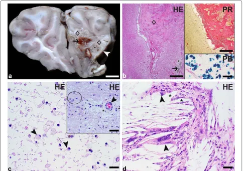
At necropsy, the carcass was found in a good nutri-tional condition. The subcutis, the parietal and pul-monary pleura as well as the heart showed multiple endo- and epicardial petechial to ecchymotic hemor-rhages. The myocardium, pulmonary artery, aorta, liver and kidneys showed no gross lesions. The abdominal cav-ity contained 40 mL of serosanguinous effusion. There was a moderate fibrinous to fibroblastic peritonitis with adhesions between the intestinal loops as well as well as the stomach. Multifocal moderate acute serosal hemor-rhages were observed as well. The mucosa of the 2nd and 3rd stomach compartment (C2 and C3) was diffusely dark red indicative of acute gastritis. Jejunum, ileum and colon displayed a marked acute to subacute diphtheroid-necrotizing enteritis. Mesenteric lymph nodes were mod-erately enlarged. Lungs were incompletely collapsed with acute alveolar edema. The right frontal lobe contained a 1.5 × 1.0 × 1.7 cm lesion with loss of normal cortical and medullary architecture that extended within the menin-ges. In the center of the lesion, the brain tissue was beige and soft with multifocal mild cavitation and contained a few small foci of brownish discoloration. The peripheral parts of the lesion were composed of firm whitish-beige tissue interpreted as fibrous connective tissue (Fig. 1a).
Samples of multiple organs including the stomach com-partments C2 and C3, small and large intestines, mesen-teric lymph nodes, lungs and brain (cerebrum with the lesion, additional parts of the ipsi- and contralateral cer-ebrum, hippocampus, cerebellum, midbrain and medulla oblongata) were fixed in 10% neutral buffered formalin, processed routinely and embedded in paraffin for micro-scopic examination. Two µm-thick sections were cut with a HM 400R Manual Sliding Microtome (Microm, Heidelberg, Germany). The microscopic evaluation was performed on hematoxylin–eosin stained tissue sections. For a better characterization of the brain lesions, a picro-sirius red staining [14] for the detection of fibrous con-nective tissue and a Prussian blue staining [15] to identify hemosiderin pigment were used.
Histopathological findings
associated with intravascular fibrin thrombi and a mul-tifocal mild fibrinous serositis. Additionally, some intral-esional vessels showed segmental to diffuse fibrinoid necrosis of their walls. Enlarged mesenteric lymph nodes had a diffuse marked acute suppurative lymphadenitis. Within the lung tissue, alveolar walls were lined by hya-line membranes and their lumens contained some fibrin stands and edema fluid.
In the right frontal lobe, the lesion was located imme-diately adjacent to the anterior horn of the right ventricle and extended into the lateral and medial leptomenin-ges. It was composed of immature and mature fibrous
connective tissue with a moderate vascular proliferation and several areas of liquefactive necrosis; neurons were completely lost. The fibrous connective tissue was high-lighted by a picrosirius red staining. Necrotic areas were infiltrated by moderate numbers of gitter cells as well as by a few gemistocytes and contained variably-sized cystic spaces. There were multifocal small aggregates of needle-like clear structures (consistent with cholesterol crystals) surrounded by a moderate granulomatous inflammation characterized by the presence of histiocytic cells includ-ing several multinucleate giant cells. The grossly noted brownish discoloration was attributed to the presence
Fig. 1 Focal brain lesion in an alpaca (Vicugna pacos): macroscopic and light microscopic findings. a Transverse section through the frontal lobes: The right frontal lobe contains a focal lesion that extends immediately to the leptomeninges of the lateral and medial sites (white arrowhead). The lesion is characterized by the loss of normal brain architecture within cortex and medulla. It consists of firm whitish-beige tissue (diamonds) as well as small areas of soft beige tissue (asterisks) and shows multifocal brownish discoloration (arrow). Bar: 1 cm. b By the microscopic examination, the firm whitish-beige tissue is identified as vascularized fibrous connective tissue (diamond) and the soft beige areas represent liquefactive necrosis (asterisk). The multifocal brown discoloration is attributed to the presence of sheets of siderocytes (arrow) revealed by hematoxylin–eosin (HE) staining. Bar: 200 µm. Upper inset: The fibrous connective tissue is highlighted in red color using a picrosirius red stain. Bar: 200 µm. Lower inset: The siderocytes are marked in blue color with a Prussian blue stain. Bar: 20 µm. c The area of liquefactive necrosis is infiltrated by moderate numbers of gitter cells (arrowheads) as shown by HE staining. Bar: 20 µm. Inset: Areas of necrosis and scar tissue formation are bordered by proliferated astrocytes (circle). An intralesional vessel (arrowhead) shows moderate perivascular edema (asterisk) visualized by HE staining. Bar: 40 µm. d
Page 4 of 9 Schöniger et al. Acta Vet Scand (2019) 61:1
of sheets of mononuclear cells (microglia/macrophages) containing intracytoplasmic brown coarse pigment that was identified as hemosiderin by the Prussian blue stain-ing. The lesion was rimmed by proliferated astrocytes (Fig. 1b–d). Intralesional vessels were characterized by surrounding empty spaces consistent with moderate perivascular edema. However, intralesional vessels and vessels of examined unaltered brain areas showed no evi-dence of intravascular thrombi, vasculitis or degenerative changes. A thorough microscopic evaluation of the entire brain area surrounding the lesion as well as representa-tive locations of the remaining grossly unaltered brain revealed also no vascular changes; in particular, no vas-cular obstruction was detected. The immediately adjacent cortex contained some neurons with mild satellitosis.
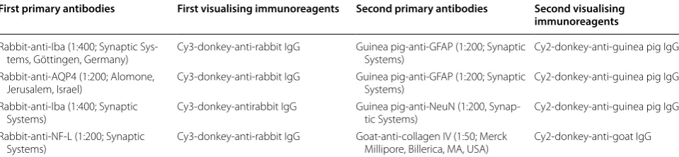
To characterize in detail the cellular and vascular alter-ations, brain sections containing the lesion and adjacent brain parenchyma were examined by triple fluorescence staining. The following primary markers were applied (Table 1): ionized calcium binding adapter protein 1 (Iba) as a marker of microglia/macrophages, Solanum tubero-sum lectin (STL) to visualize endothelial cells and micro-glia/macrophages, glial fibrillary acidic protein (GFAP) to stain astrocytic cell bodies, aquaporin 4 (AQP4) to detect astrocytic endfeet, collagen IV as a component of the vascular basement membrane, neuronal nuclear antigen (NeuN) to label neuronal nuclei and neurofilament light chain (NF-L) to verify the expression of this structural protein.
After deparaffinization, sections were heated for 25 min at 94 °C in 0.1 M citrate buffer, pH 6, for antigen retrieval. After three rinses with 0.1 M Tris-buffered saline (TBS), pH 7.4, tissue sections were incubated with different combinations of three primary markers (diluted in the blocking solution) for 20 h at room temperature (around 20 °C) as summarized in Table 1. Subsequently, tissue
sections were washed with TBS and a mixture of three different fluorochromated secondary immunoreagents (Table 1) was applied for 1 h followed by extensive rins-ing with TBS and two short washes with distilled water. Finally, sections were rinsed in distilled water, air dried and coverslipped with Entellan in toluene (Merck, Darm-stadt, Germany). The omission of primary antibodies in control experiments resulted in the expected absence of any cellular labeling. The stained sections were screened with an Axioplan fluorescence microscope (Zeiss). All pictures were made with a Biorevo BZ-9000 microscope (Keyence, Neu-Isenburg, Germany).
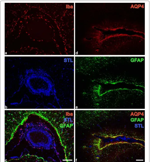
Four variants of triple fluorescence labeling contributed to the further histopathological characterization of the lesions. The affected tissues contained ameboid micro-glia/macrophages as revealed by Iba-immunolabeling (Fig. 2a) and lectin-histochemical STL-staining (Fig. 2b) primarily visualizing endothelial cells, as well as periph-erally located, activated GFAP-immunopositive astroglia (Fig. 2c). Further, the lesioned tissue displayed strong AQP-4-immunolabeling of astroglial endfeet (Fig. 2d) allocated with GFAP-marked cell bodies (Fig. 2e) around a large STL-positive vessel (Fig. 2f). Additionally, regions exhibiting ameboid microglia/macrophages (Fig. 3a, b) were largely devoid of NeuN-immunopositive neurons as shown in Fig. 3c. Moreover, enhanced immunoreac-tivities for collagen IV and NF-L became visible in the affected tissue (Fig. 3d, e), further evidencing remarkable changes to the vascular and neuronal compartment of the neurovascular unit.
Discussion and conclusions
The focal brain lesion that was incidentally found in this alpaca exhibited histopathological features includ-ing intralesional cellular and vascular changes that are
Table 1 Lectin-histochemical staining with biotinylated STL and Cy5-streptavidin combined with immunofluorescence double labelling of alpaca forebrain tissue sections
Biotinylated STL was obtained from Vector Laboratories (Burlingame, CA, USA) and applied at 20 µg/mL for 20 h. All fluorescent immunoreagents were supplied by Dianova (Hamburg, Germany) and used at 20 µg/mL for 1 h
STL Solanum tuberosum agglutinin (= potato lectin), Iba ionized calcium binding adapter molecule-1, GFAP glial fibrillary acidic protein, AQP4 aquaporin-4, NeuN
neuronal nuclei, NF-L neurofilament, light chain
First primary antibodies First visualising immunoreagents Second primary antibodies Second visualising immunoreagents
Rabbit-anti-Iba (1:400; Synaptic
Sys-tems, Göttingen, Germany) Cy3-donkey-anti-rabbit IgG Guinea pig-anti-GFAP (1:200; Synaptic Systems) Cy2-donkey-anti-guinea pig IgG Rabbit-anti-AQP4 (1:200; Alomone,
Jerusalem, Israel) Cy3-donkey-anti-rabbit IgG Guinea pig-anti-GFAP (1:200; Synaptic Systems) Cy2-donkey-anti-guinea pig IgG Rabbit-anti-Iba (1:400; Synaptic
Systems) Cy3-donkey-antirabbit IgG Guinea pig-anti-NeuN (1:200, Synap-tic Systems) Cy2-donkey-anti-guinea pig IgG Rabbit-anti-NF-L (1:200; Synaptic
Fig. 2 Focal brain lesion in an alpaca (Vicugna pacos): Fluorescence labeling to characterize intralesional microglia/macrophages and astroglia. a–c
Page 6 of 9 Schöniger et al. Acta Vet Scand (2019) 61:1
strongly suggestive for a stroke with an either ischemic or hemorrhagic nature.
Generally, an ischemic infarct is typically caused by arterial occlusion with a thrombus or an embolus [11]. In the present case, a thrombotic event appears unlikely, since the vessel walls in the alpaca brain showed nei-ther inflammatory nor degenerative changes. Instead, an embolus is regarded as the most likely cause for the observed lesion. Besides a blood clot, a septic (bacte-rial) or a parasitic embolus has to be considered as well [11]. Elaeophora schneideri, the arterial worm of mule deer and black-tailed deer, has been suspected as possi-ble causative agents for brain infarctions in alpacas and llamas as well [16]. This parasite, however, is restricted to North America, and to our knowledge no confirmed cases of brain infarcts related to intravascular parasites have been reported in alpacas or llamas. The failure to detect a thromboembolic event at the time of the post mortem investigation may be explained by the chronic-ity of the lesion [10].
Nevertheless, a primary hemorrhagic stroke needs to be considered as the main differential diagnosis given the fact of absence of an embolus. It appears unlikely, however, that the brain lesion represents the chronic stage of a focal septic encephalitis due to the absence of neutrophilic infiltrates and/or abscessation [17]. In addition, it is unlikely attributed to parasite migra-tion tracts, as these are usually characterized by foci of necrosis and/or fibrosis with associated mixed cellular inflammation including lymphocytes, plasma cells and eosinophils; intralesional parasites may be observed [18]. The lesion in the alpaca brain, however, was devoid of lymphocytes, plasma cells and eosinophils.
In contrast to the observed lesions, i.e. occasional cholesterol crystal deposits and associated granuloma-tous inflammation within a larger area of necrosis and fibrosis, xanthogranulomas are inflammatory tumor-like lesions consisting of foamy histiocytes (xanthoma cells), cholesterol crystals, multinucleated giant cells, siderocytes and fibrosis [19].
Notably, the described histopathological alterations were also observed in previous own studies focused on the neurovascular unit after experimental stroke in rodents, sheep and human autoptic tissue. In detail, applying two rat models of stroke, ischemia-induced micro- and astrogliosis with enhanced GFAP- and AQP4-staining was shown simultaneously to dras-tically diminished NeuN-immunopositive neurons in infarcted regions [20, 21]. Further, a remarkable increase of collagen IV-immunoreactivity as a histo-pathological feature of altered vessel integrity due to ischemia was described in filament-based stroke mod-els applied to the mouse [22] and the rat [21]. Concom-itantly enhanced immunolabeling of NF-L had been found after experimental stroke in rodents and sheep as well as in human stroke cases [23, 24], indicating a relevant affection of cytoskeletal elements due to the ischemic stimulus. Instead, such histological alterations strongly resembled ischemic stroke lesions in human beings [25] and different animal models [26, 27]. How-ever, based on the presence of hemosiderin-laden microglia/macrophages, the hemorrhage nature of the found lesion needs to be discussed, but such cells were also observed in experimentally induced ischemic brain infarcts in sheep [26].
The term stroke refers to an ischemic or hemorrhagic brain injury in association with overt neurological defi-cits such as sensory and motor defidefi-cits and/or pain [10,
11, 25]. Notably, the presented alpaca did not show obvi-ous neurological symptoms, which is likely attributed to the location of the brain lesion. In detail, if ischemia or hemorrhage occurs in brain regions that do not impact motoric or sensory functions, typical clinical symptoms are missing. For these cases, the designation silent stroke might be applied [28]. In human beings, injury to the prefrontal cortex can result in cognitive dysfunction and changes of personality traits [29, 30]. Accordingly, after experimental frontal lobotomy, dogs showed behavioral changes as well in particular an alleviation of aggressive behavior towards other dogs [31].
Fig. 3 Focal brain lesion in an alpaca (Vicugna pacos): Fluorescence labeling to illustrate lesions at the border of the damaged tissue (a–c) as well as in in the core of affected neocortical brain regions (d, e). (a–c): Double staining of microglia/macrophages (Iba/STL) and vascular endothelial cells (STL) combined with the detection of neuronal nuclei (NeuN). The dashed line (a, b) separates the altered tissue (upper left part) from nonaffected brain parenchyma. In the upper left part (a), Iba-immunolabeling (Cy3, red fluorescence) reveals ameboid microglia/macrophages, simultaneously visualized in (b) by STL-binding sites (Cy5-staining, color-coded in blue). Additionally, the lectin-histochemical Cy5-staining (color-coded in blue) detects some endothelial cells. The merged staining patterns of Iba, STL and NeuN in (c) clearly show the neuronal loss in the gliotic tissue. (d, e): Simultaneous detection of the lesion markers collagen IV as component of the vascular basement membrane and neuronal NF-L counterstained by STL detecting vascular endothelial cells. In d, strong Cy2-immunostaining of collagen IV (Coll, Cy2, green) indicates lesioned tissue, whereas in (e) the merged staining patterns of Coll (Cy2, green) and STL (Cy5, color-coded in blue) results in the turquoise appearance of vessels. These are located in close vicinity to pyramidal cells displaying enhanced NF-L-immunoreactivity (Cy3, red). Scale bars: c (also valid for a and b): 100 µm, d (also valid for e): 150 µm
In the present case, the brain lesion was not acute due to the marked intralesional fibrosis and vascular proliferation. Similar brain lesions were described in cerebral infarcts of sheep that were induced 43 days previously by experimental
Page 8 of 9 Schöniger et al. Acta Vet Scand (2019) 61:1
tissue was likely attributed to a breach in the meninges that allowed fibroblast invasion into the necrotic tissue [32]. In addition, the survival of pericytes within the infarcted area may have contributed to the fibrosis as well [27]. Similar as observed in the present case, cholesterol crystal deposi-tion may also occur in necrotic areas of the human brain [33]. The cholesterol is mainly released from injured myelin sheaths and evokes a chronic inflammatory response that was also detected in the alpaca brain and further augments stroke associated brain injury [33].
The incidentally found focal brain lesion in this alpaca strongly suggests for a stroke of an either ischemic or hem-orrhagic etiology, while the observed neuropathological hallmarks on the cellular level with reference to the neuro-vascular unit concept more emphasize the ischemic nature of the lesion. These results support further translational research, as they may help to explore stroke-caused cellular reactions beyond the rodents’ brain in more detail.
Abbreviations
AQP4: aquaporin 4; C2 and C3: 2nd and 3rd stomach compartment; GFAP: glial fibrillary acidic protein; Iba: ionized calcium binding adapter protein 1; NeuN: neuronal nuclear antigen; NF-L: neurofilament light chain; STL: Solanum tuberosum lectin; TBS: Tris-buffered saline.
Authors’ contributions
The necropsy and histopathology were performed by ES and SS. MK examined the animal clinically. WH and JP conducted the histochemical labeling. JP, SS, ES and DM generated the figures. SS and WH wrote the manuscript; DM made critical revisions to the manuscript. All authors have read and approved the final manuscript.
Author details
1 Institute of Veterinary Pathology, Faculty of Veterinary Medicine, Leipzig University, Leipzig, Germany. 2 Department of Neurology, Medical Fac-ulty, Leipzig University, Leipzig, Germany. 3 Paul Flechsig Institute for Brain Research, Medical Faculty, Leipzig University, Leipzig, Germany. 4 Clinic for Ruminants and Swine, Faculty of Veterinary Medicine, Leipzig University, Leipzig, Germany.
Acknowledgements
The authors thank Maritta Wipplinger, Peggy Schleinitz, Brigitte Uhlemann and Christiane Pleitz for excellent technical support.
Competing interests
The authors declare that there are no competing interests.
Funding
The authors acknowledge support from the German Research Foundation (DFG) and Leipzig University within the program of Open Access Publishing.
Publisher’s Note
Springer Nature remains neutral with regard to jurisdictional claims in pub-lished maps and institutional affiliations.
Received: 18 August 2018 Accepted: 27 December 2018
References
1. Donnan GA, Fisher M, Macleod M, Davis SM. Stroke. Lancet. 2008;371:1612–23. https ://doi.org/10.1016/S0140 -6736(08)60694 -7.
2. Radu RA, Terecoasă EO, Băjenaru OA, Tiu C. Etiologic classification of ischemic stroke: where do we stand? Clin Neurol Neurosurg. 2017;159:93–106. https ://doi.org/10.1016/j.cline uro.2017.05.019. 3. Fluri F, Schuhmann MK, Kleinschnitz C. Animal models of ischemic
stroke and their application in clinical research. Drug Des Devel Ther. 2015;9:3445–54. https ://doi.org/10.2147/DDDT.S5607 1.
4. Durukan A, Tatlisumak T. Acute ischemic stroke: overview of major experimental rodent models, pathophysiology, and therapy of focal cerebral ischemia. Pharmacol Biochem Behav. 2007;87:179–97. https :// doi.org/10.1016/j.pbb.2007.04.015.
5. Fisher M, Feuerstein G, Howells DW, Hurn PD, Kent TA, Savitz SI, Lo EH, STAIR group. Update of the stroke therapy academic industry roundta-ble preclinical recommendations. Stroke. 2009;40:2244–50. https ://doi. org/10.1016/j.pbb.2007.04.015.
6. del Zoppo GJ. Inflammation and the neurovascular unit in the setting of focal cerebral ischemia. Neuroscience. 2009;158:972–82. https ://doi. org/10.1016/j.neuro scien ce.2008.08.028.
7. Moskowitz MA, Lo EH, Iadecola C. The science of stroke: mechanisms in search of treatments. Neuron. 2010;67:181–98. https ://doi.org/10.1016/j. neuro n.2010.07.002.
8. Cherubini GB, Rusbridge C, Singh BP, Schoeniger S, Mahoney P. Rostral cerebellar arterial infarct in two cats. J Feline Med Surg. 2007;9:246–53. 9. Altay UM, Skerritt GC, Hilbe M, Ehrensperger F, Steffen F. Feline
cerebro-vascular disease: clinical and histopathological findings in 16 cats. J Am Anim Hosp Assoc. 2011;47:89–97. https ://doi.org/10.5326/JAAHA -MS-5480.
10. Thomsen BB, Gredal H, Wirenfeldt M, Kristensen BW, Clausen BH, Larsen AE, et al. Spontaneous ischaemic stroke lesions in a dog brain: neuropathological characterisation and comparison to human ischae-mic stroke. Acta Vet Scand. 2017;59:7. https ://doi.org/10.1186/s1302 8-016-0275-7.
11. Garosi LS, McConnell JF. Ischaemic stroke in dogs and humans: a com-parative review. J Small Anim Pract. 2005;46:521–9.
12. Wessmann A, Chandler K, Garosi L. Ischaemic and haemorrhagic stroke in the dog. Vet J. 2009;180:290–303. https ://doi.org/10.1016/j. tvjl.2007.12.023.
13. Boudreau CE. An update on cerebrovascular disease in dogs and cats. Vet Clin Small Anim. 2018;48:45–62. https ://doi.org/10.1016/j. cvsm.2017.08.009.
14. Constantine VS. A combined tissue stain for the selective staining of collagen, elastic fibers and acidic carbohydrates. J Invest Dermatol. 1969;52:353–6.
15. Pallaske G, Schmidel E. Pathologisch-histologische Technik. Berlin and Hamburg: Paul Parey; 1959.
16. Cebra C, Gemensky-Metzler A. Disorders of the neurologic system and special senses. In: Cebra C, Anderson DE, Tibary A, Van Saun RJ, Johnson LW, editors. Llama and alpaca care: medicine, surgery, reproduction, nutri-tion, and herd health. St. Louis: Elsevier; 2014. p. 437–63.
17. Storts RW, Montgomery DL. The nervous system. In: McGavin MD, Carlton WW, Zachary JF, editors. Thomson’s special veterinary pathology. St. Louis: Elsevier; 2001. p. 381–459.
18. Summers BA, Cummings JF, de Lahunta A. Veterinary Neuropathology. St. Louis: Mosby; 1995.
19. Burger PC, BW Scheithauer BW. Tumors of the central nervous system. AFIP atlas of tumor pathology, series 4, Washington, DC, USA: American Registry of Pathology, in collaboration with the Armed Forces Institute of Pathology; 2007.
20. Michalski D, Grosche J, Pelz J, Schneider D, Weise C, Bauer U, et al. A novel quantification of blood-brain barrier damage and histochemical typing after embolic stroke in rats. Brain Res. 2010;1359:186–200. https ://doi. org/10.1016/j.brain res.2010.08.045.
21. Michalski D, Pitsch R, Pillai, Mages B, Aleithe S, Grosche J, et al. Delayed histochemical alterations within the neurovascular unit due to transient focal cerebral ischemia and experimental treatment with neurotrophic factors. PLoS ONE. 2017;12:e0174996. https ://doi.org/10.1371/journ al.pone.01749 96.
•fast, convenient online submission
•
thorough peer review by experienced researchers in your field
• rapid publication on acceptance
• support for research data, including large and complex data types
•
gold Open Access which fosters wider collaboration and increased citations maximum visibility for your research: over 100M website views per year
•
At BMC, research is always in progress.
Learn more biomedcentral.com/submissions
Ready to submit your research? Choose BMC and benefit from: 23. Härtig W, Krueger M, Hofmann S, Preißler H, Märkel M, Frydrychowicz
C, et al. Up-regulation of neurofilament light chains is associated with diminished immunoreactivities for MAP2 and tau after ischemic stroke in rodents and in a human case. J Chem Neuroanat. 2016;78:140–8. https :// doi.org/10.1016/j.jchem neu.2016.09.004.
24. Mages B, Aleithe S, Altmann S, Blietz A, Nitzsche B, Barthel H, et al. Impaired neurofilament integrity and neuronal morphology in different models of focal cerebral ischemia and human stroke tissue. Front Cell Neurosci. 2018;12:23. https ://doi.org/10.3389/fncel .2018.00023 . 25. Sacco RL, Kasner SE, Broderick JP, Caplan LR, Connors JJ, Culebras A, et al.
An updated definition of stroke for the 21st century: a statement for healthcare professionals from the American Heart Association/American Stroke Association. Stroke. 2013;44:2064–89. https ://doi.org/10.1161/ STR.0b013 e3182 96aec a.
26. Boltze J, Förschler A, Nitzsche B, Waldmin D, Hoffmann A, Boltze CM, et al. Permanent middle cerebral artery occlusion in sheep: a novel large animal model of focal cerebral ischemia. J Cereb Blood Flow Metab. 2008;28:1951–64. https ://doi.org/10.1038/jcbfm .2008.89.
27. Tachibana M, Ago T, Wakisaka Y, Kuroda J, Shijo M, Yoshikawa Y, et al. Early reperfusion after brain ischemia has beneficial effects beyond rescuing
neurons. Stroke. 2017;48:2222–30. https ://doi.org/10.1161/STROK EAHA.117.01668 9.
28. Lim JS, Kwon HM. Risk of “silent stroke” in patients older than 60 years: risk assessment and clinical perspectives. Clin Interv Aging. 2010;5:239–51. 29. Levin H, Kraus MG. The frontal lobes and traumatic brain injury. J
Neu-ropsychiatry Clin Neurosci. 1994;6:443–54.
30. Chow TW. Personality in frontal lobe disorders. Curr Psychiatry Rep. 2000;2:446–51.
31. Allen BD, Cummings JF, de Lahunta A. The effects of prefrontal lobotomy on aggressive behavior in dogs. Cornell Vet. 1974;64:201–16.
32. Silver J, Miller JH. Regeneration beyond the glial scar. Nat Rev Neurosci. 2004;5:146–56. https ://doi.org/10.1038/nrn13 26.