R E S E A R C H
Open Access
Intervertebral disc disease in Dachshunds
radiographically screened for intervertebral disc
calcifications
Anu K Lappalainen
1*, Elina Vaittinen
2, Jouni Junnila
3and Outi Laitinen-Vapaavuori
1Abstract
Background:Intervertebral disc disease (IDD) is a very common neurological disease, Dachshunds being the breed most often affected. In this breed, IDD has a hereditary background and is associated with intervertebral disc calcification (IDC), an indicator of severe intervertebral disc degeneration. In Finland, spinal radiography is used, when screening for IDC before breeding Dachshunds. We evaluated the association between IDC and IDD in Finnish Dachshunds radiographically screened for IDC.
A questionnaire was sent to owners of 193 radiographically screened Dachshunds aged at least ten years. Clinical signs indicative of IDD were compared with IDC grade (grade 0 = no calcifications, grade 1 = 1–2 calcifications, grade 2 = 3–4 calcifications and grade 3 = 5 or more calcifications) and with age at the time of the radiographic examination. The diagnosis of IDD was confirmed by a veterinarian.
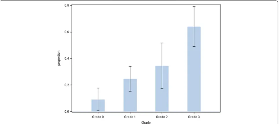
Results:IDD was common in the study population with 31% of dogs being affected. IDD and IDC were clearly connected (P< 0.001); IDD was rare in dogs with no calcifications (grade 0) and common in dogs with severe IDC (grade 3). The IDC grade was strongly positively associated with frequency of back pain periods (P< 0.001), and dogs with IDC grade 3 had frequent periods of pain. Reluctance to jump onto a sofa had a strong positive association with back pain. No association existed between age of the dog at the time of the radiographic examination and clinical signs indicative of IDD.
Conclusions:Radiographically detected IDC and IDD are common in Finnish Dachshunds and are strongly associated with one another. Spinal radiography is an appropriate screening tool for breeders attempting to diminish IDC and IDD in Dachshunds. A breeding program that screens dogs and selects against IDC can be expected to reduce the occurrence of IDD in future. Twenty-four to 48 months of age is a suitable age for screening.
Keywords:Intervertebral disc calcification, Intervertebral disc disease, Canine, Radiographic screening
Background
Intervertebral disc disease (IDD), often causing devastating back pain and neurological deficits such as paresis or paralysis, is a major medical condition in Dachshunds [1]. The peak incidence of the disease has been reported to vary between four and six years [2,3]. Studies of prevalence of IDD are scarce, but estimates have ranged from 19% to 36% [1,4-7]. In a study based on Swedish
insurance data, miniature Dachshunds had the highest mortality rate of IDD of all breeds [4].
Intervertebral disc calcification (IDC) is regarded as part of the disc degeneration process in both man and dog [8-12]. In children, it is a rare condition of unknown origin, occurring most often in the cervical spine and causing severe pain [13-15]. The calcifications usually disappear [14], but they can remain visible for several years [15]. Dachshunds are predisposed to early interver-tebral disc degeneration and calcification, and IDC can sometimes be seen macroscopically already at the age of nine months [12]. In Dachshunds, most radiographically visible disc calcifications can be seen by two years of age * Correspondence:anu.k.lappalainen@helsinki.fi
1Section of Small Animal Surgery, Department of Equine and Small Animal Medicine, Faculty of Veterinary Medicine, University of Helsinki, P.O. Box 57, FI - 00014 HU Helsinki, Finland
Full list of author information is available at the end of the article
[16]. A disc calcification can disappear after herniation, but resorption of the most degenerated discs is also possible. This has been postulated to be caused by an inflammatory response and phagocytic resorption of the calcified material in the nucleus after tearing of the degenerated annulus fibrosus [17]. Several studies have demonstrated a familial background for IDD and IDC in Dachshunds [18-23]. The hereditary basis of IDC was shown in a recent genetic study in this breed in which a major locus on chromosome 12 was found to harbour genetic variations that affected the development of IDC [24,25].
An association between the existence or number of calcifications and risk for IDD has been previously shown; in a population of Danish Dachshunds, the lower the number of calcifications, the smaller the risk for IDD [5]. In a study of Finnish miniature Dachshunds, only one out of 25 dogs without calcifications had had signs of IDD [26]. The association between IDC and IDD has been established as a tool to reduce the occurrence of IDD. Radiographic screening for IDC has been recommended in three Nordic countries (Denmark, Finland and Norway). In Finland, the protocol includes laterolateral radiographs of the cervical, thoracic and lumbar spine. IDC is graded as follows: no calcifications = free (IDC 0), 1 –2 calcifications = mild (IDC 1), 3– 4 calcifications = moderate (IDC 2) and≥5 calcifications = severe (IDC 3). The same grading is used also in Denmark and Norway. The preferred age range for screening in Finland is 24 – 42 months, but Dachshunds aged between 12 and 24 months old or older than 42 months have also been radiographed. In Denmark and Norway, the preferred age range is set at 24–48 months.
In Finland, a radiographic screening scheme for IDC in Dachshunds has been in use for over a decade. The aim of our study was to inspect the association between IDC and IDD in Finnish Dachshunds radiographically screened for IDC during a ten-year follow-up period. The hypothesis was that IDD is more common in dogs with severe IDC than in dogs only mildly affected or unaffected. The effect of age of the dog at the time of the radiographic examination on IDC and IDD were also studied.
Methods
Dachshunds aged at least ten years and screened for IDC according to the Finnish Dachshund Club’s scheme at the age of at least 12 months were included in the study. A multiple-choice questionnaire (Additional file 1), similar to one used in a previous study of incidence of IDC in Finnish miniature Dachshunds [26], was applied to determine the occurrence of IDD in Dachshunds. The questionnaire comprised the owners’ assessment of any clinical signs of IDD (ataxia of hind limbs, back or neck
pain, unexplained pain attacks, unwillingness to jump onto a sofa), and their frequency (never, seldom, some-times, often, always) in their dogs. The owners were also asked about the age at clinical sign onset and whether the dog had been diagnosed by a veterinarian and treated for the clinical signs. The dog was recorded as positive for IDD if it had had neurological deficits of the limbs or pain focusing on the back or neck, and the diagnosis had been confirmed by a veterinarian by clinical examination or in surgery. Frequency of the clinical signs indicating IDD were scored (never = 0, seldom = 1, sometimes = 2, often = 3, always = 4) and answers to questions 1 – 3 (back or neck pain, unexplained pain attacks, unwillingness to jump onto a sofa) in the questionnaire were summed up to get an overall score for symptoms indicating pain.
Altogether 386 Dachshunds met the inclusion criteria and the questionnaire was sent to their owners. Of these owners, 213 (55%) returned the questionnaire. Twelve dogs had died before ten years of age for reasons unrelated to spinal diseases, and they were excluded from the study, as were two dogs with unclear information given by their owners. Six dogs had signs indicative of IDD (pain of the back or neck, ataxia of hind limbs) without a diagnosis by a veterinarian, and they were excluded too. Thus, 193 dogs were included in our study. The IDC grade, number and the age when the radiographic screening was performed were received from the Finnish Dachshund Club open database.
The dogs were divided into three groups based on age when the radiographs were taken. This was done to explore whether age at the time of radiographic examin-ation had an effect on the associexamin-ation between IDC and owner-assessed signs indicative of IDD. In Group 1, the dogs were radiographed at the age of 12–24 months, in Group 2 at 24 – 48 months and in Group 3 at more than 48 months.
and the back or neck pain was assessed with Fisher’s exact test.
In the logistic regression and cumulative logit models, the differences between groups were quantified with odds ratios (ORs) and their 95% confidence intervals (CIs). P-values≤0.05 were considered statistically sig-nificant. All models were constructed to model the probabilities of higher/worse values in response, and the ORs were derived accordingly.
Results
The most common breed variant amongst the 193 Dachshunds was standard wire haired Dachshund (n = 63) followed by miniature long haired Dachshund (n = 38). The breed variant distribution of the dogs is presented in Table 1. The median age at the radiographic screening was 50 (range 12–116) months. Thirty dogs (15.5%) were radiographed at the age of 12–24 months (Group 1), 114 (59.1%) at 24–48 months (Group 2) and 49 (25.4%) at more than 48 months (Group 3). Forty-four dogs (22.8%) were classified as grade 0 (free of calcifications), 81 (42.0%) as grade 1 (1 – 2 calcifications), 29 (15.0%) as grade 2 (3 –4 calcifications) and 39 (20.2%) as grade 3 (≥5 calcifications). IDC grade and age group were associated (p = 0.001), as more calcifications were detected in 24 –48 months old dogs compared to the other two age groups. The detailed information of distribution of IDC grades according to age group is presented in Table 2. The median number of calcifications per dog was 1.0 (range 0–11) in Group 1, 2.0 (range 0–12) in Group 2 and 1.0 (range 0–13) in Group 3.
Of the 193 dogs, 59 (31%) had an IDD diagnosis made by a veterinarian (Table 1). Forty-three dogs (73%) had been treated conservatively with rest and non-steroidal anti-inflammatory drugs, 12 (20%) surgically and 4 (7%) had been euthanized because of the disease. One dog was operated twice and then euthanized because of the IDD, and is counted only in the surgically treated group.
The number and proportion of dogs with and without IDD in IDC grades 0–3 are presented in Figure 1.
According to the owners, the most common clinical signs of IDD were the back or neck pain (52 dogs, 88%) and unwillingness to jump onto a sofa (48 dogs, 81%). Altogether 37 dogs (63%) with IDD diagnosed by a vet-erinarian had had at least one episode of some degree of neurological deficits of the hind limbs; the rest had suffered from back pain only. Based on the information given by 28 owners, the median age for IDD was 72 months (range 30 – 132). The mean of summed score for answers to questions 1–3 in the questionnaire was 2.9 (range 0–7) for the dogs with IDD and 0.4 (range 0–6) for the dogs free of IDD. In the latter group the dog with the highest score had been diagnosed having a recur-rent otitis media, and the clinical signs were due to that disease. Of the 134 dogs free of IDD, 104 (78%) had a sum score of 0. That is, owners had answered “never” to all three questions.
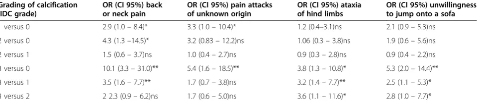
The IDC grade was positively associated (P< 0.001) with IDD in 193 Dachshunds (Table 3). Dogs with severe IDC (grade 3) had 17.9-fold odds for IDD compared with dogs without calcifications (grade 0). Only grades 1 and 2 did not differ statistically from each other. Of the 16 surgically treated or euthanized dogs, eight (50%) were graded as IDC 3, three (19%) as IDC 2 and five (31%) as IDC 1. Also the number of IDC was positively associated with IDD (P< 0.001) with OR of 1.3 (1.2–1.5) per calcified disc. IDC grade significantly explained three of the four clinical signs indicative of IDD (reluctance to jump onto a sofa, back or neck pain, pain attack for unknown reason) (Table 4). The effects were strongest between grade 3 and grade 0. For example, the proportion of dogs suffering from back pain more frequently was ten-fold when the proportion of dogs with IDC grade 3 were compared with the proportion of dogs with IDC Grade 0 (P< 0.001). The overall effect of IDC grade on ataxia of hind limbs was only slightly above significance (P= 0.061). No statistically significant difference in distribution of severity or frequency of clinical signs indicative of IDD existed between the three age groups. The frequency of back pain was positively associated (P< 0.001) with re-luctance to jump onto a sofa. In other words, dogs that experienced back pain more frequently were also more often reluctant to jump onto a sofa according to the owners’evaluation.
Discussion
The relationship between IDD and radiographically de-tected IDC was investigated in 193 Finnish Dachshunds screened for IDC. All dogs were older than ten years and had been radiographed for screening purposes ac-cording to the Finnish Dachshund Club protocol. The study was conducted as a questionnaire with a response
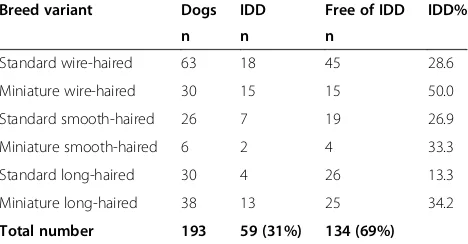
Table 1 Breed variant distribution and status of intervertebral disc disease in 193 Dachshunds radiographed for intervertebral disc calcifications
Breed variant Dogs IDD Free of IDD IDD%
n n n
Standard wire-haired 63 18 45 28.6
Miniature wire-haired 30 15 15 50.0
Standard smooth-haired 26 7 19 26.9
Miniature smooth-haired 6 2 4 33.3
Standard long-haired 30 4 26 13.3
Miniature long-haired 38 13 25 34.2
Total number 193 59 (31%) 134 (69%)
rate of 55%, which can be considered reasonable [27]. Our findings suggest that IDC grade predicts well the probability of manifesting signs indicative of IDD later in life (P< 0.001). Only four (9%) of the 44 Dachshunds without IDC (grade 0) had had signs of IDD, whereas 26 (64%) of the 39 dogs classified as grade 3 (≥5 calcifica-tions) had suffered from IDD. Additionally, the dogs with IDC grade 3 had more often and more severe signs of IDD than the dogs with no or only a few calci-fications. Our results are in accordance with a previous study conducted on 61 Dachshunds aged at least eight years where the number of radiographically detected IDC, at the age of 24 months was shown to predict well later clinical disease. Also in that study, a clear associ-ation between number of calcified discs and IDD existed (P< 0.001); in dogs with less than three calcifi-cations, IDD was rare and the clinical signs were less severe [5].
It could be assumed that IDD was more common in the dogs radiographed at the age of 24 –48 months, as dogs in this group had more calcifications than the dogs in the other two groups. However, severity and frequency of signs indicative of IDD were similarly distributed in all age groups. The reason for this remains unclear.
In this study, 31% (59/193) of the Dachshunds had had signs of IDD. The study was conducted as a questionnaire, and it can be argued that the type of the study is prone to errors of interpretation. The questionnaire has been used in a previous study [25], but it has not been validated, which can be considered a limitation of the study. How-ever, the diagnosis was confirmed by a veterinarian in all cases. It is possible that some of the cases, not confirmed by myelography, CT or MRI, were false positives. How-ever, we did not want to exclude dogs diagnosed without these modalities, since advanced imaging is seldom per-formed if the dog has mild clinical signs or the owner
Figure 1Proportion of dogs with intervertebral disc disease (IDD) in intervertebral disc calcification (IDC) Grade 0 (0 calcifications), Grade 1 (1–2 calcifications), Grade 2 (3–4 calcifications), Grade 3 (≥5 calcifications) of the 193 Dachshunds radiographed for intervertebral disc calcification.Whiskers = CI 95%.
Table 2 Number of dogs with intervertebral disc calcification grades 0–3; Grade 0 (0 calcifications), Grade 1 (1–2 calcifications), Grade 2 (3–4 calcifications), Grade 3 (≥5 calcifications) in the three age groups in 193 Dachshunds radiographed for intervertebral disc calcifications
Age (months) Grade 0 Grade 1 Grade 2 Grade 3 Total
(0 calc) (1–2 calc) (3–4 calc) (≥5 calc)
n (%) n (%) n (%) n (%)
12–24 14 (47) 10 (33) 3 (10) 3 (10) 30 (15.5)
24–48 16 (14) 49 (43) 22 (19) 27 (24) 114 (59.1)
>48 14 (29) 22 (45) 4 (8) 9 (18) 49 (25.4)
Total 44 (22.7) 81 (42.0) 29 (15.0) 39 (20.2) 193 (100)
declines surgery. Only dogs with neurological deficits of the limbs or pain focusing on the back or neck were con-sidered positive for IDD. Some of the dogs with pain of unknown origin or reluctance to jump onto the sofa might have had a mild form of IDD, and were falsely classed as negative for IDD. Also, the owners were aware of the IDC grade of their dogs, and this could have had an influence how they interpret the behavior of their dog, or how the veterinarian interpret the clinical signs.
The incidence of IDD in our study was clearly higher than the typically referred 19% [6]. In a more recent study [5], the occurrence (33%) was similar to that here. However, drawing conclusions on the occurrence of IDD in the general Finnish Dachshund population is not possible since the dogs included in the study might not be a representative sample. In our dog population, IDD appeared to be most common in miniature wire haired Dachshunds (50%) and least common in standard long haired Dachshunds (13%).
In the questionnaire, one of the questions was about a dog’s unwillingness to jump onto a sofa. This question is relevant only if the dog was allowed to jump before the clinical onset of IDD. However. none of the owners
marked this question as “always” which would indicate that the dog never does jump onto a sofa. A high posi-tive association existed between the answer to this ques-tion and the answer to the quesques-tion on back pain. It is not clear whether the owners connected unwillingness to jump onto a sofa to back pain, or whether they dealt with these as separate issues. However, the simple ques-tion of jumping onto a sofa could be useful as part of the clinical workup of Dachshunds suffering from mild clinical signs of IDD. The question on pain attacks of unknown origin might have been difficult to interpret by some owners. However, it too was associated with IDD, and dogs with several calcifications (grade 3) had them more often than dogs with no or a couple of calcifica-tions (IDC grade 0 and 1). This suggests that dogs with IDC grade 3 might have clinical signs of IDD more often than the owners recognize.
Based on the information from only 28 dogs (14%), the median age when clinical signs of IDD was manifested 72 months (range 30–132). The age was higher than that previously reported [2,3], but the result may be inaccurate, since it was based on owners’ recollection. It is possible that the owners remembered best the latest disease epi-sode if there had been several.
Our results support radiography as an effective screening tool for IDC and also by implication as a tool to diminish incidence of IDD in Dachshunds. Our results of a high positive association between IDC and IDD are in accord-ance with a previous study based on a smaller population [5]. Research on the benefits of radiographic screening as a tool to diminish IDD in Dachshunds has been lacking. However, in a recent study on Danish Dachshunds it has been shown that breeding value based on IDC indicates the risk of offspring’s IDD [7]. CT has been suggested as a screening tool since it is more sensitive than radiography in detecting small calcifications [29]. The most sensitive method for detecting intervertebral disc degeneration is MRI, which also allows degeneration of the disc without mineralization to be seen [30-32]. However, these two methods are expensive and their availability is limited,
Table 4 Effect of intervertebral disc calcification (IDC) grade on the frequency of back or neck pain, frequency of pain attacks of unknown origin, frequency of ataxia of hind limbs and frequency of unwillingness to jump onto a sofa in 193 Dachshunds radiographed for IDC
Grading of calcification (IDC grade)
OR (CI 95%) back or neck pain
OR (CI 95%) pain attacks of unknown origin
OR (CI 95%) ataxia of hind limbs
OR (CI 95%) unwillingness to jump onto a sofa
1 versus 0 2.9 (1.0–8.4)* 3.3 (1.0–10.4)* 1.2 (0.4–3.1)ns 2.1 (0.9–5.3)ns
2 versus 0 4.3 (1.3–14.5)* 3.2 (0.83–12.2)ns 1.06 (0.3–3.8)ns 1.9 (0.6–5.6)ns
2 versus 1 1.5 (0.6–3.7)ns 1.0 (0.4–2.7)ns 0.9 (0.3–2.8)ns 0.9 (0.4–2.2)ns
3 versus 0 10.1 (3.3–31.0)** 5.4 (1.6–18.5)** 3.8 (1.3–10.8)* 5.3 (2.0–14.4)**
3 versus 1 3.5 (1.6–7.7)** 1.7 (0.7–3.8)ns 3.2 (1.4–7.7)** 2.5 (1.1–5.3)*
3 versus 2 2 2.3 (0.9–6.2)ns 1.7 (0.6–5.0)ns 3.6 (1.1–11.6)* 2.8 (1.0–7.7)*
OR = odds ratio, CI = confidence interval, * =P≤0.05, ** =P≤0.01, ns = not significant.
Table 3 Effect of intervertebral disc calcification (IDC) grade on intervertebral disc disease (IDD) in 193 Dachshunds radiographed for IDC
Grading of calcification (IDC grade) OR (CI 95%)
1 versus 0 3.3 (1.0–1.4)*
2 versus 0 5.3 (1.5–19.1)**
2 versus 1 1.6 (0.6–4.0)ns
3 versus 0 17.9 (5.2–60.9)**
3 versus 1 5.4 (5.2–12.5)**
3 versus 2 3.4 (1.2–9.4)*
Disease was measured by a questionnaire where the owner was asked to report symptoms indicative of IDD and also confirmed and treated by veterinarians. Grade 0 (0 calcifications), Grade 1 (1–2 calcifications), Grade 2 (3–4 calcifications), Grade 3 (≥5 calcifications).
making them less suitable for screening purposes, where cost and access of diagnostic tools are important issues. Radiography is quite insensitive in detecting small calcifi-cations, as shown in a study comparing radiological and histopathological findings of calcifications. The sensitivity was 60% and specificity 100% for radiography when histo-pathology was used as the gold standard [28]. Moderate sensitivity of radiography could be considered as a weak-ness of screening programs, but as it is shown here and in a previous studies [5,29], the number of radiographically visible IDCs is clearly associated with IDD. This indicates that radiography is an adequate modality for screening purposes.
Conclusions
IDC and IDD are common in Finnish Dachshunds and are strongly associated with one another. Spinal radi-ography is an appropriate screening tool for breeders attempting to diminish IDC and IDD in Dachshunds. A breeding program that screens dogs and selects against IDC can be expected to reduce the occurrence the occurrence of IDD in future. Twenty-four to 48 months is a suitable age for screening.
Additional file
Additional file 1:A questionnaire for the owners of the≥10 years old Dachshunds screened for intervertebral disc calcifications.
Competing interests
The authors declare that they have no competing interests.
Authors’contributions
AL analyzed the data and drafted the manuscript. EV mailed and received the questionnaires. JJ performed the statistical analyses. OL-V participated in drafting and finalizing the manuscript. All authors have read and approved the final version of the manuscript.
Acknowledgements
We thank the Finnish Dachshund Club for financial support and for providing the owners’postal addresses.
Author details
1Section of Small Animal Surgery, Department of Equine and Small Animal Medicine, Faculty of Veterinary Medicine, University of Helsinki, P.O. Box 57, FI - 00014 HU Helsinki, Finland.2Heinolan Eläinlääkäriasema, Reumantie 2, FI - 18100 Heinola, Finland.3Pharma Ltd., Espoo, Finland.
Received: 27 March 2014 Accepted: 11 December 2014
References
1. Bonnett BN, Evenfall A, Hedhammar Å, Olson P:Mortality in over 350,000 insured Swedish dogs from 1995–2000: I. Breed-, gender-, age- and cause-specific rates.Acta Vet Scan2005,46:105–120.
2. Priester WA:Canine intervertebral disc disease—Occurrence by age, breed, and sex among 8,117 cases.Theriogenology1976,
6:293–303.
3. Gage ED:Incidence of clinical disc disease in the dog.J Am Anim Hosp Assoc1975,11:135–138.
4. Bergknut N, Egenvall A, Hagman R, Gustas P, Hazewinkel HA, Meij BP, Lagerstedt AS:Incidence of intervertebral disk degeneration-related diseases and associated mortality rates in dogs.J Am Vet Med Assoc2012,
240:1300–1309.
5. Jensen VF, Beck S, Christensen KA, Arnbjerg J:Quantification of the association between intervertebral disk calcification and disk herniation in Dachshunds.J Am Vet Med Assoc2008,233:1090–1095.
6. Ball MU, McGuire JA, Swaim SF, Hoerlein BF:Patterns of occurrence of disk disease among registered dachshunds.J Am Anim Hosp Assoc1982,
180:519–522.
7. Andersen CM, Marx T:Intervertebral disc herniation in Dachshunds;an incidence study and a follow-up study on spinal radiographic examination and the use of the number of intervertebral calcified discs and the breeding value [in Danish]. Veterinary Master Thesis.Denmark: Faculty of Health and Medical Sciences, University of Copenhagen; 2014:80.
8. Hristova GI, Jarzem P, Ouellet JA, Roughley PJ, Epure LM, Antoniou J, Mwale F:Calcification in human intervertebral disc degeneration and scoliosis.
J Orthop Res2011,29:1888–1895.
9. Karamouzian S, Eskandary H, Faramarzee M, Saba M, Safizade H, Ghadipasha M, Malekpoor AR, Ohadi A:Frequency of lumbar intervertebral disc calcification and angiogenesis, and their correlation with clinical, surgical, and magnetic resonance imaging findings.Spine2010,35:881–886.
10. Rutges JP, Duit RA, Kummer JA, Oner FC, van Rijen MH, Verbout AJ, Castelein RM, Dhert WJ, Creemers LB:Hypertrophic differentiation and calcification during intervertebral disc degeneration.Osteoarthritis Cartilage2010,18:1487–1495.
11. Benneker LM, Heini PF, Anderson SE, Alini M, Ito K:Correlation of radiographic and MRI parameters to morphological and biochemical assessment of intervertebral disc degeneration.Eur Spine J2001,4:27–35. 12. Hansen HJ:A pathologic-anatomical study on disc degeneration in dog,
with special reference to the so-called enchondrosis intervertebralis.
Acta Orthop Scan1952,11(Suppl):1–117.
13. Spapens N, Wouters C, Moens P:Thoracolumbar intervertebral disc calcifications in an 8-year-old boy: case report and review of the literature.Eur J Pediatr2010,169:577–580.
14. Dai LY, Ye H, Qian QR:The natural history of cervical disc calcification in children.J Bone Joint Surg Am2004,86:1467–1472.
15. Dhammi IK, Arora A, Monga J:Calcified thoracic intervertebral disc at two levels as a cause of mid-back pain in a child: a case report.J Orthop Sci 2002,7:587–589.
16. Jensen VF, Arnbjerg J:Development of intervertebral disk calcification in the dachshund: a prospective longitudinal radiographic study.J Am Anim Hosp Assoc2001,37:274–282.
17. Jensen VF:Asymptomatic radiographic disappearance of calcified intervertebral disc material in the Dachshund.Vet Radiol Ultrasound2001,
42:141–148.
18. Jensen VF, Christensen KA:Inheritance of disc calcification in the dachshund.J Vet Med A Physiol Pathol Clin Med2000,47:331–340. 19. Stigen O, Christensen K:Calcification of intervertebral discs in the
dachshund: an estimation of heritability.Acta Vet Scan1993,34:357–361. 20. Havranek-Balzaretti B:Contribution to the aetiology of the intervertebral disc
disease and a proposal for breeding selection [in German]. Dissertation. Switzerland: Veterinär-Medizinischen Fakultät der Universität Zürich; 1980. 21. Ghosh P, Taylor TK, Braund KG:The variation of the glycosaminoglycans of the canine intervertebral disc with ageing. I. Chondrodystrophoid breed.Gerontology1977,23:87–98.
22. Braund KG, Ghosh P, Taylor TK, Larsen LH:Morphological studies of the canine intervertebral disc. The assignment of the beagle to the achondroplastic classification.Res Vet Sci1975,19:67–172.
23. Funkquist B, Henricson B:Can frequency of intervertebral disc disease in the Dachshund be prevented by eugenics? [in German].Kleintierpraxis 1969,14:219–223.
24. Mogensen MS, Scheibye-Alsing K, Karlskov-Mortensen P, Proschowsky HF, Jensen VF, Bak M, Tommerup N, Kadarmideen HN, Fredholm M:Validation of genome-wide intervertebral disk calcification associations in dachshund and further investigation of the chromosome 12 susceptibility locus.Front Genet2012.
26. Lappalainen A, Norrgård M, Alm K, Snellman M, Laitinen O:Calcification of the intervertebral discs and curvature of the radius and ulna: a radiographic survey of Finnish miniature dachshunds.Acta Vet Scan2001,
42:229–236.
27. Baruch Y, Holtom BC:Survey response rate levels and trends in organizational research.Human Relations2008,61:1139–1160. 28. Stigen O, Kolbjørnsen O:Calcification of intervertebral discs in the
dachshund: a radiographic and histopathologic study of 20 dogs.Acta Vet Scan2007,49:39.
29. Rohdin C, Jeserevic J, Viitmaa R, Cizinauskas S:Prevalence of radiographic detectable intervertebral disc calcifications in Dachshunds surgically treated for disc extrusion.Acta Vet Scan2010,52:24.
30. Amort KH, Ondreka N, Rudorf H, Stock KF, Distl O, Tellhelm B, Kramer M, Wigger A:MR-Imaging of lumbosacral intervertebral disc degeneration in clinically sound German shepherd dogs compared to other breeds.
Vet Rad Ultrasound2012,53:289–295.
31. Bergknut N, Auriemma E, Wijsman S, Voorhout G, Hagman R, Lagerstedt AS, Hazewinkel HA, Meij BP:Evaluation of intervertebral disk degeneration in chondrodystrophic and nonchondrodystrophic dogs by use of Pfirrmann grading of images obtained with low-field magnetic resonance imaging.
Am J Vet Res2011,72:893–898.
32. Kärkkäinen M, Punto LU, Tulamo R:Magnetic resonance imaging of canine degenerative lumbar spine diseases.Vet Rad Ultrasound1993,34:399–404.
Submit your next manuscript to BioMed Central and take full advantage of:
• Convenient online submission
• Thorough peer review
• No space constraints or color figure charges
• Immediate publication on acceptance
• Inclusion in PubMed, CAS, Scopus and Google Scholar
• Research which is freely available for redistribution