HELGOI.~NDER MEERESUNTERSUCHUNGEN Helgol~inder Meeresunters. 46, 425-434 (1992).
S t r u c t u r a l e l e m e n t s o i t h e g i l l s o f t h e s h o r e c r a b
Carcinus m e d i t e r r a n e u s
L. Dalla Ve ne z i a 1, C. Zago 1, D. Siebers 2 & A. Menetto 1
1 Institute of NfaHne Biology: Riva S e t t e Martiri 1364/A, 1-30122 Venice, Italy 2 Biologische A n s t a l t Helgoland: Notkesfrafle 31, D - W - 2 0 0 0 H a m b u r g 52,
Federal R e p u b l i c o f G e r m a n y
ABSTRACT: Fine structural studies were conducted on the gills of the shore crab Carcinus mediterraneus using scanning electron microscopic techniques. The results obtained show the structural organization of crab gills from whole gills including spiny elements over the 150 lameUae to lamellar components such as cuticles, median shaft, marginal canal, afferent and efferent lameltar vessels and hemolymph cells. Enormous surface enlargement is accomplished by a variety of structural elements which allow rapid circulation of hemolymph. In the form of a relatively small organ, the gills fulfill all the necessary exchanges of specific molecules between the crab and its environment. Aggregations of ca 1-~tm particles covering the outer cuticular surfaces are considered to be bacterial colonies of unknown properties and functions.
INTRODUCTION
D u r i n g evolution, m e t a z o a n s h a v e d e v e l o p e d organs s e r v i n g a variety of s p e c i a h z e d physiological functions. C r u s t a c e a n gills are located in the interface b e t w e e n the organ- ism a n d its typically a q u a t i c e n v i r o n m e n t , m e d i a t i n g e x c h a n g e processes b e t w e e n the i n d i v i d u a l a n d its a m b i e n t m e d i u m . They are m u l t i f u n c t i o n a l organs fulfilhng a complex- ity of metabolic roles, e.g. u p t a k e of oxygen, excretion of c a r b o n dioxide, active ion transport in o s m o r e g u l a t i n g species, acid-base b a l a n c e of the h e m o l y m p h , a n d excretion of n i t r o g e n o u s e n d products. These functions a r e i n t e r c o n n e c t e d to a h i g h degree. Regarding the variety of m e t a b o l i c roles, gills are comparatively u n i q u e o r g a n s w i t h i n the a n i m a l k i n g d o m .
M a n y aspects of the f u n c t i o n i n g of crab gills h a v e b e e n thoroughly i n v e s t i g a t e d in the past decades. For a r e c e n t review, see Lucu (1990). However, descriptions of gill physiology will r e m a i n i n c o m p l e t e without k n o w l e d g e of the structural matrix e n a b l i n g the fulfillment of metabolic roles. In contrast to the large body of literature on b i o c h e m i c a l a n d physiological aspects of c r u s t a c e a n gills, the hterature d e a l i n g with the architecture a n d fine structure of this o r g a n is comparatively scarce.
Following i n v e s t i g a t i o n s o n gill physiology in the o s m o r e g u l a t i n g shore crab Car- cinus m a e n a s (Siebers et al., 1986, 1988, 1989), the p r e s e n t work d e s c r i b e s some structural e l e m e n t s of the gills of the shore crab Carcinus m e d i t e r r a n e u s u s i n g s c a n n i n g electron microscopical t e c h n i q u e s .
426 L. Dalla Venezia, C. Zago, D. S i e b e r s & A. M e n e t t o
MATERIALS A N D M E T H O D S
A d u l t m a l e crabs Carcinus mediterraneus w e r e c a u g h t b y f i s h e r m e n in t h e vicinity of the p o r t of Lido in t h e l a g o o n of Venice. I-iere the salinity is 33 + 2 %o. C r a b s w e r e t r a n s f e r r e d to the l a b o r a t o r y a n d k e p t in aquaria. T h e s e a w a t e r w a s f i l t e r e d , a n d t h e c r a b s w e r e f e d w i t h p i e c e s of b o v i n e h e a r t t h r e e times w e e k l y .
After r e m o v a l of the c a r a p a c e , o n e of the p o s t e r i o r gills, u s u a l l y gill no. 7, w a s cut a w a y from t h e b o d y wall, b l o t t e d to n e a r - d r y n e s s on p a p e r tissues a n d t r a n s f e r r e d for 3 h at 4 ~ to 2.5 % g h i t e r a l d e h y d e in 0.1 M N a + p h o s p h a t e buffer, p H 7.2. T h e gill w a s s u b s e q u e n t l y r i n s e d t h r e e times for 30 m i n at 4 ~ with 0.15 m N a + p h o s p h a t e buffer, p H 7.2. T h e r e a f t e r , the gill w a s t r a n s f e r r e d for 1 h to 1 % o s m i u m t e t r o x i d e in 0.1 M N a + p h o s p h a t e buffer p H 7.2 at 4 ~
T h e gill w a s t h e n r i n s e d four t i m e s for 4 rain at r o o m t e m p e r a t u r e w i t h t h e p h o s p h a t e b u f f e r u s e d for the s e c o n d fixation, a n d d e h y d r a t e d in g r a d e d solutions of ethanol. T h e e t h a n o l c o n c e n t r a t i o n s u s e d w e r e 30 a n d 50 % for 2 min, 70, 8 5 , 9 0 , a n d 95 % for 5 m i n a n d 100 % for hours u p to 1 day. A f t e r w a r d s , the gills w e r e d r i e d r e p e a t e d l y in a critical p o i n t (CPD) d r y i n g a p p a r a t u s (Balzers) w i t h l i q u i d CO2, w h i c h is c o m p l e t e l y m i x a b l e w i t h ethanol, until t h e r e m a i n i n g e t h a n o l w a s c o m p l e t e l y r e p l a c e d .
D r i e d gills w e r e c o a t e d with a 25 n m g o l d film in a S 150 A E d w a r d s s p u t t e r c o a t e r a p p a r a t u s a n d e x a m i n e d with a C a m b r i d g e S t e r e o s c a n 250 s c a n n i n g e l e c t r o n micro- scope.
RESULTS
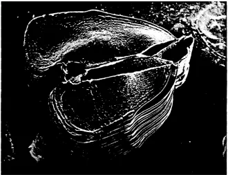
A n i n t a c t p o s t e r i o r gill (no. 7) is s h o w n in F i g u r e 1. It consists of 150 flat l a m e l l a e w h i c h are l i n e d at their outer e d g e s w i t h t h e m a r g i n a l 'canals (see also F i g s 2, 4-6). T h e y b e l o n g to t h e s y s t e m of l a m e l l a r v e s s e l s t r a n s p o r t i n g w i t h i n e a c h p l a t e l e t p a r t of the circulating h e m o l y m p h from the afferent to t h e efferent vessel. O n t h e l o w e r right side, b u l b - l i k e e n l a r g e m e n t s of the m a r g i n a l c a n a l s are s e e n (see also Fig. 4, l o w e r r i g h t side). B e t w e e n b o t h sides of the l a m e l l a e t h e afferent v e s s e l is s e e n in t h e c e n t r e of the gill.
F i g u r e 2 s h o w s the gill from the o p p o s i t e side, e x h i b i t i n g in its c e n t r e the efferent vessel. It is c o v e r e d i r r e g u l a r l y w i t h s p i n e s (see also Figs 8-10). T h e i n s e r t i o n of t h e m a r g i n a l c a n a l s of s e v e r a l l a m e l l a e into t h e efferent v e s s e l is s h o w n in F i g u r e 3. In t h e t r o u g h s a n d i n f o l d i n g s b e t w e e n t h e i n s e r t i o n p o i n t s d e n s e a s s e m b l a g e s of a p p r o x i m a t e l y 1-~tm particles, p r e s u m a b l y b a c t e r i a l colonies, a r e obvious (see "Discussion").
A t r a n s v e r s a l section of a p o s t e r i o r gill no. 7 (Fig. 4) r e v e a l s the s h a p e of an i n d i v i d u a l lamella. Its surface is c o v e r e d b y the cuticle w h i c h is c h a r a c t e r i z e d b y a specific p a t t e r n of ripples. T h e p h o t o s h o w s the afferent v e s s e l on the left a n d the efferent v e s s e l on t h e r i g h t side. Both v e s s e l s are c o n n e c t e d b y t h e m e d i a n shaft w h i c h s e p a r a t e s t h e i n d i v i d u a l p l a t e l e t into two halves. Note the b u l b - l i k e e n l a r g e m e n t s of t h e m a r g i n a l c a n a l s on t h e l o w e r right side.
F i g u r e 5, a n e n l a r g e d sector of P i g u r e 4 s h o w s the surface of a l a m e l l a with t h e m a r g i n a l c a n a l on the u p p e r right s i d e a n d the w a v e - s h a p e d p a t t e r n of c u t i c u l a r ripples. In the t r o u g h s b e t w e e n the r i p p l e s a s s e m b l a g e s of a p p r o x i m a t e l y 1-~m p a r t i c l e s are present, p r o b a b l y i d e n t i c a l to t h e b a c t e r i a l colonies in F i g u r e 3.
S t r u c t u r a l e l e m e n t s o f Carcinus mech'terraneus 4 2 7
Fig. 1. Isolated gill no. 7 w i t h its tip on t h e left side, s h o w i n g t h e l a m e l l a e a n d t h e a f f e r e n t v e s s e l in t h e c e n t r a l p a r t
4 2 8 L. D a l l a V e n e z i a , C, Z a g o , D. S i e b e r s & A. M e n e t t o
Fig, 3. Insertion of m a r g i n a l canals of individual lamellae into the efferent vessel, Note bacterial a s s e m b l a g e s especially b e t w e e n the insertions
Fig. 4. Transversal section of a posterior gill (no~ 7) s h o w i n g the surface of a n i n d i v i d u a l lamella, tlae afferent vessel on the left a n d the efferent vessel on the right side c o n n e c t e d by the m e d i a n shaft.: Note the m a r g i n a l canals at the e d g e s of the l a m e l l a e a n d the p a t t e r n of ripples o n the IameUar
S t r u c t u r a l e l e m e n t s of Carcinus mediterraneus 4 2 9
Fig. 5. Sector of Fig. 4 showing the p a t t e r n of ripples on the iamellar surface a n d bacterial a s s e m b l a g e s in the spaces b e t w e e n the ripples. The m a r g i n a l canal is s e e n on the u p p e r right side
Fig. 6. Sector of Pig. 4 with the u p p e r cellular layer b r o k e n away revealing the partially perforated lamellar septum with single h e m o l y m p h cells on its surface. On the left side the m a r g i n a l canal is
430 L. Dalla Venezia, C. Zago, D. Siebers & A. M e n e t t o
s e e n with several o p e n i n g s a l l o w i n g h e m o l y m p h passage. In a g r e e m e n t with Taylor & Taylor (1986) the light p a r t i c u l a t e structures of the surface of the l a m e l l a r septurn are identified as h e m o l y m p h ceils. T h e y ake clearly discernible from the c o n s i d e r a b l y smaller particles s e e n as d e n s e layers o n the cuticle o n the u p p e r right side of t h e photo.
The l o n g i t u d i n a l section s h o w n i n Figure 7 shows the a p p e a r a n c e of a m u l t i - f o l d e d organ. T h e large flat layers of t h e foldings reveal the n a t u r e of the gill p l a t e l e t which is formed b y a d o u b l e l a y e r of epithelial ceils. The two folded systems are s e p a r a t e d by the m e d i a n shaft (from u p p e r left to m i d d l e right side of the photo).
Fig, 7. Longitudinal section of a posterior gill (no. 7) showing both layers of each lamelIa. The surface layers of neighbouring lameUae are connected, thus building a system of multiple foldings
Figure 8 shows a sector of the efferent vessel covered with two s p i n e s w h i c h are i n s e r t e d o n a c o n e - s h a p e d basis. B e t w e e n the spines a n d on the surface of the cone- s h a p e d basis the p a t t e r n of cuticular ripples is obvious, a n d on the u p p e r l e f t side some bacterial a g g r e g a t i o n s are seen. R e m o v a l of the spines on the efferent v e s s e l allows a v i e w on the n e t w o r k of t u b e - l i k e structures forming the basis of the s p i n e s (Fig. 9). T h e y are considered to b e a sturdy m e c h a n i c a l skeleton.
S t r u c t u r a l e l e m e n t s of Carcinus mediterraneus 431
Fig. 8. E n l a r g e m e n t of spines located on the efferent vessel as s h o w n in Fig. 2
432 L. Dalla Venezia, C. Zago, D. Siebers & A. M e n e t t o
Fig. 10. Enlargement of the insertion of a spine seen from the interior of the efferent vessel. Several hemolymph cells are present on the internal surface of the vessel
D I S C U S S I O N
F i n e structural studies h a v e b e e n c o n d u c t e d on the gills of s e v e r a l d e c a p o d crusta- c e a n s such as Callinectes ( C o p e l a n d & FitzjaITel, 1968; Cioffi, 1984}, Gecarcinus {Cope-
land, 1968), Hemigrapsus a n d Pachygrapsus (Wright, 1964}, Holthuisana {Taylor &
G r e e n a w a y , 1979), Ocypode (Flemister, 1959; Storch & Welsch, 1975), Panulirus
( S t r a n g e w a y s - D i x o n & Smith, 1970), Penaeus {Talbot et a l , 1972; F o s t e r & H o w s e , 1978}, Eriocheir (Barra et al., 1983), Uca (Finol & C r o g h a n , 1983), Goniopsis (Martelo & Z a n d e r s . 1986} a n d Carcinus (Taylor & Taylor, 1986; G o o d m a n & C a v e y , 1990}. In o n l y a few of t h e s e r e p o r t s w e r e s c a n n i n g e l e c t r o n m i c r o s c o p i c t e c h n i q u e s e m p l o y e d .
T h e figures p r e s e n t e d s h o w a v a r i e t y of structural e l e m e n t s specific of c r u s t a c e a n gills. T h e gill a r c h i t e c t u r e is c h a r a c t e r i z e d b y e n o r m o u s surface e n l a r g e m e n t s a l l o w i n g r a p i d m o v e m e n t s of h e m o l y m p h o v e r t h e i n t e r n a l surfaces a n d p a s s a g e s of a m b i e n t m e d i u m o v e r t h e e x t e r n a l surfaces. T h e gills thus r e p r e s e n t in the form of a r e l a t i v e l y s m a l l o r g a n the t e c h n i c a l solution d e v e l o p e d d u r i n g evolution to a l l o w n e c e s s a r y e x c h a n g e s of specific m o l e c u l e s b e t w e e n a n o r g a n i s m w i t h a h a r d cover a n d its e n v i r o n - ment.
Structural e l e m e n t s of C a r c i n u s m e d i t e r r a n e u s 433 e n d s of the m e d i a n shaft are r e p r e s e n t e d by the afferent (Fig. 1) a n d the efferent (Figs 2, 3, 9, 10) vessel. Both vessels are c o n n e c t e d at their b a s e with the system of circulating h e m o l y m p h i n the b o d y cavity via a c o m m o n o p e n i n g i n the lateral side of the carapace. T h e afferent vessel is directed to the b o d y wall a n d the efferent vessel is l o c a t e d o n the opposite side. T h e gill is oriented with the b a s e in a lower position a n d the tip i n a n u p p e r position.
T h e l a m e l l a e consist of two u n i c e l l u l a r epithelia s e p a r a t i n g the a m b i e n t m e d i u m from the h e m o l y m p h (Fig. 7). Their outer sides are covered b y the cuticles, t h r e e - l a y e r e d structures r e l e a s e d from the b o d y together with the exuvia d u r i n g the m o u l t i n g process. B e t w e e n the b a s a l m e m b r a n e s of the two epithelial cell layers, a perforated l a m e l l a r s e p t u m is p r e s e n t (Fig. 6), a n d the h e m o l y m p h flow proceeds o n b o t h sides of it.
T h e circulating h e m o l y m p h enters the gill platelet via o p e n i n g s i n the afferent vessel. T h e h e m o l y m p h flow w i t h i n the gill proceeds within discrete canals of the i n t e r n a l l a c u n a r system a n d the m a r g i n a l c a n a l of the platelet to r e e n t e r the b o d y via v a l v e - l i k e o p e n i n g s of the afferent vessel (Taylor & Taylor, 1986).
The spines covering irregularly the efferent vessel are r e g a r d e d as e l e m e n t s of c o n s i d e r a b l e m e c h a n i c a l stability, h o w e v e r of as yet u n k n o w n physiological function. T h e p r e s e n c e of mattresses of small particulate e l e m e n t s on the cuticIes (Figs 3, 5, 6, 8) has also b e e n r e p o r t e d as "dark bodies o n the outer surface of t h e cuticle" of t h e gills of the b l u e crab, C a l l i n e c t e s s a p i d u s , b y C o p e l a n d & Fitzjarrel (1968) a n d are c o n s i d e r e d to b e "symbiontic algalike forms". T h e y have also b e e n o b s e r v e d in the form of d e n s e a g g r e g a t i o n s of p r o l o n g e d particles on the cuticles of the gills of the shore crab C a r c i n u s m a e n a s b y G o o d m a n & C a v e y (1990) who c o n s i d e r e d t h e m as "bacteria a d h e r i n g to the epicuticle". F u r t h e r work is d e s e r v e d to clarify the n a t u r e of these particles, the coexist- e n c e with their hosts as well as their potential physiologidal role i n gill functions.
LITERATURE CITED
Bara, J. A., P~queux, A. & Humbert, W,, 1083. A morphological study on the gills of a crab acchmated to fresh water. - Tissue Cell 1.5, 5 8 3 - 5 9 6 .
Cioffi, M., 1984. Comparative ultrastructure of arthropod transporting epitheha. - A n . Zool. 24, 139-156.
Copeland, D. E., 1968. Fine structure of salt and water uptake in the land-crab G e c a r c i n u s lateralis. - Am. Zool. 8, 417--432.
Copeland, D.E. & Fitzjarrel, A.T., 1968. The salt-absorbing ceils in the gills of the blue crab ( C a f l i n e e t e s s a p i d u s Rathbun) with notes on modified mitochondria. - Z. ZeUIorsch. mikrosk. Anat. 92, 1-22.
Finol, H.J. & Croghan, P.C., 1983. Ultrastructure of the branchial epithelium of an amphibious brackish-water crab. - Tissue Cell 16, 63-75.
Flemister, L J., 1959. Histophysiology of gills and kidney of the crab O c y p o d e albicans. - Biol. Bull. mar. biol. Lab., Woods Hole i16, 37-48.
Foster, C.A. & Howse, H.D., 1978. A morphological study on gills of the brown shrimp, P e n a e u s a z t e k u s . - Tissue Cell 10, 77-02.
Goodman, S.H. & Cavey, M.J., 1900. Organization of a phyllobranchiate gill from the green shore crab C a r c i n u s m a e n a s (Crustacea, Decapoda). - Cell. Tiss. Res. 260, 4 0 5 - 5 0 5 .
Lucu, C., 1990. Ionic regulatory mechanisms in crustacean frill epithelia. - Comp. Biochem. Physiol. 9 7 A , 297-306.
434 L. D a l l a V e n e z i a , C. Z a g o , D. S i e b e r s & A. M e n e t t o
Siebers, D., Lucu, C., Winkler, A., Dalla Venezia, L & Wille, H., 1986. Active u p t a k e of sodium in the gills of the hyperregulating shore crab C a r c i n u s m a e n a s . - Helgol~inder M e e r e s u n t e r s . 40, 151-160.
Siebers, D., Lucu, C. & Dalla Venezia, L., 1988. Identification of active a n d p a s s i v e ion transport processes in shore crab gills. - Comp. Biochem. Physiol. 90 A, 821 (Abstr.}.
Siebers, D., Wille, H., Lucu, C. & Dalla Venezia, L , 1989. Conductive sodium entry i n gill cells of the shore crab C a r c i n u s m a e n a s . - Mar. Biol. 101, 61-68.
Storch, V. & Welsch, U., 1975. Ober Bau u n d Funktion der Kiemen u n d L u n g e n von O c y p o d e c e r a t o p h t h a l m a (Decapoda" Crustacea). - Mar. Biol. 29, 363-371.
Strangeways-Dixon, J. & Smith, D. S , 1970. The fine structure of gill "podocytes" in P a l i n u r u s a r g u s (Crustacea}. - Tissue Cell 2, 611--624.
Talbot, P., Clark, W.H. & Lawrence, A.L., 1972. Light and electron microscopic studies on osmo- regulatory tissue in the developing b r o w n shrimp, P e n a e u s a z t e c u s . - Tissue Cell 2, 271-286. Taylor, H.H. & Greenaway, P., 1979. The structure of the gills a n d lungs of the arid-zone crab,
H o l t h u s i a n a ( A u s t r o t e l p h u s a ) transversa (Brachyura: S u n d a t e l p h u s i d a e ) i n c l u d i n g observations on arterial vessels within the gills. - J. Zool., Lond. 189, 359-384.
Taylor, H.H. & Taylor, E.W., 1986. Observations of valve-like structures and e v f d e n c e for rectifica- tion of flow within the gill lamellae of the crab C a r c i n u s m a e n a s (Crustacea, Decapoda). - Zoomorphology 106, 1-11.