RESEARCH ARTICLE
Coenzyme Q
10
defects may be
associated with a deficiency of Q
10
-independent
mitochondrial respiratory chain complexes
Konstantina Fragaki
1,2, Annabelle Chaussenot
1,2, Jean‑François Benoist
3, Samira Ait‑El‑Mkadem
1,2,
Sylvie Bannwarth
1,2, Cécile Rouzier
1,2, Charlotte Cochaud
1and Véronique Paquis‑Flucklinger
1,2*Abstract
Background: Coenzyme Q10 (CoQ10 or ubiquinone) deficiency can be due either to mutations in genes involved in
CoQ10 biosynthesis pathway, or to mutations in genes unrelated to CoQ10 biosynthesis. CoQ10 defect is the only oxida‑ tive phosphorylation disorder that can be clinically improved after oral CoQ10 supplementation. Thus, early diagnosis, first evoked by mitochondrial respiratory chain (MRC) spectrophotometric analysis, then confirmed by direct meas‑ urement of CoQ10 levels, is of critical importance to prevent irreversible damage in organs such as the kidney and the central nervous system. It is widely reported that CoQ10 deficient patients present decreased quinone‑dependent activities (segments I + III or G3P + III and II + III) while MRC activities of complexes I, II, III, IV and V are normal. We previously suggested that CoQ10 defect may be associated with a deficiency of CoQ10‑independent MRC complexes. The aim of this study was to verify this hypothesis in order to improve the diagnosis of this disease.
Results: To determine whether CoQ10 defect could be associated with MRC deficiency, we quantified CoQ10 by
LC‑MSMS in a cohort of 18 patients presenting CoQ10‑dependent deficiency associated with MRC defect. We found decreased levels of CoQ10 in eight patients out of 18 (45 %), thus confirming CoQ10 disease.
Conclusions: Our study shows that CoQ10 defect can be associated with MRC deficiency. This could be of major
importance in clinical practice for the diagnosis of a disease that can be improved by CoQ10 supplementation.
Keywords: Mitochondrial disease, CoQ10 deficiency, Respiratory chain, Spectrophotometry, LC‑MSMS
© 2016 Fragaki et al. This article is distributed under the terms of the Creative Commons Attribution 4.0 International License (http://creativecommons.org/licenses/by/4.0/), which permits unrestricted use, distribution, and reproduction in any medium, provided you give appropriate credit to the original author(s) and the source, provide a link to the Creative Commons license, and indicate if changes were made. The Creative Commons Public Domain Dedication waiver (http://creativecommons.org/ publicdomain/zero/1.0/) applies to the data made available in this article, unless otherwise stated.
Background
Coenzyme Q10 (CoQ10 or ubiquinone) is a lipid-soluble component of the mitochondrial inner membrane that plays a central role in mitochondrial respiratory chain (MRC) function, as electrons carrier from complexes I and II to complex III, thus participating in ATP produc-tion [1].
CoQ10 deficiency encompasses several clinical phe-notypes such as encephalomyopathy, severe infantile multisystemic disease, cerebellar ataxia, isolated myopa-thy or nephrotic syndrome [2]. CoQ10 deficiency can be
primary, due to mutations in genes involved in CoQ10 biosynthesis or secondary, due to mutations in genes unrelated to CoQ10 biosynthesis [3]. Secondary CoQ10 deficiency has been described in patients with mito-chondrial DNA (mtDNA) mutations or deletions, with mtDNA depletion syndrome (MDS) [4–6] and in patients with mutations in APTX [7], ETFDH [8, 9], BRAF [10], ACADVL or NPC genes [11]. CoQ10 defect is the only oxidative phosphorylation (OXPHOS) disorder that can be clinically improved after oral CoQ10 supplementation with limitation of neurological and renal manifestations, amelioration of muscular symptoms and attenuation of histological alterations. Early treatment is crucial to pre-vent irreversible damage in organs such as the kidney and the central nervous system [12–14]. Reduced activities of CoQ10-dependent enzymes by spectrophotometric
Open Access
*Correspondence: [email protected]
1 School of Medicine, IRCAN, UMR CNRS 7284/INSERM U1081/UNS,
Nice Sophia‑Antipolis University, 28 av de Valombrose, 06107 Nice Cedex 2, France
analysis (segments I + III or G3P + III and II + III) are evocative of CoQ10 deficiency but direct measurement of CoQ10 levels is the most reliable test for diagnosis [15]. It is widely reported in the literature that, in patients with CoQ10 deficiency, enzymatic activities of MRC complexes I, II, III, IV, V are normal [16]. In a previous report, we described an 11-year-old boy presenting with a propionic acidemia who succumbed to acute heart failure in the absence of decompensation of his metabolic condition. Spectrophotometric analysis in liver identified CoQ10 -dependent activities deficiency that was associated with MRC enzymatic defect. Secondary CoQ10 deficiency was likely involved in the development of heart compli-cations in this child and we hypothesized that a CoQ10 -defect may be associated with MRC deficiency [17]. The aim of this study was to verify this hypothesis in order to improve the diagnosis of this disease.
Over a 6-year period, we analyzed by spectropho-tometry 700 tissue samples from 495 patients in whom a mitochondrial disease was suspected. Isolated CoQ10 -dependent activity deficiency led to identification of CoQ10 disease in eight cases. Eighteen patients presented CoQ10-dependent enzymatic deficiency associated with MRC defect by spectrophotometry in muscle or in fibro-blasts. In order to validate our original observation and to establish if CoQ10 quantitative defect may be associated with multiple MRC enzymatic deficiency, we measured CoQ10 in this group of 18 patients. We found decreased CoQ10 levels by liquid chromatography coupled with tan-dem mass spectrometry detection (LC-MSMS) in eight patients out of 18 (45 %), thus confirming CoQ10 disease and its association with MRC enzymatic deficiency. Fur-thermore, CoQ10 disease cannot be ruled out in all other patients insofar as the quantitative assay could not always be performed in the affected tissue.
Results
Description of patients involved in the study
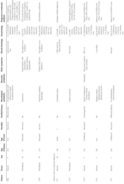
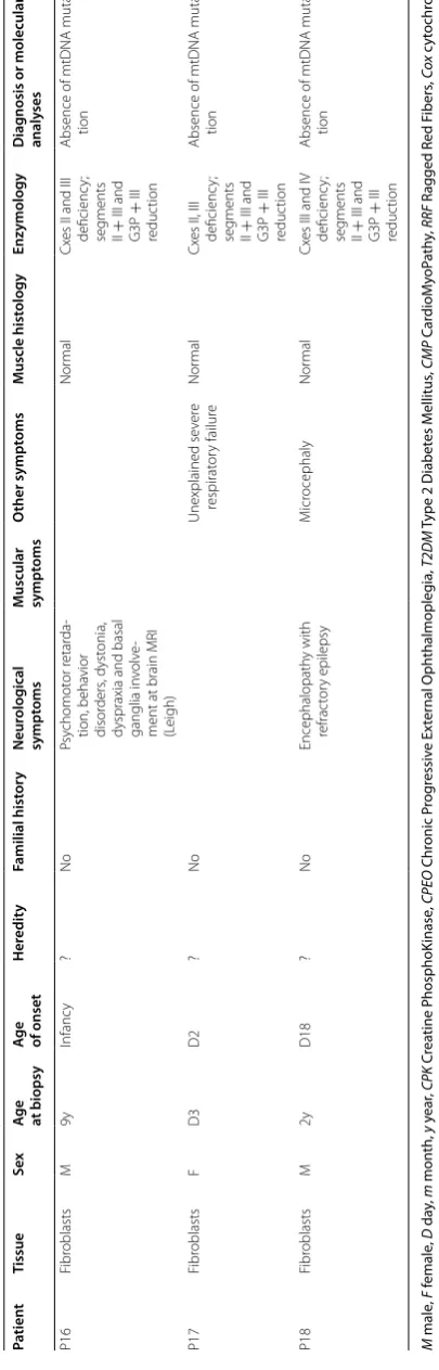
We studied 18 patients, including 10 males and eight females, ranging in age from day 1 to 76 years. Clinical presentations were very heterogeneous (Table 1). The age at onset of the disease was highly variable, ranging from (i) neonatal forms (seven cases with severe phenotypes), (ii) onset before 1 year of age (four cases with either Leigh syndrome or epileptic encephalopathy), (iii) childhood-onset (four cases including two myopathic forms and two complex phenotypes) to (iv) adult-onset (three cases with two myopathic presentations and one cerebellar ataxia). The 18 patients were divided into two different groups according to molecular results.
The first group included 10 patients with identified mutations in responsible genes (Table 1). Patient P01 pre-sented a severe neonatal multisystemic disease secondary
to a homozygous missense mutation in the CoQ2 gene [18]. Spectrophotometric analysis in fibroblasts revealed a CoQ10-dependent activities defect (segments II + III and G3P + III reduction) associated with a complex IV deficiency (Table 2). Six patients (P02–P07) presented a mitochondrial disease or dysfunction secondary either to mtDNA abnormalities (P02 and P03) or to mutations in nuclear genes (P04–P07). Patient P02 had a large hetero-plasmic mtDNA deletion responsible for Kearns–Sayre syndrome and patient P03 presented with a severe neo-natal polyvisceral failure secondary to a heteroplasmic mtDNA mutation in the MT-CYB gene. Patients P04 and P05 presented with sensory ataxia neuropathy dys-arthria and ophthalmoplegia (SANDO) phenotype asso-ciated with recessive mutations in POLG. Patient P06 had a neonatal encephalopathy with lactic acidosis and mild methylmalonic aciduria linked to mutations in the SUCLG1 gene. P07 had a diagnosis of multiple acyl-CoA dehydrogenation deficiency (MADD) with ETFDH muta-tion. The last three patients in the first group presented malignant migrating partial seizures with mutations in TBC1D24 (P08), CDG syndrome type Iq with SRD5A3 -CDG mutations (P09) and 1p36 deletion syndrome (P10). Patients P02–P10 had a CoQ10-dependent activities defi-ciency (segments I + III or G3P + III and II + III reduc-tion) associated with a multiple MRC defect in muscle or in fibroblasts (Table 2).
The second group included eight patients suspected of CoQ10 deficiency with an absence of molecular diagno-sis. Except for individual P11, who developed cerebellar ataxia during adulthood, all patients had an early-onset disease ranging from neonatal period to childhood. They presented severe neurological symptoms including two Leigh syndromes (P12 and P16) and one child had an unexplained severe respiratory failure at birth (P17). In the second group, all patients presented a CoQ10 -dependent enzymatic deficiency associated with MRC defect in muscle or in fibroblasts (Table 2).
Confirmation of CoQ10disease in eight patients by CoQ10 quantification
Table 1 C linic al phenot yp es of pa tien ts pr esen ting C oQ 10 -dep enden t enz yma tic deficienc y asso cia ted with MR C def ec t Pa tien t Tissue Sex A
ge at biopsy
A
ge of onset
Her edit y Familial hist or y Neur olog ical sympt oms
Muscular sympt
oms O ther sympt oms Muscle hist ology Enz ymology D iag
nosis or molecular
analy
ses
Patients with molecular diagnosis P01
a Fibr oblasts M D1 Neonatal Recessiv e A ffec ted br other Neonatal poly vis ‑ ceral failur e Not done
Cx IV deficienc
y; seg ments II + III and G3P + III reduc tion COQ2 : homo zy gous mutation ( c.437G > A; p.S er146A sn) P02 M uscle M 54y 25y Sporadic No
Brain MRI: mild atr
o‑
ph
y and lacunar
str
ok
es
CPEO
T2DM, hepatic st
eat osis , dyslipi ‑ demia RRF (5–10 %) and Co x‑ fibers Cx
es I, II, IV and V deficienc
y; seg ments I + III and II + III r educ ‑ tion Lar ge
‑scale deletion of
mtDNA P03 a Fibr oblasts M D1 Neonatal de no vo No H ypot onia, epilepsy
and diffuse brain lesions
Neonatal poly vis ‑ ceral failur e: r es ‑ pirat or y distr ess , hepatic failur e, hyper tr ophic CMP , lac tic acido ‑ sis ++ Not done Cx
es II and III deficienc
y; seg ments II + III and G3P + III reduc tion MT ‑ CYB : het er oplas ‑
mic mtDNA muta
‑ tion (m.15635T > C; p.S er297P ro) P04 a Fibr oblasts M 15y 11y Recessiv e No A taxic sensor y ax onal neur opath y CPEO
RRF and C
ox
‑fibers
(40
%)
Cx
es I, II, III and IV deficienc
y; seg ments II + III and G3P + III reduc tion
SANDO with multiple mtDNA deletions and homo
zy gous mutation in POL G : ( c.911T > G; p.L eu304Ar g) P05 M uscle F 54y 45y Recessiv e No A taxic sensor y ax onal neur opath y CPEO Lipid accumula ‑
tion, RRF and Cox‑
fibers
(20
%)
Cx
es I, II, III, IV and V deficienc
y; seg ments I + III and II + III r educ ‑ tion
SANDO with multiple mtDNA deletions and compound het
er
oz
y‑
gous mutations in
POL G : (c .752C > T/c .2452G > A; p.T hr251I le/p .Gly848S er) P06 a Fibr oblasts M D1 Neonatal Recessiv e No Encephalopath y and hypot onia Se ver e lac tic acidosis , meth yl ‑ malonic acidur ia Not done Cx
es II, III and IV deficienc
y; seg ments II + III and G3P + III reduc tion SUCL G1 : compound het er oz ygous mutations c.97 + 3G > C/c .509C > G (p .P ro170Ar g) P07 M uscle F 18y 4y Recessiv e
Blindness in pat
er nal family Bilat eral pt osis , pr oximal m yopath y, dysphonia, dysphag ia, ex er cice int oler ‑ ance Retinitis pig men ‑ tosa, c yclic vomiting , h yper ‑ CPK emia Lipid accumula ‑ tion Cx
es I and III deficienc
y; seg ments I + III and II + III r educ ‑ tion M
Table 1 c on tinued Pa tien t Tissue Sex A
ge at biopsy
A
ge of onset
Her edit y Familial hist or y Neur olog ical sympt oms
Muscular sympt
oms O ther sympt oms Muscle hist ology Enz ymology D iag
nosis or molecular
analy ses P08 M uscle F 4 m 4 m Recessiv e A ffec ted sist er Encephalopath y with refrac tor y mig rat ‑ ing par tial seizur es Lipid accumula ‑ tion
Cx I, II, III and IV deficienc
y; seg ments I + III and II + III r educ ‑ tion M alig nant mig rating par tial seizur es with compound het er oz ygous mutations in TBC1D24 : (c .468C > A/c .686C > T ; p.C ys156X/p .P he229S er) P09 Fibr oblasts M D1 Neonatal Recessiv e No H ypot onia H yper tr ophic CMP , dysmor phic , hepatic c yt olysis , hypospadia Gly cogenic accu ‑ mulation Cx
es II, III, IV and V deficienc
y; seg ments II + III and G3P + III reduc tion CDG syndr ome t
ype Iq :
homo zy gous mutation in SRD5A3 : ( c.620T > G; p.M et207Ar g) P10 a Fibr oblasts M 3 m Neonatal de no vo No H ypot onia, epilepsy , dysphag ia Dilat ed CMP , aor tic dilatation Gly cogenic accu ‑ mulation Cx
es III and IV deficienc
y; seg ments II + III and G3P + III reduc tion
1p36 deletion syndr
ome
Patients with no molecular diagnosis P11
M uscle M 76y A dult ? No Cer ebellar ataxia
2 RRF and C
ox
‑
fibers (20
‑30
%)
Cx IV defi
‑ cienc y; seg ‑ ments I + III and II + III reduc tion M
ultiple mtDNA deletions
P12 a Fibr oblasts F 7 m 6 m ? No Leigh syndr ome Not done
Cx II defi
‑ cienc y; seg ments II + III and G3P + III reduc tion
mtDNA depletion, absence of mtDNA and
POL G, SUCL A2, TK2 mutation P13 a M uscle F 41y Childhood Recessiv e Consanguinit y Spastic t etrapar esis , chor ea, mental retar dation M yopath y Glaucoma, catarac t, lac tic acidosis RRF ++
Cx I deficienc
y; seg ments I + III and II + III r educ ‑ tion
Absence of mtDNA and POL
G, OP A1, OP A3 muta ‑ tion P14 M uscle F 33y 6 m ? No Epilepsy , spastic dipleg ia, dyst onia, dysk inesia, tr emor 1 C ox ‑fiber Cx
es III and
V deficienc y; seg ments I + III and II + III r educ ‑ tion
Absence of mtDNA and POL
G, T TC19, D YT5 muta ‑ tion P15 M uscle F 28 y Childhood Recessiv e A ffec ted siblings Encephalopath y, mental r etar dation Nor mal Cx
es II, III and V deficienc
y; seg ments I + III and II + III r educ ‑ tion
Absence of mtDNA muta
‑
Table
1
c
on
tinued
Pa
tien
t
Tissue
Sex
A
ge at biopsy
A
ge of onset
Her
edit
y
Familial hist
or
y
Neur
olog
ical
sympt
oms
Muscular sympt
oms
O
ther sympt
oms
Muscle hist
ology
Enz
ymology
D
iag
nosis or molecular
analy
ses
P16
Fibr
oblasts
M
9y
Infanc
y
?
No
Psy
chomot
or r
etar
da
‑
tion, beha
vior
disor
ders
, dyst
onia,
dyspraxia and basal ganglia in
volv
e‑
ment at brain MRI (Leigh)
Nor
mal
Cx
es II and III deficienc
y;
seg
ments
II
+
III and
G3P
+
III
reduc
tion
Absence of mtDNA muta
‑
tion
P17
Fibr
oblasts
F
D3
D2
?
No
Unexplained se
ver
e
respirat
or
y failur
e
Nor
mal
Cx
es II, III deficienc
y;
seg
ments
II
+
III and
G3P
+
III
reduc
tion
Absence of mtDNA muta
‑
tion
P18
Fibr
oblasts
M
2y
D18
?
No
Encephalopath
y with
refrac
tor
y epilepsy
M
icr
ocephaly
Nor
mal
Cx
es III and IV deficienc
y;
seg
ments
II
+
III and
G3P
+
III
reduc
tion
Absence of mtDNA muta
‑
tion
M
male
,
F
female
,
D
da
y,
m
mon
th,
y
y
ear
,
CPK
C
rea
tine P
hosphoK
inase
,
CPEO
Chr
onic P
rog
ressiv
e Ex
ter
nal Oph
thalmopleg
ia,
T2DM
T
ype 2 Diabet
es M
ellitus
,
CMP
C
ar
dioM
yoP
ath
y,
RRF
R
agged R
ed F
ibers
,
Cox
c
yt
ochr
ome
c
o
xy
dase
,
cx
c
omple
x,
mtDNA
mit
ochondr
ial DNA,
SANDO
S
ensor
y A
taxia Neur
opa
th
y D
ysar
thr
ia and Oph
thalmopleg
ia,
MADD
M
ultiple A
cyl-CoA D
eh
ydr
ogena
tion D
eficienc
y,
CDG
C
ar
boh
ydr
at
e-D
eficien
t Gly
copr
ot
ein
a P
atien
t dec
and/or other signs. In the first group, the very low CoQ10 level observed in the fibroblasts of patient P01 confirmed the primary CoQ10 defect associated with the c.437G > A homozygous missense mutation (p.Ser146Asn) in the CoQ2 gene, involved in CoQ10 biosynthesis [18]. In the four other patients in the same group, CoQ10 defect was clearly secondary because the responsible genes were unrelated to CoQ10 biosynthesis. Three patients had a mitochondrial disease linked to a large mtDNA dele-tion (patient P02) or to mutadele-tions in POLG (patient P04) or SUCLG1 (patient P06). Patient P08 alone did not have a mitochondrial disease, her encephalopa-thy with refractory malignant migrating partial seizures being linked to mutations in the TBC1D24 gene. In the second group, low CoQ10 levels were found in three patients with no molecular diagnosis. Two patients were strongly suspected of having a mitochondrial disease: patient P11, who had a cerebellar ataxia with 20–30 %
of COX-negative fibers and multiple mtDNA deletions in muscle, and patient P16 who presented with a Leigh syndrome. The last patient (P15) had an encephalopa-thy with intellectual disability but no histological sign of mitochondrial myopathy.
Discussion
While primary CoQ10 defects are rare, secondary defects have been observed in various pathologies. In a pre-vious work, we suspected for the first time a second-ary CoQ10 defect in a child with propionic acidemia, who succumbed to acute heart failure in the absence of decompensation of his metabolic condition [17]. CoQ10 deficiency was not evoked at the outset because CoQ10 -dependent activities deficiency was associated with mul-tiple MRC deficiency in the liver of the patient and it had been widely reported that enzymatic activities of MRC complexes are normal in CoQ10 disease [16]. However, it
Table 2 Biochemical analysis of patient fibroblasts and muscle biopsies
Respiratory chain enzyme activities were measured spectrophotometrically. Results are expressed as absolute values for controls or patients (in nanomoles of substrate per minute per milligram of protein). CoQ10 quantity was measured by LC-MSMS. Results are expressed as absolute values for controls or patients (in
picomoles per milligram of protein). Abnormal values are shown in italics
OXPHOS oxidative phosphorylation; LC-MSMS liquid chromatography coupled with tandem mass spectrometry detection
OXPHOS activities
(spectrophotometry)I II III IV V G3P + III II + III CS CoQ10 quantity (LC-MSMS) CoQ10
Fibroblast measurements Control values (nmole/
min/mg of proteins)
9.0–27.1 21.0–54.0 62.0–176.2 109.9–350.0 22.0–46.2 6.5–23.0 15.0–37.2 74.7–161.1 Control values
(pmole/mg of proteins)
43.0–120.8
P01 11.2 27.7 89.7 29.2 33.5 2.3 7.5 156.2 P01 1.4
P03 11.5 18.5 21.3 177.7 34.1 4.1 12.5 106.7 P03 65.4
P04 7.5 18.6 40.2 108.2 30.5 4.7 8.6 95.0 P04 9.7
P06 11.6 20.6 47.9 78.1 37.6 5.5 10.8 116.6 P06 5.9
P09 12.0 13.4 53.4 57.0 15.8 4.1 8.9 80.9 P09 62.0
P10 13.5 22.9 54.4 65.0 28.3 5.4 12.0 148.2 P10 55.9
P12 10.9 17.6 76.6 173.3 25.0 4.4 10.1 102.5 P12 62.1
P16 11.2 20.7 57.4 134.9 38.3 6.1 14.5 124.0 P16 5.7
P17 12.9 20.0 61.5 181.7 29.3 5.6 14.7 130.3 P17 58.1
P18 14.4 22.5 39.4 78.9 39.3 5.5 13.2 147.0 P18 58.2
Muscle biopsy measurements Control values (nmole/
min/mg of proteins) 11.0–32.0 22.0–65.0 109.0–236.0 93.0-347.0 40.0–89.0 14.0–50.0 20.0–50.0 82.0–234.0 Control values (pmole/mg of
proteins)
17.8 –22.2
P02 6.7 21.5 130.4 56.9 32.5 7.4 10.8 113.4 P02 16.0
P05 5.7 21.2 28.9 59.4 12.7 10.9 15.2 122.2 P05 35.5
P07 4.2 28.6 108.8 170.4 50.0 10.5 16.7 272.4 P07 25.9
P08 10.9 14.1 102.5 92.8 63.3 10.8 17.0 116.5 P08 5.7
P11 25.7 29.7 157.6 80.2 45.0 13.6 19.6 109.9 P11 6.2
P13 7.6 28.9 112.7 212.7 58.4 9.4 13.4 192.6 P13 22.4
P14 15.5 26.6 31.6 154.5 32.8 13.7 13.2 100.5 P14 22.2
is likely that a secondary CoQ10 defect was involved in the development of heart complications leading to the child’s death and that oral CoQ10 supplementation would have been able to prevent cardiac failure if results had been obtained before acute clinical aggravation. This hypoth-esis is supported by a recent study, which describes a successful reversal of propionic acidemia-associated car-diomyopathy after treatment [19]. The child in this case presented with myocardial CoQ10 quantitative defect associated with signs of mitochondrial dysfunction such as enlarged mitochondria with atypical cristae and low MRC complex IV activity [19]. Several studies performed on cellular models of CoQ10 defect suggested a possible association with mitochondrial dysfunction: PDSS2 and COQ9 mutant fibroblasts presented a markedly reduced ATP synthesis and COQ2 mutant fibroblasts presented a partial defect in ATP synthesis, as well as significantly increased ROS production and oxidation of lipids and proteins [20, 21]. In 2013, Duberley and colleagues estab-lished the first pharmacologically-induced CoQ10 defi-cient cellular model in neuroblastoma-derived SH-SY5Y cells by using para-aminobenzoic acid (PABA). They showed that, after PABA treatment, SH-SY5Y cells pre-sented a progressive decrease in the activities of CoQ10 -dependent II + III segment but also a deficiency in MRC complexes I and IV. They also reported a concomitant decrease in the level of total cellular ATP with an increase of mitochondrial oxidative stress [22]. Lastly, deficiency of complexes I, II, III and/or IV has also been previously reported in association with CoQ10 defect in the patient’s fibroblasts, muscle or kidney [8, 11, 18, 23].
Today, in most diagnostic laboratories, a spectropho-tometric deficiency in one or several MRC enzymes associated with a decrease in CoQ10-dependent activi-ties is not considered to be a sign of a CoQ10 disease, leading to a possible under-estimation of the frequency of this disorder. With the aim of achieving a better diag-nostic approach, we quantified CoQ10 by LC-MSMS in 18 patients presenting a CoQ10-dependent enzymatic deficiency associated with a MRC defect by spectro-photometry. CoQ10 quantitative analysis in muscle or in fibroblast cells confirmed CoQ10 disease in eight patients (45 %). These data show that a primary CoQ10 defect can be associated with MRC enzymatic deficiency because patient P01, who carried a deleterious homozygous muta-tion (c.437G > A; p.Ser146Asn) in the CoQ2 gene, also presented a complex IV deficiency in muscle. Our data also confirm that a secondary CoQ10 defect can be asso-ciated with mitochondrial disease. Indeed, three other patients with a low CoQ10 level presented a respiratory chain deficiency linked to mtDNA deletion (patient P02) or to mutations in POLG and SUCLG1 genes (patients P04 and P06). Secondary CoQ10 defect has already been
reported in patients with mitochondrial diseases or dysfunctions including Kearns–Sayre syndrome [24], mtDNA depletion and PEO [5] or mutations in ETFDH coding for electrontransferring-flavoprotein dehydroge-nase and causing MADD [8, 9]. Secondary CoQ10 defect has also been described in non-mitochondrial disorders linked to genes such as APTX coding for aprataxin and causing ataxia occulomotor-apraxia [7], BRAF coding for serine/threonine-protein kinase B-Raf and causing car-diofaciocutaneous syndrome [10], ACADVL causing very long-chain Acyl-CoA dehydrogenase deficiency or NPC causing Niemann-Pick-type C disease [11]. Here, we report for the first time a secondary CoQ10 defect asso-ciated with mutations in the TBC1D24 gene, leading to malignant migrating partial seizures (Patient P08). The mechanisms linking CoQ10 defect and decreased activ-ity of MRC complexes are unknown. Studies in patients with metabolic diseases showed an increase in oxidative stress-markers and a decrease in antioxidant defences [25]. More specifically, ubiquinol depletion in patient tis-sues may lead to increased reactive oxygen species activ-ity [26] and, since all enzymes of the MRC are susceptible to free radical induced oxidative damage [27], we can hypothesize that CoQ10-independent MRC dysfunction may result from a high level of mitochondrial oxidative stress creating an imbalance with the CoQ10 antioxidant capacity, as previously evoked [25]. In parallel, a possi-ble reason for a secondary CoQ10 defect resulting from a primary MRC deficiency is that the enzymes involved in CoQ10 biosynthesis are found in a supercomplex in the inner mitochondrial membrane [28]. We hypothesize that the increased oxidative stress resulting from a pri-mary MRC deficiency may inhibit these enzymes result-ing in a secondary CoQ10 defect.
Conclusions
diagnosis of a disease that can be improved by CoQ10 supplementation.
Methods Patients
All patients were explored in the Reference Centre for Mitochondrial Disease (CHU of Nice, France). Selection of the 18 patients was based on the following inclusion criteria: (1) availability of a muscle sample or fibroblast culture and (2) spectrophotometric deficiency of CoQ10 -dependent activities (reduction of segments I + III or G3P + III and II + III) associated with MRC defect in muscle or in fibroblasts. The following data were system-atically collected: sex, age at biopsy, age of onset, hered-ity, familial history, clinical presentation, brain MRI, metabolic screening, mitochondrial enzymatic studies, histological and molecular analyses. The age of onset of clinical symptoms ranged from neonatal period to 45 years of age. Blood and tissue samples were obtained after adult patients and parents of affected children had given informed consent.
Patients were divided into two groups (Table 1), according to the results of molecular analysis: (1) indi-viduals with a molecular diagnosis, carrying mutations in mtDNA or in nuclear genes and, (2) individuals with no molecular diagnosis.
Cell culture
Primary fibroblast cultures were obtained from patient skin punches, using standard procedures, in RPMI medium supplemented with 10 % Fetal Bovine Serum, 45 μg/ml uridine and 275 μg/ml sodium pyruvate. Cul-tures were incubated at 37 ℃ with 5 % CO2.
OXPHOS spectrophotometric measurements
Enzymatic spectrophotometric measurements of the OXPHOS respiratory chain complexes and citrate syn-thase were performed at 37 ℃ on muscle crude homoge-nates or fibroblasts according to standard procedures [31]. Proteins were measured according to Bradford microassay [32] and results were expressed as nmole/ min/mg of proteins.
Coenzyme Q10 quantification
Total coenzyme Q10 was extracted from tissues and ana-lyzed by reverse phase liquid chromatography separation (column C18 symmetry 150 × 2.1 mm, 3.5 µm, Waters, France) as previously described [33]. Detection and quantification were done by mass spectrometry using an API 3000 tandem mass spectrometer (ABSciex, France) equipped with an APCI source. CoQ10 and CoQ9 were analyzed in the positive mode using the following m/z 864 → 197 and 796 → 197 transitions. CoQ9 was used
as internal standard for quantification. External calibra-tion was performed using CoQ10 solutions. A stock solu-tion was prepared by dissolving 10 mg of CoQ10 in 4 ml of methanol/chloroform (98:2 v/v). This solution was sta-ble for 3 months at −80 °C. The working solutions were prepared daily by diluting the stock solution into metha-nol to provide a range of 0.05–1 µmol/L. The intra-assay and inter-assay CV’s were, respectively, 5.7 and 6.3 % for a CoQ10 concentration of 0.25 µmol/L.
Author’s contributions
Study conception and design: KF, AC, VP‑F. LC‑MSMS experiments: JFB. Molec‑ ular analysis: SA, SB, CR. Biochemical explorations: KF, CC. Data collection and analysis: KF, AC, JFB, SA, SB, CR, VP‑F. Manuscript drafting: KF, AC, VP‑F. Study supervision: VP‑F. All authors read and approved the final manuscript.
Author details
1 School of Medicine, IRCAN, UMR CNRS 7284/INSERM U1081/UNS, Nice
Sophia‑Antipolis University, 28 av de Valombrose, 06107 Nice Cedex 2, France.
2 Department of Medical Genetics, Nice Teaching Hospital, National Centre
for Mitochondrial Diseases, Nice, France. 3 Department of Biochemistry, Robert
Debre Hospital, Paris, France.
Acknowledgements
Véronique Paquis‑Flucklinger received support from the CHU of Nice. We thank George Morgan for careful reading of the manuscript.
Competing interests
The authors declare that they have no competing interests.
Received: 24 September 2015 Accepted: 30 December 2015
References
1. Turunen M, Olsson J, Dallner G. Metabolism and function of coenzyme Q. Biochim Biophys Acta. 2004;1660:171–99.
2. Quinzii CM, Hirano M. Coenzyme Q and mitochondrial disease. Dev Disabil Res Rev. 2010;16:183–8.
3. Desbats MA, Lunardi G, Doimo M, Trevisson E, Salviati L. Genetic bases and clinical manifestations of coenzyme Q10 (CoQ 10) deficiency. J Inherit Metab Dis. 2015;38:145–56.
4. Matsuoka T, Maeda H, Goto Y, Nonaka I. Muscle coenzyme Q10 in mito‑ chondrial encephalomyopathies. Neuromuscul Disord. 1991;1:443–7. 5. Sacconi S, Trevisson E, Salviati L, Aymé S, Rigal O, Redondo AG, et al.
Coenzyme Q10 is frequently reduced in muscle of patients with mito‑ chondrial myopathy. Neuromuscul Disord. 2010;20:44–8.
6. Montero R, Grazina M, López‑Gallardo E, Montoya J, Briones P, Navarro‑
Sastre A, et al. Coenzyme Q10 deficiency in mitochondrial DNA depletion
syndromes. Mitochondrion. 2013;13:337–41.
7. Quinzii CM, Kattah AG, Naini A, Akman HO, Mootha VK, DiMauro S, et al. Coenzyme Q deficiency and cerebellar ataxia associated with an aprataxin mutation. Neurology. 2005;64:539–41.
8. Gempel K, Topaloglu H, Talim B, Schneiderat P, Schoser BG, Hans VH, et al. The myopathic form of coenzyme Q10 deficiency is caused by mutations in the electron‑transferring‑flavoprotein dehydrogenase (ETFDH) gene. Brain. 2007;130:2037–44.
9. Liang WC, Ohkuma A, Hayashi YK, López LC, Hirano M, Nonaka I, et al. ETFDH mutations, CoQ10 levels, and respiratory chain activities in patients with riboflavin‑responsive multiple acyl‑CoA dehydrogenase deficiency. Neuromuscul Disord. 2009;19:212–6.
10. Aeby A, Sznajer Y, Cavé H, Rebuffat E, Van Coster R, Rigal O, et al. Cardi‑ ofaciocutaneous (CFC) syndrome associated with muscular coenzyme Q10 deficiency. J Inherit Metab Dis. 2007;30:827.
11. Buján N, Arias A, Montero R, García‑Villoria J, Lissens W, Seneca S,
• We accept pre-submission inquiries
• Our selector tool helps you to find the most relevant journal
• We provide round the clock customer support
• Convenient online submission
• Thorough peer review
• Inclusion in PubMed and all major indexing services
• Maximum visibility for your research
Submit your manuscript at www.biomedcentral.com/submit
Submit your next manuscript to BioMed Central
and we will help you at every step:
with primary and secondary CoQ10 deficiency. J Inherit Metab Dis.
2014;37:53–62.
12. Geromel V, Darin N, Chrétien D, Bénit P, DeLonlay P, Rötig A, et al. Coen‑ zyme Q(10) and idebenone in the therapy of respiratory chain diseases: rationale and comparative benefits. Mol Genet Metab. 2002;77:21–30. 13. Trevisson E, DiMauro S, Navas P, Salviati L. Coenzyme Q deficiency in
muscle. Curr Opin Neurol. 2011;24:449–56.
14. García‑Corzo L, Luna‑Sánchez M, Doerrier C, Ortiz F, Escames G, Acuña‑Castroviejo D, et al. Ubiquinol‑10 ameliorates mitochondrial encephalopathy associated with CoQ deficiency. Biochim Biophys Acta. 2014;1842:893–901.
15. Rustin P, Munnich A, Rötig A. Mitochondrial respiratory chain dysfunction caused by coenzyme Q deficiency. Methods Enzymol. 2004;382:81–8. 16. Rötig A, Mollet J, Rio M, Munnich A. Infantile and pediatric quinone
deficiency diseases. Mitochondrion. 2007;7(Suppl):S112–21.
17. Fragaki K, Cano A, Benoist JF, Rigal O, Chaussenot A, Rouzier C, et al. Fatal heart failure associated with CoQ10 and multiple OXPHOS deficiency in a child with propionic acidemia. Mitochondrion. 2011;11:533–6.
18. Diomedi‑Camassei F, Di Giandomenico S, Santorelli FM, Caridi G, Piemonte F, Montini G, et al. COQ2 nephropathy: a newly described inherited mitochondriopathy with primary renal involvement. J Am Soc Nephrol. 2007;18:2773–80.
19. Baruteau J, Hargreaves I, Krywawych S, Chalasani A, Land JM, Davison JE, et al. Successful reversal of propionic acidaemia associated cardiomyo‑ pathy: evidence for low myocardial coenzyme Q10 status and secondary mitochondrial dysfunction as an underlying pathophysiological mecha‑ nism. Mitochondrion. 2014;17:150–6.
20. Quinzii CM, López LC, Von‑Moltke J, Naini A, Krishna S, Schuelke M, et al. Respiratory chain dysfunction and oxidative stress correlate with severity of primary CoQ10 deficiency. FASEB J. 2008;22:1874–85.
21. Quinzii CM, López LC, Gilkerson RW, Dorado B, Coku J, Naini AB, et al. Reactive oxygen species, oxidative stress, and cell death correlate with level of CoQ10 deficiency. FASEB J. 2010;24:3733–43.
22. Duberley KE, Abramov AY, Chalasani A, Heales SJ, Rahman S, Hargreaves IP. Human neuronal coenzyme Q10 deficiency results in global loss of mitochondrial respiratory chain activity, increased mitochondrial oxidative stress and reversal of ATP synthase activity: implications for pathogenesis and treatment. J Inherit Metab Dis. 2013;36:63–73. 23. Lalani SR, Vladutiu GD, Plunkett K, Lotze TE, Adesina AM, Scaglia F. Isolated
mitochondrial myopathy associated with muscle coenzyme Q10 defi‑ ciency. Arch Neurol. 2005;62:317–20.
24. Zierz S, Jahns G, Jerusalem F. Coenzyme Q in serum and muscle of 5 patients with Kearns‑Sayre syndrome and 12 patients with ophthalmo‑ plegia plus. J Neurol. 1989;236:97–101.
25. Mc Guire PJ. Parikh A, Diaz GA. Profiling of oxidative stress in patients with inborn errors of metabolism. Mol Genet Metab. 2009;98:173–80. 26. Genova ML, Pich MM, Biondi A, Bernacchia A, Falasca A, Bovina C, et al.
Mitochondrial production of oxygen radical species and the role of Coen‑ zyme Q as an antioxidant. Exp Biol Med (Maywood). 2003;228:506–13. 27. Zhang Y, Marcillat O, Giulivi C, Ernster L, Davies KJ. The oxidative inactiva‑
tion of mitochondrial electron transport chain components and ATPase. J Biol Chem. 1990;265:16330–6.
28. Hsieh EJ, Gin P, Gulmezian M, Tran UC, Saiki R, Marbois BN, et al. Sac‑ charomyces cerevisiae Coq9 polypeptide is a subunit of the mito‑ chondrial coenzyme Q biosynthetic complex. Arch Biochem Biophys. 2007;463:19–26.
29. Montero R, Artuch R, Briones P, Nascimento A, García‑Cazorla A, Vilaseca MA, et al. Muscle coenzyme Q10 concentrations in patients with prob‑ able and definite diagnosis of respiratory chain disorders. BioFactors. 2005;25:109–15.
30. Miles MV, Miles L, Tang PH, Horn PS, Steele PE, DeGrauw AJ, et al. Sys‑ tematic evaluation of muscle coenzyme Q10 content in children with mitochondrial respiratory chain enzyme deficiencies. Mitochondrion. 2008;8:170–80.
31. Rustin P, Chretien D, Bourgeron T, Gérard B, Rötig A, Saudubray JM, et al. Biochemical and molecular investigations in respiratory chain deficien‑ cies. Clin Chim Acta. 1994;228:35–51.
32. Bradford MM. A rapid and sensitive method for the quantitation of micro‑ gram quantities of protein utilizing the principle of protein‑dye binding. Anal Biochem. 1976;72:248–54.