Zhang et al. Cell Biosci (2019) 9:40
https://doi.org/10.1186/s13578-019-0303-1
RESEARCH
Functional characterization of SLC26A3
c.392C>G (p.P131R) mutation in intestinal
barrier function using CRISPR/CAS9-created cell
models
Nini Zhang
1,2,3,4, Daniel P. Heruth
2, Weibin Wu
2,3,8, Li Qin Zhang
2,5*, Marianne N. Nsumu
2,3, Katherine Shortt
2,6,
Kelvin Li
7, Xun Jiang
4*, Baoxi Wang
4, Craig Friesen
1, Ding‑You Li
1and Shui Qing Ye
2,3,6*Abstract
Background: Congenital chloride diarrhea (CCD) in a newborn is a rare autosomal recessive disorder with life‑threat‑ ening complications, requiring early diagnostics and treatment to prevent severe dehydration and infant mortality. SLC26A3 rs386833481 (c.392C>G; p.P131R) gene polymorphism is an important genetic determinant of CCD. Here, we report the influence of the non‑synonymous SLC26A3 variant rs386833481 gene polymorphism on the function of the epithelial barrier and the potential mechanisms of these effects.
Results: We found that P131R‑SLC26A3 increased dysfunction of the epithelial barrier compared with wild type SLC26A3 in human colonic Caco‑2 and mouse colonic CMT‑93 cells. When P131R‑SLC26A3 was subsequently reverted to wild type, the epithelial barrier function was restored similar to wild type cells. Further study demonstrated that variant P131R‑SLC26A3 disrupts function of epithelial barrier through two distinct molecular mechanisms: (a) decreas‑ ing SLC26A3 expression through a ubiquitination pathway and (b) disrupting a key interaction with its partner ZO‑1/ CFTR, thereby increasing the epithelial permeability.
Conclusion: Our study provides an important insight of SLC26A3 SNPs in the regulation of the epithelial perme‑ ability and indicates that SLC26A3 rs386833481 is likely a causative mutation in the dysfunction of epithelial barrier of CCD, and correction of this SNP or increasing SLC26A3 function could be therapeutically beneficial for chronic diar‑ rhea diseases.
Keywords: Single‑nucleotide polymorphism (SNP), Chloride transport, Epithelial cell, Inflammation, Intestinal epithelium
© The Author(s) 2019. This article is distributed under the terms of the Creative Commons Attribution 4.0 International License (http://creat iveco mmons .org/licen ses/by/4.0/), which permits unrestricted use, distribution, and reproduction in any medium, provided you give appropriate credit to the original author(s) and the source, provide a link to the Creative Commons license, and indicate if changes were made. The Creative Commons Public Domain Dedication waiver (http://creat iveco mmons .org/ publi cdoma in/zero/1.0/) applies to the data made available in this article, unless otherwise stated.
Open Access
Cell & Bioscience
*Correspondence: [email protected]; [email protected]; [email protected]
4 Department of Pediatrics, Tangdu Hospital, Fourth Military Medical University, Xi’an, Shaanxi, Chin
a5 Department of Biomedical Sciences, University of Missouri Kansas City School of Medicine, Kansas City, MO, USA
6 Division of Cell Biology & Biophysics, University of Missouri Kansas City School of Biological Sciences, Kansas City, MO, USA
Page 2 of 12 Zhang et al. Cell Biosci (2019) 9:40
Introduction
Globally, diarrhea is a leading cause of death among all ages (1.31 million deaths in 2015), as well as a lead-ing cause of the disability-adjusted life years (DALYs) because of its disproportionate impact on young chil-dren (71.59 million DALYs, range from 66.44 million to 77.21 million), especially in developing countries [1]. The etiology and underlying molecular pathogenesis of diar-rhea are complicated and not fully understood though it is increasingly recognized that genetic predisposition may play a significant role in an individual’s susceptibil-ity to chronic diarrhea [2, 3]. Understanding the genetic contributions to disease biology can help identify at-risk individuals, guide more effective personalized treatment approaches, and illuminate new targets and pathways for therapeutic development and intervention.
Chronic diarrhea diseases can be classified into inflam-matory, malabsorptive, osmotic, secretory and motility disorders [4]. Congenital chloride diarrhea (CCD-OMIM 214700) is an autosomal recessive disorder character-ized by life-long, severe diarrhea with intestinal Cl−
mal-absorption. Postnatal clinical diagnosis is based on the presentation of dehydration and failure to thrive in the setting of hypokalemic metabolic alkalosis, with acidic stool pH and elevated stool chloride (> 90 mM) measured after normalization of systemic volume status and serum electrolytes. Untreated disease leads to chronic systemic volume depletion, nephrocalcinosis and impaired renal function sometimes progressing to end-stage renal dis-ease. Additional clinical manifestations later in life have included intestinal inflammation, hyperuricemia, ingui-nal hernia, and impaired male fertility [3].
CCD is caused by mutations in the gene encoding SLC26A3 [3, 5, 6], with 21 exons spanning ~ 38 kb on chromosome7q31.1. SLC26A3 is a Cl−/HCO
3− exchanger that contributes to intestinal fluid absorption and entero-cyte acid/base balance [7, 8], which has been unequivo-cally demonstrated to be a Cl−/HCO
3− exchanger with a 2:1 transport stoichiometry [9, 10]. The transport func-tion of SLC26A3 is thought to play an important role in Cl− absorption and HCO
3− secretion in the colon and perhaps in the pancreas [11, 12].
Single nucleotide polymorphisms (SNPs) are the most common type of genetic variation among humans. Some SNPs have been proven to be directly associated with human diseases. SNPs that lead to amino acid substitu-tions in proteins are of particular interest because they are responsible for nearly half of the known genetic varia-tions related to inherited diseases in human [13, 14]. Pre-vious studies have found a number of SNPs in SLC26A3, including the damaging missense mutation rs386833481 (c.392C>G; p.P131R), from patients with CCD [3]. Whether any of these SNPs is a causative mutation has
been unproven. Furthermore, patients with diarrhea associated with inflammatory bowel disease (IBD), either ulcerative colitis (UC) or Crohn’s disease (CD), exhibit reduced SLC26A3 expression [15, 16]. Xiao et al. [17] previously identified that SLC26A3 deficiency is associ-ated with the absence of a firmly adherent mucus layer and mucus barrier impairment in mice. This change in mucus layer renders SLC26A3−/− mice susceptible
to dextran sulfate sodium (DSS)-induced colitis. These studies imply reduced SLC26A3 expression, leads to increased dysfunction of the epithelial barrier. However, little is known about whether these genetic variants could lead to the dysfunction of the epithelial barrier and about the potential mechanisms of these effects.
SLC26A3 interacts with cystic fibrosis transmembrane conductance regulator (CFTR) and they reciprocally regulate each other through binding of the R domain of CFTR and the STAS domain of SLC26A3 [18, 19]. Mean-while, there is an increased permeability of the small intestine both in CF humans and in CF mice (Cftr knock-out mouse model) [20], and CFTR interacts with ZO-1 to regulate tight junctions [21]. The importance of both SLC26A3 and CFTR functions in the physiology of tight junctions (TJs) is supported by their molecular interac-tion. These findings prompted us to study whether SNPs in SLC26A3 disturb its normal interaction with ZO-1/ CFTR and increase intestinal epithelial permeability.
In this study, we dissected the functional consequences of the P131R variant and SLC26A3 expression level on intestinal epithelial permeability and functionally charac-terized the interaction between SLC26A3 SNP encoded protein or WT SLC26A3 protein and ZO-1/CFTR in human colonic Caco-2 cells. Further, we evaluated the therapeutic potential of correcting this SNP mutation of SLC26A3 by testing the function of epithelial barrier of Caco-2 cells. Our study provides solid evidence that SLC26A3 SNP rs386833481 (c.392C>G; p.P131R) is a likely causative mutation in the dysfunction of epithelial barrier of CCD. Our biochemical study has also provided a lead to the underlying molecular mechanism.
Results
Construction of the P131R‑SLC26A3 genetic variant
Page 3 of 12 Zhang et al. Cell Biosci (2019) 9:40
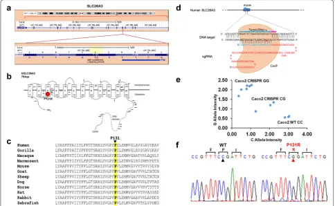
and Sift (score 0.001; cutoff: 0.05) web server tools for predicting the functional effect of amino acid sub-stitutions. Amino acid residue P131 resides within the polytopic transmembrane domain of SLC26A3 (Fig. 1b). Although the membrane domains of SLC26 polypeptides are of unknown topographical disposi-tion, hydropathy profiling has predicted a location for P131 at the putative transmembrane span3. This residue is conserved among SLC26A3 orthologs in primates, rodents, goat, sheep, dog, horse, rabbit and zebrafish (Fig. 1c). Until now, there is little informa-tion and indicainforma-tion of this SLC26A3 genetic vari-ant being linked to human diarrhea susceptibility. To further explore whether the SLC26A3 genetic variant
alters its function and expression, we adapted an HDR-mediated modification strategy using the CRISPR/ Cas9 system in both human (Caco-2, Fig. 1d) and murine colonic epithelial (CMT-93, Fig. 6a) cell lines. After the SLC26A3 c.392C>G (p.P131R) mutation was generated in both cell lines, they went though a week-long puromycin selection for a single clone that carries the exact mutation. TaqMan SNP Genotyping (Fig. 1e) and Sanger Sequencing (Fig. 1f) both were used to validate the accurate construction of P131R-SLC26A3. These results indicated that we successfully recreated SLC26A3 SNP rs386833481 (c.392C>G; p.P131R), providing the foundation for functional analysis of its effect on intestinal epithelial cell permeability.
Page 4 of 12 Zhang et al. Cell Biosci (2019) 9:40
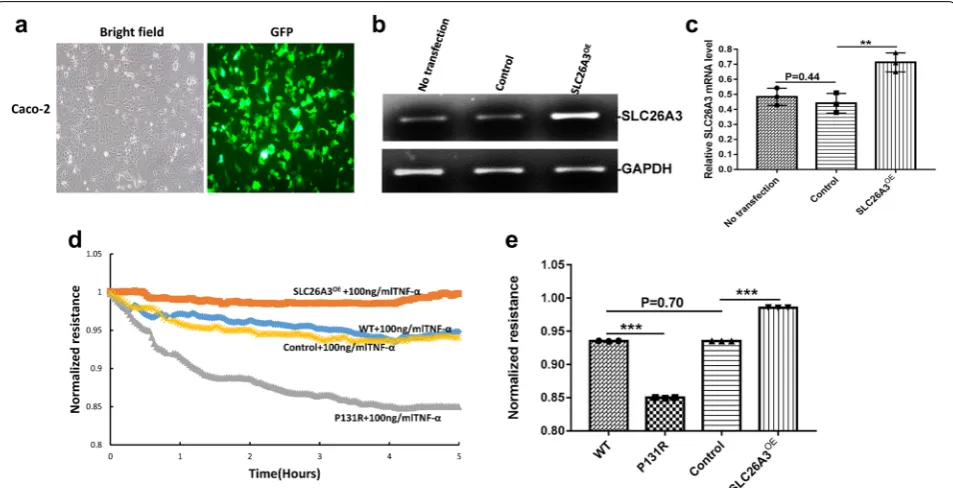
P131R‑SLC26A3 weakens the epithelial barrier and augments TNF‑α‑induced damage
To determine the role of P131R-SLC26A3 in epithelial barrier function, we also upregulated SLC26A3 expres-sion by transfecting Caco-2 cells with either SLC26A3-pCS6 or the empty vector SLC26A3-pCS6 control (Fig. 2b, c). A GFP expression vector was used to monitor transfection efficiency (Fig. 2a). Previous work showed that SLC26A3 expression is down-regulated in a TNF-α overexpressing mouse model and that TNF-α can affect the expression of tight junction proteins [22, 23]. We therefore meas-ured transepithelial electric resistance (TEER) in P131R-SLC26A3, SLC26A3-overexpressing and normal Caco-2 cells. Upon TNF-α treatment, P131R-SLC26A3 cells showed significantly lower TEER values compared with normal cells. Consistent with these results, the TEER value in SLC26A3-overexpressing (SLC26A3OE) cells was higher than that in control cells (Fig. 2d). The maxi-mum TEER induction are 0.85 ± 0.006 vs. 0.94 ± 0.003 in P131R-SLC26A3 vs. WT (P < 0.001), as well as 0.99 ± 0.003 vs. 0.95 ± 0.001 in SLC26A3OE vs. control
cells (P < 0.001), respectively. There was no significant dif-ference between WT and control cells (P = 0.70) (Fig. 2e). These results indicated that P131R-SLC26A3 weakened the epithelial barrier and augmented TNF-α-induced damage. Further, overexpression of SLC26A3 prevented TNF-α induced epithelial barrier dysfunction.
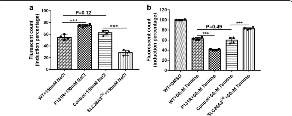
P131R‑SLC26A3 is involved in the [Cl−]
i decrease induced by Tenidap and the epithelial barrier dysfunction induced by osmotic stress
Previously, Chávez et al. [24] reported that db-cAMP elevated [Cl−]
i in noncapacitated sperm, but this increase was inhibited by Tenidap, a SLC26A3 antag-onist [25, 26]. Since P131R-SLC26A3 weakened the epithelial barrier, we explored whether this SNP was involved in the [Cl−]
i decrease induced by Tenidap. Intracellular Cl− measurements with MQAE (10 mM)
revealed that treatment with Tenidap (50 µM) sig-nificantly decreased [Cl−]
i in both P131R-SLC26A3 (42 ± 1%) and WT (63 ± 2%) cells compared to the DMSO treated controls (100 ± 1%; P < 0.001) (Fig. 3b). Fig. 2 P131R‑SLC26A3 weakens the epithelial barrier and augments TNF‑α‑induced damage. a Representative GFP for overexpressing SLC26A3. b
Representative images of SLC26A3 mRNA levels in Caco‑2 cells transfected with pCS6 (empty vector) and SLC26A3‑pCS6 (SLC26A3OE) measured
by RT‑PCR. GAPDH expression was presented as control. c Relative RT‑PCR quantification of SLC26A3 mRNA levels in Caco‑2 cells transfected with pCS6(empty vector) and SLC26A3OE. d Caco‑2 cells onto array chambers containing 40 gold electrodes per well (8W10E+) pretreated with 10 mM
cysteine and coated with fibronectin (20 µg). The experiments were initiated when the cells reached confluence, as determined by a capacitance of 10 nF at 32,000 Hz, the monolayers were starved from serum for 2 h, then treated with 100 ng/ml of TNF‑α. Representative figure of transepithelial electric resistance (TEER) data from ECIS analysis at 500HZ of wild‑type Caco‑2 cells, Caco‑2 cells containing the rs386833481(c.392C>G; p.P131R) SNP generated by CRISPR/Cas9 knock‑in, SLC26A3 overexpressing Caco‑2 cells (SLC26A3OE) and Caco‑2 cells transfected with empty vector pCS6
Page 5 of 12 Zhang et al. Cell Biosci (2019) 9:40
To investigate the function of SLC26A3 in response to hyperosmotic stress, we exposed confluent monolayers of Caco-2 cells to 150 mM sodium chloride (NaCl). The treatments provoked increased epithelial barrier dys-function in P131R-SLC26A3 cells (75% ± 5%) compared with WT cells (56% ± 4%, P < 0.001), while overexpres-sion of SLC26A3 lowered epithelial barrier dysfunc-tion (29 ± 2%) compared with pCS6 vector control cells (62 ± 4%; P < 0.001) (Fig. 3a). These results indi-cated that P131R-SLC26A3 was involved in the [Cl−]
i decrease induced by Tenidap and the epithelial barrier dysfunction induced by osmotic stress.
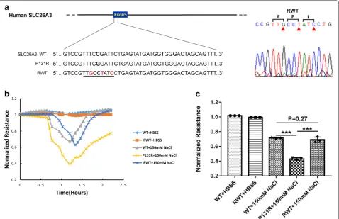
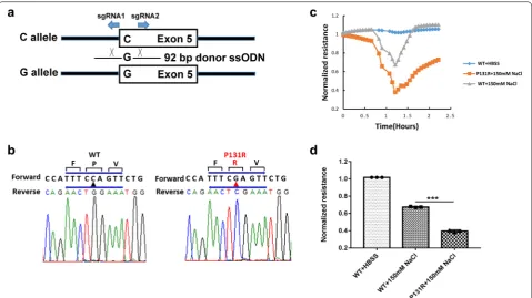
Correction of P131R‑SLC26A3 to WT restored the epithelial barrier function
To investigate if normal function of SLC26A3 could be restored by changing the P131R-SLC26A3 sequence back to a WT sequence, we employed CRISPR/Cas9 gene edit-ing usedit-ing a novel ssODN in Caco-2 cells. We designed an ssODN that coded for the WT-SLC26A3, but utilized unique codons for the three amino acid sequence (F-P-I) that spanned the wild-type Proline. This allowed us to differentiate the newly constructed WT gene from the original gene. Sanger sequencing validated the correc-tion of P131R-SLC26A3 to WT-SLC26A3 (Fig. 4a). In order to investigate the function of SLC26A3 corrected P131R-SLC26A3 (RWT) cells, we exposed confluent
monolayers of Caco-2 cells to 150 mM NaCl. The treat-ments provoked reduction in TEER in P131R-SLC26A3 cells relative to WT-SLC26A3 cells that indicated a sig-nificant decrease in epithelial barrier function (Fig. 4b). However, when SLC26A3-P131R was reversed back to wild type a similar TEER and epithelial barrier function was observed in the corrected cells (Fig. 4b). The maxi-mum TEER inductions were 0.43 ± 0.02 vs. 0.72 ± 0.01 in P131R-SLC26A3 vs. WT (P < 0.001) and 0.69 ± 0.04 vs. 0.72 ± 0.01 in RWT vs. WT (P = 0.27) (Fig. 4c). These effects were transient and did not induce significant cel-lular loss, because TEER values recovered after with-drawal of the osmotic challenge. These results indicated that reverting P131R-SLC26A3 to WT can restore the epithelial barrier function.
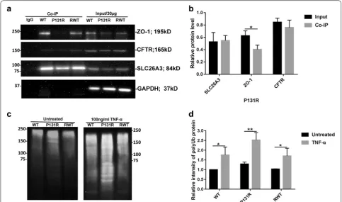
ZO‑1/CFTR mediates the epithelial barrier dysfunction induced by TNF‑α and osmotic stress and P131R‑SLC26A3 promotes SLC26A3 ubiquitination
Since TJ disruption is considered a vital event in the pathogenesis of intestinal inflammation, and there is increased permeability of the small intestine both in CF humans and in CF mice (Cftr knockout mouse mode) [20], we explored the physiological func-tions of P131R-SLC26A3 on the TJ protein ZO-1 and CFTR. Endogenous co-IP assays revealed that ZO-1 Fig. 3 P131R‑SLC26A3 is involved in the intestinal barrier dysfunction induced by osmotic stress and the [Cl−]
i decrease induced by Tenidap. a
Caco‑2 cells were seeded and cultured in growth medium until a monolayer was formed, the monolayers were starved for 1 h with Hank’s balanced salt solution (HBSS), then treated with HBSS or 150 mM of NaCl in HBSS. P131R‑SLC26A3 significantly increased the 150 mM NaCl‑induced increase fluorescence value (485/535 nm excitation/emission) compared with WT Caco‑2 cells (P = 7.4 × 10⁻⁴), but SLC26A3 over expression prevented the
NaCl‑induced increase in fluorescence value compared with the empty vector pCS6 (Control) cells. NaCl treatment resulted in similar increase of fluorescence values between WT and control cells (N = 4). b Intracellular Cl− measurements with MQAE (10 mM) were performed in CRISPR/Cas9 edited Caco‑2 cells. SLC26A3 inhibitor Tenidap (50 µM) significantly decreased [Cl−]
i in P131R‑SLC26A3 compared with WT cells. While SLC26A3
over‑expressing cells partly prevented decrease of [Cl−]
i induced by Tenidap compared with empty vector (pCS6) control cells (N = 4). *P < 0.05,
Page 6 of 12 Zhang et al. Cell Biosci (2019) 9:40
was immune-precipitated by the SLC26A3 antibody. In addition, SLC26A3 and ZO-1 protein levels are also decreased in P131R-SLC26A3 cells. Moreover, the inter-action between SLC26A3 and ZO-1/CFTR are both decreased (Fig. 5a, b). To examine the detailed mecha-nism by which P131R-SLC26A3 induced lower levels of SLC26A3, we detected SLC26A3 degradation changes. As shown in Fig. 5c, d, P131R-SLC26A3 in Caco-2 cells resulted in increased ubiquitination of SLC26A3, which was intensified by TNF-α treatment. Correction of P131R-SLC26A3 to WT displayed similar ubiquitination status as the original WT. These results indicated that ZO-1/CFTR mediated the epithelial barrier dysfunction induced by TNF-α and osmotic stress, and that P131R-SLC26A3 further promoted P131R-SLC26A3 ubiquitination.
Construction and function of Slc26a3 P131R genetic variant on murine colonic epithelial cells
To lay the foundation for in vivo experiments we inves-tigated the role of P131R-Slc26a3 in murine epithelial
barrier function. We adapted a similar HDR-mediated modification strategy using the CRISPR/Cas9 system to edit the Slc26a3 gene sequence in a murine colonic epi-thelial cell line (CMT-93, Fig. 6a). CMT-93 cells were transiently transfected with the mixture of two sgRNA constructs and an ssODN. Sanger sequencing assay (Fig. 6b) validated the construction of P131R-Slc26a3. To characterize the role of P131R-Slc26a3 in murine epi-thelial barrier function, we measured TEER in P131R-Slc26a3 and WT CMT-93 cells. Upon 150 mM NaCl treatment, P131R-Slc26a3 cells showed lower TEER val-ues compared with WT cells (Fig. 6c). The maximum TEER inductions were 0.38 ± 0.01 in P131R-Slc26a3 vs. 0.67 ± 0.02 in WT (P < 0.001) (Fig. 6d). Notably, simi-lar results were obtained when we constructed P131R-Slc26a3 and analyzed effect of this SNP on permeability in murine epithelial cells.
Page 7 of 12 Zhang et al. Cell Biosci (2019) 9:40
decreasing SLC26A3 expression through ubiquitination pathway and (b) disrupting a key interaction with ZO-1/ CFTR, thereby increasing the epithelial permeability and induced epithelial barrier dysfunction.
Discussion
In this study, we employed the CRISPR/Cas9 genomic editing tool to create human colonic epithelial Caco-2 cells containing the SLC26A3 SNP rs386833481 (c.392C>G; p.P131R) and then reverted the SNP back to its wild type sequence to investigate its effects on intes-tinal epithelial cell permeability. SNP rs386833481 was identified in patients with congenital chloride diarrhea (CCD), but its functional consequence was unknown. We have provided several lines of solid evidence that SNP rs386833481 caused the increased permeability in intesti-nal epithelial cells, indicating it is a likely causative muta-tion for diarrhea. We have also demonstrated that this mutation caused increased ubiquitination mediated deg-radation of SLC26A3, leading to decreased protein levels of SLC26A3. Our findings, along with other reports [27],
demonstrated that SLC26A3 overexpression enhances intestinal epithelial cell barrier function and may explain why SNP rs386833481 mutation caused increased intes-tinal epithelial cell permeability. Our study is the first to supply the evidence that SLC26A3 SNP rs386833481 (c.392C>G; p.P131R) is a likely causative mutation for diarrhea and has also provided a molecular mechanism underlying this observation.
Here, we investigated the influence of P131R-SLC26A3 on the epithelial barrier, and the mechanisms of regulation in human colonic epithelial cells (Caco-2). Functional analysis showed that P131R-SLC26A3 was associated with increased dysfunction of the epi-thelial barrier induced by TNF-α (Fig. 2) and osmotic stress (Fig. 3a), while overexpression of SLC26A3 pro-tected the epithelial barrier against TNF-α (Fig. 2). Moreover, P131R-SLC26A3 was involved in the [Cl−]
Page 8 of 12 Zhang et al. Cell Biosci (2019) 9:40
suggesting that P131R-SLC26A3 might be critical for development of chronic diarrhea diseases caused by impaired epithelial barrier associated with disruption of TJ proteins. We further presented two mechanisms through which chronic diarrhea-risk-associated variant at 7q31.1 lead to increased dysfunction of the epithe-lial barrier by lower levels and activity of SLC26A3: (a) P131R-SLC26A3 reduced SLC26A3 expression through an enhanced ubiquitination mediated degradation pathway, and (b) a disrupted interaction with ZO-1/ CFTR protein (Fig. 5), resulting in increased epithelial permeability and induced epithelial barrier dysfunc-tion. Both mechanisms point to reduced function of SLC26A3 as a mechanism for disease pathogenesis.
We also recreated this SNP and investigated its influ-ence on the function of epithelial barrier in murine colonic epithelial cells (CMT-93). The result similarly indicated that P131R-Slc26a3 caused increased intesti-nal epithelial cell permeability induced by osmotic stress (Fig. 6). Our ongoing studies are pursuing recreation and correction of this point mutation in an in vivo mouse
model by the AAV-CRISPR system to evaluate its utility for therapeutic development in chronic diarrhea.
Conclusions
Page 9 of 12 Zhang et al. Cell Biosci (2019) 9:40
congenital chloride diarrhea, and with applicability to complex IBD, which exhibits reduced SLC26A3 expres-sion and harbors some SNPs in its SLC26A3 [32, 33].
Methods Cell culture
The Caco-2 cells (ATCC ® HTB-37™) and CMT-93 cells (ATCC ® CCL-223™) were obtained from ATCC. Caco-2 cells, which are human colorectal adenocarcinoma epi-thelial cells, were maintained in ATCC-formulated Eagle’s Minimum Essential Medium (EMEM, Catalog#: 30-2003), supplemented with 20% fetal bovine serum (Catalog#:S11150, Atlanta Biologicals, GA, USA) and 100 U/ml penicillin/streptomycin (Catalog#: 15140122, Thermo Fisher, Waltham, MA, USA). CMT-93 cells, which are murine colonic epithelial cells, were main-tained in ATCC-formulated Dulbecco’s Modified Eagle’s medium (DMEM, Catalog#: 30-2002), supplemented with 10% FBS and penicillin/streptomycin. All cells were cultured at 37 °C in a humidified atmosphere of 5% CO2, 95% air. Cells from each primary flask were detached with 0.25% trypsin–EDTA (Catalog No. 25200056, Thermo Fisher, Waltham, MA, USA), re-suspended in fresh cul-ture medium, and seeded into 6-well plates for the fol-lowing experiments.
CRISPR target sequence design
Guide sequences for CRISPR/Cas9 gene editing were designed as previously detailed [34], chemically synthe-sized, and RNase-Free HPLC purified by Integrated DNA Technologies
(Coralville, IA, USA). Single-strand ODN was chemi-cally synthesized and standard desalted by Integrated
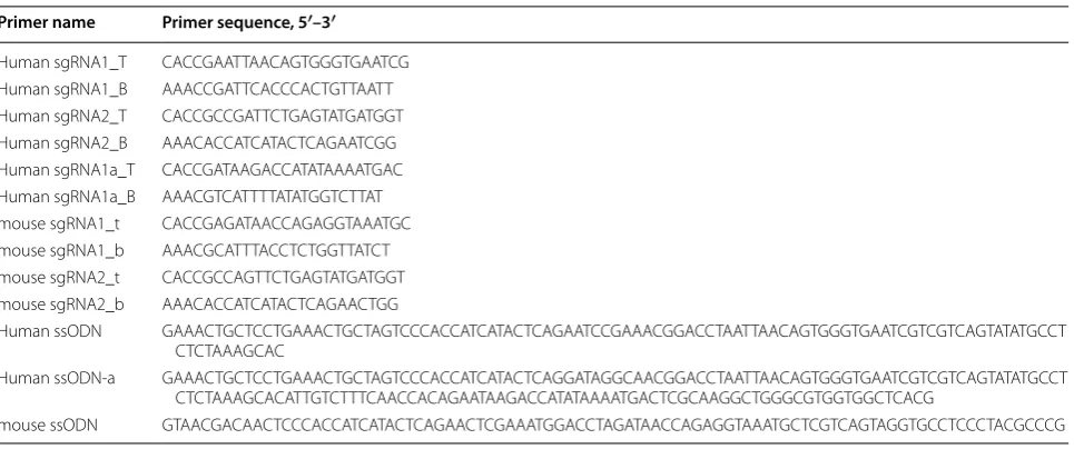
DNA Technologies (Coralville, IA, USA). All sequences are listed in Table 1.
We designed 2 guide RNAs (a forward and a reverse) that flank the SNP and a unique ssODN for a human epithelial cell line (Human sgRNA1, Human sgRNA2 and Human ssODN) and for a mouse epithelial cell line (mouse sgRNA1, mouse sgRNA2 and mouse ssODN), respectively. Each gRNA has a Top and Bottom oligo for cloning. For reverting the SNP to WT, we used different Forward sgRNA and a novel ssODN (Human sgRNA1a, Human sgRNA2 and Human ssODN-a) that code for the same amino acids as of WT SLC26A3, but use unique codons for the F-P-I triAA sequence flanking the wild-type Proline that was changed to Arginine and now back to Proline.
Plasmid construction
Sense and antisense oligonucleotides for each sgRNA were annealed and inserted into a BbsI site of the pX462 plasmid expressing Cas9/gRNA scaffold [35]. pSpCas9n (BB)-2A-Puro (PX462) V2.0 was a gift from Feng Zhang (Addgene plasmid #62987). Cloning of annealed oligonu-cleotides was confirmed by Sanger sequencing analysis using the following primer: pLKO.1.5 FW, 5′-GAC TAT CAT ATG CTT ACC GT-3′ (Lot: 14868230).
SNP models in human Caco‑2 cells and murine CMT‑93 cells
A total of 200,000 cells were seeded in 6-well plate overnight in the regular growth medium, so that they would be 80–90% confluent at the time of transfection. One hour prior to transfection, media was removed and 750 µl of pre-warmed reduced serum OptiMEM
Table 1 Primer sequence for sgRNA cloning
Primer name Primer sequence, 5′–3′
Human sgRNA1_T CAC CGA ATT AAC AGT GGG TGA ATC G Human sgRNA1_B AAA CCG ATT CAC CCA CTG TTA ATT Human sgRNA2_T CAC CGC CGA TTC TGA GTA TGA TGG T Human sgRNA2_B AAA CAC CAT CAT ACT CAG AAT CGG Human sgRNA1a_T CAC CGA TAA GAC CAT ATA AAA TGA C Human sgRNA1a_B AAA CGT CAT TTT ATA TGG TCT TAT mouse sgRNA1_t CAC CGA GAT AAC CAG AGG TAA ATG C mouse sgRNA1_b AAA CGC ATT TAC CTC TGG TTA TCT mouse sgRNA2_t CAC CGC CAG TTC TGA GTA TGA TGG T mouse sgRNA2_b AAA CAC CAT CAT ACT CAG AAC TGG
Human ssODN GAA ACT GCT CCT GAA ACT GCT AGT CCC ACC ATC ATA CTC AGA ATC CGA AAC GGA CCT AAT TAA CAG TGG GTG AAT CGT CGT CAG TAT ATG CCT CTC TAA AGC AC
Human ssODN‑a GAA ACT GCT CCT GAA ACT GCT AGT CCC ACC ATC ATA CTC AGG ATA GGC AAC GGA CCT AAT TAA CAG TGG GTG AAT CGT CGT CAG TAT ATG CCT CTC TAA AGC ACA TTG TCT TTC AAC CAC AGA ATA AGA CCA TAT AAA ATG ACT CGC AAG GCT GGG CGT GGT GGC TCA CG
Page 10 of 12 Zhang et al. Cell Biosci (2019) 9:40
media (Catalog#: 31985070, Thermo Fisher, Waltham, MA, USA) was added to each well. Transfection was performed using Lipofectamine 3000 and P3000 rea-gent (Catalog#: 3000015, Thermo Fisher, Waltham, MA, USA). For each well, 5 µl of P3000 reagent was diluted in 125 µl OptiMEM with pX462-gRNA plasmids (500 ng) and the donor plasmid (500 ng) containing a synthe-sized sequence for SNP. 5 µl of Lipofectamine 3000 was diluted in 125 µl OptiMEM and, after 3 min, it was added to the mixture of DNA and P3000 reagent. The complete mixture was incubated 15 min before being added to cells. After 6 h, the media was changed to 2 ml complete medium. GFP plasmid was used to monitor transfection efficiency. The puromycin concentration used for SNP selection was determined prior to cell selection by meas-uring cell sensitivity (1 µg/ml for Caco-2 cells and 2 µg/ ml for CMT-93 cells). Puromycin selection was initiated 24 h post transfection for 72 h until WT control cells were all dead. Then transfected cells were cultured in reg-ular medium with 0.1 µg/ml puromycin for 4-7 days and harvested for Taqman genotyping and sequence analysis.
Sanger sequencing
Genomic DNA (gDNA) was extracted from puromycin selected cells using the Gentra Puregene Cell Kit (Cata-log#: 51306, QIAGEN, Toronto, Canada) according to the manufacturer’s instruction. 50 ng of the isolated genomic DNA was used as template to amplify DNA by Platinum Taq (Catalog#: 10966-026, Thermo Fisher, Waltham, MA, USA) PCR. The amplicon DNA after PCR was verified by 2% agarose gel and gel purified using the Nucleospin Gel and PCR Clean-up kit (Catalog#: 740609.250, Clontech, Mountain View, CA, USA). The concentration and purity of DNA was determined by measuring absorbance at 260 and 280 nm using Take3 microspot plate reader (BioTek), and the nucleotide sequence of individual colonies was determined by sequencing using the following primer: Human SLC26A3_R: 5′-TCC CAA AGT GCT GGG ATT AC-3′ (Lot: 162860013); Mouse Slc26a3_R: 5′-TAC TGA TGC AGC CAC CAT TAC-3′ (Lot: 190941198).
ViiA7 TaqMan SNP genotyping assay
Genomic DNA (gDNA) was extracted from puromycin selected cells using the Gentra Puregene Cell Kit, accord-ing to the manufacturer’s instruction. The TaqMan fluo-rescently labeled probes (Catalog#: 4351379, Thermo Fisher, Waltham, MA, USA) targeting the studied rs386833481 SNP and genotyping Master Mix (Catalog#: 4371355, Thermo Fisher, Waltham, MA, USA) were used for DNA amplification in the ViiA7 Sequence Detection System (Applied Biosystems, USA). Genotyping was per-formed blinded to sample status. A non-template reac-tion (using water instead of DNA) was used as negative
control and a sample of known genotype was used as positive control.
Measurement of TEER (ECIS assay)
Cellular barrier properties (Transepithelial electric resist-ance, TEER) were measured using an electrical cell-sub-strate impedance sensing system (ECIS Ztheta; Applied Biophysics, Troy, NY, USA). Caco-2 or CMT-93 cells were seeded onto array chambers containing 40 gold electrodes per well (8W10E+, Applied Biophysics) pre-treated with 10 mM cysteine and coated with fibronectin (20 µg) according to the manufacturers’ specifications. The experiments were initiated when the cells reached confluence, as determined by a capacitance of 10 nF at 32,000 Hz. Caco-2 or CMT-93 cells were starved for 2 h, then treated with 100 ng/ml of TNF-α (Catalog#: 210-TA and 410-MT-010, R&D Systems Inc., Minneapolis, MN, USA) or 150 mM NaCl (Catalog#: IB07072, IBI Scientific, Peosta, IA,USA). The data are presented as normalized resistance vs. time at 500 HZ. Resistance was averaged over the 40 electrodes per chamber and normalized so the time 0 resistance was 1.0.
In vitro cell permeability assays
In vitro cell permeability assays were carried out according to the protocol of the CHEMICON In Vitro Vascular Per-meability Assay kit (Catalog#: ECM644; Millipore, Biller-ica, MA, USA). Briefly, cells (1 × 10⁵) were seeded into the culture inserts of permeability chambers that were coated with collagen and incubated at 37 °C until a monolayer was formed. After the cells were starved from serum for 1 h with Hank’s balanced salt solution (HBSS), then treated with HBSS (control) or 150 mM of NaCl in HBSS. Cells were incubated for another 30 min at 37 °C, followed by addition of 75 μl of FITC-Dextran to each insert for 20 min at room temperature (RT), and then 100 μl of the solution in the bottom chamber was transferred to a black 96-well opaque plate. Absorbance at 485 and 535 nm was meas-ured in a TriStar Multimode Reader (LB 941, Berthold Technologies GmbH & Co. KG, Bad Wildbad, Germany). Reagent control wells were treated with HBSS only. Blank inserts without plated cells were also included as controls.
Intracellular Cl− measurements in Caco‑2 cells
The [Cl−]
i was measured in Caco-2 cells using MQAE (Catalog#: E3103, Thermo Fisher, Waltham, MA, USA), a Cl− sensitive fluorescent dye, as previously described
[20]. Briefly, Caco-2 cells were incubated with 10 mM MQAE for 30 min at 37 °C. Excess MQAE was removed by changing with fresh medium. The influence of SLC26A3 inhibitor (50 µM Tenidap, Catalog#: PZ0196, Sigma, St. Louis, MO, USA) on [Cl−]
Page 11 of 12 Zhang et al. Cell Biosci (2019) 9:40
excitation/emission) for 1–3 min, and measuring for a further 5–10 min after the addition of Tenidap. Two controls were performed: (1) DMSO (drug solvent) was added while the fluorescence was recorded; and (2) MQAE fluorescence without cells was recorded, and the Tenidap were added. No significant fluorescence changes were observed after performing both controls.
Co‑immunoprecipitation
For co-IP assays, Caco-2 cells were lysed on ice with non-denaturing lysis buffer for 1 min, then were scraped and gently transferred into a chilled microcentrifuge tube. The cells were mixed on a rotary mixer for 30 min at 4 °C. After centrifugation, the concentration of supernatants were assayed by BCA assay and incubated overnight at 4 °C with the SLC26A3 antibody (Catalog#: GTX34204, GeneTex, Irvine, CA, USA). We used 500 µg protein and 2 µg SLC26A3 antibody in 500 µl Lysis Buffer containing the protease inhibitor for WT, P131R and RWT at the same time. After antibody binding, add 25 µl of protein A/G Sepharose® beads slurry to each tube and incubate for 1 h at 4 °C. The beads were then washed three times with 1× wash buffer, and the precipitates were eluted with sample buffer, separated by 7.5% SDS/PAGE, and analyzed by immunoblotting.
We had validated all antibodies in Caco-2 cells, CMT-93 cells and HCT116 cells, which are all colonic epithelial cells.
Western blotting
Caco-2 cell lysates were collected on ice in RIPA buffer and isolated by centrifugation at 13,000 RPM for 10 min at 4 °C. Protein was quantified by Pierce BCA (Catalog#: 23225, Thermo Fisher, Waltham, MA, USA). 30 μg pro-tein was boiled with sample buffer prior to loading on a polyacrylamide gel. SLC26A3 antibody diluted 1:1000 in TBS-T + 5% milk (Catalog#: GTX34204, GeneTex, Irvine, CA, USA), CFTR antibody diluted 1:500 in TBS-T + 5% milk (Catalog#:sc-376683, Santa Cruz Biotechnol-ogy, Dallas, TX, USA), ZO-1 antibody diluted 1:1000 in TBS-T + 5% milk (Catalog#: GTX108613, GeneTex, Irvine, CA, USA), GAPDH antibody diluted 1:2000 in TBS-T + 5% milk (Catalog#: Sc-25778, Santa Cruz Bio-technology, Dallas, TX, USA). Goat anti-rabbit HRP antibody diluted 1:10,000 in TBS-T + 5% milk (Catalog#: PI-1000, Vector Biolabs, Malvern, PA, USA) and horse anti-mouse HRP antibody diluted 1:10,000 in TBS-T + 5% milk (Catalog#: P1-2000, vector Biolabs Malvern, PA, USA) were used to visualize westerns. Bands were visual-ized by ECL (Pierce ECL Western Blotting Substrate, Cat-alog#: 32106, Thermo Fisher,Waltham, MA, USA) with a FluorChem M Imager (ProteinSimple, San Jose, CA, USA) and quantified by AlphaView Software SA, v.3.4.0.0.
Statistical analyses
Statistical analyses were carried out using the Sigma Stat (ver.4.0, SysTest Software, Inc., San Jose, CA). All data were expressed as mean ± SD (standard deviation) of at least three independent experiments. Two group com-parisons were done by an unpaired Student’s t test. Dif-ferences between groups were considered statistically significant at P < 0.05.
Abbreviations
SLC26A3: solute carrier family 26 member 3; CCD: congenital chloride diar‑ rhea; SNP: single nucleotide polymorphism; ssODN: single‑stranded DNA oligonucleotide; ECIS: electrical cell‑substrate impedance sensing system.
Acknowledgements
We would like to thank Natasha Kibiryeva for her assistance in Sanger sequencing and Dr. Mark D. Nichols for reviewing our manuscript.
Authors’ contributions
SQY, NZ, DPH, and LQZ designed the study; NZ, WW, MN, KS and KL performed experiments; NZ, SQY, DPH and LQZ analyzed data; NZ, SQY and DPH wrote the manuscript; NZ, MN, XJ, BW, CF, DYL, LQZ, SQY reviewed and revised the manuscript. All authors read and approved the final manuscript.
Funding
This study is in part supported by the start‑up fund and William R Brown/ Missouri State endowments of The Children’s Mercy Hospital, University of Missouri Kansas City School of Medicine and Missouri State (S.Q.Y.) and GI Research funds of The Children’s Mercy Hospital, Kansas City, MO (DYL and CF).
Availability of data and materials Not applicable.
Ethics approval and consent to participate Not applicable.
Consent for publication Not applicable.
Competing interests
The authors declare that they have no competing interests.
Author details
1 Division of Gastroenterology, Department of Pediatrics, Children’s Mercy Hospitals and Clinics, Kansas City, MO, USA. 2 Division of Experimental and Translational Genetics, Department of Pediatrics, Children’s Mercy Hospi‑ tals and Clinics, Kansas City, MO, USA. 3 Department of Biomedical and Health Informatics, University of Missouri Kansas City School of Medicine, Kansas City, MO, USA. 4 Department of Pediatrics, Tangdu Hospital, Fourth Military Medical University, Xi’an, Shaanxi, China. 5 Department of Biomedical Sciences, University of Missouri Kansas City School of Medicine, Kansas City, MO, USA. 6 Division of Cell Biology & Biophysics, University of Missouri Kansas City School of Biological Sciences, Kansas City, MO, USA. 7 Department of Global Biostatistics and Data Science, Center for Bioinformatics and Genomics, Tulane University, New Orleans, LA, USA. 8 Department of Neonatology, Nanfang Hospital, Southern Medical University, Guangzhou, China.
Received: 11 March 2019 Accepted: 7 May 2019
References
Page 12 of 12 Zhang et al. Cell Biosci (2019) 9:40
•fast, convenient online submission •
thorough peer review by experienced researchers in your field • rapid publication on acceptance
• support for research data, including large and complex data types •
gold Open Access which fosters wider collaboration and increased citations maximum visibility for your research: over 100M website views per year •
At BMC, research is always in progress.
Learn more biomedcentral.com/submissions
Ready to submit your research? Choose BMC and benefit from: 2. Chatterjee I, Kumar A, Castilla‑Madrigal RM, Pellon‑Cardenas O, Gill
RK, Alrefai WA, et al. CDX2 upregulates SLC26A3 gene expression in intestinal epithelial cells. Am J Physiol Gastrointestin Liver Physiol. 2017;313(3):G256–64.
3. Wedenoja S, Pekansaari E, Höglund P, Mäkelä S, Holmberg C, Kere J. Update on SLC26A3 mutations in congenital chloride diarrhea. Hum Mutat. 2011;32(7):715–22.
4. Ziki MDA, Verjee MA. Case report: rare mutation in the SLC26A3 trans‑ porter causes life‑long diarrhoea with metabolic alkalosis. BMJ Case Rep. 2015. https ://doi.org/10.1136/bcr‑2014‑20684 9.
5. Matsunoshita N, Nozu K, Yoshikane M, Kawaguchi A, Fujita N, Morisada N, et al. Congenital chloride diarrhea needs to be distinguished from Bartter and Gitelman syndrome. J Hum Genet. 2018;63(8):887–92.
6. Höglund P, Haila S, Socha J, Tomaszewski L, Saarialho‑Kere U, Karjalainen‑ Lindsberg M‑L, et al. Mutations of the down‑regulated in adenoma (DRA) gene cause congenital chloride diarrhoea. Nat Genet. 1996;14(3):316–9. 7. Xia W, Yu Q, Riederer B, Singh AK, Engelhardt R, Yeruva S, et al. The distinct
roles of anion transporters Slc26a3 (DRA) and Slc26a6 (PAT‑1) in fluid and electrolyte absorption in the murine small intestine. Pflügers Archiv‑Eur J Physiol. 2014;466(8):1541–56.
8. Ishiguro H. HCO3− secretion by SLC26A3 and mucosal defence in the colon. Acta Physiol. 2014;211(1):17–9.
9. Shcheynikov N, Wang Y, Park M, Ko SB, Dorwart M, Naruse S, et al. Cou‑ pling modes and stoichiometry of Cl−/HCO
3− exchange by slc26a3 and slc26a6. J Gen Physiol. 2006;127(5):511–24.
10. Dorwart MR, Shcheynikov N, Baker JM, Forman‑Kay JD, Muallem S, Thomas PJ. Congenital chloride‑losing diarrhea causing mutations in the STAS domain result in misfolding and mistrafficking of SLC26A3. J Biol Chem. 2008;283(13):8711–22.
11. Janecke AR, Heinz‑Erian P, Müller T. Mechanisms underlying dysregulation of electrolyte absorption in inflammatory bowel disease‑associated diar‑ rhea. Inflamm Bowel Dis. 2016;22(6):E17–8.
12. Kumar A, Chatterjee I, Gujral T, Alakkam A, Coffing H, Anbazhagan AN, et al. Activation of nuclear factor‑κB by tumor necrosis factor in intestinal epithelial cells and mouse intestinal epithelia reduces expression of the chloride transporter SLC26A3. Gastroenterology. 2017;153(5):1338–50. 13. Zhang W, He T, Wang Q, Li X, Wei J, Hou X, et al. IL‑1 receptor‑associated
kinase‑2 genetic variant rs708035 increases NF‑κB activity through pro‑ moting TRAF6 ubiquitination. J Biol Chem. 2014;289(18):12507–19. 14. Krawczak M, Ball EV, Fenton I, Stenson PD, Abeysinghe S, Thomas N,
et al. Human gene mutation database—a biomedical information and research resource. Hum Mutat. 2000;15(1):45–51.
15. Asano K, Matsushita T, Umeno J, Hosono N, Takahashi A, Kawaguchi T, et al. A genome‑wide association study identifies three new suscepti‑ bility loci for ulcerative colitis in the Japanese population. Nat Genet. 2009;41(12):1325–9.
16. Wojtal KA, Eloranta JJ, Hruz P, Gutmann H, Drewe J, Beglinger C, et al. Changes in mRNA expression levels of solute carrier transport‑ ers in inflammatory bowel disease patients. Drug Metab Dispos. 2009;37(9):1871–7.
17. Xiao F, Yu Q, Li J, Johansson M, Singh A, Xia W, et al. Slc26a3 deficiency is associated with loss of colonic HCO3− secretion, absence of a firm mucus layer and barrier impairment in mice. Acta Physiol. 2014;211(1):161–75. 18. Fong P. CFTR–SLC26 transporter interactions in epithelia. Biophys Rev.
2012;4(2):107–16.
19. Ko SB, Zeng W, Dorwart MR, Luo X, Kim KH, Millen L, et al. Gating of CFTR by the STAS domain of SLC26 transporters. Nat Cell Biol. 2004;6(4):343–50. 20. De Lisle RC. Disrupted tight junctions in the small intestine of cystic
fibrosis mice. Cell Tissue Res. 2014;355(1):131–42.
21. Ruan YC, Wang Y, Da Silva N, Kim B, Diao RY, Hill E, et al. CFTR interacts with ZO‑1 to regulate tight junction assembly and epithelial differentia‑ tion via the ZONAB pathway. J Cell Sci. 2014;127(Pt 20):4396–408. 22. Juric M, Xiao F, Amasheh S, May O, Wahl K, Bantel H, et al. Increased epi‑
thelial permeability is the primary cause for bicarbonate loss in inflamed murine colon. Inflamm Bowel Dis. 2013;19(5):904–11.
23. Ding X, Li D, Li M, Tian D, Yu H, Yu Q. Tumor necrosis factor‑α acts reciprocally with solute carrier family 26, member 3,(downregulated‑in‑ adenoma) and reduces its expression, leading to intestinal inflammation. Int J Mol Med. 2018;41(3):1224–32.
24. Chávez JC, Hernández‑González EO, Wertheimer E, Visconti PE, Darszon A, Treviño CL. Participation of the Cl−/HCO
3− exchangers SLC26A3 and SLC26A6, the Cl− channel CFTR, and the regulatory factor SLC9A3R1 in
mouse sperm capacitation. Biol Reprod. 2012;86(1):1–14.
25. Chernova MN, Jiang L, Shmukler BE, Schweinfest CW, Blanco P, Freedman SD, et al. Acute regulation of the SLC26A3 congenital chloride diar‑ rhoea anion exchanger (DRA) expressed in Xenopus oocytes. J Physiol. 2003;549(1):3–19.
26. Uchiyama H, Hayashi H, Suzuki Y. Functional characterization of Cl−/
HCO3−exchange in villous cells of the mouse ileum. Biomed Res. 2006;27(6):265–74.
27. Ding X, Li D, Li M, Wang H, He Q, Wang Y, et al. SLC26A3 (DRA) prevents TNF‑alpha‑induced barrier dysfunction and dextran sulfate sodium‑ induced acute colitis. Lab Invest. 2018;98(4):462–76.
28. Kim E, Koo T, Park SW, Kim D, Kim K, Cho H‑Y, et al. In vivo genome editing with a small Cas9 orthologue derived from Campylobacter jejuni. Nat Commun. 2017;8:14500–11.
29. Yang Y, Wang L, Bell P, McMenamin D, He Z, White J, et al. A dual AAV system enables the Cas9‑mediated correction of a metabolic liver disease in newborn mice. Nat Biotechnol. 2016;34(3):334–8.
30. Johansen AK, Molenaar B, Versteeg D, Leitoguinho AR, Demkes CJ, Spanjaard B, et al. Postnatal cardiac gene‑editing using CRISPR/Cas9 with AAV9‑mediated delivery of short guide RNAs results in mosaic gene disruption. Circ Res. 2017;121(10):1168–81.
31. Platt RJ, Chen S, Zhou Y, Yim MJ, Swiech L, Kempton HR, et al. CRISPR– Cas9 knockin mice for genome editing and cancer modeling. Cell. 2014;159(2):440–55.
32. Ye BD, McGovern DP. Genetic variation in IBD: progress, clues to pathogenesis and possible clinical utility. Expert Rev Clin Immunol. 2016;12(10):1091–107.
33. Brant SR, Okou DT, Simpson CL, Cutler DJ, Haritunians T, Bradfield JP, et al. Genome‑wide association study identifies African‑specific susceptibility loci in African Americans with inflammatory bowel disease. Gastroenter‑ ology. 2017;152(1):206–17.
34. Ran FA, Hsu PD, Wright J, Agarwala V, Scott DA, Zhang F. Genome engineering using the CRISPR–Cas9 system. Nat Protocol. 2013;8(11):2281–308.
35. Cong L, Ran FA, Cox D, Lin S, Barretto R, Habib N, et al. Multiplex genome engineering using CRISPR/Cas systems. Science. 2013;339(6121):819–23.
Publisher’s Note