Please cite this article as Nasiri K, Kazemi-Fard M, Rezaei M & Yousefi S. 2019. Supplementation of Sodium Selenite and Methionine on Concentration of Selenium in Egg and Serum, Antioxidant Enzymes Activity and Immune Response of Iranian Native Broiler Breeders. Poult. Sci. J. 7(2): 119-129.
© 2019 PSJ. All Rights Reserved ISSN: 2345-6604 (Print), 2345-6566 (Online) http://psj.gau.ac.ir
DOI: 10.22069/psj.2019.16438.1427
Supplementation of Sodium Selenite and Methionine on Concentration of Selenium in
Egg and Serum, Antioxidant Enzymes Activity and Immune Response of Iranian Native
Broiler Breeders
Nasiri K, Kazemi-Fard M, Rezaei M & Yousefi S
Department of Animal Science, Sari Agricultural Sciences and Natural Resources University, Sari, Iran
Poultry Science Journal 2019, 7(2): 119-129
Introduction
Trace elements are very important to production, health and activities of major antioxidant enzymes. Selenium is an essential trace element that plays important role in many activities, including in the production, health and many antioxidant enzymes activity such as GSH-Px and thioredoxin reductase. This trace element also has fundamental roles in human health. Sodium selenite (SS) has been the major source of selenium in animals’ diets to warrant a most favorable supply (Hoffmann et al., 2008). In the last few years many researchers found better
replacement source for SS that have a high bioavailability and low toxicity such as seleno-yeast and seleno-methionine [Se]Met (Yoon et al., 2007; Reis et al., 2009). Organic selenium is found in the form of selenium yeast and [Se]Met. Organic selenium consumption is safe for humans and has a toxic effect at 0.5 mg/kg of diet.
Your body can metabolize and use all three forms of selenium, but the amount absorbed varies. About 90 % of [Se]Met and 50 % of selenite are absorbed. Selenium Methionine can replace methionine in the body tissue instead of methionine in case of need,
Abstract
Keywords
Methionine Broiler breeder Sodium selenite Immune response Antioxidant enzyme
Effects of sodium selenite (SS) in offspring of broiler breeders has been reported, but the comparison between SS and different level of methionine on offspring has received limited, so this study was conducted to investigate sodium selenite and methionine effects on concentration of selenium in egg and serum of Iranian native broiler breeders. An experiment was conducted in 3 × 3 factorial experiment with three levels of sodium selenite (0.0, 0.3, and 0.5 mg/kg of diet) and three levels of methionine (0.23, 0.31 and 0.33 % of diet) to investigate reproductive performance and immune status of broiler breeders (64-74 wks). The higher methionine levels increased the egg weight until the dose of 0.31% of diet (P < 0.05). In this experiment, 0.5 mg/kg SS with 0.31 % methionine in the diet was found to increase egg weight (P < 0.05). The highest salable chicks were obtained by supplementing 0.3 mg/kg SS (P= 0.07). Increasing the level of methionine from 0.23 to 0.33% without SS and also, supplementation SS at 0.3 mg/kg with 0.33 % methionine exhibited higher serum selenium, although, no effect of methionine levels was observed on concentration of selenium in serum and transfer rate. Concentration of selenium in eggs and serum were significantly affected by interaction of SS and methionine levels in diet. An increment in glutathione peroxidase (GSH-Px) level tended to increase with SS (0.5 mg/kg) and methionine (0.33%) interaction (P=0.06). The results showed that increasing the level of methionine increases the immune response against influenza (AI) and Newcastle disease (ND) (P = 0.0001). These results suggest that higher level of methionine at 0.31% could improve egg weight and Influenza and ND titers, SS at dose of 0.5 could increase concentration of selenium in serum, whereas increasing of SS levels significantly raised embryonic mortality.
Corresponding author Mohammad Kazemi Fard [email protected]
Article history
however, the incorporation of seleno-methionine into tissue proteins and keratin in the cattle, birds and fish causes alkali disease. Two pools of reserve selenium have been detected in the body (Waschulewski and Sunde, 1988). The first is as [Se]Met that reportedly is different from methionine in terms of physiological function. The second reserve pool is the selenium found in liver GSH-Px. Following direct metabolism, the consumed selenite, selenate, and selenocysteine are all turned into selenide, which is the reduced form of selenium. According to Knight and Sunde (1988), selenium was less available for GSH-Px synthesis from dietary [Se]Met consumed less than 0.5 mg Se/kg in rats having a methionine-deficient diet. These researchers offered that [Se]Met had been preferentially entered into body proteins when methionine was limiting. Contrary to increased tissue selenium levels, The tissue GSH-Px activity decreased in a methionine deficient diet when the rate
received methionine in a [Se]Met form
(Waschulewski and Sunde. 1988)
Waschulewski and Sunde (1988) showed that methionine supplementation in rat for 8 wk resulted in a 1.8-fold increase in erythrocyte GSH-Px, suggesting that the additional methionine increased the availability of dietary [Se]Met or stored [Se]Met. Selenium absorption efficiency is relatively high (89±4) and depends on the dietary sources ( Susanne et al., 2008). In contrast to inorganic forms [Se]Met is absorbed very rapidly and enhanced at low methionine intake in the intestinal (Vendeland et al., 1996).
According to above reports, we assume that perhaps the different levels of Methionine could affect availability of selenium in the inorganic resources. The one objective of this experiment was to compare GSH-Px plasma and egg selenium concentrations in naive broiler breeders that fed with different levels of methionine and selenium. We wanted to determine if the methionine that had been supplied in higher dose could be improved selenium transfer rate for egg and serum (reflected in antioxidants enzyme) when selenium intake is in different levels.
Therefore, the present experiment was designed to evaluate the antioxidant activity and selenium levels in serum and eggs of Iranian native broiler breeders that were fed diets supplemented with different doses of selenium as SS form and methionine during a 56-d period.
Material and Methods
Experimental Design and Diets
All the procedures used in this study were based on the approval of the Animal Rights Protection Committee of Sari Agricultural and Natural Resources University. A completely randomized design was applied in a 3 × 3 factorial arrangement,
with three levels of SS (0.0, 0.3, and 0.5 mg/kg of diet) and three levels of methionine (0.23, 0.31, and 0.33 % of diet). At 64 wk of age, the birds were distributed among the pens based on body weight and egg production (2,215 ± 25.8 g). The experiment started two wks after adaptation period at 66 wk of age and lasted for 8 wks.
A total of 270 Iranian native broiler breeders were randomly allocated to nine groups with three replicates. Briefly, 10 females and 1 male were assigned to each of 27 pens. All birds were housed in breeder pens (120 × 80 cm; length × width) and had free access to feed and water. House temperature tried to keep at 24 ± 3ºC and the length of lighting was16 h per day. The experimental diets were based on corn and soybean meal formulated according to Iranian native broiler breeder’s guide to meet all nutrients requirement except selenium and methionine. Sodium selenite was supplemented to corn basal diet (According to table 1) at the levels of 0, 0.3, 0.5 mg/kg. Methionine was added at the level of 0.0027 and 0.0057 % (Evonik Degussa GmbH, Hanau-Wolfgang, Germany) to the basal diets to provide 0.23, 0.31, and 0.33 % (Table 1).
Data collection
Productive Performance
Egg production and settable eggs were recorded daily. Percentage of settable eggs, egg production, and egg weight were measured as indicated by Kazemi-Fard et al. (2013). Archimedes method (Hempe et al., 1988) was used for specific gravity assay. Egg shell thickness, egg quality and egg shell surface area (SA) were measured as indicated by Tahmasbi et al. (2012) and Ousterhout (1980), respectively. The shell weight per unit of surface area (SWUSA) was calculated by dividing the dried shell weight by the surface area of each egg. Haugh unit calculated based on albumen height and egg weight using the formula: HU = 100 log10 (H − 1.7 W0.37 + 7.56), (Carter, 1975). Where: HU= Haugh unit, H= height of the albumen (mm) and W= egg weight (g).
Hatch Characteristics and Embryonic Mortality
Poultry Science Journal2019, 7(2): 119-129
mid-dead (8-16 d), late dead (17-21 d), and pips. The rate of fertile eggs to total eggs set was set to explain fertility. After 19 days, the eggs were put into baskets; then, these baskets were put into the hatchery cabinets and the hatcher was adjusted to 36.44 ̊C dry bulb and 32.18 ̊ C wet bulb temperatures. After 21.5 days of incubation, the number of hatched eggs was
logged. The number of hatched chicks to fertile eggs was set to explain the hatchability of fertile eggs and the percentage of hatched chicks to the total eggs set was used to explain the cumulative hatchability. As explained by Dziaczkowska (1980), chick quality was determined based on normal and abnormal chick.
Table 1. Feed ingredients and chemical composition of experimental diets
item sodium selenite (0 mg/kg)
sodium selenite (0.3 mg/kg) sodium selenite (0.5 mg/kg) Methionine level (%)
methionine level (%) Methionine level (%) Ingredients (%) 0.21 0.31 0.33 0.21 0.31 0.33 0.21 0.31 0.33 Corn 69.87 69.86 69.86 69.57 69.56 69.56 69.37 69.36 69.36 Soybean meal 22.22 22.22 22.22 22.22 22.22 22.22 22.22 22.22 22.22 Calcium carbonate 6.2 6.2 6.2 6.2 6.2 6.2 6.2 6.2 6.2 Di-calciu phosphate 0.81 0.81 0.81 0.81 0.81 0.81 0.81 0.81 0.81 Sodium chloride 0.37 0.37 0.37 0.37 0.37 0.37 0.37 0.37 0.37 L-Lysine 0.03 0.03 0.03 0.03 0.03 0.03 0.03 0.03 0.03 DL-MET - 0.0027 0.0055 - 0.0027 0.0055 - 0.0027 0.0055 Sodium selenite
(mg/kg) - - - 0.3 0.3 0.3 0.5 0.5 0.5
Vitamin premix‡ 0.25 0.25 0.25 0.25 0.25 0.25 0.25 0.25 0.25 Mineral premix# 0.25 0.25 0.25 0.25 0.25 0.25 0.25 0.25 0.25 Chemical analysis
for selenium (mg/kg) 0.20 0.20 0.02 0.401 0.401 0.401 0.468 0.468 0.468 Calculated Feed analysis
) kcal / kg ( ME
2850 2850 2850 2850 2850 2850 2850 2850 2850
CP (%) 15.5 15.5 15.5 15.5 15.5 15.5 15.5 15.5 15.5 (%)
a
P 0.35 0.35 0.35 0.35 0.35 0.35 0.35 0.35 0.35
Ca (%) 2.7 2.7 2.7 2.7 2.7 2.7 2.7 2.7 2.7
Met (%) 0. 21 0. 31 0. 33 0. 21 0. 31 0. 33 0. 21 0. 31 0. 33 SAA (%) 0.45 0.60 0.64 0.45 0.60 0.64 0.45 0.60 0.64 ‡Vitamin premix (per kg of diet) supplied: vitamin A: 12000 IU; vitamin E: 100 IU; vitamin K3: 7 mg; vitamin B1: 3 mg;
vitamin B2: 12 mg; vitamin B3: 12 mg; Nicotinic acid: 40 mg; vitamin B6: 4 mg; vitamin B9: 1.5 mg; vitamin B12: 0.04 mg; vitamin B19: 0.25 mg; Choline chloride: 200 mg.
Vitamin premix: vitamin D3 was added at the rate of 0, 3500, and 4200 IU/kg of diet to provide three vitamin D3 diets. #Mineral premix provided (mg/kg of diet): Mn: 60; Fe: 60; Zn: 100; Cu: 10; Co: 0.2; I: 0.5 and Se: 0.4.
Selenium Concentrating in Diet, Egg and Serum Content
Total selenium concentrations in diet, egg, and serum samples were determined by inductively coupled plasma–mass spectrometry (ICP–MP, AES 4100; Australia). We added the 1g homogenized sample to the PTFE vessel, then added 5 mL nitric acid 65% and 2.5 mL Hydrogen peroxide 30% to allow the sample to predigest for at least 2h at 120 ºC. Then sample was diluted up to 25 mL, 10 mL of sample was transferred to a 25 mL volume flask and 5 mL concentrated HCl was added. The solution was taken at water bath at 80 ºC for 30 minutes. Transfer percentage of Se from the feed to the eggs and sera was considered as a transfer rate (Delezie et al., 2014).
Antioxidant Enzymes Activities in Plasma
To specify GSH-Px and SOD activity, six blood samples were taken from each treatment in the middle and end of experiment (at 7:00 am). Plastic tubes containing EDTA were used to hold the blood taken
from the left brachial vein. Samples were kept on ice up to the time when plasma was divided by centrifuging (1500 × g for 20 minutes) within 60 min of collection. For further analysis, plasma was put for harvesting and was kept at -20°C until. The commercial RANSEL and RANSOD kit was used to specify the concentrations of GSH-Px and SOD
(RANDOX Laboratories Ltd., London, UK)
2-(4-iodophenyl-3-(4-nitrophenol)-5-phenyltetrazolium chloride. The degree of inhibition of this reaction determines the superoxide dismutase activity.
Newcastle and Influenza Immunity
Following the administration of the dead vaccine, two blood samples (2 to 3 mL) were gathered as two birds from the left brachial vein in each pen at 4, 6, and 8 wk. All blood samples underwent centrifuging at 1000 × g for 5 min to isolate sera; then the sera was put for harvesting and kept at -20 ̊C until analysis. The hemagglutination inhibition (HI) method was applied to determine the presence of Newcastle and Influenza antibodies in sera samples.
Statistical Analysis
Data were analyzed according to a completely
randomized design with a 3 × 3 factorial arrangement using the GLM procedure of SAS (2003) software. Newcastle and Influenza antibody titers data were analyzed using the repeated measurement procedure of the SAS in PROC MIXED procedure. Differences among treatment means were measured by Duncan’s multiple range test and considered significant at P < 0.05.
Results and Discussion
Analyzed selenium contents in the experimental diets were slightly greater than calculated values. No significant effect was found for treatments on the egg shell thickness, shell weight unit per surface area, shape index, yolk and albumin weight, thin shell and Haugh unit (data not shown).
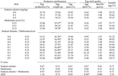
Table 2. The effect of sodium selenite, methionine level and their interaction on productive performance,
eggshell quality and settable egg of Iranian native broiler breeder
Settable egg (%) Egg shell quality
Productive performance Item Specific gravity Egg shell (%) Egg mass (%) Egg weight (g) Egg production (%) Sodium selenite (mg/kg)
92.25 1.08 8.62 19.82 55.04 32.76 0.0 93.62 1.08 8.50 21.31 57.20 35.11 0.3 92.47 1.08 8.38 19.56 56.23 32.31 0.5 Methionine level (%)
91.77 1.07 8.56 19.30 55.47ab 32.06 0.23 91.65 1.08 8.51 21.07 58.23a 34.22 0.31 94.92 1.08 8.42 20.31 54.78b 33.90 0.33 Sodium Selenite × Methionine level
91.22 1.07 8.95 19.84 56.78ab 31.81 0.23 0.0 89.85 1.08 8.52 20.79 56.99ab 33.47 0.31 0.0 95.67 1.08 8.39 18.38 51.35b 33.00 0.33 0.0 93.85 1.08 8.50 21.07 57.93a 34.81 0.23 0.3 92.09 1.08 8.50 20.73 56.68ab 34.47 0.31 0.3 94.92 1.07 8.48 22.11 56.98ab 36.04 0.33 0.3 90.23 1.07 81.23 16.99 51.68b 29.57 0.23 0.5 93.02 1.08 8.51 21.70 61.01a 34.71 0.31 0.5 94.15 1.08 8.40 19.99 56.00ab 32.66 0.33 0.5 P-value 0.71 0.63 0.67 0.61 0.31 0.67 Sodium selenite 0.14 0.51 0.86 0.64 0.05 0.78 Methionine 0.71 0.51 0.78 0.76 0.017 0.97
Sodium selenite × Methionine
2.19 0.001 0.097 0.69 0.72 1.18 SEM
Means in a same column with different superscripts significantly differ (P < 0.05).
Productive Performance
Performance of Iranian native broiler breeders fed diets supplemental with different levels of SS and methionine during the whole trial period is showed in Table 2. Selenium and methionine supplementation did not influence (P > 0.05) egg production, egg mass, shell weight, specific gravity, and settable egg. This results for egg production agree with Payne and southern (2005) indicated no difference in percentage hen-day production when breeders were fed different levels of SS. Jing et al. (2015) showed that the
researchers showed that increased dietary methionine intake couldn’t improve egg production, specific gravity and egg mass (Bunchasak and Silapasorn, 2005; Nassiri-Moghaddam et al., 2012). Petersen et al. (1983) found that increasing dietary methionine increased egg weight without affecting egg production (Petersen et al., 1983). Safaa et al. (2008) showed that an increase in methionine content of the
diet from 0.31 to 0.36% did not affect hen performance at any age. Similar to our experiment Jackson et al. (1987) observed that egg weight increased by increasing dietary methionine. Also, Petersen et al. (1983) reported that reducing dietary methionine declined egg weight without any effect on the egg production.
Table 3. Effect of sodium selenite, methionine level and their interaction on the hatch, embryonic mortality and
salable chicks in the Iranina native broiler breeders (%)
Salable chicks Embryonic mortality (%)
Hatch. of
fertile egg Hatch. of
set egg Item
18 to 21.5 d
10 to 17 d 1 to 9 d
Sodium selenite (mg/kg)
b 92.59 24.18
0.28
0.00 81.652 75.534
0
a 96.24 19.66
1.16 0.89 81.008
78.293 0.3
ab 92.89 31.91
0.26 0.53
80.779 71.771
0.5
Methionine level (%)
92.37 27.40
0.64 0.53
78.034 71.425
0.23
95.03 23.12
0.75 0.46
83.430 79.144
0.31
94.32 25.23
0.31 0.42
81.974 75.029
0.33
Sodium selenite× Methionine level
89.55 1.11
0.00 0.00
78.034 71.425
0.23 0
94.73 0.85
0.85 0.00
83.430 79.144
0.31 0
93.50 0.79
0.00 0.00
81.974 75.029
0.33 0
94.39 3.09
1.14 0.00
74.00 74.00
0.23 0.3
96.63 0.69
1.39 1.39
86.54 85.71
0.31 0.3
97.68 2.38
0.94 1.29
85.08 81.98
0.33 0.3
93.16 0.79
0.79 1.59
78.75 78.10
0.23 0.5
93.72 0.00
0.00 0.00
71.38 71.38
0.31 0.5
91.78 0.84
0.00 0.00
75.84 75.84
0.33 0.5
P-value
0.07 0.08
0.32 0.42
0.972 0.136
Selenite sodium
0.27 0.72
0.77 0.98
0.367 0.069
Methionine
0.62 0.43
0.88 0.40
0.843 0.630
Sodium selenite × Methionine
0.71 2.23
0.24 0.26
4.000 3.794
SEM
Means in a same row with different superscripts significantly differ (P < 0.05).
Hatchability and Embryonic Mortality
The results of hatchability, embryonic mortality and salable chicks are shown in Table 3. Different levels of SS and methionine had not significant effect on the hatchability and embryonic mortality in early and mid-phase of incubation. Our findings were in agreement with the results of Lesson et al. (2008) who did not find any positive effect of concentration and source of selenium hatchability in broiler breeders. It has been reported that supplementation of selenium at 1 mg/kg had not any significant effect on hatchability (Pappas et al., 2006). Some researchers have shown that, when SS levels rose in the diet, hatchability percentage decreased (Ort and Latshaw, 1978). Xue et al. (2017) showed that methionine has a positive effect on the hatchability.
They found this effect may be due to the presence of methionine and cysteine in the GSH-Px
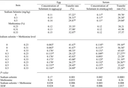
The amount of selenium in the diet plays a decisive role in the concentration of selenium in the egg (Kenyon and Spring, 2003). Similar to our results (Table-4), Boruta et al., 2007) also showed that increasing the amount of selenium in the diet led to increase of selenium concentration in eggs and blood sera. Selenium plays immunostimulatory effects on the lymphoid tissue of the immune system in chickens (Szeleszczuk et al., 2004). High levels of selenium or deficiency have a negative effect on
reproductive traits. Excess selenium is considered embryo toxic (Boruta et al., 2007), which is also visible (late embryonic mortality) in our experiment results (Boruta et al., 2007). The results of Table 4 showed that high levels of selenium or its deficiency increased embryonic mortality in the late phase of incubation. In pheasants, excess selenium caused abnormal behavior in its female sex, decreased embryonic development, hatchability and chick quality (Latshaw et al., 2004).
Table 4. The effect of sodium selenite, methionine level and their interaction on concentration of selenium on
egg, serum and transfer rate of Iraninan native broiler breeders
Serum Egg
Item
Transfer
rate (%) Concentration of
Selenium in erum(µg/ml) Transfer rate
(%) Concentration of
Selenium in egg(µg/g)
Sodium Selenite (mg/kg)
a 59.78 B
0.11 57.22A
0.11 0
b 28.38 B
0.11 38.31B
0.15 0.3
b 29.04 A
0.13 29.87B
0.14 0.5
Methionine (%)
38.21 0.12
35.35a 0.12
0.23
41.98 0.12
37.98a 0.13
0.31
37.57 0.12
52.07b 0.15
0.33
Sodium selenite × Methionine level
ab 59.10 bc
0.118 41.67b
0.083b 0.23
0
b 56.56 bc
0.113 41.67b
0.083b 0.31
0
a 65.65 b
0.131 88.33a
0.176a 0.33
0
dce 27.27 bc
0.109 28.12b
0.113ab 0.23
0.3
e 23.29 c
0.093 43.83b
0.176a 0.31
0.3
dc 31.19 b
0.125 43.00b
0.173a 0.33
0.3
dce 28.26 b
0.132 36.27b
0.170a 0.23
0.5
c 33.62 a
0.157 28.45b
0.133ab 0.31
0.5
de 25.24 bc
0.118 24.89b
0.116ab 0.33
0.5
P-value
0.0001 0.002
0.001 0.17
Sodium selenite
0.26 0.68
0.033 0.26
Methionine
0.008 0.003
0.006
0.038 Sodium selenite × Methionine
2.017 0.006
7.68 0.024
SEM
Means in a same column with different superscripts significantly differ (P < 0.05).
Selenium Content in Egg and blood serum
The influence of SS, methionine and their interaction on the selenium concentration in eggs, blood serum, and their transfer rate are shown in Table 4. A significant interaction between SS and methionine was found on concentration of selenium in egg (P < 0.05). The concentration of selenium increased to 0.176 (µg/g) in the group with 0.0 mg SS/kg and 0.33% Methionine compared with the control group (0.083; 0.0 mg SS/kg and 0.23% Methionine), the transfer rate of Se for egg was affected (P < 0.05) by SS, methionine, and their interaction. Supplementing the diet with SS reduced its transmission in eggs and serum, also adding of methionine more than 0.23 % increased egg transfer rate. The results of this table showed adding methionine at dose 0.33 % increased egg transfer rate when the concentration of SS was
supplementing the lowest level of selenium independent. Selenium absorption efficiency is relatively high (50-95%) and depends on the dietary sources (Robinson and Thomson, 1983). In contrast to inorganic forms [Se]Met absorbed very rapidly and enhanced at low methionine intake in the intestinal (Vendeland et al., 1992). There is a dynamic mechanism comparable to that of its S-analogue
methionine (Wolffram et al., 1989). The
concentration of selenium in the plasma was higher in the birds that received 0.3 mg/kg diet than the other group without selenium supplementation (Jing et al., 2015). Serum selenium concentrations was decreased in broilers received the corn soybean meal diet without selenium supplementation in compare to hens fed corn soybean meal with supplemental selenium in diets, regardless of source (Payne et al., 2005). Previous researchers have showed that selenium supplementation in to diets for broiler breeders or laying hens enhanced the selenium content of eggs.
These finding are in agreement of our experiment. One of main dietary factor influencing availability of selenium includes methionine, (Fairweather-Tait and Hurrell, 1996). The quantity of selenium intake determines its resorption from the gastrointestinal tract, its retention, and metabolism in the body. The liver does not store selenium. Insufficient selenium decreases its serum level significantly. The resorbed selenium is transferred by the bloodstream that ultimately binds to plasmatic proteins and integrates to all tissues (Cousins and Cairney, 1961). Liver have central role in the selenium metabolism and homeostasis, and protein plasma mediated it for absorption and metabolize. According to this document methionine is one of the main components to synthesis of protein that bound to selenium and transport selenium to tissue such as eggs. Skrivan et al. (2010) indicated that supplementation of either form ([Se] Met, SS) of selenium significantly increased the selenium concentration in eggs.
Table 5. The effect of sodium selenite, methionine level and their interaction on antioxidant enzymes in plasma of Iraninan native broiler breeders
Superoxide Dismutase (U/mL) Glutathione Peroxidase
(U/g of Hb) Item overall SOD 2 SOD 1 SOD overall Px -GSH 2 Px -GSH 1 Px -GSH Sodium selenite (mg/kg)
153.04 154.93 151.14 250.30 282.36 218.2 0 146.84 144.56 149.13 262.53 279.55 245.5 0.3 147.81 145.27 150.98 332.74 264.79 400.7 0.5
Methionine level (%)
149.52 149.01 150.20 241.64 289.93 193.4 0.23 149.17 149.00 149.33 285.82 276.56 295.1 0.31 149.36 147.39 151.72 318.11 260.21 376.0 0.33
Sodium selenite × Methionine level
149.91 150.53 149.29 ab 269.5 436.0 102.9 0.23 0 157.13 168.19 146.08 ab 275.1 264.4 285.7 0.31 0 152.07 146.08 158.06 b 206.4 146.6 266.1 0.33 0 151.08 147.94 154.23 b 217.0 243.1 190.9 0.23 0.3 144.17 139.17 149.17 ab 387.8 390.0 385.5 0.31 0.3 145.28 146.58 143.98 b 182.8 205.5 160.1 0.33 0.3 146.61 148.37 147.07 b 238.5 190.7 286.3 0.23 0.5 146.20 139.66 152.75 b 194.6 175.2 213.9 0.31 0.5 151.42 150.59 153.12 a 565.1 428.5 701.8 0.33 0.5 P-value 0.33 0.19 0.92 0.54 0.97 0.21 Sodium selenite 0.99 0.97 0.91 0.63 0.93 0.25 Methionine 0.64 0.16 0.56 0.06 0.07 0.13
Sodium selenite × Methionine
5.17 7.02 6.87 97.60 100.17 131.09 SEM
Means in a same column with different superscripts significantly differ (P < 0.05) GSH-Px = glutathione peroxidase; SOD = superoxide dismutase
GSH-Px1= at 70 wk GSH-Px2= at 74 wk SOD1= at 70 wk SOD1= at 74 wk
Glutathione Peroxidase and Superoxide Dismutase Activity
As can be seen from Table 5, different levels of SS and methionine did not significant effect on GSH-Px and SOD activity (P > 0.05). Moreover, the results of this study revealed that interaction between SS and
name belonging to the enzyme family performing peroxidase activity. It plays a biological role by guarding an organism against all oxidative damages. They also contribute to the decreased free hydrogen peroxide to water (Brigelius-Flohé and Medicine, 1999). One of the reasons why it is possible to justify this conclusion is that methionine which is an analog to the [Se]Met compounds plays important roles to support cysteine for GSH synthesis. In opposition to the results obtained in this experiment, Leeson et al. (2008) showed GSH-Px activity in plasma was affected by selenium concentration.
In consist of our experiment Waschulewski and Sunde (1988) found that when rats fed 0.5 mg selenium as [Se]Met/kg tissue selenium increased significantly. The concentration of selenium in the erythrocyte and muscle in rats when fed with methionine deficiency was higher than methionine-supplemented to diet, while the concentration of enzyme was increased when methionine was added to the diet.
Immune Response
The data showed that the antibody titer against influenza didn't change during wks 70 to 74 at 0.3 mg / kg and 0.31% methionine (Table-7), Also this trend was observed for 0.0 mg / kg SS with 0.33 % methionine (P < 0.05), but during the time, this effect was meaningful. The results indicate that GSH-Px levels were highest when 0.5 mg / kg SS with 0.31% methionine and 0.3 mg / kg / mL with 0.31% methionine were placed in the diet. In treatments of 0.5 mg / kg SS and 23% of methionine, concentrations of selenium in eggs and serum as well as GSH-Px enzyme were higher. The results of this study showed that, increasing the SS content in the range of 0.3 to 0.5 mg / kg with added levels of methionine in the range of 0.23 to 0.31% to diet can be increased amount of selenium in eggs and serum. In addition, increase in the amount of GSH-Px in plasma was observed which can be led to strengthening the immune system and antioxidants. Spallholz (1990) reported that with balanced of dietary selenium concentration in broiler chickens,
increasing of peroxidase concentration was observed in the blood. In some studies, the effect of selenium on the immune system with enhancement of both cell-mediated and humoral immune has been demonstrated.
It has been reported that in some viral diseases, the effect of selenium is not due to virus removal, but prevent genetic adaptations in the viral genomic RNA. It has been reported that over 20 types of seleno-proteins have been identified in the human body, which expressed by 25 genes in the human genome (Hatfield and Gladyshev., 2002; Lescure et al., 2002). Except for GSH-Px, other enzymes such as thioredoxin reductases and three iodothyronine deiodinases are also part of the selenoenzyme’s family. Seleno-proteins P and W are other seleno-proteins that involved in antioxidant functions. Due to the immune function, there is also a specific seleno-protein in T cells called 15-kDa seleno-seleno-protein. Seleno-phosphate synthetase is another protein having selenium that speeds up the production of seleno-phosphate. This seleno-phosphate is involved as the main inorganic element to produce selenocysteine from the serine during seleno-protein synthesis. The afore-mentioned seleno-proteins could be suitable options for the involvement of selenium in the immune system. (Arthur, 2001; McKenzie et al., 2002). Lymphocytes that have selenium deficiency are less able to proliferate in response to mitogen, leukotriene B4 is produced in macrophages which is essential for the activity of neutrophils, and the deficiency of selenium causes destruction of its production (McKenzie etal., 2002; Spallholz, 1990). It has been argued that selenium increases the production of IFN gamma and other cytokines, proliferation of T cell and increased the number of T helper cells (Broome et al., 2004). Reffett et al. (1988) Reported that adding selenium may increase IgM level in calves. The influenza virus can be hampered by nutritional elements such as lysine, proline, ascorbic acid, green tea extract, N-acetyl cysteine, and selenium through the activity of neuraminidase (Willemsen et al., 2011).
Table 6. The effect of treatment (sodium selenite and methionine level interaction) during the time on Antibody
titer against Newcastle of Iranian native broiler breeders (Anti log2)
Time Item
74 wk 72 wk
70 wk Sodium selenite × Methionine level
cd 106 384abc
17.3d 0.23
0
bcd 341 512bc
18.7d 0.31
0
bcd 298 426bcd
3.3d 0.33
0
cd 47.7 298bcd
8.7d 0.23
0.3
bcd 192 1066a
13.3d 0.31
0.3
cd 106 298bcd
16.0d 0.33
0.3
d 53.3 106cd
4.00d 0.23
0.5
cd 128 597b
12.0d 0.31
0.5
cd 106 298bcd
8.70d 0.33
0.5
P-value
0.0001 0.0001
0.0001 Treatment × Time
Means with different superscripts significantly differ (P < 0.05).
Table 7. The effect of treatment (sodium selenite and methionine level interaction) during the time on Antibody titer against Influenza of Iranian native broiler breeders (Anti log2)
Time Item
74 wk 72 wk
70 wk
Sodium selenite × Methionine level
cd 128 170.7cd
18.7d 0.23
0
cd 256 682.7a
26.7d 0.31
0
d 96.0 85.3d
10.7d 0.33
0
d 64.0 384bc
18.7d 0.23
0.3
cd 176 256cd
18.7d 0.31
0.3
d 106 170.7cd
64d 0.33
0.3
d 64.0 64d
16d 0.23
0.5
cd 213 512ab
21.3d 0.31
0.5
d 74.7 213.3cd
50.7d 0.33
0.5
P-value
0.0001 0.0001
0.0001 Treatment × Time
Means with different superscripts significantly differ (P < 0.05)
It is concluded that 0.3 SS mg/kg with 0.33% methionine can be effectively used to increase salable chick without detrimental effects on performance and embryonic mortality. The results from this experiment showed that SS with methionine result in greater deposition of selenium in serum. High selenium and
methionine contents of diet have been correlated with higher activity of GSH-PX in plasma. The current study demonstrated that supplementation of methionine to inorganic source of selenium could improve bioavailability of selenium and transfer rate of it.
References
Abdalla A, Yakout HM & Khalifah MM. 2005. Determination of lysine and methionine requirements of gimmizah strain during egg production stage. Proc. 3rd Inter. Poultry Conference Egypt.
Abd-EL-Samee MO, EL-Sheikh TM & Ali AM. 2007. An attempt to alleviate heat stress and improve laying hens performance and egg quality during summer season by using some amino acids or vitamins. Egypt. Poultry Science, 27: 875- 891. Arthur JR. The glutathione peroxidases. 2000.
Cellular and Molecular Life Sciences, 57: 1825– 1835.
Boruta A, Oewierczewska E & Roszkowski T. 2007. Effect of organic and inorganic forms of selenium on the morphological composition of eggs and selenium content in egg mass. Medycyna Weterynaryjna (In Polish), 63: 238-241.
Brigelius-Flohe R.1999. Tissue-specific functions of individual glutathione peroxidases. Free Radical Biology and Medicine, 27: 951–965. DOI: 10.1016/S0891-5849(99)00173-2
Broome CS, McArdle F, Kyle JA, Andrews F, Lowe NM & Hart CA. 2004. An increase in selenium intake improves immune function and poliovirus handing in adults with marginal selenium status. The American Journal of Clinical Nutrition, 80: 154-62. DOI: 10.1093/ajcn/80.1.154
Bunchasak C & Silapasorn T. 2005. Effects of adding methionine in low-protein diet on production performance, reproductive organs and chemical liver composition of laying hens under tropical
Science, 4: 301-308. DOI:10.3923/ ijps.2005. 301.308
Carter TC. 1975. The hen’s egg: Estimation of shell superficial area and egg volume, using measurements of fresh egg weight and shell length and breadth alone or in combination. British Poultry Science, 16: 541-543. DOI: 10.1080/00071667508416224
Cousins FB & Cairney M. 1961. Some aspects of selenium metabolism in sheep. Australian Journal of Agricultural Research, 12: 927–943. DOI: 10.4141/cjas68-045
Delezie E, Rovers M, Van der Aa A, Ruttens A, Wittocx S & Segers L. 2014. Comparing responses to different selenium sources and dosages in laying hens. Poultry Science, 93: 3083–3090. DOI: 10.3382/ps.2014-04301
Dziaczkowska L. 1980. The pattern for the assessment of turkey chicks for fattening. In polish.; Centre for Poultry Research and Development, Center for Water Fowl. Poznan. Fairweather-Tait S & Hurrell RF. 1996.
Bioavailability of minerals and trace elements. Nutation Research Review, 9: 295–324. DOI: 10.1079/NRR19960016
Hoffmann PR & Marla JB. 2008. The influence of selenium on immune responses. Molecular Nutrition and Food Research, 52: 1273–1280. DOI: 10.1002/mnfr.200700330
Hatfield DL & Gladyshev VN. 2002. How selenium has altered our understanding of the genetic code. Molecular and Cell Biology, 22: 3565–3576. DOI: 10.1002/mnfr.200700330
Hempe JM & Laukxen RC. 1988. Savage JE. Rapid determination of egg weight and specific gravity using a computerized data collection system. Poultry Science, 67: 902-907. DOI: 10.3382/ ps.0670902
Jackson ME, Hellwig HM & Waldroup WP. 1987. Shell quality: Potential for improvement by dietary means and relationship with egg size. Poultry Science, 66: 1702–1713. DOI: 10.3382/ ps.0661702
Jing CL, Dong XF, Wang ZM, Liu S & Tong JM. 2015. Comparative study of DL-[Se] Met vs SS and seleno-yeast on antioxidant activity and selenium status in laying hens. Poultry Science, 94:965–975. DOI: 10.3382/ps/pev045
Kazemi Fard M, Kermanshahi H, Rezaei M & Golian A. 2013. Effect of different levels of fennel extract and vitamin D3 on performance, hatchability and immunity in post molted broiler breeders. Iranian Journal of Animal Science, 3: 735-745.
Kenyon S & Spring P. 2003. Egg selenium concentrations in breeder’s hens fed Na – selenite or Sel Plex® supplemented diets. XthEuropean Symp. On the quality of eggs and egg poducts, September 23-26, Saint – Brieuc – Ploufragan, Cotes d’ Armor France, pp. 182.
Knight SAB & Sunde RA. 1988. Effect of selenium repletion on glutathione peroxidase protein in rat liver. Journal of Nutrition, 118: 853-58
Kurz B, Jost B & Sch¨unke M. 2002. Dietary vitamins and selenium diminish the development of mechanically induced osteoarthritis and increase the expression of antioxidative enzymes in the knee joint of STR/1N mice. Osteoarthritis and cartilage, 10: 119–126. DOI: 10.1053/joca. 2001.0489
Latshaw JD, Morishita TY, Sarver CF & Thilsted J. 2004. Selenium toxicity in breeding Ring-Necked pheasants (Phasianus colchicus). Avian Diseases, 48: 935-939. DOI: 10.1637/7177-030904R Leeson S, Namkung H, Caston L, Durosoy S &
Schlegel P. 2008. Comparison of selenium levels and sources and dietary fat quality in diets for broiler breeders and layer hens. Poultry Science, 87: 2605–2612. DOI: 10.3382/ps.2008-00174 Lescure A, Gautheret D & Krol A. 2002. Novel
selenoproteins identified from genomic sequence data. In: Methods in Enzymology 347 (Sies, H. and Packer, L., eds.), pp.; 57–70. Academic Press, San Diego, CA.
McConnell KP & Cho GJ. 1965. Transmucosal movement of selenium. American Journal of Physiology, 208: 1191–1195. DOI: 10.1152/ ajplegacy.1965.208.6.1191
McKenzie RC, Arthur JR & Beckett GJ. 2002. Selenium and the regulation of cell signaling, growth, and survival: molecular and mechanistic
aspects. Antioxidants and Redox. Signaling, 4: 339–351. DOI: 10.1089/152308602753666398 Nassiri-Moghaddam H, Kazemi-Fard M, Agah MJ,
Hosseini SJ & Mirakzehi MT. 2012. Effect of different levels of methionine, protein and tallow on the productive performance and egg quality of laying hens in the late-phase production. Brazilian Journal of Poultry Science, 14: 149-158. DOI: 10.1590/S1516-635X2012000200010
Naulia AH & Singh MA. 2002. Effect of dietary protein and supplemental fat on performance of laying hens. International Journal of Poultry Science, 4: 1986-1989. DOI: 10.3923/ijps. 2005.986.989
Ort JF & Latshaw JD.1978. The toxic level of SS in the diet of laying hens. Journal of Nutrition, 108: 1114–1120.http:// doi:10.1093/jn/108.7.1114 Ousterhout LE. 1980. Effects of calcium and
phosphorus levels on egg weight and egg shell quality in laying hens. Poultry Science, 59: 1480-1484. DOI: 10.3382/ps.0591480
Pappas AC, Acmovic T, Sparks NHC, Surai PF & McDevitt RM. 2006. Effects of supplementing broiler breeders diets with organoselenium compounds and polyunsaturated fatty acids on hatchability. Poultry Science, 85: 1584–1593. DOI: 10.3382/ps.0591480
Payne RL & Southern LL. 2005. Changes in GSH-Px and tissue selenium concentrations of broilers after consuming a diet adequate in selenium.
Poultry Science, 84: 1268–1276. DOI:
10.1093/ps/84.8.1268
Payne RL, Lavergne TK & Southern LL. 2005. Effect of inorganic versus organic selenium on hen production and egg selenium concentration. Poultry Science, 84: 232–237. DOI: 10.1093/ ps/84.2.232
Petersen CF, Sauter EA, Steele EE & Parkinson JF. 1983. Use of methionine intake restriction to improve egg shell quality by control of egg weight. Poultry Science, 62: 2044–2047. DOI: 10.3382/ps.0622044
Reffett JK, Spears JW & Brown TT. 1988. Effect of dietary selenium on the primary and secondary immune response in calves challenged with infectious bovine rhinotracheitis virus. Journal of Nutrition, 118: 229–235. DOI: 10.1093/ jn/118.2.229
Reis RN, Vieira SL, Nascimento PC, Pena JE, Barros R & Torres CA. 2009. Selenium contents of eggs from broiler breeders supplemented with SS or zinc-l-selenium methionine. Journal of Applied Poultry Research, 18: 151–157. DOI: 10.3382/ japr.2008-00069
Safaa HM, Serrano MP, Valencia DG, Arbe X, Jime ́nez-Moreno E, La ́zaro R & Mateos GG. 2008. Effects of the levels of methionine, linoleic acid, and added fat in the diet on productive performance and egg quality of brown laying hens in the late phase of production. Poultry Science, 87: 1595–1602. DOI: 10.3382/ps.2008-00005 SAS Institute. 2003. SAS/STAT 9.1.3 User’s Guide.
SAS Inst. Inc., Cary, NC.
Susanne B, Erik HL, Jens JS, Knut F, Kim
O, Lars CS & Sven M. 2008. Absorption,
excretion, and retention of selenium from a high selenium yeast in men with a high intake of selenium. Food Nutrition Research, 52: 10.3402.
Skrivan M, Bubancova I, Marounek M & Dlouha G. 2010. Selenium and alpha-tocopherol content in eggs produced by hens that were fed diets supplemented with selenomethionine, SS and vitamin E. Czech. Journal of Animal Science, 55: 388–397. DOI: 10.17221/92/2010-CJAS
Spallholz JE. 1990. Selenium and glutathione peroxidase: essential nutrient and antioxidant component of the immune system. Advances in Experimental Medicine and Biology, 262: 145– 158.
Szeleszczuk P, Karpinska E, Bielecki W, Kosowska G &Borzemska W. 2004. Evaluation of lithium and selenium toxicity for chicken embryos and one-day-old chicks. Medycyna Weterynaryjna, 60: 492-495.
Tahmasbi AM, Mirakzehi MT, Hosseini SJ, Agah MJ & Kazemi-Fard M. 2012. The effects of phytase and root hydroalcoholic extract of Withania somnifera on productive performance and bone mineralization of laying hens in the late phase of production. British Poultry Science, 53: 204-214. DOI: 10.1080/00071668.2012.662628
Utterback PL, Parsons CM, Yoon I & Butler J. 2005. Effect of supplementing selenium east in diets of laying hens on egg selenium content. Poultry Science, 84: 900–1901. DOI: 10.1093/ps/ 84.12.1900
Vendeland SC, Butler JA & Whanger PD. 1992. Intestinal absorption of selenite, selenate, and selenomethionine in the rat. Journal of Nutrition Biochemistry, 3: 359-365. DOI: 10.1016/0955-2863(92)90028-H
Vendeland SC, Deagen JT & Whanger PD. 1996. Uptake of selenotrisulfides of glutathione and cysteine by brush border membranes from rat intestines. Journal of Inorganic Biochemistry, 47: 131-140.
Waschulewski H & Sunde RA. 1988. Effect of dietary methionine on utilization of tissue selenium from dietary selenomethionine for glutathione peroxidase in the rat. Journal of Nutrition, 118: 367-374. DOI:10.1093/jn/ 118.3.367
Waschulewski H & Sunde RA. 1988. Effect of dietary methionine on tissue selenium and glutathione peroxidase (EC 1.11.1.9) activity in rats given selenomethionine. British Journal of Nutrition, 60: 57-68. DOI: 10.1093/jn/118.3.367 Willemsen H, Swennen Q & Everaert N. 2011.
Effects of dietary supplementation of methionine
and its hydroxy analog
DL-2-hydroxy-4-methylthiobutanoic acid on growth performance, plasma hormone levels, and the redox status of broiler chickens exposed to high temperatures. Poultry Science, 90: 2311-20. DOI: 10.3382/ ps.2011-01353
Wolffram S, Berger B, Grenacher B & Scharrer E. 1989. Transport of selenoamino acids and their sulfur analogues across the intestinal brush border membrane of pigs. Journal of Nutrition, 119: 706-712. DOI: 10.1093/jn/119.5.706
Xue X, Wang Y, Liu W, Ju T & Zhan X. 2017. Effects of different methionine sources on production and reproduction performance, egg quality and serum biochemical indices of broiler breeders. Asian-Australian Journal of Animal Science, 30: 828–833. DOI: 10.5713/ajas.16.0404 Yoon I, Werner T & Butler J. 2007. Effect of source