1535-9778/10/$12.00 doi:10.1128/EC.00134-10
Copyright © 2010, American Society for Microbiology. All Rights Reserved.
␣
-1,6-Mannosylation of N-Linked Oligosaccharide Present on Cell
Wall Proteins Is Required for Their Incorporation into
the Cell Wall in the Filamentous Fungus
Neurospora crassa
䌤
†
Abhiram Maddi
2and Stephen J. Free
1*
Department of Biological Sciences, SUNY, University at Buffalo, Buffalo, New York,1and
Department of Oral Biology, SUNY, University at Buffalo, Buffalo, New York2
Received 2 June 2010/Accepted 10 September 2010
The enzyme␣-1,6-mannosyltransferase (OCH-1) is required for the synthesis of galactomannans attached to the N-linked oligosaccharides ofNeurospora crassacell wall proteins. TheNeurospora crassa och-1mutant has a tight colonial phenotype and a defective cell wall. A carbohydrate analysis of the och-1 mutant cell wall revealed a 10-fold reduction in the levels of mannose and galactose and a total lack of 1,6-linked mannose residues. Analysis of the integral cell wall protein from wild-type andoch-1 mutant cells showed that the mutant cell wall had reduced protein content. Theoch-1mutant was found to secrete 18-fold more protein than wild-type cells. Proteomic analysis of the proteins released by the mutant into the growth medium identified seven of the major cell wall proteins. Western blot analysis of ACW-1 and GEL-1 (two glycosylphosphatidy-linositol [GPI]-anchored proteins that are covalently integrated into the wild-type cell wall) showed that high levels of these proteins were being released into the medium by theoch-1mutant. High levels of ACW-1 and GEL-1 were also released from theoch-1mutant cell wall by subjecting the wall to boiling in a 1% SDS solution, indicating that these proteins are not being covalently integrated into the mutant cell wall. From these results, we conclude that N-linked mannosylation of cell wall proteins by OCH-1 is required for their efficient covalent incorporation into the cell wall.
The fungal cell wall is an important organelle that protects the cell from various environmental stresses. It is a dynamic structure that interacts with the environment and is modified to accommodate growth, cell division, and development. Fun-gal cell walls have been shown to contain-1,3-glucan,␣ -1,3-glucan, -1,6-glucan, mixed -1,3/-1,4-glucans, chitin, and mannan/galactomannan (6, 19). These polysaccharide poly-mers constitute 80 to 85% of the cell wall mass, while glyco-proteins constitute the remaining 15 to 20% (6). The cell wall glycoproteins are required for vital functions, like structural support, signal transduction, biofilm formation, and cell wall biosynthesis. In the case of pathogenic fungi, the cell wall is critical for the invasion of host tissues (8). Because of their accessibility and the crucial functions they perform, cell wall proteins could be important targets for the development of antifungal therapeutics.
The glucan and chitin cell wall polymers are synthesized by enzyme complexes (glucan synthases and chitin synthases) that are associated with the plasma membrane. Glucan and chitin are vectorially passed into the cell wall space during synthesis and cross-linked together in the cell wall space. The mannan and galactomannan present in the cell wall are found as gly-coconjugates on cell wall proteins. Mannosylation of cell wall
proteins occurs in the endoplasmic reticulum (ER) and Golgi apparatus at O-linked and N-linked glycosylation sites. In
Sac-charomyces cerevisiae, mannosylation of N-linked glycosylation
is initiated by the addition of an␣-1,6-linked mannose residue by Och1p (33). In the filamentous fungusNeurospora crassa, the structure of the galactomannan associated with N-linked sites has not been definitively determined, butN. crassa has most of the enzymes defined in yeast for the mannosylation of N-linked oligosaccharides (14). An analysis of N-linked oligo-saccharides fromN. crassaglycoproteins showed that the gly-coproteins are modified by the addition of short␣ -1,6-man-nans with short␣-1,2-mannose branches that are terminated by galactofuranose residues (31, 32). TheN. crassa posttransla-tional modifications appear to differ from those found inS.
cerevisiaeby having shorter mannan chains and by the presence
of terminal galactofuranose residues.
Mannosylation of glycoproteins has been extensively studied in yeast. InS. cerevisiae, OCH1encodes the ␣ -1,6-mannosyl-transferase enzyme that mediates the addition of the initial ␣-1,6-mannose in the synthesis of long mannans which are attached to the N-linked oligosaccharides (22, 33). Knockout mutants ofOCH1are viable but exhibit a temperature-sensi-tive growth pattern and are sensitemperature-sensi-tive to cell wall perturbation reagents (34). Mutants for Candida albicans homologs of
OCH1had near-normal growth rates but were much less vir-ulent (3). Mass spectrometry analysis of glycoproteins from the
S. cerevisiae och1andC. albicans och1mutants showed that the
␣-1,6-mannose core was absent (3, 33). InKluyveromyces lactis, the KlOCH1 gene has been shown to be important for cell wall organization and to give a hypersecretion phenotype (37). * Corresponding author. Mailing address: University at Buffalo,
De-partment of Biological Sciences, Cooke Hall, Room 109, Buffalo, NY 14260. Phone: (716) 645-4935. Fax: (716) 645-2975. E-mail: free @buffalo.edu.
† Supplemental material for this article may be found at http://ec .asm.org/.
䌤Published ahead of print on 24 September 2010.
1766
on September 8, 2020 by guest
http://ec.asm.org/
OCH1 mutants have also been identified in Pichia angusta,
Yarrowia lipolytica, Pichia pastoris, and Schizosaccharomyces
pombe, and these mutants have cell wall-related phenotypes (2,
9, 17, 38). However, a recent report ofOCH1knockout mu-tants ofAspergillus fumigatusindicates that these mutants do not have a cell wall-defective phenotype (18).
Mannosylation of cell wall proteins has not been extensively studied in filamentous fungi. We report on the characterization of theN. crassaknockout mutant of the␣ -1,6-mannosyltrans-ferase, och-1. The mutant was generated by the Neurospora
genome knockout project (10). TheN. crassa och-1mutant has a severe growth defect and exhibits a tight colonial phenotype. We demonstrate that theoch-1mutant exhibits a defect in cell wall biosynthesis. A carbohydrate analysis of the mutant cell wall showed a drastic reduction in mannose and galactose content with a compensatory increase in the glucose content.
The och-1 cell wall also showed a reduced cell wall protein
content as assessed by a Coomassie brilliant blue dye binding assay and by proteomic analysis. Protein secretion assays showed that the mutant releases large amounts of cell wall protein into the growth medium. We demonstrate that the
och-1mutant is defective in covalently cross-linking known cell
wall proteins into the cell wall matrix. Our data demonstrate that the N-linked galactomannan, which is built upon the man-nose residue added by OCH-1, is required for the incorpora-tion of cell wall proteins into the cell wall matrix.
MATERIALS AND METHODS
Strains and culturing conditions.N. crassawild-type strain 74-OR23-IV A and
och-1knockout mutant strains were maintained in Vogel’s medium with 2%
sucrose at room temperature (12). Theoch-1knockout mutant was generated
during theNeurosporagenome project by replacing the␣
-1,6-mannosyltrans-ferase gene with a hygromycin resistance cassette (10). Theoch-1 knockout
mutant was obtained from the Fungal Genetics Stock Center (Kansas City, MO). Cloning, sequencing, and transformation experiments were done as described by Colot et al. (10) and by Maniatis et al. (27).
Complementation ofoch-1knockout mutation.Knock-in experiments
target-ing the insertion of a wild-type copy ofoch-1downstream of thehis-3locus were
performed using the pBM61 plasmid as described by Margolin et al. (28). The
och-1gene (NCU00609) is 1,194 bases in length from the start site to the stop
site, including introns, as given in theNeurosporagenome database. For
trans-formation purposes,⬃1,500 bases upstream of the start site and⬃500 bases
downstream of the stop site were included as part of the gene. Primers containing added XbaI and XmaI restriction sites (forward primer, ACTATCTAGAGCC CGATTTCATGCTGGAACAG; reverse primer, CTTACCCGGGTTCGGTG ACGATTTCGGACC) were used in amplifying the gene and inserting it as an
XbaI/XmaI fragment into pBM61. A plasmid carrying theoch-1gene was
se-quenced to ensure that it contained no mutations and was used to transform an
och-1 his-3mutant as described by Margolin et al. (28).
Cell wall stress tests.Theoch-1mutant and wild-type control were subjected to various cell wall stress conditions. The strains were grown on slants of Vogel’s medium with 2% sucrose agar or in Vogel’s liquid medium with 2% sucrose containing a cell wall stress reagent. The growth of the mutant was observed at 30°C over a period of 72 h and compared to the growth of the wild type. Cell wall stress reagents included 10% NaCl (salt stress), 2 M glycerol (hyperosmolarity), 0.05 mM hydrogen peroxide (oxidative stress), 0.01% SDS (detergent), 10 mg/ml
calcofluor white (CFW) (chitin inhibitor), and 10g/ml caspofungin (glucan
inhibitor). Caspofungin acetate was obtained as a kind gift from Merck Research Laboratories (Rahway, NJ). The optimum concentration of the cell wall stress reagents for testing the mutant was determined by subjecting the wild-type strain to a range of concentrations and identifying a concentration which was slightly below the concentration at which the wild-type cells were unable to grow. Apart from subjecting the mutant to cell wall stress reagents, it was also subjected to growth at 18°C and 37°C to test for a temperature-sensitive growth phenotype.
Cell wall preparations forN. crassa.Cell walls were prepared as described
previously (26). Seven- to 10-day-old conidia from wild-type andoch-1strains
were used to inoculate 1 liter of Vogel’s liquid medium containing 2% sucrose and cultured at 25°C in a shaking incubator for 12 to 14 h. Vegetative hyphae were harvested by filtration through a filter paper placed on a Buchner funnel. The harvested vegetative hypha cultures were then frozen with liquid nitrogen in a mortar and ground to a fine powder with a pestle while intermittently adding liquid nitrogen. The ground hyphae were then resuspended in an extraction buffer consisting of phosphate-buffered saline (PBS) (27).
To purify cell walls, disrupted hypha preparations were first subjected to a
centrifugation step (4,000⫻gfor 5 min) to separate the cell walls from the
cytosolic proteins. The crude cell walls were washed twice with PBS and resus-pended in PBS containing 1% SDS. The resusresus-pended cell wall pellets were boiled in the SDS-containing extraction buffer for 15 min, allowed to cool, and collected
by centrifugation (10,000⫻g for 5 min). The supernatant was collected as
“SDS-solubilized fraction” and was used to examine the proteins that are non-covalently associated with the cell wall in Western blot analyses. The cell wall pellet was washed twice with ice-cold PBS and once with distilled water. The
centrifugation step for these washes was carried out at 10,000⫻gfor 5 min. The
washed pellet was considered “purified cell wall” and was subjected to deglyco-sylation using trifluoromethanesulfonic acid (TFMS), which completely solubi-lized the cell wall. To identify integral cell wall proteins, the TFMS-solubisolubi-lized cell wall was subjected to nano-liquid chromatography/tandem mass spectrom-etry (LC/MS-MS) analysis. The purified cell wall fraction was also subjected to an
aniline blue binding analysis to assess the levels of-1,3-glucan present and
Coomassie brilliant blue dye binding analysis to assess the levels of cell wall protein and sent to the Complex Carbohydrate Research Center (CCRC; Ath-ens, GA) for carbohydrate analyses.
Protein secretion assay.Wild-type and mutant cells were grown in Vogel’s liquid medium with 2% sucrose for 12 to 14 h at room temperature in a shaking incubator. Under sterile conditions, the cells were collected by filtration over a Buchner funnel, resuspended into fresh Vogel’s liquid medium with 2% sucrose, and allowed to grow for an additional 2 h on the shaking incubator. The cells were collected again by filtration on a Buchner funnel, and the medium was collected for the analysis of secreted proteins. Trichloroacetic acid (TCA) pre-cipitation was used to collect the secreted proteins from the medium. Acetone and TCA were added to the medium to a final concentration of 50% acetone and
12.5% TCA, and the proteins were allowed to precipitate for 24 h at⫺20°C. The
precipitated proteins were collected by centrifugation, washed twice with⫺20°C
acetone, and lyophilized. Samples of the lyophilized protein were then subjected to SDS-PAGE, Western blot analysis, and TFMS digestion.
Carbohydrate and glycosyl linkage analysis of cell walls.Glycosyl composition of cell walls was analyzed by combined gas chromatography/mass spectrometry
(GC/MS) of the per-O-trimethylsilyl (TMS) derivatives of the monosaccharide
methyl glycosides produced from the sample by acidic methanolysis at the CCRC. For glycosyl linkage analysis, the samples were permethylated, depoly-merized, reduced, and acetylated, and the partially methylated alditol acetates (PMAAs) were analyzed at the CCRC by GC/MS.
TFMS digestion of cell walls and secreted protein.To analyze the secreted protein and the proteins covalently linked to the cell walls, the samples were treated with TFMS using a procedure described previously (26). TFMS, anisole, and pyridine were obtained from Sigma-Aldrich Chemical Company (St. Louis, MO). Secreted protein samples and cell wall pellets were lyophilized overnight to ensure complete dryness of the samples. To maintain anhydrous conditions during the TFMS treatment, all glass tubes and syringes used were dried under a vacuum, and the digestion was performed in a chamber being continually purged with liquid nitrogen. Initially, a solution of 16% anisole in TFMS was prepared and 1.25 ml of this mixture was added to 20 mg of the purified cell wall sample or to a sample of secreted protein collected from 500 ml of culture medium. The samples were then purged with liquid nitrogen, quickly covered
with Parafilm, and placed in the N2-filled chamber at 4°C. During the course of
the digestion, the samples were periodically mixed with a Pasteur pipette, purged
with N2gas, and covered again with Parafilm. The cell wall samples and the
samples of secreted protein were completely solubilized in 5 h, as assessed by visual inspection. After the cell walls had been solubilized, 3.75 ml of a solution
of pyridine-methanol-H2O (3:1:1) was added in a dropwise fashion to each of the
digests, which were continually swirled in a dry ice-ethanol bath. The samples were then left in the dry ice-ethanol bath for 20 min, followed by incubation for
another 20 min at⫺20°C. The samples were then removed and allowed to thaw,
and 1 ml of 5% ammonium bicarbonate solution was added to each sample. The released proteins were then precipitated by adding 6 ml of 25% TCA in acetone
and incubating at⫺20°C for 24 h. The precipitated proteins were collected by
centrifugation at 10,000⫻g, washed three times in ice-cold 100% acetone, and
briefly dried. The proteins were then resuspended in PBS with 1% SDS and subjected to a 10-min boiling treatment. Protein concentrations of all samples
on September 8, 2020 by guest
http://ec.asm.org/
were determined using the Bio-Rad DC protein assay kit. The amount of protein released from up to 5 mg of starting cell wall material was separated by SDS-PAGE on a 4 to 12% Bis-Tris NuSDS-PAGE gel and visualized using the SilverQuest silver staining kit (Invitrogen Life Technologies, Carlsbad, CA) or with Coomas-sie blue (27).
Nano-LC/MS-MS analysis of peptides.Samples containing 20g of integral
cell wall protein released by TFMS digestion or 20g of TFMS-digested secreted
protein were briefly subjected to SDS-PAGE. The gel was then stained with Coomassie brilliant blue, and the stained part of the gel was excised and sent to Midwest Bio Services (Overland Park, KS) for nano-LC/MS-MS-based identifi-cation.
Data analysis (MS-MS database search).Cell wall proteins were identified by matching the peptide sequences obtained from the MS-MS analysis with the
protein sequences from theNeurosporagenome available at the Broad Institute
(www.broad.mit.edu) using the TurboSEQUEST software. Only those proteins with multiple peptides and/or single peptides with an XC (correlation coefficient)
score of⬎2.5 for 2 ions or⬎3.0 for 3 ions were accepted as accurate
identifi-cations in this analysis.
Western blot analysis of cell wall and secreted protein samples.Western blot analysis of cell wall protein, secreted protein, SDS-soluble cell wall protein, and cytosolic protein samples was done using ACW-1 antibody as described previ-ously (7). Polyclonal antibody directed against purified GH72-5/GEL-1 was raised in rabbits.
Aniline blue binding assay. As a measure of the amounts of-1,3-glucan present in the wild-type and mutant cell walls, we assessed the ability of the purified cell wall to bind aniline blue. The assay was carried out as previously described (7).
Coomassie brilliant blue dye binding assay.To compare the protein contents of the wild-type and mutant cell walls, the ability of the wall to absorb Coomassie brilliant blue dye (Bio-Rad, Hercules, CA) was assessed. Lyophilized purified cell wall material from wild-type and mutant cells was washed with distilled water and resuspended in a 15-ml Corex tube at a concentration of 2 mg/ml in water. The cell wall material was subjected to vortexing, and aliquots of cell wall material were taken with a micropipette outfitted with a large orifice tip into microcentrifuge tubes. Distilled water was added to the tube to give a volume of
0.5 ml, and 0.5 ml of 1⫻Coomassie dye reagent (Bio-Rad Laboratories,
Her-cules, CA) was added. The samples were incubated with agitation at room temperature for 30 min. The samples were then centrifuged to pellet the cell wall, and the amount of Coomassie blue remaining in the supernatant was
determined at an optical density of 465 nm (OD465). The amount of dye
ab-sorbed was determined by comparing the OD465of the samples with the OD465
of a control sample without cell wall material.
RESULTS
Theoch-1mutant exhibits morphological defects.Theoch-1
mutant was generated by the Neurospora genome knockout project (10) and obtained from the Fungal Genetics Stock Center (Kansas City, MO). It was identified as being an inter-esting mutant during a screening of the knockout library for
morphological mutants. TheN. crassa och-1gene (NCU00609) is a homolog of the OCH1 gene of S. cerevisiae. The gene encodes a 373-amino-acid type II transmembrane protein with a nucleotide sugar binding motif between residues 150 and 210.
TheS. cerevisiaeOch1p has been shown to be localized to the
cis-Golgi apparatus (30). The knockout mutant was generated by replacing the gene with a hygromycin resistance cassette as described previously (10). The presence of the hygromycin resistance cassette in the och-1 mutant was verified by PCR amplification of the region between 500 bases upstream of the hygromycin cassette insertion site and 500 bases into the hy-gromycin resistance cassette. TheN. crassa och-1mutant grew with a tight colonial morphology, as shown in Fig. 1. Micro-scopic examination of the mutant showed that the hyphae exhibited a tendency to grow in a globular fashion with an abnormal branching pattern (Fig. 2). The mutant was unable to produce normal aerial hyphae and produced abnormal conidia close to the colony surface. The conidia that are produced have a conidial separation phenotype but are viable. The och-1
mutant produces protoperithecia, the female mating structure, and following mating the protoperithecia mature into perithe-cia with ascospores. The mutant is unable to release the asco-spores from the perithecia, but the ascoasco-spores can be collected from the perithecia and are viable. Theoch-1mutant colonial phenotype was found to cosegregate with the hygromycin re-sistance gene used to create the knockout mutation, suggesting that the mutant phenotypes are the result of the loss of the
och-1gene.
To verify that the mutant phenotypes were the result of the loss of theoch-1-encoded␣-1,6-mannosyltransferase, comple-mentation experiments were performed in which a wild-type copy ofoch-1was used to transform theoch-1knockout mu-tant. Wild-type morphology was restored to the mutant upon transformation with a copy of theoch-1 gene, demonstrating that the mutant phenotypes were due to the loss of the OCH-1 ␣-1,6-mannosyltransferase activity (Fig. 1).
Theoch-1mutant is defective in cell wall biosynthesis.The enzyme encoded byoch-1is a close homolog of theS. cerevisiae
Och1p and C. albicans Och1p (␣-1,6-mannosyltransferases), which have been found to be required for the synthesis of the long polymannose outer chains on N-linked oligosaccharides attached to cell wall glycoproteins (13, 23). The yeast Och1p FIG. 1. Growth pattern of the wild-type and theoch-1mutant colonies. The wild type, theoch-1knockout mutant, and theoch-1knockout mutant that was transformed with the wild-type copy of theoch-1gene (knock-in) were grown in Vogel’s medium with 2% sucrose agar in plates and slants for a period of 6 to 10 days.
on September 8, 2020 by guest
http://ec.asm.org/
␣-1,6-mannosyltransferase adds the initial mannose residue to the N-linked oligosaccharides on cell wall and secreted pro-teins, as shown in Fig. 3 (22, 33).S. cerevisiaeandC. albicans och1mutants have a defect in cell wall biogenesis (3, 11, 21). In order to test whether theN. crassamutant was affected in cell wall biogenesis, we examined the growth of theoch-1mutant under various cell wall stress conditions. Calcofluor white (CFW) is a known inhibitor of cell wall biosynthesis which functions by binding to chitin and preventing chitin polymer-ization (8). Theoch-1 mutant was unable to grow in Vogel’s liquid medium with 2% sucrose containing 10 mg/ml CFW, a concentration in which the wild type is able to grow. When tested for growth in other cell wall stress conditions, including 2 M glycerol (hyperosmolarity), 10% NaCl (salt stress), 0.01% SDS (detergent), and 0.05 mM hydrogen peroxide (oxidative stress), the och-1 mutant was unable to grow. Caspofungin acetate (Merck) is a potent antifungal agent that inhibits -1,3-glucan synthase (5). Theoch-1mutant was unable to grow in
Vogel’s liquid medium with 2% sucrose containing 10g/ml caspofungin. The och-1 mutant was also unable to grow at elevated (37°C) temperatures. It has been shown previously that sorbitol can be used for osmotic stabilization of cell wall-defective growth inS. cerevisiae(24, 36). When grown on an agar medium supplemented with sorbitol, theN. crassa och-1
mutant continued to grow in a colonial manner but had an improved growth rate. From these results, we conclude that the
och-1mutant has a severe defect in cell wall biosynthesis.
The och-1 mutant has altered cell wall carbohydrate con-tent.To characterize the cell wall defect of theoch-1mutant, we examined the cell wall carbohydrate composition. In S.
cerevisiae, Och1p is known to add an initial mannose residue to
the N-linked oligosaccharide, which is then further modified to generate a large branched mannan structure. The yeast Och1p specifically recognizes the N-linked oligosaccharide (N -acetyl-glucosamine 2; mannose 8 structure) as a substrate for man-nosylation, as shown in Fig. 3 (33). In theS. cerevisiae och1
mutant, the high-molecular-mass mannans are lost from se-creted glycoproteins (34). The N. crassa cell wall does not contain the large mannans characteristic of S. cerevisiae. As shown in Fig. 3, the available data suggest that N-linked oli-gosaccharides are modified by shorter ␣-1,6-mannans that have ␣-1,2-mannan side chains capped by  -1,5-galacto-furanose residues (20, 32, 33). If theN. crassa och-1encoded the ␣-1,6-mannosyltransferase that mediates the addition of the initial mannose residue for the galactomannan, then we would expect to see a reduction in galactomannan in the mu-tant cell wall. A carbohydrate analysis of neutral sugars in the lyophilized cell walls from theoch-1mutant and the wild type was performed by GC/MS. The analysis showed that the mu-tant cell wall had an ⬃90% reduction in mannose and an ⬃75% reduction in galactose (Table 1). This large reduction in cell wall mannose and galactose suggests that the modification of N-linked oligosaccharides is defective in theoch-1mutant. We saw an increase in the glucose levels of the cell wall car-FIG. 2. Differential interference contrast (DIC) microscopy images
of the growing edge of the colony from wild-type cells and och-1 mutant cells at 40⫻resolution (bar represents 10m).
FIG. 3. Representations of the N-linked oligosaccharides fromS. cerevisiae(left structure) andN. crassa(right structure). InS. cerevisiae, Och1p adds the initial␣-1,6-mannose of a long␣-1,6-mannose chain to the N-linked oligosaccharide (GlcNac 2/mannose 8 structure). Side branches containing 2␣-1,2-mannoses and a terminal␣-1,3-mannose are added to the mannoses in the␣-1,6-chain. InN. crassa, OCH-1 adds the initial␣-1,6-mannose of a short chain of␣-1,6-mannoses to the N-linked oligosaccharide (GlcNac 2/mannose 8 structure). Approx-imately 30% of the mannoses in the chain have side branches contain-ing two␣-1,2-mannoses and two galactofuranoses.
on September 8, 2020 by guest
http://ec.asm.org/
bohydrate in the mutant, from 87.1% to 98.5% (Table 1), which suggests that the mutant may increase glucan synthesis as part of a compensatory mechanism in response to a weak-ened cell wall. These results indicate that theoch-1mutant has an altered cell wall composition.
To further characterize the carbohydrates of the mutant cell wall, samples of mutant and wild-type cell walls were subjected to a glycosyl linkage analysis. The major sugars identified in both samples were 1,3-linked glucose and 1,4-linked glu-cose (Table 1). In agreement with the carbohydrate analysis, the mutant had an increase in the levels of 1,3-linked glucose (glucan), and the various types of mannose linkages and the terminal galactose linkages were dramatically reduced. The increased 1,3-glucan could be due to an increase in ␣ -1,3-glucan,-1,3-glucan, or both. An aniline blue binding experi-ment shows that the levels of-1,3-glucan are elevated in the
och-1mutant (see Fig. S1 in the supplemental material). More
importantly, the mutant cell wall was devoid of 1,6-linked man-nose, providing verification that the och-1 gene encodes an ␣-1,6-mannosyltransferase. The major reduction in the levels of 1,6-linked mannose, 1,2-linked mannose, and terminal ga-lactofuranose strongly suggests that in the absence of OCH-1 activity, the synthesis of N-linked galactomannan is compro-mised. The remaining levels of mannose and galactose present in the mutant cell wall could be the results of O-linked galac-tomannan synthesis and are consistent with a loss of N-linked mannosylation.
Theoch-1mutant has an altered cell wall protein composi-tion.Because the galactomannan component of the cell wall is part of the glycosyl modification of the cell wall glycoproteins,
we were interested in learning how the loss of mannosylation affects the protein composition of the cell wall. As one way of asking if the protein composition of the cell wall was affected in theoch-1mutant, a proteomic analysis of integral cell wall proteins released by TFMS, from cell walls of vegetative hy-phae, was performed using nano-LC/MS-MS as previously de-scribed (26). TFMS not only cleaves the cell wall carbohydrate matrix to release the cell wall glycoproteins but also deglyco-sylates the glycoproteins to enhance their identification. SDS-PAGE analysis of cell wall proteins released by TFMS from 5 mg of lyophilized cell wall fromoch-1 mutant and wild-type cells (Fig. 4, lanes 1 and 3) showed that the mutant cell wall seemed to contain fewer identifiable protein bands and that the amount of protein in the bands was smaller than that in the wild-type cell wall. A nano-LC/MS-MS analysis of the protein extracts from the mutant cell wall identified only four pro-teins with N-terminal signal sequences (Table 2), and these proteins were identified by a limited number of peptides (see Table S1 in the supplemental material). A parallel analysis of the wild-type cell wall identified 15 proteins. We have carried out an extensive proteomic analysis of the wild-type cell and generally identify 15 to 20 cell wall pro-teins in an analysis (26). Three of the propro-teins identified in the mutant cell wall, ACW-9 (NCU06185), NCW-2 (NCU01752), and -glucosidase (NCU09326), were pro-teins that we routinely identify in the wild-type cell wall. The other protein we identified in the mutant cell wall was a glycosylphosphatidylinositol (GPI)-anchored cell wall pro-tein (NCU00175). The GPI-anchored cell wall propro-tein (NCU00175) was identified as a cell wall protein for the first time in our analysis of the mutant cell wall. This cell wall
FIG. 4. Proteomic analysis of cell wall proteins and secreted pro-teins. Lane 1,och-1mutant cell wall proteins from 2 mg of cell wall; lane 2,och-1mutant secreted proteins (20g); lane 3, wild-type cell wall proteins from 2 mg of cell wall; lane 4, wild-type secreted proteins (20g).
TABLE 1. Glycosyl composition analysis of the cell wall and linkage analysis of cell wall carbohydrates
Glycosyl residue or linkage type
% molecular weight of total
cell wall⫾SD
Wild type och-1
mutant
Residues
Mannose (Man) 7.5⫾0.42 0.8⫾0.07
Galactose (Gal) 5.2⫾0.63 0.7⫾0.42
Glucose (Glc) 87.1⫾0.0 98.5⫾0.35
Linkage types
Terminally linked mannopyranosyl residue (t-Man)
3.4 1.4
2-Linked mannopyranosyl residue (2-Man)
5.4 0.6
6-Linked mannopyranosyl residue (6-Man)
2.0 0.0
2,3-Linked mannopyranosyl residue (2,3-Man)
1.7 0.2
2,6-Linked mannopyranosyl residue (2,6-Man)
2.3 1.0
Terminally linked galactofuranosyl residue (t-Galf)
6.8 2.0
Terminally linked galactopyranosyl residue (t-Gal)
1.6 1.3
3-Linked galactopyranosyl residue (3-Gal)
0.0 4.3
3-Linked glucopyranosyl residue (3-Glc) 40.1 55.2 4-Linked glucopyranosyl residue (4-Glc) 18.9 16.5 6-Linked glucopyranosyl residue (6-Glc) 0.0 0.0
on September 8, 2020 by guest
http://ec.asm.org/
protein may have been produced as part of the compensa-tory mechanism in response to a weakened cell wall by the mutant. What is striking about the proteomic analysis is the fact that most of the cell wall proteins we routinely identify in the wild-type cell wall were not identified in the mutant, suggesting that the och-1 mutant has an altered cell wall protein composition.
To determine if the och-1 mutant cell wall had a reduced level of protein incorporated into the cell wall matrix, we developed a Coomassie blue dye binding assay to examine the protein levels within wild-type and mutant cell walls. The assay consists of incubating increasing amounts of purified lyophi-lized cell wall with Coomassie blue dye and allowing the cell wall proteins to bind the dye. Following dye absorption, the cell walls were collected by centrifugation and the amount of unbound dye was determined. Although the assay is not linear with increasing cell wall samples, it is clear that the mutant cell wall does not absorb as much dye as the wild-type cell wall (Fig. 5). Based on the dye binding assay (Fig. 5), SDS-PAGE analysis (Fig. 4), and proteomic analysis (Table 2), we conclude that the mutant cell wall is deficient in protein.
Large amounts of cell wall protein are released into the medium by theoch-1mutant.With a reduced level of protein being incorporated into the cell wall, we considered whether cell wall proteins were being released into the growth medium. To assess whether cell wall proteins were being lost to the medium, we examined the amount of protein being released into the medium during a secretion assay. We grew the wild type and mutant in a liquid medium, collected the cells, and transferred them into fresh medium for a 2-h period of time. The cells and the medium were collected, and TCA was used to precipitate proteins from the growth medium. We also pre-pared cellular extracts from the collected cells. Protein assays of the cellular extracts and TCA-precipitated protein from the growth medium showed that wild-type cells secreted 0.5g of protein into the medium per mg of cellular protein. Theoch-1
mutant secreted 9.1g of protein per mg of cellular protein, an 18-fold increase in the amount of secreted protein. Clearly, the mutant cell released a larger amount of protein into the me-dium. This increase in protein released into the medium could be due to an increased secretion of cell wall protein, cell lysis, or a combination of the two.
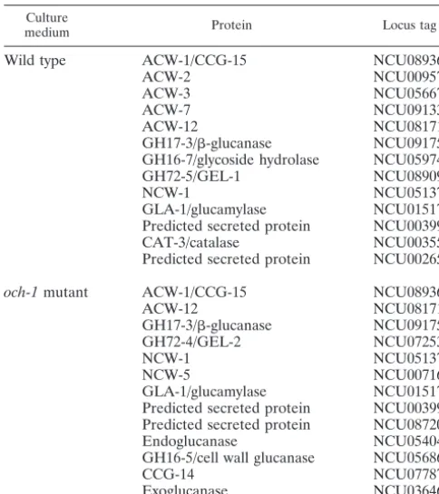
To identify what types of proteins were being released into the medium, we carried out a proteomic analysis of theoch-1
mutant secretome and compared it with the wild-type sectome. Secreted proteins are typically glycoproteins, and to re-move the carbohydrates and maximize the identification of these proteins in SDS-PAGE and by proteomic analysis, we treated secreted protein samples from mutant and wild-type cells with TFMS (26). Examination of the secretome from
och-1mutant cultures by gel electrophoresis showed that there
were more than 20 proteins being secreted (Fig. 4, lane 2). A nano-LC/MS-MS analysis of the secretome proteins from the
och-1mutant is given in Table 3. The mutant secreted 7
pro-teins that we normally see in the wild-type cell wall fraction (26): ACW-1 (NCU08936), ACW-12 (NCU08171), GH17-3/ -glucanase (NCU09175), GH72-4/GEL-2 (NCU07253), NCW-1 (NCU05137), NCW-5 (NCU00716), and GH16-5/cell wall glu-canase (NCU05686). Most of these proteins were identified by the presence of a number of peptide fragments (see Table S2 in the supplemental material).
The och-1 mutant releases elevated levels of ACW-1 and GEL-1 into the medium.As a third way to follow the release of cell wall proteins into the medium, we chose to look at the secretion of two well-known cell wall proteins, ACW-1 and GEL-1, in a secretion assay. ACW-1 is a major GPI-anchored cell wall protein inN. crassa(7) and a homolog of the yeast Ecm33p cell wall protein. The release of ACW-1 into the medium was examined in a Western blot assay from the sam-ples of the 2-h secretion assay described above. Figure 6 shows that ACW-1 was easily detected in the cellular extracts of both wild-type and mutant cells and that the levels of ACW-1 in the mutant cells were elevated, suggesting that ACW-1 expression is upregulated as part of the compensatory response to a weak-ened cell wall.C. albicans och1mutants have been shown to activate the cell wall integrity pathway in response to a weak-ened cell wall (3). Examination of the secreted proteins shows that small amounts of ACW-1 were released into the medium
FIG. 5. Coomassie brilliant blue dye assay of cell wall protein. Wild-type andoch-1mutant cell walls were incubated in the presence of Coomassie blue, and the amounts of Coomassie blue absorbed by the cell wall were determined.
TABLE 2. Cell wall proteins identified by nano-LC/MS-MS in the TFMS-deglycosylated cell wall fraction of vegetative hyphae
Cell wall
fraction Protein Locus tag
Wild type ACW-1/CCG-15 NCU08936
ACW-2 NCU00957
ACW-3 NCU05667
ACW-5 NCU07776
ACW-6 NCU03530
ACW-7 NCU09133
ACW-8 NCU07277
ACW-9 NCU06185
GH17-3/-glucanase NCU09175 GH16-1/mixed linked glucanase NCU01353 GH16-7/glycoside hydrolase NCU05974
GH72-4/GEL-2 NCU07253
-Glucosidase NCU09326
NCW-1 NCU05137
NCW-2 NCU01752
och-1mutant ACW-9 NCU06185
-Glucosidase NCU09326
NCW-2 NCU01752
GPI-anchored cell wall protein NCU00175
on September 8, 2020 by guest
http://ec.asm.org/
by the wild-type cell but that the mutant cell released large amounts of ACW-1 (Fig. 6). The N. crassa GH72-5/GEL-1 gene encodes a GPI-anchored cross-linking enzyme with ho-mology to theS. cerevisiaeGas1p and theA. fumigatusGel1 -1,3-glucanosyltransferase (26, 29). Gel1 has been shown to be an important cell wall biosynthetic enzyme (29). Western blot experiments using antibody directed against purified GEL-1 showed that the levels of GEL-1 in the cytosol were elevated in the mutant and that the mutant released easily detectable levels of GEL-1 into the medium, while the levels of GEL-1 released from the wild-type cell were below the level of detection in our system (Fig. 7).
The results of our measurements showing that the mutant released much more protein into the medium than the
wild-type cell, the proteomic analysis showing the presence of cell wall proteins in the medium from the mutant cells (Table 3), and the Western blot analyses demonstrating that the mutant released elevated levels of the ACW-1 and GEL-1 cell wall proteins into the medium (Fig. 6 and 7) clearly demonstrate that in the och-1 mutant, the cell wall glycoproteins move through the intracellular secretory pathway and pass through the cell wall without being cross-linked into the cell wall glu-can-chitin matrix. This suggests that the proper mannosylation of the cell wall proteins is required for their recognition by cell wall cross-linking enzymes and that, in its absence, the incor-poration of the proteins into the cell wall glucan-chitin matrix is compromised.
Theoch-1 mutant is defective in cross-linking ACW-1 into the cell wall.Once cell wall proteins are released into the cell wall space by secretory vesicles, they become covalently cross-linked into the cell wall glucan-chitin matrix (5, 8). This process is expected to take some time, and cell wall proteins that have been secreted but not yet cross-linked into the wall matrix could be extracted from the cell wall. Our procedure for pre-paring the purified cell walls used throughout this paper in-cludes a step in which the cell wall fraction is subjected to boiling in a buffer containing 1% SDS to remove proteins that are not covalently attached to the glucan-chitin matrix. If the
och-1 mutant is defective in the incorporation of cell wall
proteins into the matrix, we expected that boiling the cell wall in 1% SDS would release large amounts of cell wall protein. To directly test whether theoch-1mutant is defective in the in-corporation of cell wall proteins, we carried out a Western blot analysis of ACW-1 in the protein fraction released from the cell walls during the treatment step in which the cell wall fraction was boiled in SDS. As shown in Fig. 8, some ACW-1 is released from wild-type cells by the treatment. We ascribe this to the fact that it takes some time for ACW-1 to be TABLE 3. Cell wall proteins identified by nano-LC/MS-MS in the
TFMS-deglycosylated protein released into the growth medium by vegetative hyphae
Culture
medium Protein Locus tag
Wild type ACW-1/CCG-15 NCU08936
ACW-2 NCU00957
ACW-3 NCU05667
ACW-7 NCU09133
ACW-12 NCU08171
GH17-3/-glucanase NCU09175 GH16-7/glycoside hydrolase NCU05974
GH72-5/GEL-1 NCU08909
NCW-1 NCU05137
GLA-1/glucamylase NCU01517
Predicted secreted protein NCU00399
CAT-3/catalase NCU00355
Predicted secreted protein NCU00265
och-1mutant ACW-1/CCG-15 NCU08936
ACW-12 NCU08171
GH17-3/-glucanase NCU09175
GH72-4/GEL-2 NCU07253
NCW-1 NCU05137
NCW-5 NCU00716
GLA-1/glucamylase NCU01517
Predicted secreted protein NCU00399 Predicted secreted protein NCU08720
Endoglucanase NCU05404
GH16-5/cell wall glucanase NCU05686
CCG-14 NCU07787
Exoglucanase NCU03646
FIG. 6. Secretion of ACW-1 is elevated in theoch-1mutant. West-ern blot analysis of protein using antibody directed against ACW-1. Lane 1, wild-type cytosolic fraction (30g of cytosolic protein); lane 2, och-1cytosolic fraction (30g of cytosolic protein); lane 3, wild-type secreted protein fraction (normalized to the amount of secreted pro-tein from 30g of cytosolic protein); lane 4,och-1secreted protein fraction (normalized to the amount of secreted protein from 30g of cytosolic protein); lane 5, wild-type secreted protein fraction (normal-ized to the amount of secreted protein from 500g of cytosolic pro-tein); lane 6, och-1 secreted protein fraction (normalized to the amount of secreted protein from 500g of cytosolic protein).
FIG. 7. Secretion of GEL-1 is elevated in theoch-1mutant. West-ern blot analysis using antibody directed against GEL-1. Lane 1, wild-type cytosolic fraction (30g of cytosolic protein); lane 2,och-1 cyto-solic fraction (30g of cytosolic protein); lane 3, wild-type secreted protein fraction (normalized to the amount of secreted protein from 30
g of cytosolic protein); lane 4,och-1secreted protein fraction (nor-malized to the amount of secreted protein from 30g of cytosolic protein).
FIG. 8. The covalent incorporation of ACW-1 into the cell wall is reduced in theoch-1 mutant. Western blot analysis using antibody directed against the cell wall protein ACW-1. Lane 1, wild-type cyto-solic fraction (30g of cytosolic protein); lane 2,och-1cytosolic frac-tion (30g of cytosolic protein); lane 3, wild-type SDS-soluble cell wall protein fraction (normalized to 30g of cytosolic protein); lane 4, och-1secreted protein fraction (normalized to 30g of cytosolic pro-tein).
on September 8, 2020 by guest
http://ec.asm.org/
covalently incorporated into the wall in wild-type cells. In the wild-type cells, a comparison of the levels of unincorporated, cell wall-associated ACW-1 with the levels of ACW-1 in the cellular extract (representing ACW-1 in transit to the cell wall) shows that the level of unincorporated, cell wall-associated ACW-1 is slightly higher than the level of ACW-1 in transit. Figure 8 also shows that ACW-1 levels are upregulated in the
och-1cells as part of the compensatory mechanism turned on
by the weakened state of the mutant cell wall, as mentioned earlier. Most importantly, theoch-1mutant cell wall has large amounts of unincorporated ACW-1, much more than could be explained as arising from the upregulation of ACW-1 expres-sion. Figure 8 shows that the mutant is compromised in its ability to incorporate ACW-1 into the cell wall. A similar analysis for GEL-1 showed that theoch-1mutant cell wall had elevated levels of unincorporated GEL-1 (data not shown). Based on these results, along with the results of the secretion experiments, we conclude that the mannosylation of cell wall proteins by OCH-1 is required for the efficient covalent incor-poration of the proteins into the cell wall matrix.
DISCUSSION
TheN. crassacell wall is composed of polysaccharides and
proteins. The wall is known to contain -1,3-glucan, ␣ -1,3-glucan, chitin, and a number of identified cell wall glycopro-teins (5, 7, 26). In the ER, the cell wall glycoproglycopro-teins are modified by N-linked glycosylation, and many cell wall proteins receive a GPI anchor. Subsequently, galactomannans are added to the N-linked oligosaccharides. As the proteins pass through the secretory pathway, O-linked galactomannans are also added. After being secreted into the cell wall space, inte-gral cell wall proteins are then cross-linked into the cell wall glucan-chitin matrix. The cross-linking of cell wall proteins into the cell walls of filamentous fungi has not been carefully ex-amined.
Different species of fungi have been found to modify their glycoproteins differently (16).S. cerevisiaehas been shown to modify glycoproteins by the addition of a large polymannose structure, while filamentous fungi generate shorter galacto-mannan structures (14). The posttranslational modifications of glycoproteins can be important targeting determinants, as demonstrated by the tagging of lysosomal protein for import into that organelle by the addition of a posttranslationally added mannose-6-phosphate residue. We demonstrate that in
N. crassathe addition of an␣-1,6-mannose residue to N-linked
oligosaccharides is necessary for their covalent incorporation into the cell wall. This suggests that the incorporation of cell wall proteins into the glucan-chitin matrix may occur between N-linked galactomannan and the matrix.
In this report, we characterize theN. crassa och-1knockout mutant. Theoch-1mutant was identified in a screening of the
Neurosporaknockout library, and the gene (NCU00609) was
found to be a homolog of known␣-1,6-mannosyltransferases involved in initiating the addition of mannose to N-linked oligosaccharides. The encoded protein, OCH-1, contains 373 amino acids with a classical DXD sugar binding motif.
Examination of theoch-1mutant showed that it was a mor-phological mutant with a slow growth rate and a tight colonial phenotype (Fig. 1 and 2). A microscopic examination of the
mutant showed that it has an abnormal pattern of hyphal growth and branching. Two interesting aspects of the mutant’s phenotype are that when used as a female in mating, it is unable to “throw” the ascospores, and that during the asexual phase of the life cycle, it produces abnormal conidia that do not readily separate from each other. A variety of cell wall stress tests demonstrated that the mutant has a cell wall defect and is compromised in its ability to survive any of the cell wall stress reagents we tested. The altered morphology and growth characteristics of the mutant are likely to be the direct result of the weakened state of the mutant cell wall.
OCH1mutants have been isolated from a number of differ-ent fungi. InS. cerevisiae,C. albicans, K. lactis,P. angusta,Y.
lipolytica,S. pombe, andP. pastoris, loss ofOCH1gives rise to
morphologies that correlate with alterations in the cell wall (2, 3, 9, 17, 33, 35, 37, 38). In contrast with these yeasts, knockout mutations of the OCH1 genes in the filamentous fungus A.
fumigatusdo not result in a cell wall-defective phenotype (18).
TheN. crassa och-1mutant has a phenotype more in line with
the phenotypes seen inS. cerevisiae. There are, however, some important differences between theN. crassa och-1mutant and
theS. cerevisiaemutant. InS. cerevisiae, GPI-anchored cell wall
proteins can be cross-linked into the wall through the attach-ment of -1,6-glucan to GPI-anchored oligosaccharides (15, 25), and the yeast mutant has not been reported as having a large-scale loss of cell wall protein into the growth medium. In
N. crassa, which lacks-1,6-glucan (4) (Table 1), there is no
evidence for the cross-linking of GPI anchors into the cell wall, and theoch-1mutant releases GPI-anchored proteins into the growth medium.
To characterize the carbohydrate polymers in theoch-1 mu-tant cell wall, we examined the carbohydrate composition and the linkages present between the different sugars in wild-type and mutant cell walls. We found that the mutant cell wall had an altered carbohydrate composition, with a major decrease in mannose and galactose residues. This suggests that most of the mannose and galactose present in the N. crassa cell wall is associated with cell wall glycoproteins. Linkage analysis showed that the mutant had a complete lack of 1,6-linked mannose residues. The␣-1,6-mannosylation of cell wall pro-teins has been shown to be associated with N-linked oligosac-charides inS. cerevisiaeandC. albicans(13, 14). The carbohy-drate analysis also showed a major decrease in mannose residues with 1,2-linkages and galactose residues. The data suggest that OCH-1 is required for the synthesis of N-linked galactomannan (Fig. 3). A comparable cell wall carbohydrate analysis has not been done for the yeastOCH1mutants, but it has been shown that the cell wall and secreted proteins from the mutants lack the high-mannan structure shown in Fig. 3 (1, 3, 34).
The most important conclusion from our experiments is that the mannosylation of N-linked oligosaccharides is required for the incorporation of cell wall proteins into the cell wall matrix. Using such techniques as SDS-PAGE of TFMS-released cell wall protein, proteomic analysis of cell wall protein, and a Coomassie blue dye binding assay, we found that theoch-1
mutant cell wall had less protein than the wild-type cell wall. This provides a partial explanation for the reduced levels of mannose and galactose in the carbohydrate analysis of the
och-1cell wall. Mannose and galactose are incorporated into
on September 8, 2020 by guest
http://ec.asm.org/
the wall as oligosaccharides associated with cell wall protein. Proteomic analysis of purified mutant cell walls failed to iden-tify a number of proteins we have routinely found in wild-type cell walls.
We also found that when grown in a liquid medium, the
och-1 mutant released large amounts of proteins into the
medium. Proteomic analysis of the secreted proteins demon-strated that the growth medium contained a number of pro-teins that we previously identified as cell wall propro-teins in wild-type cell walls. Western blot analysis of two well-known cell wall proteins, ACW-1 and GEL-1 (26), demonstrated that large amounts of these cell wall proteins were being released into the medium by the mutant, while the levels of ACW-1 and GEL-1 released from wild-type cells were small or negligible (Fig. 6 and 7). The data indicate that in the absence of OCH-1-directed␣-1,6-mannosylation, cell wall proteins are not be-ing covalently incorporated into the cell wall glucan-chitin ma-trix and are being released into the medium.
We directly tested whether the incorporation of cell wall proteins into the cell wall matrix was compromised in theoch-1
mutant and found that the levels of unincorporated, cell wall-associated ACW-1 were elevated in the mutant cell wall. As shown in Fig. 8, the mutant cell wall does contain elevated levels of ACW-1 that have not been covalently linked into the cell wall matrix. We conclude that in the absence of ␣ -1,6-mannosylation, the proteins are not efficiently cross-linked into the wall. Our results support a model for cell wall biogenesis in which the cross-linking of cell wall protein into the wall re-quires some elements of the N-linked galactomannan, whose synthesis begins with the OCH-1-mediated addition of an ␣-1,6-linked mannose residue to the N-linked oligosaccharide. The enzymes used to cross-link the proteins into the cell wall may recognize some aspects of the N-linked oligosaccharide-galactomannan structure and mediate the cross-linking of the structure into the glucan-chitin matrix.
ACKNOWLEDGMENTS
This work was funded by NIH grant R01 078589. The carbohydrate analysis was supported in part by the Department of Energy-funded Center for Plant and Microbial Complex Carbohydrates (DE-FG09-93ER-20097).
We are grateful to David Toczynski for the polyclonal rabbit anti-body directed against GH72-5/GEL-1. We thank Alan Siegel and James Stamos for assistance in preparing figures.
REFERENCES
1.Ballou, C. E.1990. Isolation, characterization, and properties of Saccharo-myces cerevisiae mnnmutants with nonconditional protein glycosylation
de-fects. Methods Enzymol.185:440–479.
2.Barnay-Verdier, S., A. Boisrame, and J.-M. Beckerick.2004. Identification
and characterization of two␣-1,6-mannosyltransferases, Anl1p and Och1p,
in the yeastYarrowia lipolytica. Microbiology150:2185–2195.
3.Bates, S., H. B. Hughes, C. A. Munro, W. P. H. Thomas, D. M. MacCallum, G. Bertram, et al.2006. Outer chain N-glycans are required for cell wall
integrity and virulence ofCandida albicans. J. Biol. Chem.281:90–98.
4.Borkovich, K. A., L. A. Alex, O. Yarden, M. Freitag, G. E. Turner, N. D. Read, et al.2004. Lessons from the genome sequences ofNeurospora crassa: tracing the path from genomic blueprint to multicellular organism.
Micro-biol. Mol. Biol. Rev.68:1–108.
5.Bouffard, F. A., R. A. Zambias, J. F. Dropinski, J. M. Balkovec, M. L. Hammond, G. K. Abrazzo, et al.1994. Synthesis and antifungal activity of
novel cationic pneumocandin B(o) derivatives. J. Med. Chem.37:222–225.
6.Bowman, S. M., and S. J. Free.2006. The structure and synthesis of the
fungal cell wall. Bioessays28:799–808.
7.Bowman, S. M., A. Piwowar, M. Al Dabbous, J. Vierula, and S. J. Free.2006. Mutational analysis of the glycosylphosphatidylinositol (GPI) anchor
path-way demonstrates that GPI-anchored proteins are required for cell wall
biogenesis and normal hyphal growth inNeurospora crassa. Eukaryot. Cell
5:587–600.
8.Chaffin, W. L.2008.Candida albicanscell wall proteins. Microbiol. Mol. Biol.
Rev.72:495–544.
9.Choi, B.-K., P. Bobrowicz, R. C. Davidson, S. R. Hamilton, D. H. Kung, H. Li, et al.2003. Use of combinatorial genetic libraries to humanize N-linked
glycosylation in the yeastPichia pastoris. Proc. Natl. Acad. Sci. U. S. A.
100:5022–5027.
10.Colot, H. V., G. Park, G. E. Turner, C. Ringelberg, C. M. Crew, L. Litvinkova, et al.2006. A high-throughput gene knockout procedure for
Neurosporareveals functions for multiple transcription factors. Proc. Natl.
Acad. Sci. U. S. A.103:10352–10357.
11.Cullen, P. J., J. Schultz, J. Horecka, B. J. Stevenson, Y. Jigami, and G. F. Sprague.2000. Defects in protein glycosylation cause SHO1-dependent
ac-tivation of STE12 signaling pathway in yeast. Genetics155:1005–1018.
12.Davis, R. H., and F. J. DeSerres.1970. Genetic and microbiological research
techniques forNeurospora crassa. Methods Enzymol.27:79–143.
13.Dean, N.1999. Asparagine-linked glycosylation in yeast Golgi. Biochim.
Biophys. Acta1426:309–322.
14.Deshpande, N., M. R. Wilkins, N. Packer, and H. Nevalainen.2008. Protein
glycosylation pathways in filamentous fungi. Glycobiology18:626–637.
15.Fujii, T., H. Shimoni, and Y. Iimura.1999. Structure of the glucan-binding
sugar chain of Tip1p, a cell wall protein ofSaccharomyces cerevisiae.
Bio-chim. Biophys. Acta1427:133–144.
16.Gemmill, T. R., and R. B. Trimble.1999. Overview of N- and O-linked oligosaccharide structures found in various yeast species. Biochim. Biophys. Acta1426:227–237.
17.Kim, M. W., E. J. Kim, J.-Y. Kim, J.-S. Park, D.-B. Oh, Y.-I. Shimma, et al.
2006. Functional characterization of the Hansenula polymorphaHOC1,
OCH1, and OCR1 genes as members of the yeast OCH1
mannosyltrans-ferase family involved in protein glycosylation. J. Biol. Chem.281:6261–6272.
18.Lambou, K., S. Perkhofer, T. Fontaine, and J. P. Latge.2010. Comparative
functional analysis of the OCH1 mannosyltransferase families inAspergillus
fumigatusandSaccharomyces cerevisiae. Yeast27:625–636.
19.Latge, J.-P.2007. The cell wall: a carbohydrate armour for the fungal cell.
Mol. Microbiol.66:279–290.
20.Leal, J. A., J. Jimenez-Barbero, B. Gomez-Miranda, A. Prieto, J. Dome-nech, and M. Bernabe. 1996. Structural investigation of the cell-wall
galactomannan fromNeurospora crassaandN. sitophila. Carbohydr. Res.
283:215–222.
21.Lee, B. N., and E. A. Elion.1999. The MAPKKK Ste11 regulates vegetative growth through a kinase cascade of shared signaling components. Proc. Natl.
Acad. Sci. U. S. A.96:12679–12684.
22.Lehle, L., A. Eiden, K. Lehnert, A. Haselbeck, and E. Kopetzki.1995.
Gly-coprotein biosynthesis inSaccharomyces cerevisiae:ngd29, an N-glycosylation
mutant allelic tooch1having a defect in the initiation of outer chain
forma-tion. FEBS Lett.370:41–45.
23.Lesage, G., and H. Bussey.2006. Cell wall assembly inSaccharomyces cer-evisiae. Microbiol. Mol. Biol. Rev.70:317–343.
24.Levin, D. E.2005. Cell wall integrity signaling inSaccharomyces cerevisiae.
Microbiol. Mol. Biol. Rev.69:262–291.
25.Lu, C.-F., R. C. Montijn, J. L. Brown, F. Klis, J. Kurjan, H. Bussey, and P. N. Lipke.1995. Glycosylphosphatidylinositol-dependent cross-linking of
alpha-agglutinin and beta-1,6-glucan in theSaccharomyces cerevisiae cell wall.
J. Cell Biol.128:333–340.
26.Maddi, A., S. M. Bowman, and S. J. Free.2009. Trifluoromethansulfonic acid-based proteomic analysis of cell wall and secreted proteins of the
asco-mycetous fungiNeurospora crassaandCandida albicans. Fungal Genet. Biol.
46:768–781.
27.Maniatis, T., E. F. Fritsch, and C. Sambrook.1982. Molecular cloning: a laboratory manual. Cold Spring Harbor Press, Cold Spring Harbor, NY. 28.Margolin, B. S., M. Freitag, and E. U. Selker.1997. Improved plasmids for
gene targeting at thehis-3locus ofNeurospora crassa. Fungal Genet. Newsl.
44:34–36.
29.Mouyna, I., T. Fontaine, M. Vai, M. Monod, W. A. Fonzi, M. Diaquin, et al.
2000. Glycosylphosphatidylinositol-anchored glucanosyltransferases play an
active role in the biosynthesis of the fungal cell wall. J. Biol. Chem.275:
14882–14889.
30.Munro, S.2001. What can yeast tell us about N-linked glycosylation in the
Golgi apparatus? FEBS Lett.498:223–227.
31.Nakajima, T., M. Yoshida, N. Hura, and K. Matsuda.1984. Structure of the
cell wall proteogalactomannan fromNeurospora crassa. I. Purification of the
proteoheteroglycan and characterization of alkali-labile oligosaccharides.
J. Biochem.96:1005–1011.
32.Nakajima, T., M. Yoshida, M. Nakamura, N. Hura, and K. Matsuda.
1984. Structure of the cell wall proteogalactomannan fromNeurospora
crassa. II. Structural analysis of the polysaccharide part. J. Biochem.
96:1013–1020.
33.Nakanishi-Shindo, Y., K. Nakayama, A. Tanaka, Y. Toda, and Y. Jigami.1993.
Structure of the N-linked oligosaccharides that show the complete loss of␣
on September 8, 2020 by guest
http://ec.asm.org/
polymannose outer chain fromoch1,och1 mnn1, andmnn1 alg3mutants of
Saccharomyces cerevisiae. J. Biol. Chem.268:26338–26345.
34.Nakayama, K., T. Nagasu, Y. Shimma, J. Kuromitsu, and Y. Jigami.1992.
OCH1 encodes a novel membrane bound mannosyltransferase: outer
chain elongation of asparagines-linked oligosaccharides. EMBO J.11:
2511–2519.
35.Ohashi, T., Y. Ikeda, N. Tanaka, S. Nakakita, S. Natsuka, Y. Giga-Hama, and K. Takegawa.2009. Theoch1mutant ofSchizosaccharomyces pombe
produces galactosylated core structures of N-linked oligosaccharides. Biosci.
Biotechnol. Biochem.73:4070–4414.
36.Scrimale, T., L. Didone, K. L. de Mesy Bentley, and D. J. Krysan.2009.
The unfolded protein response is induced by the cell wall integrity mito-gen-activated protein kinase signaling cascade and is required for cell
wall integrity inSaccharomyces cerevisiae. Mol. Biol. Cell20:164–175.
37.Uccelletti, D., F. Farina, S. Rufini, P. Magnelli, C. Abeijon, and C. Palleschi.
2006. TheKluyveromyces lactis␣-1,6-mannosyltransferase KlOch1p is
re-quired for cell-wall organization and proper functioning of the secretory
pathway. FEMS Yeast Res.6:449–457.
38.Yoko-o, T., K. Tsukahara, T. Watanabe, N. Hata-Sugi, K. Yoshimatsu, T. Nagasu, and Y. Jigami.2001.Schizosaccharomyces pombeoch1⫹encodes
␣-1,6-mannosyltransferase that is involved in outer chain elongation of
N-linked oligosaccharides. FEBS Lett.489:75–80.