A Quality-Improvement Collaborative Project to Reduce
Pressure Ulcers in PICUs
abstract
BACKGROUND AND OBJECTIVE:Pediatric patients are at risk for de-veloping pressure ulcers (PUs) and associated pain, infection risk, and prolonged hospitalization. Stage III and IV ulcers are serious, reportable events. The objective of this study was to develop and implement a quality-improvement (QI) intervention to reduce PUs by 50% in our ICUs.
METHODS: We established a QI collaborative leadership team, mea-sured PU rates during an initial period of rapid-cycle tests of change, developed a QI bundle, and evaluated the PU rates after the QI implementation. The prospective study encompassed 1425 patients over 54 351 patient-days in the PICU and NICU.
RESULTS:The PU rate in the PICU was 14.3/1000 patient-days during the QI development and 3.7/1000 patient-days after QI implementation (P, .05), achieving the aim of 50% reduction. The PICU rates of stages I, II, and III conventional and device-related PUs decreased after the QI
intervention. The PU rate in the NICU did not change significantly
over time but remained at a mean of 0.9/1000 patient-days. In the postimplementation period, 3 points were outside the control limits, primarily due to an increase in PUs associated with pulse oximeters and cannulas.
CONCLUSIONS: The collaborative QI model was effective at reducing PUs in the PICU. Pediatric patients, particularly neonates, are at risk for device-related ulcers. Heightened awareness, early detection, and identification of strategies to mitigate device-related injury are necessary to further reduce PU rates.Pediatrics2013;131:e1950–e1960
AUTHORS:Marty Visscher, PhD,a,fAlice King, MD,b,fAnn
Marie Nie, MSN,b,fPat Schaffer, MSN,cTeresa Taylor, BSN,c
David Pruitt, MD,d,gMary Jo Giaccone, MSN,cMarshall
Ashby, MHSM,eand Sundeep Keswani, MDb,f
aSkin Sciences Program, Plastic Surgery,bPediatric Advanced Wound Care and Skin Service,cPatient Services,dPhysical Medicine and Rehabilitation,eJames M. Anderson Center for Health Systems Excellence, Cincinnati Children’s Hospital Medical Center, Cincinnati, Ohio; and Departments offSurgery, and gPediatrics, College of Medicine, University of Cincinnati, Cincinnati, Ohio
KEY WORDS
pressure ulcer, skin, wound, device, quality improvement, bundle, intervention, neonatal, ICU
ABBREVIATIONS
CWOCN—certified wound ostomy and continence nurse DTI—deep tissue injury
NIPPV—noninvasive positive pressure ventilation PU—pressure ulcer
QI—quality improvement
QIC—quality improvement collaborative TOC—test of change
Dr Visscher made a substantial contribution to the conception and design, acquisition of data, and analysis and interpretation of data and in drafting the article with critical revision for important intellectual content; Dr Leung made a substantial contribution to the design, analysis, and interpretation of data and in drafting the article with critical revision for important intellectual content; Mss Nie, Schaffer, and Taylor, Dr Pruitt, and Ms Giaccone made substantial contributions to the conception and design, acquisition of data, and interpretation of data; Mr Ashby made a substantial contribution to the conception and design, analysis of data, and interpretation and in drafting of article with critical revision for important intellectual content; Dr Keswani made a substantial contribution to conception and design, analysis and interpretation of data and in drafting the article with critical revision for important intellectual content; and all authors hadfinal approval of the version to be published.
www.pediatrics.org/cgi/doi/10.1542/peds.2012-1626
doi:10.1542/peds.2012-1626
Accepted for publication Feb 13, 2013
Address correspondence to Marty O. Visscher, Skin Sciences Program, Division of Plastic Surgery, Cincinnati Children’s Hospital Medical Center, 3333 Burnet Ave, Cincinnati, OH 45229. E-mail: [email protected]
Pediatric patients are at risk for pres-sure ulcers (PUs) due to immature skin, compromised perfusion, decreased mobility, altered neurologic respon-siveness,fluid retention, moisture, and
medical devices.1 PUs can develop
from the surface or from below, at the level of muscle and dermal tissue in-teraction and compression.2,3PUs are
classified by the depth and severity. Stage I is nonblanchable erythema-tous skin that may be painful, soft, and warmer or cooler than adjacent tis-sue. Stage II involves partial dermal loss (eg, shallow open ulcer or intact blister). Stage III has dermal loss wherein subdermal elements (eg, subcutaneous fat) are visualized. Stage IV is full-thickness tissue loss with exposed bone, tendon, or mus-cle. Unstageable ulcers are full-thickness wounds covered by slough and/or eschar. Deep tissue injuries (DTIs) have grossly intact skin with obvious underlying injury related to pressure.4
Stage III and IV PUs are serious report-able events, considered“never events” by several national benchmarking
or-ganizations.5 PU incidence is higher
in critically ill patients,6with increased
pain, infection, and prolonged hospital-ization.7 Reductions in reimbursement
for health care-acquired PUs have been implemented by the Centers for Medi-care & Medicaid Services for adult institutions.8 They extend to Medicaid
recipients, including pediatrics, as of July 2012.
More than 70% of adult PUs are“
con-ventional” ulcers from pressure over
bony prominences, such as the sacrum, shoulder, and heels.9,10 Up to 34% are
associated with medical devices, such as nasal cannulas and facemasks.11,12
While PUs are relatively well studied in adults, the pediatric literature is sparse, perhaps due to a lack of awareness and/or an incomplete understanding of neonatal/pediatric skin physiology.13–15
Both conventional and device-related PUs have been reported.16–19
We established an interdisciplinary quality-improvement (QI) collaborative (QIC) focused on reduction of PUs in the pediatric and neonatal intensive care units and based on the Institute for Healthcare Improvement Breakthrough Series Collaborative.20We hypothesized
that a QIC focused on pressure-related skin injury would reduce the PU rate. We describe the development, imple-mentation and effectiveness of a QI in-tervention for reducing PUs.
METHODS
Setting
Cincinnati Children’s Hospital Medical Center is a 577-bed, free-standing qua-ternary care academic facility that serves patients from regional, national, and international locations. The 53-bed level III NICU serves infants who require surgery, have complex congenital
con-ditions, or need specific diagnostic
procedures. The 42-bed PICU treats older critically ill patients.
Human Subjects Protection
The institutional review board approved the quality improvement project and waived the requirements to obtain writ-ten parental permission and child assent.
QIC Leadership Team
The leadership team was established in August 2007 after a hospitalwide, single-day study revealed a PU preva-lence rate of.10%, higher than repor-ted for pediatric institutions.10The team
included key clinical personnel and subject matter experts from hospital departments; physicians, nursing lead-ers from the NICU and PICU, nursing and respiratory bedside staff, nurse educa-tors, certified wound ostomy and con-tinence nurses (CWOCNs), and skin researchers. The team was supported by QI consultants and outcome managers.
As a strategic priority, the collaborative received senior-level sponsorship from the board of trustees.
The leadership team generated an aim
statement based on specific,
measur-able, attainmeasur-able, relevant, and time-bound21goals: to reduce the PU rate in
the NICU and PICU by 50% by 1 year after implementation of a QI intervention. The team met every 2 weeks to review the detailed findings from the initial survey, published literature, experi-ences at other institutions, and unit-level processes.
Prework
Because the quarterly prevalence sur-vey represented a single time point, we collected additional data to understand PU incidence, severity, and etiology. Designated nursing staff from each ICU received training on PU physiology, skin evaluation, and data collection. They performed head-to-toe skin evaluations on all inpatients on 1 day every 2 weeks beginning in September 2007. Patient clothing was removed, and all areas, including under devices, were exam-ined for compromise and PUs. Anatomic location, suspected cause, and pre-liminary PU stage were documented.
Suspected cause was classified as
conventional pressure related or di-rectly attributed to device use. Stage was verified by a CWOCN using the Na-tional Pressure Ulcer Advisory Panel staging system.4The team reviewed the
data every 2 weeks. The QI consultants provided training throughout the pro-cess. Progress was reviewed with the hospital Patient Safety Program. Stage III and IV PUs were reported monthly to management. Team minutes and PU rates were shared will all ICU patient care and management staff.
QI Intervention Development Process
From the data on location, stage and
key drivers: (1) skin assessments, (2) patient skin care, (3) patient care in-directly related to skin (pain control, nutrition, hydration), (4) products re-lated to pressure, and (5) patient/family involvement (Fig 1). The key driver se-lection stemmed from the importance of close monitoring and early detection of skin compromise due to pressure and excess moisture. Our improvement model used rapid-cycle small tests of change (TOCs) to construct and refine the QI intervention.22 TOCs were
con-ducted among small groups of patients and staff and evaluated for clarity, ease of completion, and impact on work
flow. Modifications were made and
subsequent TOCs were conducted. TOCs were performed on risk assessment (ie, selection of a validated tool), repo-sitioning frequencies (eg, 4-hour inter-vals), daily head-to-toe skin and device assessment, moisture management, and education of staff and parents. Pressure redistribution mattresses were
used. Staff members were trained to use positioning aids and plan-do-study-act cycles were conducted.
Three months after QIC initiation, the skin assessment data continued to show that PUs occurred at multiple body sites. More than 50% were asso-ciated with medical devices, including facemasks for noninvasive positive pressure ventilation (NIPPV), tracheos-tomy tubes and ties, casts, and endotra-cheal tubes. The frequent, comprehensive skin assessments needed to examine the skin under devices. Multiple plan-do-study-act cycles were required to develop a systematic method for identi-fication and documentation of “ abnor-mal” skin features (eg, nonblanchable erythema, blistering) that were asso-ciated with pressure. Assessments
were time consuming and difficult to
conduct for skin under critical devices (eg, masks, tracheostomies, tubes). There was uncertainty about the cause of “abnormal” skin findings. However,
nonblanchable erythema and blistering were noted, suggesting that early-stage skin injury was being detected by bed-side staff. The feedback suggested a need for detailed training on skin com-promise and PU cause. The CWOCNs, unit-nursing educators, and content experts developed 6 online training modules (Table 1). Completion with de-monstration of competency was man-datory for all patient care staff.
The importance of early detection of skin injury prompted the implementa-tion of unit “skin champions.” These staff members, with interests in skin/ wound care, underwent additional skin training and became resources to all unit staff. Several became wound care certified by the National Alliance of Wound Care. They conducted rounds twice weekly on all patients to identify skin issues, discussed patients with the CWOCNs, and conducted a bedside skin assessment. A patient-specific skin and wound plan was created, ordered
FIGURE 1
by the physician or advanced practice
nurse, and documented. “Skin
inte-grity”was added to the individual plan of care. Unit educators provided the skin integrity goals for families. A skin champion consulted one-on-one with staff when suspected PUs were found and reinforced the skin assessment procedures.
Table 2 shows thefinal QI intervention implemented for all patients and staff in both ICUs in November 2008. Skin redness or compromise, observed during daily skin and device assess-ments, was recorded in the medical record along with modified Braden Q23
risk levels of#16 (high risk), 17 to 21 (moderate risk), and 22 to 25 (mild risk)
for patients $28 days of age. NICU
patients were considered to be at high risk as there was no validated risk
assessment tool for infants. If a PU was detected, the stage was noted and reported to unit management. At QI implementation, unit champions and managers conducted an in-depth event cause analyses for all stage III PUs, unstageable PUs, and DTIs within 48 hours of detection. Event forms were developed to determine factors associ-ated with failures including changes in
patient risk, compliance with QI
for assessment, positioning, and use of intervention elements (Table 3). Results were discussed with the QIC team, and
specific interventions or processes
were modified via TOC cycles.
Outcomes
The primary outcome was PUs/1000 patient-days from the every-2-week head-to-toe skin assessments. New
PUs occurring after admission24were
counted and reported as rate (ie, number/1000 patient-days), which was calculated from the length of hospital stay summed for all evaluated patients. Key process measures (ie, daily skin
assessments, daily device
assess-ments, and moisture management) were determined quarterly from a re-view of the medical records.24
Statistical Analysis
We calculated monthly PU rates by dividing the total number by the mean patient-days of all evaluated patients (ie, during QI development or after
implementation) 3 1000. Statistical
run charts25,26 were used to
deter-mine the effect of the QI intervention on PU rates over time. This method is also used to show special-cause
variation.27 Age, weight, length of
stay, and gender were compared for patients before and after QI
imple-mentation using t test procedures
(SigmaStat; SPSS, IBM Corporation, Somers, NY) with significance levels of P,.05.
RESULTS
Table 4 shows patient demographics. The mean hospital stay was longer and weights and ages were lower after QI
implementation than during
de-velopment in the PICU (P ,.01). The
gender distributions were similar for both periods. There were no differ-ences for NICU patients in length of stay, weight, gestational age, adjusted age, or gender during and after QI de-velopment.
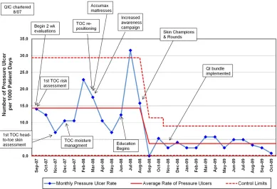
In the PICU, the mean PU rate during QI development was 14.3/1000 patient-days (100 PUs; 7979 patient-days) After the QI bundle was fully implemented, the rate was 3.7/1000 patient-days (51 PUs; 14 729 days). This represented a down-ward shift, defined as$8 consecutive points below the centerline, and a sig-nificant decrease in rate, as shown in
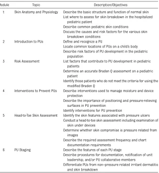
TABLE 1 PU Education Modules
Module Topic Description/Objectives
t Skin Anatomy and Physiology Describe the basic structure and function of normal skin List where to assess for skin breakdown in the hospitalized
pediatric patient
Describe common pediatric skin conditions
Discuss the causes and risk factors for the various skin breakdown conditions
2 Introduction to PUs Define and recognize a PU
Locate common locations of PUs on a child’s body Describe risk factors of PU development in the pediatric
population
3 Risk Assessment List factors that contribute to PU development in pediatric patients
Determine an accurate Braden Q assessment on a pediatric patient
Identify those patients who do not meet the criteria for using the modified Braden Q
4 Interventions to Prevent PUs Describe interventions used to manage moisture and device protection
Describe the importance of positioning and pressure-relieving surfaces in PU prevention
Identify interventions for PU prevention
5 Head-to-Toe Skin Assessment Identify the skin features associated with pressure ulcers Conduct a head-to-toe skin assessment including examination of
skin under devices
Determine whether skin compromise is pressure related from images
Describe the required assessment frequency and chart documentation requirements
6 PU Staging Describe the features of each PU stage
Describe procedures for documentation, notification of unit leadership, and/or PU collaborative members
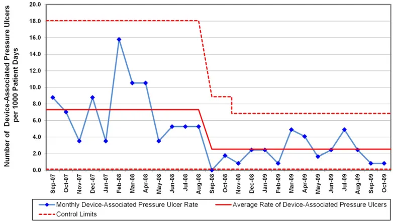
the run chart (Fig 2). The change began in September 2008, toward the end of the baseline period, and was stable during the postintervention period. The PICU rates of stage I, stage II (Fig 3), and severe (stage III, unstageable, DTI) conventional and device-related (Fig 4) PUs were all lower after the QI intervention. There were no stage IV PUs.
In the NICU, the mean PU rate was 0.9/ 1000 patient-days (18 PUs; 19 939 days during development and 31 PUs; 11 704 days after development) and did not change over time. The mean did not increase, but there were 3 points above the upper control limit, indicating the process was out of control (Fig 5). The NICU staff and QIC team completed event forms to identify potential causes.
Stage II and III PUs from pulse oxi-meters and extracorporeal membrane oxygenation cannulas were noted. PUs from these devices had not been ob-served in the NICU during QI develop-ment. The skin under pulse oximeters had been assessed every 12 hours, and devices had been replaced at least once in 48 hours. There were no re-ports of increased edema, difficulty in obtaining an oxygen saturation read-ing, or device tightening (ie, increased pressure). The hospital had changed the pulse oximeter supplier before the months when PUs occurred. No specific problem with the pulse oximeter could be identified. There were no stage IV PUs
A high number of PUs were associated with medical devices both before and after QI implementation, particularly from NIPPV facemasks and tracheos-tomy tubes and ties (Table 5). Of patients with facemask PUs, 46% had diagnoses associated with craniofacial anomalies (eg, micrognathia).
Process Measure: Compliance
Compliance with daily head-to-toe skin assessments averaged 81% in the PICU and 50% in the NICU after implemen-tation. Conformity with skin assess-ments under devices was 57% and 50% in the PICU and NICU, respectively. Use of products to manage moisture was 92% and use of nutrition consults was 100% in both units.
DISCUSSION
We assembled a leadership team to reduce the PU rate by 50% in the PICU and NICU. Using iterative, rapid-cycle TOCs, the QIC developed a QI in-tervention based on the cornerstones of (1) frequent, thorough skin assess-ments, (2) preventative interventions, (3) comprehensive education, (4) clin-ical staff empowerment (eg, skin champions), and (5) systems change
TABLE 2 Elements of the PU QI Bundle
Risk assessment for all patientsa
$28 d of age, complete a modified Braden Q risk assessment on admission and daily to classify PU risk level as low, moderate, or highb
#28 d of age, treat as high risk All NICU patients, treat as high risk Skin assessment for all patientsa
Daily head-to-toe evaluation of entire body surface
Document specific body areas of nonblanchable erythema or skin trauma that may be due to pressure Medical device assessment for all patientsa
Examine skin under each device every 12 h
Document specific areas of nonblanchable erythema or skin trauma that may be due to pressure Implement specific interventions for prevention of PUs based on risk assessment level of low, moderate,
or high
Reposition at a minimum of every 2 h formoderate to high riskand 4 h forlow risk
Use repositioning devices for allrisk levelsand pressure reduction surfaces for chairs and adult beds Float heels
Use appropriate seat cushion for chair-bound patients and shift weight every 15 min Reduce moisture forall risk levels
Check common moisture sites every 2–4 h
Use diapers with breathable outer cover (avoid plastic) Remove moisture from under devices
Keep skin under casts, splints, braces, and collars dry and clean In diaper area forall risk levels
Keep skin clean and dry and change diapers as soon as they are wet Apply protective cream
Avoid diapers/products with nonbreathable plastic backing Manage friction and shear forall risk levels
Use draw sheet to reposition
When bed elevated, use positioning products and/or elevate knees to prevent friction and shearing Optimize nutrition via ongoing nutrition consults for allmoderate- to high-riskpatients Education/awareness
All unit bedside staff (nurses, respiratory therapists, physical therapists) required to complete 6 PU-focused training modules and demonstrate competency via score on posttests
All unit bedside staff required to complete annual refresher training
Physician staff (attending, fellows, residents) participated in training provided by the QIC leadership team physician and CWOCN
Results of every 2-wk skin assessments shared with all staff including continuous improvements to the QI bundle
Provide an educational brochure regarding pressure ulcers and skin assessments for families Bedside staff actively engaged families in assessments and discussed observations with them Implement skin champions and conduct collaborative skin rounds
Identify specific unit staff to serve as skin champions, resources to other unit staff Participate in weekly collaborative skin rounds
Develop skin and wound plans for identified patients
aThe results of the daily or every 12-h risk skin and device assessments are charted in the medical record for the locations
where the skin exhibits any redness, compromise, or damage. If a PU is detected, the stage is noted and reported to the unit management.
bRisk levels from the modified Braden Q scale are as follows:#16, high risk; 17–21, moderate risk; and 22–25, mild risk. The
(eg, skin rounds). In the PICU, PUs de-creased from 14.3/1000 patient-days during QI development to 3.7/1000 patient-days during the first year af-ter implementation, thereby achieving the aim of a 50% reduction. In the NICU, the prebundle mean rate of 0.9 PU/1000 days did not change. The majority of PUs were stage II during both periods.
This is one of a limited number of studies to implement an effective in-tervention to reduce PUs in pediatrics.
Boesch et al19developed a QI
preven-tion bundle for tracheostomy patients, including a redesigned device, and sig-nificantly reduced the rate of patients
who had a tracheostomy-related PU.19
Others have reported PUs as inci-dence, ranging from 0.8% to 17.5% in
a large study of 9 PICUs28 and up to
53% in smaller studies.16,29,30The NICU
rate of 0.9 PUs/1000 patient-days is lower than reported by other institu-tions31and lower than our PICU rates.
The literature on neonatal PUs is sparse, with limited studies with small sample sizes. One multicenter na-tional pediatric survey included 82 neonates and reported a 13% preva-lence.10
We noted a high proportion of PUs as-sociated with medical devices. Our PICU proportions of 51% pre and 69% post QI implementation are higher than pre-vious reports wherein 50% to 62% of patients had device-associated PUs.16–29 TABLE 3 PU Event Review Form
Patient MRN Primary Diagnosis–––––––––––––––––––– Wt (kg)––––––––––––––––––––
Date PU found PU location PU stage
Device related Y N If yes, list device Position related Y N
Before Discovery
Risk assessment Category: high, moderate, low Score
Hospital location 48 h before Mean oxygen saturation previous 24 h
Circulatory status at PU site Capillary refill skin turgor Temperature pulse
Risk Factors
Skin issues on admission Nutrition type If NPO, how long
Patient edematous Y N Patient diaphoretic Y N Skin moist Y N
Was skin team aware? Y N Time team notified Incontinent Y N
Moisture-prone device(s) Y N List devices Incontinence treated Y N
List, interventions in place
Position Related
List surface used under patient Describe how used Repositioned as indicated by risk level? Y N
Z-flo used under? Y N What between Z-flo and patient? Patient at angle? Y N
Patient immobilized? Y N Patient in surgery Y N Time immobilized
Device Related
Pulse oximeter Changed past 48 h? Y N Difficult to obtain oxygen saturation Y N Other issues
Line: PICC, CVC, IV Placed at bedside? Y N Extravasation Y N How secured?
No attempts Treatment Dressing type
Mask: BiPAP, CPAP Washed daily? Y N Brand Flow rate
Alternated with other masks? Y N Size Leaks Y N
Anything under? Y N What used under?
Tracheostomy (old) Brand Anything under? Y N Brand of ties
Size Anything under ties? Y N Ties changed?
What? Y N
Tracheostomy (new) Date placed————–Did ENT loosen ties? Y N Brand ENT loosened tape? Y N
Size
Tracheostomy with vent Product to prop tubing? Y N Product to secure tracheostomy to vent? Y N
ETT Taped? Y N What specific tape? Was product under?
Where was it taped? Y N If Y, what was used
Orthotic device Type Removed? Y N Other
Other Issues: describe
NPO, nothing by mouth; BiPAP, bilevel positive airway pressure; CPAP, continuous positive airway pressure; ETT, endotracheal tube; IV, intravenous; N, no; Y, yes. Z-flo is the trade name for fluidized positioners made by Sundance, White Plains, NY.
The NICU levels of 61% and 90% have not been previously described. The high device-related PUs in pediatrics is dis-tinct from that of adults16,29,32,33and may
indicate a susceptibility to iatrogenic injury in pediatrics, perhaps resulting from physiologic differences between adult and pediatric skin.13–15,34–38 Our
NICU stage II device-related PU level is
higher than in other reports. Fisher et al39found 88% stage I ulcers from
facemasks. Our stage II rates are con-cerning in light of reports suggesting that device-related stage II PUs have the propensity to progress to stage III or IV ulcers, already classified as “never
events,” compared with conventional
stage II PUs.12
We noted a decrease in the PU rate even before the QI intervention in the PICU (Fig 2). These changes during QI
de-velopment could reflect increased
awareness of PUs, focused attention on early indicators of skin compromise by PICU staff, and the implementation of some of the bundle elements. The NICU PU rate did not change after the QI implementation, during which 3 points were above the control limits. The in-crease was attributed to PUs from pulse oximeters and coincided with a change in manufacturer, suggesting that a single device can have a mea-sureable impact on PU injury.
While designed to evaluate QI bundle efficacy, limitations of the study were the inability to (1) control for increased detection from heightened attention and organizational focus and (2) isolate the effects of the total QI bundle from changes that occurred during the TOCs and the QI process itself. We did not investigate specific patient-related characteristics that may contribute to
FIGURE 2
A control chart of PUs in the PICU shows a sustained reduction from the period of QI development to implementation.
TABLE 4 Characteristics (mean6SD of the mean) for Patients With and Without PUs During the Period of QI Development and After QI Implementation in the PICU and the NICU
During QI Development After QI Implementation
PICU
No. of patients 293 391
Total patient-days 7979 14 729
Length of stay, d (median, range) 33.0653.0 (14, 1–383) 41.2657.1* (20, 0–365) Weight, kg (median, range) 38.8625.0 (34.6, 2.2–160) 18.3621.0* (10.2, 2.5–122) Age, y (median, range) 10.866.3 (10.5, 0.2–28.0) 4.565.4* (2.0, 0.1–25.2)
Gender, females/males 46%/54% 44%/56%
NICU
No. of patients 461 280
Total patient-days 19 939 11 704
Length of stay, d (median, range) 44.5650.8 (28, 10–372) 44.3647.2 (27, 1–278) Weight, kg (median, range) 3.9061.4* (4.44, 1.0–5.4) 2.6061.0 (2.59, 0.5–7.03) Gestational age, wk (median, range) 34.464.5 (36.0, 23–41) 34.064.6 (36, 23–41) Age, wk (median, range) 39.865.9 (39.0, 25–91) 38.865.1 (39.0, 24–63)
Gender, females/males 44%/56% 44%/56%
development of PUs (eg, sepsis, edema, and hypoperfusion).40–42 Examination
of these factors is necessary to better predict PU risk in pediatrics.
We noted multiple groups at increased risk for PUs, including patients with lengthy hospital stays, patients using NIPPV facemasks, and neonates on ex-tracorporeal membrane oxygenation. Craniofacial anomalies among patients
with facemask PUs may impact the maskfit, resulting in uneven pressure distribution. Poorly fitting masks may result in air leaks, a situation that may be remedied clinically with tighter mask application. Our results are con-sistent with reports of nasal trauma in 42.5% of infants on NIPPV for whom PUs developed despite interventions, in-cluding massage and ointments.39These
interventions may be ineffective, as they fail to address a basic designflaw, the facemasks are not designed for pediatric faces and may disregard nor-mal developmental changes in cranio-facial morphology. Our low NICU facemask PU rate may be due to low use of NIPPV. However, use of this intervention in neonates is increasing and may increase PU risk.
FIGURE 3
A control chart of stage II PUs in the PICU shows a sustained reduction from QI development to implementation.
FIGURE 4
The QI intervention was not especially effective for facemask-associated PUs. This highlights the need for application of novel measures to identify early indicators of at-risk areas of PU de-velopment. For example, patients with excess moisture are associated with more frequent and more severe ulcers (stage II).43 Moist skin has a higher
frictional coefficient,44 thereby
exac-erbating the effects of mechanical
trauma.45Color imaging of erythema
and 3-dimensional imaging of shape may topographically identify patients at increased risk for PUs, particularly mask-related ulcers. We are currently
using this quality improvement
strategy, along with color and 3-dimensional imaging, to implement interventions to reduce pressure-re-lated injury from NIPPV masks, pulse oximeters, and cannulas. A redesign of pediatric facemasks may be re-quired to effectively decrease the in-cidence and severity of mask-related injury.
CONCLUSIONS
We have demonstrated an under-appreciated number of pediatric PUs and the association with medical device use. We implemented a QI bundle that
led to a significant decrease in the PU rate in the PICU. While this initial
in-tervention has proved to be effi
ca-cious, we need to use established skin evaluation methods, identify early tissue changes, and test additional interventions to reduce harm from medical devices. The unanticipated increase in PUs from pulse oximeters indicates that new products must be evaluated before widespread use. We recommend rapid-cycle TOCs for all devices that have the potential to damage the skin, because the impact of design changes cannot presently be determined without such trials. Sub-stantial reduction in device-associated PU rates remains an essential strate-gic focus.
ACKNOWLEDGMENTS
The authors thank Diana Bailey, MSN, Pattie Bondurant, DNPc, Christine Myers, BSN, Mary Stange, MSN, and the clinical staff in the ICUs for their diligence in conducting the skin assessments and the leadership of Cincinnati Children’s
TABLE 5 PUs Associated With Medical Devices During QI Development and After Implementation
During QI Development After QI Implementation
PICU
Overall percentage of total PUs 51 69
No. 51 35
Associated with NIPPV, % 24 40
Associated with casts, % 16 0
Associated with tracheostomy tubes and ties, % 8 26
NICU
Overall percentage of total PUs 61 90
No. 11 18
FIGURE 5
Hospital Medical Center for their orga-nizational support of this work. We
thank Pamela Schoettker, MS, of the James M. Anderson Center for Health
Systems Excellence for her expert edi-torial assistance.
REFERENCES
1. Schindler CA, Mikhailov TA, Fischer K, Lukasiewicz G, Kuhn EM, Duncan L. Skin integrity in critically ill and injured chil-dren.Am J Crit Care. 2007;16(6):568–574
2. Bouten CV, Oomens CW, Baaijens FP, Bader DL. The etiology of pressure ulcers: skin deep or muscle bound? Arch Phys Med Rehabil. 2003;84(4):616–619
3. Stekelenburg A, Strijkers GJ, Parusel H, Bader DL, Nicolay K, Oomens CW. Role of ischemia and deformation in the onset of compression-induced deep tissue injury: MRI-based studies in a rat model.J Appl Physiol. 2007;102(5):2002–2011
4. National Pressure Ulcer Advisory Panel. Updated Staging System: National Pressure Ulcer Advisory Panel. 2007. Available at: http://www.npuap.org/. Accessed May 15, 2009
5. National Quality Forum. Serious reportable events (SREs). Transparency, accountability critical to reducing medical errors and harm. www.qualityforum.org/Publications/ 2008/10/Serious_Reportable_Events.aspx. 2006. Accessed October 30, 2011
6. Nijs N, Toppets A, Defloor T, Bernaerts K, Milisen K, Van Den Berghe G. Incidence and risk factors for pressure ulcers in the in-tensive care unit.J Clin Nurs. 2009;18(9): 1258–1266
7. Keller BP, Wille J, van Ramshorst B, van der Werken C. Pressure ulcers in intensive care patients: a review of risks and pre-vention. Intensive Care Med. 2002;28(10): 1379–1388
8. Centers for Medicare and Medicaid Serv-ices (CMS), Health and Human ServServ-ices. Medicaid program; payment adjustment for provider-preventable conditions including health care-acquired conditions. Final rule.
Fed Regist. 2011;76(108):32816–32838
9. Sayar S, Turgut S, Dogan H, et al. Incidence of pressure ulcers in intensive care unit patients at risk according to the Waterlow scale and factors influencing the de-velopment of pressure ulcers.J Clin Nurs. 2009;18(5):765–774
10. McLane KM, Bookout K, McCord S, McCain J, Jefferson LS. The 2003 National Pediatric Pressure Ulcer and Skin Breakdown Preva-lence Survey: a multisite study. J Wound Ostomy Continence Nurs. 2004;31(4):168–178
11. Black JM, Cuddigan JE, Walko MA, Didier LA, Lander MJ, Kelpe MR. Medical device
re-lated pressure ulcers in hospitalized patients.Int Wound J. 2010;7(5):358–365
12. Apold J, Rydrych D. Preventing device-related pressure ulcers: using data to guide statewide change.J Nurs Care Qual. 2012;27(1):28–34
13. Nikolovski J, Stamatas GN, Kollias N, Wiegand BC. Barrier function and water-holding and transport properties of infant stra-tum corneum are different from adult and continue to develop through thefirst year of life. J Invest Dermatol. 2008;128(7): 1728–1736
14. Visscher MO, Chatterjee R, Ebel JP, LaRuffa AA, Hoath SB. Biomedical assessment and instrumental evaluation of healthy infant skin.Pediatr Dermatol. 2002;19(6):473–481
15. Visscher MO, Chatterjee R, Munson KA, Pickens WL, Hoath SB. Changes in diapered and nondiapered infant skin over thefirst month of life.Pediatr Dermatol. 2000;17(1): 45–51
16. Curley MA, Quigley SM, Lin M. Pressure ulcers in pediatric intensive care: in-cidence and associated factors. Pediatr Crit Care Med. 2003;4(3):284–290
17. Waterlow JA. Pressure sore risk assess-ment in children.Paediatr Nurs. 1997;9(6): 21–24
18. Willock J, Hughes J, Tickle S, Rossiter G, Johnson C, Pye H. Pressure sores in chil-dren—the acute hospital perspective. J Tissue Viability. 2000;10(2):59–62
19. Boesch RP, Myers C, Garrett T, et al. Pre-vention of tracheostomy-related pressure ulcers in children.Pediatrics. 2012;129(3). Available at: www.pediatrics.org/cgi/con-tent/full/129/3/e792
20. Kilo CM. Improving care through collabora-tion.Pediatrics. 1999;103(1 suppl E):384–393
21. Langley GJ, Moen R, Nolan KM, Nolan TW, Norman CL, Provost LP. The Improvement Guide: A Practical Approach to Enhancing Organizational Performance, 2nd ed. San Francisco, CA: Jossey-Bass; 2009
22. Langley GJ, Nolan K, Nolan M, Norman TW, Provost CL. The Improvement Guide: A Practical Approach to Enhancing Organiza-tional Performance. San Francisco, CA: Jossey-Bass; 1996
23. Curley MA, Razmus IS, Roberts KE, Wypij D. Predicting pressure ulcer risk in pediatric patients: the Braden Q Scale. Nurs Res. 2003;52(1):22–33
24. Berlowitz D, Lukas CV, Parker V, et al. Pre-venting pressure ulcer in hospitals: a tool-kit for improving quality of care. www.ahrq. gov/research/ltc/pressureulcertoolkit. 2011. Accessed August 28, 2012
25. Amin SG. Control charts 101: a guide to health care applications. Qual Manag Health Care. 2001;9(3):1–27
26. Benneyan JC, Lloyd RC, Plsek PE. Statistical process control as a tool for research and healthcare improvement. Qual Saf Health Care. 2003;12(6):458–464
27. Moen RD, Nolan TW, Provost LP.Quality Im-provement Through Planned Experimenta-tion, 2nd ed. New York: McGraw-Hill; 1999
28. Schindler CA, Mikhailov TA, Kuhn EM, et al. Protecting fragile skin: nursing interven-tions to decrease development of pressure ulcers in pediatric intensive care.Am J Crit Care. 2011;20(1):26–34, quiz 35
29. Willock J, Harris C, Harrison J, Poole C. Identi-fying the characteristics of children with pres-sure ulcers.Nurs Times. 2005;101(11):40–43 30. Zollo MB, Gostisha ML, Berens RJ, Schmidt
JE, Weigle CG. Altered skin integrity in chil-dren admitted to a pediatric intensive care unit.J Nurs Care Qual. 1996;11(2):62–67
31. Baharestani MM, Ratliff CR. Pressure ulcers in neonates and children: an NPUAP white paper. Adv Skin Wound Care. 2007;20(4): 208–, 210, 212, 214, 216, 218–220
32. Ayoub AF, Xiao Y, Khambay B, Siebert JP, Hadley D. Towards building a photo-realistic virtual human face for craniomaxillofacial diagnosis and treatment planning.Int J Oral Maxillofac Surg. 2007;36(5):423–428
33. Noonan C, Quigley S, Curley MA. Skin integrity in hospitalized infants and children: a preva-lence survey.J Pediatr Nurs. 2006;21(6):445– 453
34. Eichenfield LF, Hardaway CA. Neonatal der-matology. Curr Opin Pediatr. 1999;11(5): 471–474
35. Visscher M, Odio M, Taylor T, et al. Skin care in the NICU patient: effects of wipes versus cloth and water on stratum corneum in-tegrity.Neonatology. 2009;96(4):226–234
36. Visscher M, deCastro MV, Combs L, et al. Effect of chlorhexidine gluconate on the skin integrity at PICC line sites.J Perinatol. 2009;29(12):802–807
38. Visscher MO, Utturkar R, Pickens WL, et al. Neonatal skin maturation—vernix caseosa and free amino acids. Pediatr Dermatol. 2011;28(2):122–132
39. Fischer C, Bertelle V, Hohlfeld J, Forcada-Guex M, Stadelmann-Diaw C, Tolsa JF. Na-sal trauma due to continuous positive airway pressure in neonates. Arch Dis Child Fetal Neonatal Ed. 2010;95(6):F447– F451
40. O’Sullivan KL, Engrav LH, Maier RV, Pilcher SL, Isik FF, Copass MK. Pressure sores in
the acute trauma patient: incidence and causes.J Trauma. 1997;42(2):276–278
41. Pinchcofsky-Devin GD, Kaminski MV Jr. Correlation of pressure sores and nutri-tional status.J Am Geriatr Soc. 1986;34(6): 435–440
42. Linares HA, Mawson AR, Suarez E, Biundo JJ. Association between pressure sores and immobilization in the immediate post-injury period.Orthopedics. 1987;10(4):571–573
43. Bates-Jensen BM, McCreath HE, Pongquan V. Subepidermal moisture is associated
with early pressure ulcer damage in nursing home residents with dark skin tones: pilot findings. J Wound Ostomy Continence Nurs. 2009;36(3):277–284
44. Visscher MO, Robinson M, Fugit B, Rosenberg RJ, Hoath SB, Randall Wickett R. Amputee skin condition: occlusion, stratum corneum hy-dration and free amino acid levels. Arch Dermatol Res. 2011;303(2):117–124
45. Zimmerer RE, Lawson KD, Calvert CJ. The effects of wearing diapers on skin.Pediatr Dermatol. 1986;3(2):95–101
(Continued fromfirst page)
PEDIATRICS (ISSN Numbers: Print, 0031-4005; Online, 1098-4275).
Copyright © 2013 by the American Academy of Pediatrics
FINANCIAL DISCLOSURE:Following thefindings of this project, Marty Visscher has provided CareFusion with education regarding skin damage from respiratory devices; the other authors have indicated they have nofinancial relationships relevant to this article to disclose.
DOI: 10.1542/peds.2012-1626 originally published online May 6, 2013;
2013;131;e1950
Pediatrics
Pruitt, Mary Jo Giaccone, Marshall Ashby and Sundeep Keswani
Marty Visscher, Alice King, Ann Marie Nie, Pat Schaffer, Teresa Taylor, David
Services
Updated Information &
http://pediatrics.aappublications.org/content/131/6/e1950 including high resolution figures, can be found at:
References
http://pediatrics.aappublications.org/content/131/6/e1950#BIBL This article cites 39 articles, 6 of which you can access for free at:
Subspecialty Collections
sub
http://www.aappublications.org/cgi/collection/quality_improvement_ Quality Improvement
e_management_sub
http://www.aappublications.org/cgi/collection/administration:practic Administration/Practice Management
following collection(s):
This article, along with others on similar topics, appears in the
Permissions & Licensing
http://www.aappublications.org/site/misc/Permissions.xhtml in its entirety can be found online at:
Information about reproducing this article in parts (figures, tables) or
Reprints
DOI: 10.1542/peds.2012-1626 originally published online May 6, 2013;
2013;131;e1950
Pediatrics
Pruitt, Mary Jo Giaccone, Marshall Ashby and Sundeep Keswani
Marty Visscher, Alice King, Ann Marie Nie, Pat Schaffer, Teresa Taylor, David
PICUs
A Quality-Improvement Collaborative Project to Reduce Pressure Ulcers in
http://pediatrics.aappublications.org/content/131/6/e1950
located on the World Wide Web at:
The online version of this article, along with updated information and services, is
by the American Academy of Pediatrics. All rights reserved. Print ISSN: 1073-0397.