COM BINED Nd YAG LASER AND EXTERNAL BEAM RADIOTHERAPY FOR PALLIATION IN
GASTROINTESTINAL CANCER
Ian R Sargeant
A thesis for the degree of Doctor of Medicine
ProQuest Number: 10105176
All rights reserved
INFORMATION TO ALL USERS
The quality of this reproduction is dependent upon the quality of the copy submitted.
In the unlikely event that the author did not send a complete manuscript and there are missing pages, these will be noted. Also, if material had to be removed,
a note will indicate the deletion.
uest.
ProQuest 10105176
Published by ProQuest LLC(2016). Copyright of the Dissertation is held by the Author.
All rights reserved.
This work is protected against unauthorized copying under Title 17, United States Code. Microform Edition © ProQuest LLC.
ProQuest LLC
789 East Eisenhower Parkway P.O. Box 1346
ABSTRACT
The Nd YAG laser offers rapid relief of symptoms advanced gastrointestinal
cancers but deep tumour is inaccessible. External beam radiotherapy alone can be
effectively used to treat all local tumour but its effect is delayed and effective
recanalisation is often not achieved. There is very little data on how these treatments
can best be combined to achieve the optimum clinical result. This thesis reports a
series of studies which focus on the combination of endoscopic laser therapy and
external beam radiotherapy for palliation of GI cancers.
Laser is effective in palliation of dysphagia in oesophageal cancer but regular repeat
treatments are required to maintain symptomatic relief. We have studied the additional use of external beam radiotherapy to reduce frequency of follow up
treatments in this group and have identified patients most likely to benefit. We have
also studied the effect on quality of life and cost of the additional radiotherapy in
patients undergoing laser treatment.
The use of external beam radiotherapy in patients with rectal cancer undergoing
laser has been studied. This approach reduces the need for follow up procedures more dramatically than with oesophageal cancers.
The role of endoprostheses in combination with laser and radiotherapy for
oesophageal cancer is assessed. Patients with late complications such as fistulae and
perforation are probably seen more commonly after laser/radiotherapy
combinations. The use of cuffed tubes in this group is described. Standard
oesophageal prostheses are useful later in palliation when laser is no longer
effective. Tube overgrowth can subsequently present a problem in such patients,
but is usually controllable with laser recanalisation.
Conclusion: Laser has an important role in endoscopic palliation of advanced
ACKNOWLEDGEMENTS
I wish to acknowledge the contributions of then many colleagues and friends
without whom this work would not have been completed.
Professor Stephen Sown who helped teach me the basics of endoscopic laser
therapy, who provided the facilities, who oversaw the studies and who provided
encouragement and enthusiasm.
Dr Louis Loizou, my predecessor, who also helped to teach me endoscopic laser
therapy and who performed many studies which laid down the foundations for this thesis.
Sally Thorpe, Mary Tulloch and Di Rampton the research sisters who tirelessly
helped collect the data on which this thesis is based and who provided invaluable
support to the patients.
Dr Jeff Tobias, Dr Glenn Blackman and the radiotherapy team who provided a rapid opinion for patients considered for inclusion in the relevant studies and who
contributed enthusiasm and support for the management of the patients with cancer.
Mandy Jones for her help with administrative matters and for sharing her expertise
in the use of the Macintosh computers.
Zahir Amin, Simon Harries and Roy Lawrence, the research fellows who shared
the office and who provided constant support and encouragement.
CONTENTS
SRCnON A : BACKGROUND
Page
CHAPTER 1 : BACKGROUND TO LASER ENDOSCOPY
1.1 The history of cautery 18
1.2 Tissue effects of heat 18
1.3 Endoscopic therm al methods 19
1.3.1 Monopolar electrocoagulation 19
1.3.2 Bipolar electrocoagulation 21
1.3.3 Heater probe 21
1.4 The development of lasers 21
1.4.1 Light Theories 22
1.4.2 Atomic Structure 23
1.4.3 Spontaneous emission of radiation 24
1.4.4 Stimulated emission of radiation 24
1.4.5 The optical resonator 25
1.4.6 Maiman's Ruby Laser 25
1.4.7 Laser light : Monochromatic, Coherent and Collimated 26
1.5 Optical Fibres 26
1 .6 Lasers in Gastroenterology 27
1.6.1 The Argon Laser 27
1.6.2 The neodymium yttrium aluminium garnet (Nd YAG) laser 27
1 .7 Laser-tissue interactions 28
1.8 Therapeutic techniques with Nd YAG laser 29
1.8.1 Oesophageal cancer 29
1.8.2 Rectosigmoid tumours 30
CHAPTER 2 : REVIEW OF LASER ENDOSCOPY FOR CANCER AND
COMPARISON WITH OTHER THERAPIES
2 .1 Oesophagogastric cancer 32
2.1.1 Introduction 32
2.1.2 Staging of oesophageal cancer 33
2 .1 .3 Techniques for palliation 35
2.1.3.1 Intubation 36
2.1.3.2 Nd YAG Laser 37
2.1.3.3 Bipolar electrocoagulation 38
2.1.3.4 Alcohol injection 38
2.1.3.5 Comparative studies of laser and tube 39
2.1.3.6 External Beam Radiotherapy 42
2.1.3.7 Brachytherapy 44
2.1.3.8 Radiotherapy combination treatments 45
2.1.4 Comments 47
2 .2 Rectosigmoid Cancer 48
2.2.1 Why minimally invasive treatment? 48
2.2.2 Clinical experience 50
2.2.2.1 Nd YAG -laser 50
2.2.2.2 Comparison of Nd YAG Laser with surgery 56
2.2.2.3 External Beam Radiotherapy 57
2.2.2.4 Intracavitary/interstitial irradiation 61
2.2.2.5 Electrocoagulation 62
2.2.2.Ô Transanal Resection 63
1.1 2 .1 Local Excision and Cryosurgery 64
SECTION B : CLINICAL STIJDTES FOR CANCERS
CHAPTER 3 : COMBINED LASER AND EXTERNAL BEAM RADIOTHERAPY FOR PALLIATION OF MAUGNANT DYSPHAGIA : A PILOT STUDY
3 .1 Aims and rationale 68
3 .2 M ethods 69
3.2.1 Patient selection 69
3.2.2 Ethical aspects 71
3.2.3 Techniques 71
3.2.4 Follow-up 72
3.2.5 Statistical methods 72
3 .3 R esults 72
3.3.1 Dysphagia grades 72
3.3.2 Dysphagia controlled interval (DCI) and further therapy 73
3.3.3 Survival 75
3.3.4 Complications 77
CHAPTER 4 : COMBINED LASER AND EXTERNAL BEAM RADIOTHERAPY
FOR PALLIATION OF MALIGNANT DYSPHAGIA : A RANDOMISED
STUDY
4 .1 Aims and rationale 79
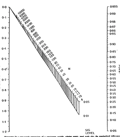
4.1.1 Sample size estimation 79
4.1.2 Significance level 79
4.1.3 Power 80
4.1.4 An estimate of the variability of the response variable 80
4.1.5 The clinically relevant difference in treatments 80
4.1.6 Calculating sample size 80
4 .2 M ethods 82
4.2.1 Patient selection 82
4.2.2 Ethical aspects, randomisation and administration 85
4.2.3 Techniques 85
4.2.3.1 Therapeutic 85
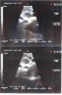
4.2.3.2 Endoluminal ultrasound 86
4.2.3.4 Follow-up 88
4.2.3.5 Statistical methods 88
4 .3 R esu lts 88
4.3.1 Hospital stay 88
4.3.2 Dysphagia 88
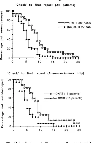
4.3.3 'Dysphagia controlled interval' and 'treatment interval' 89
4.3.4 Endoluminal ultrasound 93
4.3.5 Survival 102
4.3.6 Complications and Intubation 105
4.3.7 Patients with bulky Tumours and Métastasés 106
CHAPTER 5 : A QUALITY OF LIFE ANALYSIS IN PATIENTS RANDOMISED TO LASER OR LASER AND RADIOTHERAPY FOR
OESOPHAGEAL CANCER
5 .1 Aims and rationale 111
5 .2 M ethods 112
5.2.1 Prospective evaluation of quality of life 112
5.2.2 Retrospective evaluations 115
5.2.3 Statistical analysis 116
5 .4 R esu lts 116
5.4.1 The effect of radiotherapy 116
5.4.2 Correlation between quality of life measures 119
5.4 3 Correlation between quality of life measures and DG 122
5.4.4 Change in quality of life with time 124
5 .5 D iscussion 125
CHAPTER 6 AN ECONOMIC EVALUATION OF THE USE OF RADIOTHERAPY IN ADDITION TO LASER IN PALLIATION OF
MALIGNANT DYSPHAGIA
6 .1 B ackground and Rationale 128
6 .2 M ethods 129
6.2.1 Assessing resource use 130
6.2.1.1 Diagnostic resource use 130
6.2.1.2 Endoscopic procedures 130
6.2.1.3 External beam radiotherapy 130
6.2.1.4 Follow up and terminal/supportive care 131
6.2.2 Valuing resource use 131
6 .3 R esu lts 135
CHAPTER 7 : RADIATION ENHANCEMENT OF LASER
PALLIATION FOR ADVANCED RECTAL AND RECTOSIGMOID
CANŒR : A PILOT STUDY
7 .1 B ackground 139
7 .2 Patients and methods 140
7.2.1 Patient selection 140
7.2.2 Techniques 143
7.2.3 Patient follow up and evaluation 143
7.2.4 Statistical methods 144
7 .3 R esults 144
7.3.1 Endoscopic 149
7.3.2 Symptomatic 149
7.3.3 Treatment requirements 151
7.3.4 Survival and Complications 151
7 .4 D iscussion 154
SECTION C : ASSESSMENT OF ENDOPROSTHESES IN COMBINATION
WITH LASER /RADIOTHERAPY FOR OESOPHAGEAL MALIGNANCY
CHAPTER 8 : A CUFFED TUBE FOR PALLIATION OF COMPLICATIONS OF OESOPHAGEAL MALIGNANCY AND ITS TREATMENT WITH
LASER/RADIOTHERAPY
8 .1 B ackground 156
8 .2 M ethods 156
8 .3 R esults 159
8.3.1 Oesophago-respiratory Fistula 159
8.3.1.1 Patient details 159
8.3.1.2 Case Report A (oesophageal primary) 160
8.3.2 Oesophageal perforation/tear 163
8.3.2.1 Case report B 163
8.3.3 Group 3 Life threatening bleeding 165
8.3.3.1 Case report C 166
8 .4 D iscussion 168
CHAPTER 9 : RECANALISATION OF TUBE OVERGROWTH : AN
ADDITIONAL USE FOR LASER IN PATIENTS WHERE LASER AND/OR
RADIOTHERAPY HAS PREVIOUSLY FAILED
9 .1 Aims and rationale 172
9 .2 M ethods 174
9.2.1 Patients 174
9.2.2 Techniques 174
9.2.3 Statistics 175
9 .3 R esu lts 176
SECTION D
CHAPTER 10: SUMMARY AND DISCUSSION
1 0 .1 Palliation of oesophageal cancer 183
1 0 .2 Palliation of rectosigmoid cancer 186
10.3 Collaberation with radiotherapists 187
1 0 .4 The laser and tube - a complimentary duo 187
1 1 .5 The future 190
REFERENCES 192
APPENDICES 207
1 Oesophageal pilot study raw data
2 Oesophageal randomised study raw data
3 Oesophageal ultrasound data
4 Quality of life data
5 Cost data
6 Rectal cancer study raw data
7 Tube overgrowth raw data
8 Publications and reviews arising from the studies described in this thesis
9 Statement describing the Author’s individual contribution to the studies
in this thesis
LIST OF TABLES
Background
2.1 Details of all 'head to head' comparisons of Nd YAG laser and tube
2.2 Symptoms relieved and serious complications with Nd YAG laser
treatment for rectal cancers
2.3 Symptoms of advanced rectal cancer relieved with external beam
radiotherapy
Laser and external beam radiotherapy pilot study
3.1 Patient details
3.2 Dysphagia controlled interval (DCI) in weeks after 'check' endoscopy
3.3 Survival data according to subgroup
Laser and external beam radiotherapy randomised study
4.1 Demographic details of laser and laser with radiotherapy groups
4.2 Tumour details of laser and laser with radiotherapy groups
4.3 Frequency of therapeutic endoscopy required to control symptoms
according to histology and randomisation group
4.4 Endoluminal ultrasound data
4.5 Survival according to tumour extent at presentation
Quality of life analysis
5.1 QL Index before, during and immediately after radiotherapy
5.2 LAS A before, during and immediately after radiotherapy
5.3 Correlation between quality of life measures
5.4 Correlation between quality of life measures and dysphagia grades
Economie evaluation of laser v laser + DXRT
6.1 Unit costs of the endoscopic procedures used
6.2 Mean costs of laser and laser + 30Gy DXRT
Laser and external beam radiotherapy for rectal cancer
7.1 Endoscopic and symptom results
7.2 Treatment interval data in weeks before and after radiotherapy
7.3 Laser energy requirements before and after radiotherapy (excluding initial
recanalisation)
Use of cuffed oesophageal tubes
8.1 All patients with oesophagorespiratory fistulae
8.2 Patients treated for oesophageal perforation or tear 8.3 Patients treated for life-threatening bleeding
8.4 Published series of oesophageal prostheses for the treatment of fistulae
LIST OF FIGURES
Laser and external beam radiotherapy pilot study
3.1 Survival curves laser + 30 Gy and historical laser only controls
3.2 Survival curves laser + 30 Gy and laser + 40 Gy
Laser and external beam radiotherapy randomised study
4.1 Nomogram for sample size calculation
4.2 Illustration of Aloka curved linear array probe
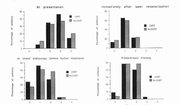
4.3 Dysphagia grades with time for all patients
4.4 Initial dysphagia controlled interval for all patients and according to
histology and trial arm
4.5 Endoluminal ultrasound images
4.6 Sequential endoluminal ultrasound images
4.7 Survival curves according to histology and trial arm
Quality of life assessments
5.1 QL index assessment form
5.2 LAS A assessment form
5.3 Correlation between quality of life measures
5.4 Correlation between quality of life measures and dysphagia grades
Economic Evaluation
6.1-6.3 Important costings for resource use evaluation
6.4 Unit costs of endoscopic procedures
6.5 Oveall costing of laser v laser + DXRT trial
7.2 Results of laser followed by radiotherapy for patients with rectal
bleeding
7.3 Results of laser followed by radiotherapy for patients with diarrhoea
8.1 A 10.4cm (shaft) Wilson -Cook cuffed tube with the cuff inflated and
deflated ready for insertion.
8.2 An example of an oesophagorespiratory fistula.
8.3 A contrast swallow to show a cuffed tube in situ in the oesophagus
9.1 A polypoid tumour overgrowing a Celestin tube before and after laser
9.2 A second example of a tube overgrowth
9.3 A third tube overgrowth before and after laser including a contrast swallow before treatment
ABBREVIATIONS
DCI Dysphagia controlled interval
DXRT Deep external beam radiotherapy
LASA Linear analogue self assessment
MP Monopolar electrocoagulation
Nd YAG Neodymium Yttrium Aluminium Garnet
QL-index Quality of life index (Spitzer 1981)
CHAPTER 1
B ACKGROIJND TO I.ASER ENDOSCOPY
1.1 The history of cautery
The application of heat for cauterization of tumours is ancient, the oldest reference
being in the Edwin Smith surgical Papyrus which has been carbon dated to 1700
b.c. The original manuscript was probably produced 1000 years earlier. At that time
the Egyptians used the hot iron for treatment of breast cancers. References to
cautery for tumour destroying properties can be found again from the 6th century
onwards but the technique did not advance until the 1940's when high frequency
diathermy was introduced. Direct application of heat by a pre-heated iron instrument
was replaced by the direct generation of heat by electric current. It was not until the
1970's that probes which could be introduced either along side an endoscope or
down the instrument channel opened up the gastrointestinal tract for electrocoagulation. At about the same time fibres became available which would
allow transmission of laser light and thus both endoscopic electrocoagulation and
laser therapy was possible.
1.2 Tissue effects of heat
Raising the temperature of living tissue to around 60 degrees Centigrade results in desiccation, contraction and protein coagulation, at 100 degrees Centigrade water
boils causing cells to explode and at higher temperatures the tissue becomes
carbonized and subsequently Vapourises (Cummings 1983). Coagulation is an
irreversible dénaturation or conformational change of structural proteins and
enzymes. It is readily observed macroscopically as whitening of tissue. A detailed
histological study of the coagulative change produced by the Nd YAG laser in dog
stomach (Kelly 1983) showed initial hyperaemia and oedema followed by thermal
contraction with larger amounts of energy. Contraction was thought to be the initial
mechanism of haemostasis rather than thrombus formation within vessels which
only occurred as a secondary phenomenon. There are several endoscopic devices
designed to induce coagulation of tissue but vaporisation can only be safely
achieved with the non-contact technique using laser light.
1.3 Endoscopic thermal methods
1.3.1 Monopolar electrocoagulation
Diathermy is a crude form of monopolar electrocoagulation. An electric current is
passed through tissue from an electrode (in this case diathermy forceps) to a metal plate usually attached to the patients leg, this causes heat generation within the
tissue. The current is rapidly alternating preventing depolarisation of cells which
would occur with a direct current. Current density and thus heating is maximal at
the point where the electrode touches tissue and the effect rapidly falls off with
distance from the electrode as the current is distributed through increasing volumes
of tissue. At open operation quite extensive damage occurs to the tissue adjacent to the point of diathermy which could be dangerous in a thin walled organ.
The simplest endoscopic device consists of a narrow electrode which can be passed
down the biopsy channel of an endoscope and pressed against the tissue to be
coagulated. It is connected to an electrosurgical generator and a radiofrequency
current (> 300,000Hz) is passed between the electrode and a plate strapped to the
patients leg. The inactivated probe is applied and then activated, a pre set quantity of
energy being delivered and the procedure is then repeated at multiple sites. Good
contact between the probe and tissue is essential for the passage of current and this results in adherence to coagulated tissue. The monopolar probe is thus difficult to
use in bleeding because the coagulum is often tom of with removal of the probe. To
overcome this problem 'liquid' electrodes were developed which use water or saline
between the electrode and tissue. This technique, known as 'wet'
electrocoagulation, gives better electrical contact and minimises tissue adherence.
affecting the extent of coagulation. The distance between the ground plate and probe
varies between individuals as does the electrical conductivity of the tissue. Most
importantly the contact surface between probe and tissue varies. The current density is therefore unpredictable and the risk of excessive tissue damage leading to
perforation has limited clinical use. Treatment of rectal cancer is however safe as the
rectum is retroperitoneal and thus full thickness damage or small perforations are
unlikely to have important clinical consequences. Most authors advise general or
caudal anaesthesia and treatment is applied via an operating sigmoidoscope. The
treatment technique itself varies between centres. An electrocoagulator with a spherical electrode 4 mm in diameter at the tip is commonly used but a needle point
electrode which will induce deeper necrosis is an alternative. The sphere is applied
to the tumour to be treated and electric current is applied for a few seconds until the
tissue treated begins to boil. Fulguration (holding the tip of the electrode over the
tumour and destroying the surface by sparking) is avoided by most groups. This practice causes carbonization of the surface and tissue damage is superficial. Some
authors treat the tumour surface only once on each occasion (Hoekstra 1985) but
others (Madden 1983) prefer to clear the coagulum and retreat up to 5 or 6 times at
the same session until all tumour has been coagulated, often treating down to the
extrarectal fat.
Loop diathermy or endoscopic transanal resection of rectal cancer (ETAR) can be
achieved with a urological resectoscope which is a monopolar probe in the form of a
'loop'. This technique is very similar to transurethral resection of the prostate.
Restriction to oral fluids for 24 hours prior to operation and a phosphate enema is all
the preparation that is required. The procedure is performed under spinal or general
anaesthetic in the lithotomy position. The resectoscope is inserted and the resection
performed with a continuous infusion of 1.5% glycine. Resection is deeper , the
limits being judged by the appearance of circular or longitudinal muscle fibres.
1.3.2 Bipolar electrocoagulation
These probes were introduced in an attempt to minimise the risk of full thickness
damage seen with monopolar probes. The positive and negative poles are located
close to each other on the operating probe. This dispenses with the need for a plate
or dispersive electrode. Multiple electrodes can be incorporated into a single device.
There are usually 2 or 3 pairs (BICAP) of bipolar electrodes allowing diathermy
with tip angulation. The probes come in various sizes early probes being 2.3mm
diameter and later ones 3.2mm or 3.4mm diameter, with a central irrigation
channel. Early power sources deliver 25 watts, the later units 50 watts. The
technique for coagulation is identical to that employed with the monopolar and
heater probes. Large tumour probes can be inserted alongside the endoscope.
1.3.3 Heater probe
The heater probe was developed in 1978 (Protell). It was designed to apply
pressure and heat simultaneously to a bleeding vessel. The probe comprises a
hollow aluminium cylinder with an inner heater coil and an outer coating of non
stick teflon. It also contains a separate thermocouple element in the tip to measure
its temperature and can be heated to a maximum of 250 degrees C. The probe
temperature is maintained until a preset amount of energy is dehvered. There is also
a proximal irrigation port to allow washing of the target even when the probe is
forcibly applied to tissue. The technique was not designed for use in cancers although it could potentially be used for this application.
1.4 The development of lasers
The development of lasers was dependent on our modem understanding of the dual
1.4.1 Light Theories
In the 17th Century these properties were explained by two different theories. Isaac
Newton postulated a corpuscular theory which envisaged light as a stream of
particles and Robert Hooke and Christian Huygens considered light as a wave
propagating through an all-pervading elastic medium they called the 'aether'.
Newton felt that the wave theory could explain neither the rectilinear propagation of
light nor polarisation and his view prevailed until the early 19th Century. Young
and Fresnel then revived the wave theory after work on interference and diffraction
of polarised light. Young was able to show that light does bend into shadows, if only by very small degrees and he suggested that light was a transverse wave, the
medium through which light travelled being disturbed in a direction perpendicular
to the direction of propagation. The speed of light was first measured by Fizeau in 1849 and was found to be less in water than air. Newton had predicted the opposite
and the corpuscular theory fell from grace. In 1867 Maxwell generated a set of
equations which described many properties of electricity and magnetism exactly. He
was able to calculate that the speed of propagation of a disturbance in an
electromagnetic field was equal to the speed of hght and concluded that hght was an
electromagnetic wave. Subsequently Hertz confirmed this theory by demonstrating
that long wavelength non-visible electromagnetic waves could be refracted in
exactly the same way as hght.
Electromagnetic wave theory however does not explain all the properties of light. In
particular there were problems explaining the emission and absorption of light.
Classical wave theory predicted that the intensity of radiation emitted should
increase with decreasing wavelength, this would result in emission of light of
infinite intensity as the wavelength decreased into the ultraviolet. This 'ultraviolet
catastrophe' does not occur in practice. Max Plank provided the basis of an
explanation. He proposed that energy is imparted into the electromagnetic field in
finite quantities or quanta and not in a continuous fashion. Another phenomenon
observed by Hertz could not be explained by wave theory. He noticed that a spark
was more readily formed between two electrodes when they were illuninated with
ultraviolet light. Further study showed that the light prompted release of electrons
from the metallic surface of the cathode. This phenomenon is the 'photoelectric
effect.' Contrary to expectation of the wave theory this effect persisted even at very
low light intensities, there was no threshold below which it could not be observed.
Einstein subsequently provided an explanation for the 'photoelectric effect' using
Planks ideas. He treated light as being made up of discrete packets of energy or
'photons', the energy of each photon being inversely proportional to the wavelength of the light. Ultraviolet light has a short wavelength with photons of
high energy, even at low intensity the energy of each photon imparts enough energy
to knock an electron off the surface of a metallic cathode and reduce the voltage
required to initiate a spark. It was for this explanation of the 'photoelectric effect'
and his application of the quantum theory to light that Einstein was awarded the Nobel prize for Physics in 1921.
The concept of the photon whereby light can take the form of both a particle and a
wave, formed the basis of the quantum theory of matter. It had therefore become
apparent that both corpuscular and wave theory were required to explain all the
properties of light; the propagation of light is best explained using the
electromagnetic wave theory.
1.4.2 Atomic Structure
Frauenhofer (1797-1826) showed that the wavelength of light emitted or absorbed
by a particular element was confined to a number of narrow bands particular to the
element. Rutherford (1871-1937) postulated that the atom was comparable to the
solar system with electrons orbiting the nucleus as the planets orbit the sun. In 1913
Bohr used the new quantum theory to update the atom model. He argued that
Electrons in orbit were postulated to contain a 'quanta' of energy which was
smallest for orbits of small radius and largest for orbits of large radius. A specific
quanta of energy would be required to move the electron from an orbit with a low
energy state to an orbit with a high energy state or larger radius. The new theory
accurately predicted the wavelengths of the emission spectra of hydrogen.
1.4.3 Spontaneous emission of radiation
The quantum of energy required to move an electron to a higher energy state must
come from absorbing external energy such as electricity or light and similarly when an electron falls from a high energy level to a lower one there is a spontaneous
emission of in the form of a photon of light. The energy of the photon which is
spontaneously emitted is equal to the difference between the two energy levels. The
wavelength of the light emitted is inversely proportional to its energy and thus light
of a certain wavelength is emitted. There may be many potential energy states for a
given electron in an atom and thus several given wavelengths may be emitted from a given atom. The emission and absorption of light is best considered using the
photon and quantum theory of light.
1.4.4 Stimulated emission of radiation.
Einstein predicted in 1917 that emission of a photon from an excited electron within
the atom could be stimulated by another photon. The stimulated photon would
travel in the same direction as the stimulating photon and would be of equal energy,
wavelength, phase and polarity. The stimulating photon would remain unchanged.
It was later realised that it may be possible to start a chain reaction whereby the
stimulated photon subsequently stimulated emission of other photons from other
excited atoms so that the photon flux was amplified. An essential pre-requisite for
this process would be the the presence of more excited than ground state orbiting
electrons. Unless this applies then photons would simply be absorbed. This situation is called a 'population inversion' and was first achieved by Townes of
Columbia University in 1953. He produced a device which produced
electromagnetic waves from the microwave part of the spectrum and was termed a
'MASER' (microwave amplication by stimulated emission of radiation). In 1958
Townes went on to define the prerequisites for construction of the first 'LASER'
(Light amplification by stimulated emission of radiation).
1.4.5 The optical resonator
Population inversion is difficult to achieve and this explains why the amplification
of stimulated emission does not occur easily. It can now be obtained in a variety of
substances including solids, liquids and gases in atomic, ionic and molecular forms
and is achieved by pumping with energy usually as light or electricity. However in
addition to a medium capable of stimulated emission of radiation the other essential component of a laser is an optical resonator. This consists of two mirrors facing
each other so that multiple reflections can occur between them. The mirrors are
positioned at a distance equivalent to an integral number of half wavelengths apart
so that constructive interference occurs. This results in resonance or the production
of a standing wave.
1.4.6 Maiman’s Rubv Laser
The first laser was constructed by Maiman in 1960. The medium was a 1 cm crystal
of synthetic ruby on which 2 parallel faces were polished and coated with silver to
form 2 mirrors facing each other to form the optical resonator. The chromium ions
of ruby were excited using a high power flash lamp to achieve a highly unstable
state which quickly decays to a semistable state and it is with this state that the
population inversion occurs. When the semistable state returns to ground state
emission of a photon occurs at a wavelength of 694 nm. This photon can then
stimulate emission of other identical photons and so on. One of the mirrors is only
partially silvered so that 5% of the light escapes through it in the form of an intense
1.4.7 Laser light : Monochromatic. Coherent and Colliinated
These are the special characteristics of laser light which follow directly from the
principals discussed. Many lasing mediums can emit light with a number of discrete
wavelengths. Unwanted wavelengths can be blocked by coating the reflective
mirrors to block reflection of unwanted bands. Thus laser light can be
monochromatic. The beam is also at the same phase at any given point (coherent)
and is non-divergent (collimated) and the spot size thus remains fairly constant over large distances.
1.5 Optical Fibres
The development of optical fibres capable of transmitting laser light was an essential
prerequisite for the use of lasers in gastroenterology. These fibres are elongated
glass rods of narrow diameter which are coated. Transmission of light along an
optical fibre occurs due to total internal reflection which the coating facilitates. Light travelling along a quartz (glass) fibre which strikes the interface at an angle less than
the critical angle is totally internally reflected. Multiple such reflections occur until
the light emerges at the far end of the fibre. Laser light is coupled to the fibre by
focusing it onto its proximal end. Each photon undergoes different reflections
within the fibre and thus the beam is neither coherent nor collimated when it
emerges. This has clinical implications as the emerging beam is divergent and thus
the power density falls with increasing distance from the tip. Quartz fibres used in
gastroenterology can transmit light in the wavelength range 380-1300nm.
1.6 L asers in Gastroenterology
1.6.1 The Argon Laser
This was the first laser to find extensive clinical use and the first to be used in
gastroenterology. It is a gaseous ion laser which is one of the most inefficient of the
lasers. This is because the lasing medium is a gas which normally exists in its
atomic state. High currents are required to convert the atoms into ions (by removal
of an electron). The argon ions are then excited further to obtain a population
inversion. Typically 5 kilowatts input power is required for a laser output of 5
watts. The high power required for these lasers places constraints on the materials
required for construction of the discharge tube which is required to withstand high
temperatures. Argon lasers do offer outputs at a number of discrete wavelengths as
there are several energy levels that excited electrons can fall to. A typical output wavelength for these lasers is 514 or 488 nm. These wavelengths are in the green
and blue regions of the visible spectrum and are highly absorbed in vascular tissue
and thus protein coagulation can readily be achieved at low powers of a few watts.
1.6.2 The neodymium yttrium aluminium garnet (Nd YAG) laser
This is also an ion laser but is not normally described as such as the neodymium
(Nd) ions are locked into particular positions by a crystalline host structure. It is
thus a ’solid state laser’. The most satisfactory host is the crystal yttrium aluminium
garnet (YAG). The laser medium is formed as a rod 10cm in length. Pumping or
excitation is achieved by the absorption of incoherent light from a bright incoherent
source which is a krypton arc lamp. This lamp is chosen as a larger proportion of
its broadband output falls within the narrow band of wavelengths that neodymium
ions can absorb. Using such a lamp this laser can achieve an efficiency of 1-2%.
This is 10-20 times more efficient than the argon laser. The lamp is also far cheaper
to replace than the argon laser discharge tube. The Nd-YAG laser operates in the
infrared at 1.06 micrometers. This wavelength is only weakly absorbed in tissue
endoscopie use and thus the input power of around 5 killowatts is similar to that required for the argon laser.
1.7 Laser-tissue interactions
Lasers are sophisticated light sources which can deliver energy with great precision. The delivery of energy to tissue can thus be readily controlled. Within tissue the
light can be reflected, transmitted, scattered or absorbed. Biological effects are due
to the absorption of light but the other factors determine where the light is
disseminated and subsequently absorbed. The absorption characteristics of soft
tissues vary enormously between wavelengths giving rise to quite different effects,
however studies have shown that the extent of damage depends closely on the
energy dissipated (Bown 1980 and 1983). The tissue effects of heat have been discussed in section 1.2. In summary heating initially causes thermal contraction
and protein coagulation, as tissue shrinks small vessels are sealed, thrombosis then
occurs as a secondary effect. If the volume heated is large vessels up to 1mm in diameter can be sealed (Kelly 1983). If further energy is dissipated carbonization
and subsequently vaporisation occurs. At higher Nd YAG laser powers several of
these effects can be seen in the same piece of tissue, vaporisation occurs
immediately below the beam, deeper there is necrosis with subsequent sloughing
and scarring. The extent of different tissue effects can be varied by altering
treatment parameters. The Nd YAG wavelength is poorly absorbed by tissue and
more of the light is transmitted deeper into the tissue. The Nd YAG laser has a
greater power output, and with deeper tissue penetration it is not surprising that
superior haemostatic efficiency has been documented in experimental studies than
the argon laser (Bown 1980). The bluegreen argon laser light is readily absorbed by
vascular tissue and its effects are relatively superficial.
None of the other techniques for coagulation offers the precision of the laser
however this may not be necessary for all indications for endoscopic therapy. An
important advantage of the Nd YAG laser over other techniques is the ability to
vaporise tumour with rapid removal of tumour bulk. Many groups have reported
excellent results with this laser (chapter 2).
1.8 Therapeutic techniques with Nd YAG laser
1.8.1 Oesophageal cancer
Sedation and analgesia are achieved with diazemuls/pethidine. A designated laser
endoscope with a large diameter working channel is used, and we find the Olympus
IT 20 (Keymed Ltd, Southend) ideal for this application. It is specially adapted with
a safety filter in the eyepiece. We use a 'Flexilase' Nd:YAG laser (Living
Technology Glasgow) which can generate up to lOOW at a wavelength of 1064nm.
It is usually set at 50-70W with a pulse duration of 1 second for treatment of
oesophageal cancers. Its output is focused onto the end of a 0.6mm quartz fibre contained in a 2.2mm teflon catheter which stiffens the fibre and allows insertion
down the working channel of the endoscope. It also allows a co-axial stream of
carbon dioxide or air to be passed which keeps the fibre tip clean and cool and clears the target of blood and debris. The fibre is three metres in length and its distal end is
protected by a metal guard. An aiming beam is provided by a low power helium-
neon laser which is coupled into the beam path of the Nd YAG. We use a 'non-
contact' technique, shaving back exophytic nodules by vaporisation, and treating flat
areas by coagulation alone. Insufflated gas and smoke generated by vaporisation is
vented by connecting the working channel of the endoscope to an underwater drain
using a two way valve which can be closed when endoscopic suction is required. If
the procedure lasts more than thirty minutes the scope is removed in order to clean
debris from the end, to flush out the suction channel and check patency of the air and
water channels. This practice reduces the risk of scope blockage and ensures a better
view as the gradual deterioration in vision as smoke and debris adheres to the end of
the scope is often not fully appreciated. Necrosed tumour sloughs to a depth of 2-
period if necessary. Laser treatment is only possible in tumours with exophytic
(intraluminal) component; however most tumours come into this category at
presentation. Luminal narrowing due to compression by extrinsic tumour is usually
dealt with by intubation. Where possible treatment is commenced at the distal tumour
margin and proceeds in a retrograde fashion. This avoids the problem of oedema in
tissue adjacent to that vaporised limiting forward progress and the lumen is always
visualised thus reducing the risk of perforation. Impassable strictures are normally
dilated prior to laser treatment so that retrograde tumour destruction can be
performed. Occasionally a guide wire cannot be passed and only in these
circumstances is laser treatment carried out in a prograde (forward) direction.
1.8.2 Rectosigmoid tumours
A sodium phosphate enema is usually adequate preparation for these lesions, more
proximal lesions requiring a rectal washout or occasionally a full bowel preparation.
Sedation and analgesia are often not necessary but diazemuls and/or pethidine are administered intravenously if required. Access is obtained using a flexible
sigmoidoscope or colonoscope with a safety filter in the eyepiece. Air is insufflated
and the tumour is washed as necessary to obtain a good view and document the
extent of disease as far as possible. The basic instrumentation and technique is
exactly as that for treating oesophageal cancers however higher energy treatments are
sometimes necessary. Carbon dioxide may be used for insufflation of the bowel as
this is partially absorbed and thus reduces the risk of excessive bowel distension.
Whenever possible treatment is started at the proximal margin of the stricture and
proceeds distally towards the anus as oedema in areas of coagulated tumour can hinder progress forward. Some authors (Brunetaud 1987) advocate an argon laser to
treat small lesions up to 4 cm from the anal verge as the Nd YAG laser with its
greater power output and tissue penetration can be painful.
1.8.3 Difficulties with Non-contact Nd YAG laser for cancers
There are three main problems.
1) Contamination of the fibre tip with blood or debris results in excessive heating of
the tip with consequent fibre destruction (fibre 'bum'). In order to minimise this a
continuous gas flow is required to keep the tip clean. The flow causes bowel
distension which can be uncomfortable.
2) Smoke generated with vaporisation obscures the view until vented, and settles on
the endoscope lens in a film, causing a progressive deterioration in visual access and
clarity.
3) High power, necessary for tumour vaporisation, can cause a sensation of 'heat'
which is distressing to some patients.
Several techniques are employed to minimise these difficulties. Small areas are vaporised at a time allowing heat to dissipate and smoke to vent between shots. If
debris is seen on the tip or if the aiming beam is diminished the fibre is removed for
CHAPTER 2
REVIEW OF LASER ENDOSCOPY FOR CANCER AND COMPARISON WITH
OTHER THERAPIES
2.1 OesoDhayoyastric cancer
2.1.1 Introduction
The incidence of carcinoma of the cardia is rising (Cheng and day 1992). A recent
study in Oxfordshire (Rios-Castellanos 1992) found a rate of cardia cancer of 5.2
per 100,000 per year, this had doubled over a 20 year period. This increase is
reflected in the overall figures for oesophageal cancer (carcinoma of the cardia and
true, squamous cell oesophageal cancer). In 1980 Earlam found a rate of 8 deaths
per 100,000 of the UK population per year. More recently the WHO estimates that
oesophageal cancer is responsible for about 10 deaths per 100,000 people annually in the United Kingdom (WHO 1988). These tumours most commonly occur in the
elderly and present late in their natural history as dysphagia usually does not occur
until two-thirds of the oesophageal circumference is involved. Overall at least 60%
of all patients are unsuitable for curative treatments (Watson 1988, Desa 1988). The
historical long term results for curative surgery and radiotherapy are poor.
Earlham’s comprehensive review of 1980 found a 5 year survival of 4% and 6%
respectively for surgery and radiotherapy for squamous cell cancers.
Adenocarcinoma of the cardia has an even poorer prognosis. This review was
however historical and retrospective and unrepresentative of the current results in
specialist units. There is no doubt that better selection of cases treated in such units
gives more satisfactory results. More recent surgical series from the UK indicate an
operability rate of 20-40% with a five year survival of 10-15% in those who
underwent surgery. (Skinner 1986,Watson 1988, Desa 1988). An up to date study
study in gastric cancer (Sue-Ling 1993) shows better results with 53 % of patients
undegoing curative resection during 1985-9 and 5 year survival in such patients is
probably aroung 70%.
2.1.2 Staging oesophageal cancer
Careful staging has demonstrated its crucial importance on prognosis. Eastern
surgical series of patients with early lesions have reported five year survival rates of up to 96% (Akiyama 1981, Huang 1981). In the Western world tumours are
usually advanced at the time of presentation as screening is not generally
undertaken. In large series there are however a significant number of patients with
early disease and stratification of patients for stage can reveal surprisingly good
results. Watson (1988) noted that five year survival in patients with squamous
oesophageal cancer found to be node negative was 50% and this increased to 75% for patients with superficial lesions. Even better results are reported for early stage
gastric cancer (Sue-Ling 1993) with up to 90% 5 year survival. Looking at data
from several studies Rankin and Mason point out in a recent review on staging
(1992) that tumours restricted to the oesophageal wall have a five year survival of
40% compared to 4% for those which have progressed beyond the adventitia. Node
negative patients also have a reasonable outlook with five year survival rates of
42% but for those with nodes the figure is 3%. Accurate staging is therefore
essential in planning patient management. The finding of a tumour with a low
chance of cure does not exclude surgery as primary treatment but at least it is clear
from the outset that palliation is the main treatment aim.
Current staging is based on the TNM system. The important criteria for any
imaging technique are to determine are.
1) The depth of penetration through the oesophageal wall.
2) Direct tumour invasion into adjacent structures.
4) Distant métastasés
CT scanning has been the mainstay of staging until the recent introduction of
endoluminal ultrasound which is still only available in a few units in the UK.
However CT staging alone of gastro-oesophageal cancer is unsatisfactory
(Thompson 1983, Becker 1986). A detailed examination from several studies of CT
compared to surgical/pathological staging shows the overall accuracy to be only 39-
68% (Rankin 1992). Although CT visualises the intrathoracic oesophagus well,
stage 1 and stage 2 (invading muscularis propria) tumours cannot be differentiated
and small nodal métastasés are frequently missed resulting in understaging.
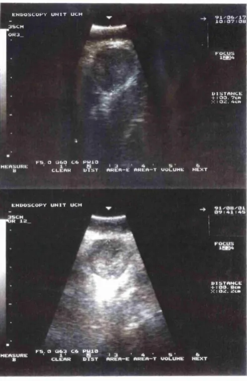
Endoluminal ultrasound uses a modified endoscope with an ultrasound probe at its tip. The probe frequency is usually 7.5 MHz which gives a depth of field of 5-
7cm; lower frequencies will give a deeper field depth but close up definition (of the
oesophageal wall) is less detailed (Botet 1991). The instrument is passed through
the tumour into stomach and scanning performed as it is withdrawn. Full examination is not possible in all patients (Tio 1990) as the probe will not always
pass the stricture. Using this technique excellent oesophageal wall images can be
obtained. There are 5 alternating hyper and hypoechoic layers and distortion of the
pattern gives the depth of tumour penetration. The characteristics of the nodes are
used to define malignant involvement (unlike CT where node size is important).
Elliptical, hypoechoic nodes with clearly deliniated margins suggest malignancy.
Rankin (1992) reviewed several studies comparing this technique with the resected
specimen and found an accuracy of 59-92% for tumour penetration and 69-88% for
mediastinal lymph nodes. Diagnosis of infiltration into invading structures is also
highly accurate (Tio 1990).
In studies which compare the two techniques (Tio 1990, Botet 1991, Tytgat 1991,
Siewert 1990) the endoluminal ultrasound is better for depth of tumour invasion
(92%v 60%) and nodal métastasés (88%v74%) but CT scanning better for distant
métastasés (90%v 70%). In preoperative staging the overall accuracy for both
techniques (86%) is better than for each alone. However for monitoring local
response to treatment such as radiotherapy or chemotherapy the ultrasound
technique should be superior.
CT and endoluminal ultrasound are complex and expensive and may not be
necessary. Rankin and Mason (1992) suggest that around 50% of patients can be
staged without undertaking such tests. Routine investigations may be enough to stage diseased adequately. Trans-abdominal ultrasound is an excellent technique for
identifying liver métastasés which can be confirmed by biopsy or cytology if
required. Tumours found to be longer than 5cm at endoscopy have
extraoesophageal spread in 90% of cases. Laparoscopy may also be useful in staging of cardia cancer (Watt 1989). Some patients have local involvement of neck
nodes and a trans cutaneous biopsy may confirm stage 4 disease in such patients.
2.1.3 Techniques for palliation
Using the figures quoted above 60% of the 5,0(X) new cases (3,(X)0 patients) of
oesophageal and cardia cancer are suitable for palliation only. Palliative resection
restores normal swallowing in 90% of patients who survive the procedure and
confers a longer survival than the other palliative procedures available, but carries a
high morbidity and a mortality of up to 30% (Watson 1982) although this figure
continues to fall with better patient selection and better perioperative care.
Many other treatment modalities are now available for palliation of this difficult
condition, including Nd YAG laser therapy, intubation with prosthetic tubes and
radiotherapy by external and intracavitary (brachytherapy) methods. Individually
there is good evidence for the efficacy of these modalities, but all have their
swallowing achieved, which is a key factor in overall quality of life (Barr 1990a,
Loizou 1991), and the price paid to achieve that quality (procedure related
complications, time in hospital for procedures, number of procedures).
Most of the studies of palliative treatment for oesophageal cancer have been
uncontrolled, and some have studied small numbers of patients. Many only
examine one treatment modality but even when treatments have been compared
patients have often not been randomised and historical controls are frequently used. This makes interpretation of results difficult. This review concentrates on the larger
more recent studies and those which have compared the important treatment
modalities.
2.1.3.1 In tubation
Surgical intubation procedures carry a high morbidity and mortality (Watson 1982,
Urschel 1991) and have been largely replaced by endoscopic methods which are
safer and equally effective (Ogilvie 1982). Despite this improvement there is a risk of procedure related perforation of 5-10% (Ogilvie 1982, Gasparri 1987, Tytgat
1986, Barr 1990, Loizou 1991a). Mortality rates are usually less than 10% but
figures as high as 31% have been reported (Diamantes 1983). Such a high rate to
some extent reflects the grave condition of many of these patients; however rates
this high have not been recorded with other palliative treatments. Potentially serious
long term complications are reported in all series and can occur in anywhere up to
50% of patients. They include tube displacement, blockage, overgrowth and late
perforation. These rather disappointing figures for morbidity and mortality with
endoscopic intubation have generated interest in endoscopic laser therapy. The other
important reason for such interest is that tubes do not fully restore swallowing,
most patients will only manage a semi-solid diet and some will not manage more
than fluids.
2.1.3.2 Nd YAG L aser
The use of the Nd YAG laser for recanalisation of oesophageal cancers was first
reported in 1982 and further details published in 1983 (Fleischer). Other groups
confirmed that this treatment was effective and safe (Krasner 1987, Bown 1987), and there is now considerable worldwide experience. A multicentre inquiry (Ell
1987) on 1359 patients showed initial success in 83% of patients and a serious
complication rate of 4% of which around half were perforations. An early study
from our unit (Bown 1987) was representative of the general experience. Around
three-quarters of 34 patients treated were able to manage at least some solid foods
after a course of treatment although most of those who survived more than a short
period needed to return for regular repeat treatments every 5 weeks or so. This has
led some authors to recommend regular repeat treatments regardless of symptoms (Krasner 1987, Barr 1990).
Others have tried to identify groups of patients who are more likely to do well with
laser. Fleischer (1985) suggested that patients with short lesions (<5cm) in a
straight segment of the mid and distal oesophagus were likely to do best. He found that those with cervical tumours did not respond well with laser. We have achieved
better results with laser in these patients but when laser fails the problem can be
successfully addressed with a modified oesophageal prosthesis (Loizou 1992).
Fleischer also suggested that patients with soft non-constricting circumferential
tumours which would not hold a prosthesis well were good candidates for laser. In
contrast obstruction secondary to extrinsic compression or infiltrating tumour and
long stenotic lesions were felt to be best treated with intubation. Naveau (1990)
expanded on this theme. Patients with adenocarcinomas, an initial length < 6 cm
and those who improved after initial laser treatment were all independently
correlated with longer 'symptom improvement duration'. The authors concluded
that patients with squamous cell cancers longer than 6 cm should be entered into
trials of other palliative techniques. In a larger more recent series (Mason 1991),
endoscopy and the authors policy was to initially treat all patients with laser
subsequently intubating those who do badly. A subsequent study from the same
unit showed that survival was better in patients with less severe dysphagia at
presentation (Derodra 1992). Our own policy is similar to that of that of Mason. If
there is polypoid tumour treatable with laser we try this approach first unless the
prognosis of the patient is felt to be very poor (Loizou 1991).
In summary laser treatment for cancer of the oesophagus and gastric cardia offers
rapid relief of dysphagia, can be performed as an out patient procedure if the
patients general condition allows, does not have systemic effects and serious complications occur in less than 5% of patients. The major drawback is the need for regular repeat treatments every 5 weeks or so to maintain adequate swallowing
2.1.3.3 Bipolar electrocoagulation
Johnson first reported use of the BICAP probe (3 pairs of bipolar electrodes) for recanalisation of oesophageal tumours in 1987. A special tumour probe was used
which is inserted into the oesophagus separately from the endoscope. Only 20 patients were treated and 4 of these developed serious complications including late
bleeding and fistula formation. Further evaluation of this probe was performed by
Jenson in 1988. Fourteen patients were treated with laser and 14 with BICAP and
results compared. Both modalities were effective at relieving dysphagia (86%
patients improved). One patient in the BICAP group developed a fistula but no
serious complications were seen with laser. The authors concluded that BICAP was
equally good for circumferential tumours but that laser was safer for exophytic non-
circumferential tumours because it could be directed endoscopically. A small study
on 30 patients comparing BICAP with oesophageal prosthesis (McIntyre 1989)
demonstrated no benefit in terms of swallowing for the probe. Complication rates
were similar, there were 2 perforations during dilatation (one in each group), one
patient perforated on tube insertion and one patient developed an
tracheooesophageal stricture after probe treatment. Patients treated with the probe
needed repeat sessions every 28 days. These studies are rather small to make a
final judgement but the relatively high rate of serious complications encountered
with this tumour probe is worrying.
2.1.3.4 Alcohol injection
Encouraging results have been reported for injection sclerotherapy with absolute
alcohol (Payne James 1990). Alcohol causes tissue death by desiccation. The
treated area subsequently sloughs to leave a recanalised lumen. The relief of
dysphagia was similar to that achieved with the Nd YAG laser and the technique appeared safe. The ability to vaporise polypoid tumour with laser cannot however
be achieved and it is likely that dysphagia is not relieved as rapidly in many
patients. Studies on alcohol injection into liver deposits undertaken at UCH
demonstrate the difficulty in controlling the distribution of injected alcohol. The extent of tissue damage is accordingly difficult to predict and extensive damage may
conceivable increase the risk of complications such as delayed fistula formation or perforation.
2.1.3.5 Comparative studies of laser and tube
At the time of writing there have been 8 studies directly comparing laser and tube.
These are documented in table 2.1 Four (Carter 1986, Buset 1987, Hahl 1991,
Loizou 1991a) were not randomised and the other 4 were (Barr 1990, Alderson
1990, Fuchs 1991, Carter 1992). Only six of the studies looked carefully at
dysphagia control (these studies looked at 253 patients in total) 4 showed laser to be
superior and 2 showed no difference. Most studies agree that the morbidity rates
with laser are substantially lower than with tube. The exact figures are given in the
table. The mortality associated with tube insertion is up to 11% (Hahl) and in the
patient who dies with laser treatment (2 patients in these series ; one in Aldersons
and one in Carter 1992).
Table 2.1 Details of all 'head to head' comparisons of Nd YAG laser
and tube
R a n d o m i s e d s t u d i e s
Patient number B est dysphagia Laser
relief com plications
Tube complicatons
Alderson 1990
Barr 1990
4 0
4 0
laser
equal
4 (20%) 3 perforations
1 bleed
2 ( 10%) 2 perforations
4(20% ) 1 perforation 2 displacements
1 overgrowth
12(60%) 4 bolus obstr 3 perforations 4 overgrowths 2 displacements
1 bleed
Fuchs 1991 4 0 equal 1(4%)
perforation
8(47%) 1 perforation 4 displacements
1 overgrowth 2 bolus obstr
Carter 1992
N o n r a n d o m i s e d
St l i d i e s
4 0 laser 5 (25%)
3 perforations 1 pneumonia
1 fistula
9 (45%) 3 bolus obst 1 overgrowth 4 pneumonia
1 failed
C artcrl986
Buset 1987
10 tube 10 laser
116 tube (14mm) 28 laser
laser
equal
4 (40%) 1 fistula 3 perforations
1 (4%) 1 perforation
5 (50%) 3 overgrowths 2 displacements
47 (40%) 9 perforations 4 haemorrhage 3 pneumonias 21 displacements
4 bolus obstr 3 overgrowth 2 oesophagitis 1 pressure necrosis
Loizou 1991
Hahl 1991
30 tube 43 laser
27 tube 69 laser
laser
equal
1 (2%) 1 perforation
6(9%) 4 perforation
2 sepsis
11 (37%) 4 perforations
7 bolus obstr/ displacem ents
13 (48%) 2 pefroration 2 massive bleed
6 bolus obstr/ displacem ents
The study from our own unit comparing laser with intubation (Loizou 1991a) has
given us a better insight into which patients are likely to benefit most, in terms of
quality of swallowing, from each of these modalities. Solids can be managed for
more than half the survival time in 1 in 3 of those treated with laser but only 1 in 10
of those intubated. In addition the proportion of patients managing fluids only is
double in patients intubated compared with patients receiving laser therapy (19% v 8%). In simple terms around 1/3 of these patients will swallow better with laser
than a tube for more than half the time. The results for tube indicate that more than
70% of those intubated will manage at least semi-solids. Overall the group receiving laser required more procedures (mean 4.6 v 1.4) but the risk of perforation was
high (13%).
Laser therapy is thus demonstrated capable of providing more effective and safer palliation than tube. The tube, however is a useful secondary therapy for patients
who do do not respond to laser or who are late laser failures. In our unit it is now standard policy to treat patients with inoperable tumours with laser as first line
treatment. Patients who are anorectic and in poor general condition who would not
undergo repeated endoscopic laser treatments easily are also advised to have a tube
as first line treatment.
2.1.3.6 External Beam Radiotherapy
External beam radiotherapy has been used extensively for many years in the
treatment of squamous cell carcinoma of the oesophagus. Most patients treated have
been considered unsuitable for surgery. Historically it has been used aggressively
with cure as the main aim. The hope was that such treatment would lead to
prolonged remission of the tumour and symptoms even if cure was not actually
achieved. An extensive review of radiotherapy for oesophageal cancer (Earlam 1980) however, revealed a one year survival of only 18% and a five year survival
of 6%, though selected cases do better. The results for radical surgery were similar.
Subsequently Earlam (1991) attempted to perform a multicentre randomised study
of radiotherapy and surgery for squamous cell cancer of the oesophagus but
unfortunately there was insufficient support from surgeons. His results for
radiotherapy (Earlam 1990) in patients with operable and inoperable squamous cell cancer of the oesophagus were impressive. One year survival in these groups was
46% an 16% and 5 year survival 14% and 4% respectively.
There is still some argument over the treatment of choice for patients with
squamous cell oesophageal cancer who are operable. Some would argue that
radiotherapy is as effective as surgery, others take the opposite view (Cuschieri,
Earlam, Khoury 1991).
A recent study from the Christie Hospital, Manchester, demonstrated a five year
survival of 16% in patients with cancer of the upper third of the oesophagus (Slevin
and Stout 1989). However earlier series have found up to 20% of patients initially
enroled for curative treatment are unable to tolerate a radical course of radiotherapy
(Wara et al 1976).
In the last 10 years the importance of palliation of symptoms has been
acknowledged and subsequent studies (of all modalities) have often considered
relief of dysphagia as well as survival times. Despite this change in emphasis there
have been relatively few publications looking at palliation of dysphagia with
radiotherapy alone. One early study did, however, note that external beam
radiotherapy usually relieves dysphagia slowly, often taking several weeks for
maximal effect and only half the patients respond (Pearson 1978).
Two studies (Mellow 1984, Karlin 1987) prospectively compared laser treated
studies were small and both focussed on survival which was longer in laser treated
patients.
Caspers et al (1988) performed a retrospective but detailed analysis in a large cohort
of patients with inoperable oesophageal cancer palliated with external beam
radiotherapy. The mean age was 64.5 years (range 36-92). They found an
improvement in dysphagia in 70% of 127 patients, and 50% remained palliated to
death. Survival was longer in patients treated with higher radiotherapy doses (more
than 50 Gy in 5 weeks). Patients who could not swallow semi-solids before
treatment started survived for a shorter period, (6.4 versus 8.7 months), although some patients whose swallowing was initially poor did improve with treatment. It is
of particular interest that patients with adenocarcinoma did as well as those with
squamous cell tumours. A second study (Cederqvist et al 1978) also found no difference in survival or radiosensitivity of the two tumours.
Earlam (1990) also reports relief of dysphagia in a group of 11 patients with non resectable adenocarcinoma of the stomach and oesophagus treated with palliative
radiotherapy (mainly 40 Gy).
2.1.3.7 Brachvtherapy
Intracavitary irradiation (brachytherapy) has been available for many years but has
attracted attention recently, as it can be applied by remote controlled afterloading
units such as the 'Selection' (Rowland 1985). This makes the procedure quick and
simple, and staff are not exposed to radiation. Using this technique a relatively high
dose of radiotherapy can be applied circumferentially to the tumour from within the
oesophagus via a nasogastric tube. As the treatment dose falls off rapidly from the
source, the volume of normal tissue exposed to the irradiated field is less than with
conventional radiotherapy thus limiting systemic side effects. Unfortunately this
also means that regional nodes cannot be treated as effectively as with external beam