Proteomic profiling of protease-primed virus-permissive Caco-2 cells display abortive-interferon pathway and deregulated thromboinflammatory SERPINS
Maryada Sharma* and Naresh K. Panda
Department of Otolaryngology and Head & Neck Surgery
Postgraduate Institute of Medical Education and Research, Chandigarh, 160012
Abstract: Emerging paradigms in interferon (IFN) biology suggest a dynamic INF induced interactome that extends through broader Interferon Stimulated Gene (ISG)- induction, which implicates interferon- ISG coordinated cross-talk with mRNA processing, post-translational modification and metabolic processes that underlie pathological (viral, autoimmune and tumor biology) and physiological (stem cell regenerative pathways) processes. INF immune responses can also be triggered by endogenous host-derived molecules that are generated in response to cellular stress or hemostasis imbalance to establish tissue repair and regeneration in first place, however, overactivation or lack of countermeasures can result in host tissue damage. The proteases are integral to viral and tumor pathology, and importantly serine proteases TMPRSS2 and trypsin have been identified as important molecular determinants underlying COVID-19 pathology, and emergence of coronaviruses cultured in vitro, respectively. We propose that pathogen associated proteases can act as novel stress-inducers to facilitate viral- competent immunomodulation. We term it as Protease Induced Transcriptomic/ epi-Transcriptomic Reshaping (PITTR) of host cells to counter cellular stress. We present a novel experimental model and our preliminary findings of trypsin- primed Caco-2 cells (CPT) that result in translational halt comparable to cells grown under serum-starvation conditions (CSS). CPT at escalating trypsin concentration (CPT- EC) induce upregulation of selective proteins that majorly map to ribosomal, RNA transport, and spliceosome ribonucleoproteins (RNPs). The inclusion of proinflammatory IL1-b to CPT (CPT- IL) resulted in global overexpression of proteins comparable to Caco-2 cells cultured in growth-factor rich serum conditions (CFBS), indicating a likely de-repression of trypsin- induced translational halt. Caco-2 cells display abortive interferon proteome under differential trypsin conditions (CPT, CPT-EC and CPT-IL), which is marked by complete lack of INF generation despite induction of intermediate ISGs, suggestive of protease (trypsin)- dependent regulation of INF response.
Viruses regulate the proteome of stress granules (SGs) that are induced to cope transient translational halt as a central adaptive response to pathogen induced cellular stress. The integral components of SGs include non-translating mRNA, ribonucleoproteins (RNPs) and RNA binding proteins (RBPs), which together form biological condensates through a biophysical process involving weak electrostatic interactions through intrinsically disordered regions in RBPs resulting in liquid- liquid phase separation. We compared the CPT- EC proteome to the Mammalian Stress Granules Proteome (MSGP) database to explore potential RBPs that could possibly regulate INF response (and could act as potential anti-viral targets). Notably, differentially upregulated RNPs and potential RBPs from ISG family including ADAR and PRKRA, and RNA helicases implicated in viral pathogenesis were found to be upregulated in the CPT- EC proteome further strengthening the role of proteases (trypsin) in regulating INF pathways independent of the pathogen. We propose that the supplementation of viable SARS-CoV-2 viral loads to trypsin- primed host cells could recapitulate an infectious disease model, which may closely phenocopy pathogen- driven inflammation and signaling events. Based on the global downregulation of seven SERPINS (serine protease inhibitors) linked to thromboinflammation in our LCMS profiling data, we support the candidature of serine protease inhibitors for protease mediated viral pathologies. COVID-19 is increasingly linked to coagulopathy and resemblance to Neutrophil Extracellular Trap (NET) related thromboinflammatory features; SERPIN A1AT (alpha 1 antitrypsin) being a potent neutrophil- elastase inhibitor and a negative regulator of coagulation complement pathway may be a promising candidate for establishing hemostasis rebalancing in COVID-19 pathology.
Key words- serine protease, interferon, ribonucleoproteins, RNA binding proteins, SERPIN, A1AT, hemostasis rebalancing
To whom correspondence should be addressed: Maryada Sharma, Department of Department of Otolaryngology and Head & Neck Surgery, Nehru Extension Block, Postgraduate Institute of Medical Education and Research, Sector 12, Chandigarh, 160012.
Email: [email protected], [email protected]
Introduction
A novel betacoronavirus, severe acute respiratory syndrome coronavirus 2
(SARS-CoV-2) is currently spreading across many countries globally (Zhou et al., 2020; Sun
et al., 2020; Li et al. 2020; Lai et al., 2020). Coronavirus disease 2019 (COVID-19) is a pandemic with no specific therapeutic agents and hence substantial mortality. The potential drugs in preclinical and clinical studies include viral protease targeting
inhibitors-Lopinavir- Ritonavir (Cao et al., 2020), lysmotrophic molecule
hydroxychloroquine (Gautret et al., 2020), ribosome and RNA metabolism modulators
-remdesivir and ribavirin (Beigel et al., 2020; Kmietowicz, 2020; Alexander et al.,
2020;Khalili et al., 2020), anticoagulant heparin (Negri et al., 2020), IL-6 neutralizing
antibody (Xu et al., 2020), triple combination of interferon-1b, lopinavir- ritonavir, and
ribavirin (Hung et al., 2020), and ribavirin in combination with anti-viral interferons a/
b (Khalili et al., 2020). Other suggested therapeutic strategies are directed toward the viral entry pathway that involves host cell receptors ACE2 and serine protease TMPRSS2. TMPRSS2 serine protease inhibitors Camostat and Nafamostat have
been implicated in COVID-19 treatment (Bittmann et al., 2020). ACE2 (virus-binding
receptor) blockers, antimalarials; antivirals; immunoenhancers, monoclonal
antibodies, and vaccines (Zhang et al., 2020) are proposed for COVID-19 treatment.
In silico approaches (Mercurio et al., 2020) driven potential drug candidates
(Balkrishna, 2020), non-human primate model for COVID-19 pathogenesis (Lu et al.,
2020), and nutritional supplements (lactoferrin) (Chang et al., 2020), are other
research guided strategies that are being actively investigated. However, exploring
COVID-19 treatment still remains a global challenge (Mohta et al., 2020), and
accelerating drug development through repurposed FDA approved drugs is encouraged for COVID-19 treatment, which involves employing pathogen- or host- directed approaches that may potentiate anti-viral immune response by interfering with
host cell factors or dampening pro-inflammatory cytokine storm (Gordy et al, 2020).
Enhanced type I interferon (IFN) susceptibility (Loukugamage et al., 2020), yet
induction of low type I interferon anti-viral responses in SARS-CoV-2 infection as compared to SARS-CoV strongly encourages clinical trials of interferon lambda to
treat early COVID-19 (O’Brien et al., 2020).
Further, the emerging studies indicate that COVID-19 is commonly complicated with
coagulopathy (Gris J-Christophe et al., 2020; Connors and Levy, 2020), marked by
higher platelet count, and elevated D-dimer (Yin et al., 2020; Zhou et al., 2020).
Continuous efforts to find potential treatments and understand the molecular
determinants of disease mechanism are underway (Wang et. al., 2020; Shen et al.,
2020; Liu et al., 2020; Yang and Chen et al., 2020; Xia et al., 2020, Dong et al.,
2020), primary efforts are geared toward understanding the host immune responses
to the viral infection (Fung et al., 2020; Merad and Martin, 2020; Bojkova et al.,
2020; Blanco-Melo et al., 2020; Kadkhoda, 2020; Lamers et al., 2020; Ortea and Bock, 2020; Tay et al., 2020). An excessive inflammatory response to SARS-CoV-2 (Mehta et al., 2020), elevated inflammatory markers in blood (including C-reactive
protein, ferritin, and D-dimers), raised neutrophil-to-lymphocyte ratio (Wu et al., 2020;
Chen et al., 2020; Zhou et al., 2020, Wang et al., 2020, Liu et al., 2020, Mo et al., 2020; Qin et al., 2020, Li et al., 2020), heightened serum levels of proinflammatory
cytokines and chemokines (Chen et al., 2020; Yang et al., 2020; Gong et al., 2020;
Qin et al., 2020), and significant T cell depletion from the secondary lymphoid organs
of patients infected with SARS-CoV-2 (Chen et al., 2020) have been reported in
innate T cells in COVID-19 (van der Heide, 2020). Neutrophil infiltration has also been
reported in pathological findings from autopsied COVID-19 patients (Fox et al., 2020;
Yao et al., 2020) implicating neutrophilia as a potential source of excess Neutrophil
Extracellular Traps (NETs) in case of COVID-19 (Barnes et al., 2020; Mozzini and
Girelli, 2020; Zuo et al., 2020, Bonow et al., 2020; Thierry and Roch, 2020). The clinical presentations of severe COVID-19 patients (including ARDS and
microthrombosis) strikingly overlap with the established NETopathies (Barnes et al.,
2020). Most notably, COVID-19 is characterized by a unique immunological profile marked by a profoundly impaired IFN type I response, which features a low interferon production and downregulation of interferon-stimulated genes (ISGs). Improved identification of therapeutic targets requires better understanding of the differential immunological responses underlying SARS-CoV-2 pathogenicity in COVID-19 disease. Intriguingly, sustained refractory interferon response (resulting in reduced viral clearance) in parallel to massive proinflammatory responses (leading to
host-tissue destruction) in COVID-19 disease (Hadjadj et al., 2020), suggests existence of
yet unexplored pathways supportive of persistent low level of type I interferon induction. Identification of the precise mechanisms that contribute to reduced type I interferon activity are proposed to be critical in the development of targeted
immunomodulatory strategies in patients with COVID-19 (Merad and Martin, 2020).
Several coronavirus proteins including MERS-CoV encoded ORF 8b (Lee et al.,
2019), SARS-CoV proteins 8b and 8ab (Wong et al., 2018), and NS7b and NS8, in
2019-nCoV (Fahmi et al., 2020), are implicated in immune modulation. The
coronavirus S glycoprotein binds host cell receptors and promotes fusion of the viral
and cellular membranes (Walls et al., 2016; 2017). The extensively N-glycosylated S
protein trimers are the main target of neutralizing antibodies and the focus of
therapeutic and vaccine design efforts (Tortorici and Veesler, 2019), the recent
comprehensive structural and functional insights into the SARS-CoV-2 spike S protein
and its interaction with host protein ACE2 and TMPRSS2 (Walls et al., 2020; Wrapp
et al., 2020, Hofmann et al., 2020; Letko et al., 2020; Yan et al., 2020; Shang et al., 2020; Lan et al., 2020) have generated a blueprint for the design of vaccines and inhibitors of viral entry. A recent study repots cross-neutralization of SARS-CoV-2 by
a human monoclonal SARS-CoV antibody S309 (Pinto et al., 2020). Interestingly,
coronavirus (CoV) proteases are also reported to have host immunomodulatory
properties, (Zhu et al., 2017; Wang et al., 2015, Mielech et al., 2014; Yuan et al.,
2015; Ma-lauer et al., 2016). Herpes Simplex Virus (HSV-1) serine protease UL24 is
known to impair NF-κB activation and modulate host antiviral immune pathways (Xu
et al., 2017). Emerging studies indicate that the viral proteases possess side-functions extending beyond the polyprotein clipping, which are linked to host immune evasion
by these viruses (Kikkert, 2020).
SARS-CoV-2 enters the host cells by using its spike protein receptor binding domain,
which binds to the host cell receptor ACE2 (carboxypeptidase) (Walls et al., 2020;
Letko et al., 2020), and a nearby serine protease TMPRSS2 later cleaves it off to
permit the cell-cell fusion and viral entry (Hoffmann et al., 2020). TMPRSS2 is also
likely key protease for SARS-CoV-2 replication (Matsuyama et al., 2020). Expression
of two mucosa-specific serine proteases, TMPRSS2 and TMPRSS4 have recently been shown to facilitate SARS-CoV-2 spike fusogenic activity highlighting the intestine
as a potential site of SARS-CoV-2 replication (Zang et al., 2020). Importantly, serine
underlying pathology and emergence of coronaviruses, respectively. Particularly trypsin-treatment (proteolytic processing) of viral spike protein has come up as potential species barrier for emergence of zoonotic coronaviruses, hence posing a potential threat for future outbreaks driven by cross-species transmission of
coronaviruses (Walls et al., 2020; Hoffmann et al., 2020; Matsuyama et al., 2020
Menachery et al., 2020; Letko et al., 2020). It was recently proposed that the small intestine additional proteases such as trypsin might enhance SARS-CoV-2 viral
infection and pathogenesis by triggering more robust cell fusion (Zang et al., 2020).
Gene expression is a tightly regulated process and integrally linked to regulation of RNA homeostasis. Cellular mRNA transcription, decay, transport, localization, translation and degradation is further regulated by specific proteins, which majorly belong to the family of ribonucleoproteins (RNPs) or RNA binding proteins (RBPs). Currently, these RNA-protein complexes are called membraneless organelles (MOs), and are focus of intensive research to understand precise regulation of gene expression under physiological and pathological conditions. RNAs and RBPs enriched MOs can form and localize to nucleus as speckles, paraspeckles or Cajal bodies, and
in the cytoplasm as P-bodies and stress granules (FU, 2020). MOs are preferred
targets of invading viruses as they exploit them for faithful replication (hiding from host immune sentinels), and in case MOs are associated with gene repression, viruses
tend to evolve mechanisms to counteract their assembly and formation (Gaete-Argel
et al., 2019) Different types of MO ribonucleoprotein granules (speckles, paraspeckles, SGs or P-bodies) have overlapping constituents however they involve distinct RBPs scaffolds to form. Interestingly recent studies report the G3BP protein homologues (G3BP1 and G3BP2) and other intrinsically disordered region (IDR) containing proteins as integral to MLO biogenesis via liquid–liquid phase separation
(LLPS) process (Deniz, 2020, Yang et al., 2020, Tauber et al., 2020; Guillen-Boixet,
2020), which is an adaptive response to environmental stresses required for cellular
function (reviewed in Fay and Anderson, 2018). Viral infections are a major trigger of
cellular stress and many classes of viruses are known to interact with host
ribonucleoproteins, RNA transport, and RNA binding proteins (Brandt et al., 2020,
Wendt et al., 2020) with implication in MO formation, regulation or counter formation.
Based on authors’ previous findings (Sharma et al., 2013; Sharma et al., 2017;
Sharma et al., 2019) and recent emerging studies, we envisaged a novel role for proteases in viral pathogenesis, which suggests Pathogen (protease) Induced Transcriptomic/ epiTranscriptomic Reshaping (PITTR) of host cells for pathogen-competent immunomodulation. We tested the following reasonable hypothesis – “Trypsin-priming of virus-permissive host cells reshapes host metabolic circuitry, which drives transcriptional and epitranscriptomic remodeling conducive to viral-competent immunomodulation. We present our preliminary findings that offer a new framework of protease induced interferon deregulation of host cells and implicate SERPINS as potential therapeutic molecules that may help trigger anti-viral immune response. Our high-throughput proteomics data links partial loss of A1AT in protease- primed host cells (SARS-CoV-2 permissive Caco-2) to induction of viral-competent immunomodulatory pathways.
Materials and Methodology:
(NCCS), Pune, India. The cells were maintained in Dulbecco’s Modified Eagle Medium with 10% fetal bovine serum and grown to confluency before using them for the desired treatments for proteomic studies. The study was approved by the Institute Ethics Committee of Postgraduate Institute of Medical Education and Research, (PGIMER) Chandigarh, India.
LC-MS and protein data analysis: For LC-MS studies, total cellular proteins were
extracted from Caco-2 cells grown under 5 different conditions (Fig.1)- i) DMEM serum
free medium- Control 1, ii) DMEM medium and 10% FBS, iii) DMEM serum free medium supplemented with varying doses of trypsin (Catalog #: 17-161E, Lonza). T1
is the maximum concentration (500µg /ml) and T2 is 1:10 of T1 (50 µg /ml). T3 is a
unique treatment, which includes the well characterized conditioned media of ARPE19
(adult retinal pigment epithelial) cell line cultured in presence of residual trypsin (50 µg
/ml). Briefly, confluent Caco-2 cells were trypsinized using 500µg /ml of trypsin
solution, the trypsinized cells were spun and 1/10th of residual trypsin was retained,
diluted 10 times in serum free media and replated at a high cell density of 1 million cells/ml for 24-48h. This conditioned media is marked by secretion of biologically
active bFGF (basic fibroblast growth factor) and interleukin IL-1b as determined by
using multiplex assays previously reported by the author (Sharma et al., 2013;
Sharma et al., 2017; Sharma et al., 2019). Therefore, T3 is ARPE19 conditioned
media containing bFGF, IL-1b and active trypsin (as qualitatively determined by gelatin
zymography- data not shown), however, absolute concentrations of trypsin were not determined using a commercial trypsin substrate. The Caco-2 cells cultured under
different conditions as aforementioned, were incubated for 48 h in standard CO2
incubator at 37°C and 5% CO2.
Sample preparation for LCMS proteomic profiling- at the end of experiment, the cells were washed in 1X TBS to remove media or any other residual buffer. 90 μl of hot 6M Guanidine Hydrochloride (GnHCl)/ 0.1M Tris (pH 8.5) was added. The mixture was thoroughly mixed and vortexed and immediately kept at 90°C for 10 minutes to achieve lysis of the cells. Brief sonication (5-10 second single pulse) of the samples was carried out followed by returning the samples to 90°C for further 5 minutes. The cell lysates were spun at maximum speed (14000rpm/20 minutes at room temperature) to allow the debris to settle completely. The clear supernatants were secured sparing the residual dirty glue of the pellet to prevent any contaminating artifact (lysis buffer 6M GnHCl). 25 μg of each sample was taken and subjected to reduction and alkylation as follows- 10 mM DTT (2 μl of 200mM stock) was added to each tube containing 20 μl protein sample. Tubes were incubated for 45 minutes in dark at 37°C, this was followed by addition of IAA (Idoacetamide) to each tube to achieve final concentration of 50mM (2 μl of 500mM stock was added to each tube) and incubated for 45 minutes in dark at 37°C.
Sample Clean up- following trypsinization, the pH was readjusted to around 2.0 using 2-3 μl of 10% TFA. The C18 clean-up columns were washed with 400 μl of methanol, spun at 2000 rpm for 1 minute (this step was repeated 2 times). It was followed by washing with 400 μl of Solution A. Composition of Solution A was as follows 80% ACN, 20% H20, 0.1% formic acid. The column was the equilibrated with 400 μl of Solution B-2% ACN, 98% H20, 0.1% formic acid. This step was repeated twice. The samples were now loaded onto the respective columns, which were spun at 1500rpm for 1 minute (the flow through was made to pass the columns thrice). The columns were washed with Solution B five times. The bound proteins were eluted with 150 μl Solution C- 50% ACN, 50% H20, 0.1% formic acid. Bound proteins were twice more eluted using Solution A (150 μl and 100 μl). At this step the total volume of eluted protein was 400 μl.
Protein identification and data analyses- the samples were concentrated by speed vacuuming that was kept on heater-off mode to prevent sample evaporation. After drying up, the samples were resuspended in 30-50ul of 0.1% formic acid and mixed well. The protein concentrations were measured using nanodrop (ThermoFisher Scientific). The samples were then bath sonicated briefly (1 minute) to break down the air bubbles if any and transferred to auto sampler vials to be loaded onto the LC-MS machine (Thermo Scientific™ Orbitrap Fusion™ Tribrid™ Mass Spectrometer, ThermoFischer Scientific) at the Central Sophisticated Instrument Cell (CSIC) at Postgraduate Institute of Medical Education and Research, Chandigarh, India.
The experiment was performed using EASY-nLC 1000 system (Thermo Fisher Scientific, Waltham, MA, USA) coupled to Thermo Fisher-Orbitrap Fusion Trybrid mass spectrometer equipped with nano-electrospray ion source. 1.0 µg of the peptide mixture was separated using a 25 cm thermos Easy-spray PepMap C18 column. Elution buffer was a 0–40% gradient of (95% acetonitrile, 0.1% formic acid) buffer at a flow rate of 300 nl/ min for 180 min. Mass Spectrometric (MS) data was acquired using a data-dependent top10 method dynamically choosing the most abundant precursor ions from the survey scan. The data was further processed for protein database search.
The system recommends loading protein samples with 1-2 μg /μl concentration, the injection volume was adjusted accordingly. The run time for sample was 180 minutes, following which the raw files were generated and were analysed in Proteome Discoverer 2.2 analysis software. Identification and quantification of proteins in complex biological samples was carried out using Thermo Scientific™ Proteome Discoverer™ software. This software simplifies a wide range of proteomics workflows, from protein and peptide identification to posttranslational modification analysis in label-free quantitation. The analyses were carried out against a standard homo sapiens database from UniprotKB. The samples were divided into desired groups/conditions/replicates and compared on intra and inter basis (to evaluate replicability and assess differentially expressed proteins, respectively). The results thus obtained reflected the PEP scores, unique peptides and more importantly the abundances of detected proteins in each sample.
(cleavage at the C terminus of “K/R: unless followed by “P”) along with maximum missed cleavages value of 2. Carbamidomethyl on cysteine amino acid was considered as fixed modification and oxidation of methionine & N-terminal acetylation were considered as variable modifications for database search. Both peptide spectrum
match and protein false discovery rate (FDR) were set to 0.01.
Results
Model of inducing protease-mediated cellular stress in Caco-2 cells:
We hypothesized that the COVID-19 associated serine proteases could act as stress- inducers and display stress- granule (SG)- like proteome, marked by compromised translational output with suppressed anti-viral interferon pathway. However, the protease- primed cellular proteome would retain the translational expression of proteins involved in the formation of biological substructures like SGs and negative regulation of interferon pathway. We established a novel experimental model of inducing cellular stress using serine protease trypsin as a stress-inducer. Caco-2 cell cells were maintained in Dulbecco’s Modified Eagle Medium with 10% fetal bovine serum and grown to confluency before using them for the desired treatments for proteomic studies. The different treatments and the cellular morphology under
respective treatment has been shown by bright field microscopy (Fig. 1A)
Bioinformatics analysis of identified proteins
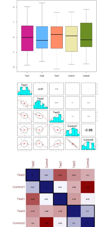
Differential analysis- Abundance values of Treated (T) and Control (C) samples were taken and differential analysis was performed to see the degree of fold change among the conditions. Abundance values were Log2 transformed and then filtered protein wise for whole samples on the basis of valid values by taking at least present in all conditions. Total 3088 proteins were filtered out from 4860 proteins. These values were then Z-score standardized. Categorical grouping was also for both the conditions: Control and Treatment- Control: (Control1:F2, Control2:F4), Treatment:
(Treatment 1: F1, Treatment 2: F3, Treatment 3: F5), as detailed (Fig S1 A-F). Student
t-test was used as the numbers of groups needed to be compared were 2 and statistical significance was considered for P value < = 0.05 and fold change > = 2.
Plots and histograms
Correlation Plot- The correlation plot shows the spearman correlation coefficient value that lies between -1 (negative correlation) to +1 (positive correlation). The colour code follows the indicated values of correlation coefficient. PCA- PCA is a widely used mathematical technique designed to extract, display and rank the variance within a data set. The overall goal of PCA is to reduce the dimensionality of a data set, simultaneously retaining the information present in the data. The PCA- biplot contains a lot of information and can be helpful in interpreting relationships between experimental samples. In this biplot, we can see that runs from each sample as representing different experimental conditions are far from each other. Screeplot: The contribution of all ten principal components to the profiling classification in plot of
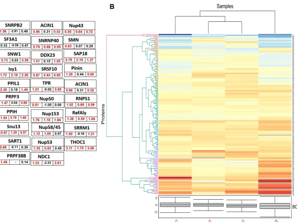
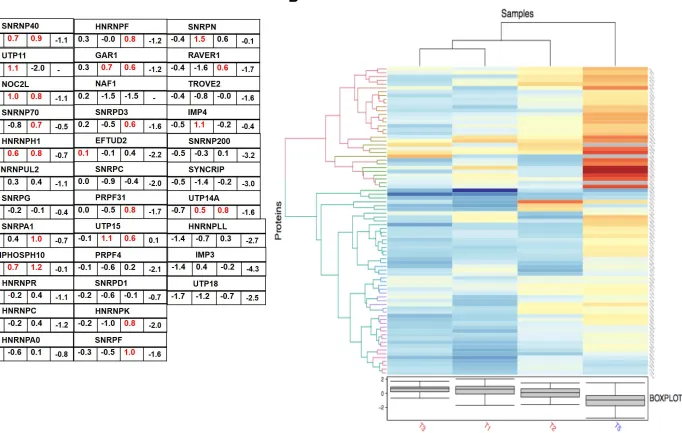
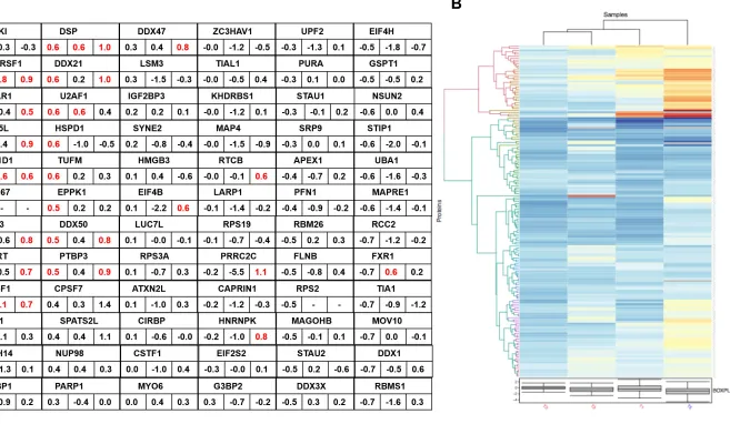
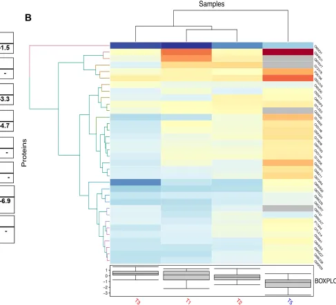
percentage explained variance of PC (Figs. 2 A-C, Fig. S2 A-F). Heatmaps-
Clustering was performed on the LFQ abundance of total 23 statistically Significant
proteins (Fig. 2D) and on all the 3088 proteins (Fig. 1B) that were filtered as identified
and z-normalized. Hierarchical clustering was performed using Euclidian distance and average linkage using In-House R script (Package: ComplexHeatmap).
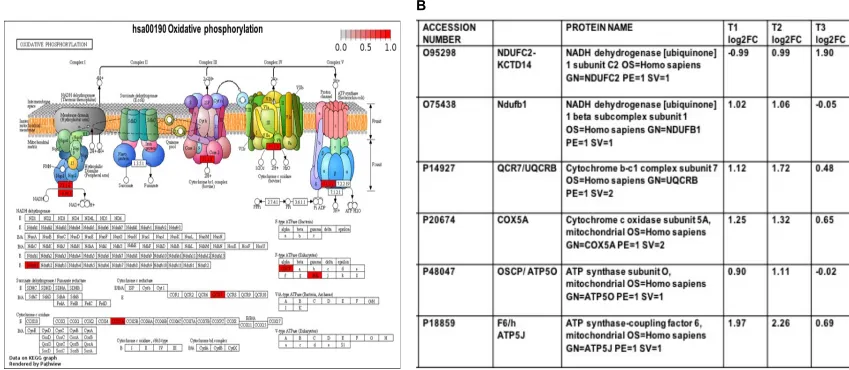
Pathway Enrichment analysis was performed on both group of Proteins filtered (3088 and 23). 3,077 out of 3,088 Proteins were successfully mapped to 3,123 Entrez Gene ID and used for pathway analysis. Out of 202 KEGG pathways gene sets were statistical significantly enriched over following pathways and highlighted in “Red” colour as identified in the KEGG pathway diagram- hsa03010 Ribosome, hsa03040
Spliceosome, hsa03013 RNA transport (Figure 5), hsa03050 proteosome and
hsa04141 protein processing in endoplasmic reticulum (data not shown). 23 Proteins were found to be T-test statistically significant. These 3088 Proteins and 23 statistically significant proteins values were then visualized using In-house R Programming Scripts. 23 out of 23 proteins were successfully mapped to 23 Entrez Gene IDs. Out of 202 KEGG pathways gene sets were statistical significantly enriched over 1 pathway and genes were highlighted in “Red” colour as Identified in the KEGG
pathway diagram- hsa00190 oxidative phosphorylation (Figure 3 A and B).
Mitochondrial proteins in trypsin-treated Caco-2 cells and implication in viral pathogenesis- We compared the upregulated proteins in oxidative phosphorylation
pathway (Figure 3) under various trypsin treatments (T1>T2>T3) compared to control
2. Interestingly, 5 out of 6 significant proteins were upregulated in T1 and T2, however
these proteins were either down regulated or modestly upregulated in T3 (Fig. 3B,
logarithmic fold changes). The upregulated proteins (NDUFB1, UQCRB, COX5A, ATP5O and ATP5J) are associated with assembly of the five oxidative phosphorylation system complexes (OXPHOS) in the inner mitochondrial membrane.
KEGG pathway analyses for biological pathways implicated in viral pathogenesis by employing colour coded expression tags
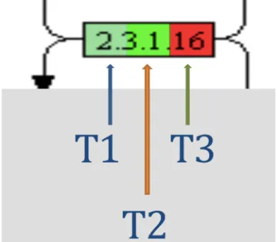
We comprehensively analysed 26 biological pathways (Figure 4A), which are linked to viral pathogenesis and generated modified respective KEGG pathways highlighting the expression of a particular pathway intermediate protein/gene using color-coded gradations (log2FC -1 to +1, green to red colour gradients), for the most relevant
pathways (Figures 5B, 11, 12, 14). This helped us to assess the dose dependent
expression (3 independent concentrations of trypsin) of the “concerned” pathway protein in color-coded read outs. However, owing to the limitation of missing independent replicates and inclusion of all the proteins for pathway analyses (as discussed above), a fraction of proteins did not accurately map to the color coding on the pathways (the missing upregulated proteins in T1 group are highlighted in blue arrows). Therefore, to get an accurate quantitative fold change value for the identified proteins we tabulated all the pathway proteins (above and below 1, on Log2 scale) including upregulated (+1 to +7) and downregulated (-1 to -6) proteins on log2 FC
scale (supplementary files 1 and 2). Interestingly, there were many proteins present
(up or own regulated) in trypsin treated Caco-2 cells as can been seen from colored
boxes in the respective pathways (Figures 5, 11, 12, 14). Further, we observed a
consistent trend in protein upregulation (red color) that was negatively correlated with trypsin concentration (T) for most of the pathway proteins. Major fraction of mapped proteins was upregulated (red color) in T3 (lowest concentration of trypsin used) and
hence most of the “red-color” is restricted to right 1/3rd of each box (for most of the
pathways), which is the position assigned to T3. The mid part of each box is T2 and
left 1/3rd is T1 (maximum concentration of trypsin) (expression tags, Figure 4B).
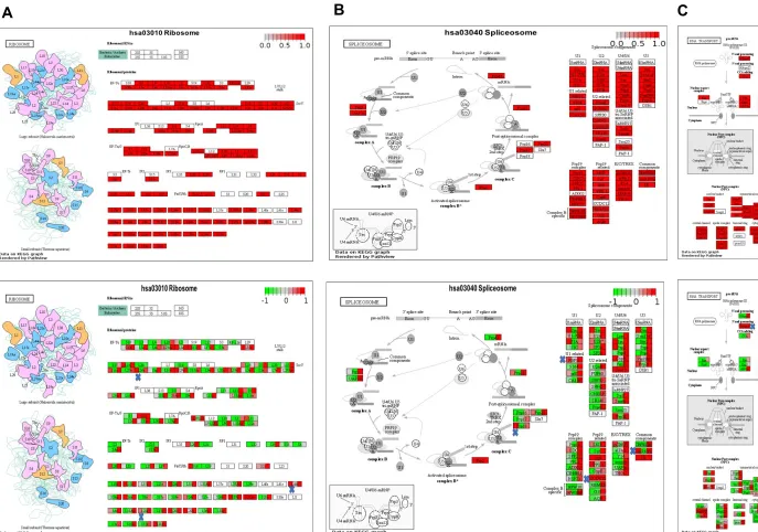
Modulation of ribosome and RNA pathways in trypsin-treated Caco-2 cells
The ribosome, spliceosome and RNA transport pathways emerged as most
significantly abundant pathways in our data set (Figure 5 A-C, upper panel). To
explore their relative abundance in T1, T2 and T3 over control 2, we did the KEGG pathways analysis employing colour coded expression tags as described above (Figure 5 A-C, bottom panel). We hypothesized that serine- proteases linked to COVID-19 pathology might act as stress-inducers to trigger biological condensate associated translational halt, involving global proteome suppression that compromises induction of anti-viral proteins, primarily the interferon pathway. We asked if trypsin treatment of Caco-2 cells was associated with the integral components of stress associated biological condensates, which include i) non-translating mRNA pool, ii) ribonucleoproteins, and iii) RNA binding proteins. In support of our hypothesis, we found the ribosome, spliceosome and RNA pathways had 68 proteins upregulated in
their T1 group (Figure 6A), implicated in RNA transport, metabolism, transcription and
translation.We also analysed the other (non-protease) stress control (control1), where
we grew Caco-2 cells in presence of DMEM under serum starvation (SS) for extended 48h. We did not observe obvious differences at the level of ribosomal proteins across
T1 and T5 (SS) (Figure 6B). However, interestingly we observed appreciable
heterogenous and small ribonucleoproteins (Figure 7 A and B) upregulated in the
trypsin treatment groups (T1 and T3) compared to stress control C1 (SS), indicating limiting ribonucleoprotein availability in SS conditions, which may discourage faithful RNP-concentration dependent formation of biological condensates. We further explored the potential of RNA binding proteins present in our dataset, which could offer potential anti-viral therapeutic value. We took leverage of the open access electronic resource The Mammalian Stress Granule Proteome (MSGP) containing all
https://msgp.pt/. We particularly compared the reported RBPs in MSGP database to our proteomic data set, and it was found that nearly three-fourth (171) of the total
RBPs were present in our database out of which nearly one-third were upregulated (~
1.4-fold, 0.5 log2) in T1 (Figure 8A and B). Most notably, we identified 2 interferon
stimulatory genes ADAR and PRKRA that were upregulated in T1 and in T1/T5
respectively (Figure 9A and B). We observed appreciable number of RNA- binding
RNA helicase proteins, which are implicated in viral pathogenesis. It is noteworthy that
we identified several helicases in our dataset (Figure 10 A and B) that could be
potential G4 interactors.
We found modest downregulation of Ras GTPase-activating protein-binding protein 2 (G3BP2) in all treated groups (T1, T2 and T3), however, G3BP2 was appreciably downregulated in T1 and T2 (log2 -1.0), indicating suppressed anti-viral response. We further searched for de-capping enzymes in our data set and found appreciable downregulation of mRNA- decapping enzyme 1A (DCP1A) in T1 (log2 -2.5) and T2 (log2 -1.0) and modest downregulation in T3. Among other cap related proteins, m7GpppX diphosphatase (log2 -2.7), mRNA cap guanine-N7 methyltransferase (log2 -0.8), cap-specific mRNA (nucleoside-2'-O-)-methyltransferase (CMTR1) (log2 -1.5)
were downregulated in T1 (supplementary file 2). There was downregulation (log2
-1.5) of CPSF3 and CPSF6 in T1 treated Caco-2 cells (supplementary file 2), and
although we could not detect CSPF30 in our total proteins, however, very interestingly, the highest upregulated protein was found to be polyadenylation pre-mRNA 3' end processing protein WDR33 in T1 (log2 7.3) and T3 (Log2 6.5), with appreciable
upregulation in T2 (Log2 3.2) as well (supplementary file 1). We also observed
upregulation of YTHDC1 protein (log2 3.2) in T1, which is implicated in m6A recognition by YTH domain. The m6A effectors include the writer protein (m6A methyltransferase complex including METTL3–METTL14), eraser proteins (RNA demethylases; FTO and ALKBH5), and reader proteins (YTHDC1, YTHDC2, YTHDF1, YTHDF2). Alpha-ketoglutarate-dependent dioxygenase FTO and RNA demethylase ALKBH5 were found to be downregulated by (log2 -1.7), and (log2 -1.5)
respectively, indicating complex regulation of RNA N6methyl adenosine recognition
and regulation. Pseudouridylate synthase 7 (PUS7) and HIV Tat-specific factor 1 (HTATSF1) were upregulated in T1 by (Log2 1 and Log2 1.5) respectively. The other highly expressed splicing factor that was upregulated in all 3 groups included Putative pre-mRNA-splicing factor ATP-dependent RNA helicase DHX16, with log fold changes
of 4.9, 3.2 and 4.2 in T1, T2 and T3 respectively (supplementary file 1).
Host cell immunomodulation in trypsin-treated Caco-2 cells
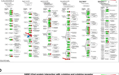
We mapped the total proteins to the known viral pathways in the KEGG data base including- 05168-Herpes simplex virus 1 infection, 05203- viral carcinogenesis, 05416- viral myocarditis and 04061-Viral protein interaction with cytokine and cytokine
receptor (Figure 11 A-D). With most proteins downregulated in these pathways under
T1 conditions (green color restricted to left), it was intriguing to observe upregulation of few proteins in T1 (red -shit toward left side of box) including the host factors Oct-1 and HCF (in 05168-Herpes simplex virus 1 infection pathway), which are known to promote viral infection in host cells (detailed in discussion). The T1 treatment appeared to be conducive to productive viral replication/ infection and, therefore, we
focused on upregulated proteins in T1 in remaining pathways (Figures 12,13) to
establish a correlation (if any) between pro-viral activity in T1 (indicated by Oct-1
cell determinants as indicated in our data set to speculate a possible hypothesis underlying protease-mediated downmodulation of immune cell pathways, and identify potential host target proteins for viral drug development. Further, the viral nucleotide sensing cytosolic proteins (RIG-1 and cGAS, see discussion for details) eventually
converge at generating antiviral interferon response (INF-a and INF-b) and
pro-inflammatory cytokines. INF-g induced activation of INFGR (interferon gamma
receptor) (upregulated in T3), which results in transcription of antiviral proteins showed upregulation of OAS interferon-stimulatory gene (ISGs) in T3, however, no IFN gene transcription was seen for any of the antiviral pathways at any of the trypsin
concentration tested (Figures 11A, 12).
Induction of abortive-interferon anti-viral immune pathways in trypsin-treated Caco-2 cells
As shown (Figure 12 A-D) the major innate immune (TLR, RIG-I, cGAS, NF-𝜅B)
pathway proteins were downregulated in T1 (green color restriction to left part of the box) as opposed to T3 (red color restriction to right part of the same box). Notably, the central (RIG-I RNA sensing pathway) protein ISG- ISG15 was appreciably downregulated in T1, modestly upregulated in T2 and over 2 fold upregulated in T3, thereby, indicating a likely “trypsin-dependent” suppression of interferon pathway intermediate/ISG15, which however fail to trigger the production of terminal effector antiviral interferons (at all the trypsin concentrations- T1, T2, T3), thereby resulting in
an “abortive-interferon” pathway (Figure 12 and 13). This is in contrast to cyclic
GAMP-AMP synthase (cGAS), which was downregulated in all the treatments (T1, T2,
T3) (Figure 13). Intriguingly, we found upregulation of interferon protein ADAR
(adenosine deaminase acting on RNA), RNA-editing enzyme in T1 (Figure 12C, 13C).
ISGs execute their inhibitory function by degrading viral RNA and/or blocking translation of viral mRNAs including 2′,5′-oligoadenylate synthetase (OAS) and latent ribonuclease L (RNase L), protein kinase R (PKR), Moloney leukemia virus 10 homolog (MOV10), and zinc-finger antiviral protein (ZAP), many of which were
downregulated in our data set (figure 13). ISG15 is the most highly induced interferon
stimulated gene, which can inhibit viral translation, replication, or egress. It is a ubiquitin-like protein with an ability to covalently attach to target proteins (ISGylation). Plethora of proteins are involved in ISGylation and deISGylation including UBE2L6, HERC5, HERC6, UBE1LA, TRIM25, and USP18. TRIM25 was downregulated in T1,
T2, and T3 (Figure 13), and HERC4 was highly downregulated in T1 (log2 -4.0)
(supplementary file 2). Since ubiquitination and phosphorylation are key regulatory post translation modification in innate immune RIG-I -ISG15-dependent signaling pathway, we tabulated all the proteins with keyword “ubiquitin” and “phosphatase” (Figure S3 and B), respectively. To our surprise we found many ubiquitinating/deubiquitinating proteins that may have potential implication in interferon response pathway including Lys-63-specific deubiquitinase BRCC36
(downregulated in T1- log2 -4.9), which is known to have role in interferon signaling
by deubiquitination of the type 1 interferon receptor alpha (IFNAR1), resulting in stabilization and cell surface expression of INFRA1 (Figure 13B).
We further did a “theme” based search with key word “Interferon” and tabulated 11
entries that were present in at least 2 groups (Figure 13B). 7/11 proteins were down
(EIF2AK2), Interferon gamma receptor 1 (IFNGR1) and Interferon-induced 35 kDa protein (IFI35) that showed highest downregulation (log2 -4.0). The study by Kerr et al., observed an increase in ribosome RPL28, which was selectively incorporated into ribosomes/polysomes during the IFN response, they further reported an increase in ISG abundance upon RPL28 knockdown (siRPL28) in cells, indicating it as a negative
regulator of ISG synthesis. We tabulated the proteins present in the heatmap (Kerr et
al., 2020; Fig. 6H) showing abundance of select well-studied ISGs in siRPL28-treated and IFN-stimulated cells compared to controls. To our surprise, out of 12 proteins in their heatmap, we were able to find 10 proteins in our proteomics data including RPL28, which was over 2 fold upregulated in T1 (log2, 1.2), unchanged in T2 and modestly upregulated in T3. Further, we could correlate the downregulation of ISGs to the expression levels of RPL28 and interestingly, increased levels of RPL28 appreciably correlated with more downregulation of ISGs in T1 compared to T2 and
T3 wrt Control 2 (Figure 13D). The most highly downregulated ISG was CD55, a
complement decay-accelerating factor that showed a remarkable downregulation (logF2 -4.9), other ISGs that were highly downregulated included anti-viral SAMHD1 (logF2 -2.49), proteasome subunit PSDM1(logF2 -1.9) and ISG15 (logF2 -1.8) in T1.
Trim21, a E3 ubiquitin ligase was reported as a negative regulator of anti-viral Nmi-IFI35 complex with implications for various autoimmune diseases associated
uncontrolled antiviral signaling (Das et al., 2015). We did not find Trim21 in our data,
however, Trim21 regulated ISG protein- Sterile alpha motif and histidine–aspartic acid
domain-containing protein 1 (SAMHD1) was downregulated in T1 (log2 -2.5) (Figure
13D). Further, we also tabulated the INF responsive proteins as reported in the mRNA
expression profiles of proteins that were significantly upregulated at the proteomic
level at 24 h of IFN stimulation (Kerr et al., 2019). We could find 17 proteins in our
data set that were present in at least 2 conditions out of 3 (T1, T2, T3), with top downregulated proteins being IFI35 (logF2 -4.0), STAT2 (logF2 -3.09), E3 ubiquitin-protein ligase RNF213 (logF2 -3.02), and E3 ubiquitin/ISG15 ligase TRIM25, which
ubiquitinates RIG-I and activates it (Figure 13E).
We further explored the spectra of ISGs present in our proteomic profiling data set by
tabulating shared ISGs (Figure 13C) as reported in (Lamers et al., 2020).
Interestingly, we could find total of 15 proteins that were present in at least 2 treatments out of 3 (T1, T2, T3 wrt control2) and 1 protein (TRIM56) was present only in T3. Most of the proteins were downregulated in T1 as (as opposed Lamers et al., results) and interestingly, modestly upregulated in T3. We observed another interesting shared result with Lamers et al., as the expression of 3 genes (downregulated in the expansion organoids/ intestinal progenitor populations)
compared to more differentiated organoids (Lamers et al., 2020) were found to be
downregulated in our cell culture model of trypsin treated Caco-2 cells. These included Transthyretin/TTR (logF2 -2.92), Apolipoprotein A-1/ APOA1 (logF2 -4.3) and Galectin-2/ LGALS2 (logF2 -2.76). The level of downregulation for APOA-1 and LGALS2 was higher in T1, however, interestingly, TTR was more downregulated in T3
(log2 -4.30) (supplementary file 2).
Extending the Interferon-Ribosomal-RNA pathway axis to Coagulation-Complement Networking via thromboinflammatory SERPINS
treatments (T1, T2, and T3) (Figure 14A-C). Encouraged by the presence of SERPIN (serine protease inhibitor) A1AT in our system, we explored other potential SERPINS (if any) present in our data set (that may have potential thromboinflammatory regulating functions) using a keyword “serine protease inhibitor and/or SERPIN”. We could find 7 serine protease inhibitors with implications in thromboinflammatory diseases. 7/7 serine protease inhibitors were downregulated in T1, and interestingly, 6/7 serine protease inhibitors except SPINT2 were downregulated in T2 and T3 (Figure 14B). SERPINS downregulation at all trypsin concentrations indicated a likely similar pattern of downmodulation as seen for abortive- interferon response in T1, T2, and T3. We further searched for serine proteases present in our data set to assess their expression pattern and draw a correlation (if any) with protease/antiprotease balance and interferon response. Total of 3 serine proteases were identified in our
dataset as opposed to 7 SERPINs (Figure 14C). The serine proteases present in the
data set included Prostasin (PRSS8), Suppressor of tumorigenicity 14 protein (ST14) encoding Matriptase, and Serine protease HTRA2, mitochondrial (HTRA2), which showed the following expression levels in T1 (log2 -4.25, log2 -0.79 and log2 1.46) respectively, indicating a more complex regulation of proteases compared to serine protease inhibitors, which were all downregulated in T1. Prostasin was recently shown to have an anti-inflammatory effect via downregulation of TLR4 expression in colonic
epithelial cells (Sugitani et al., 2020). SPINT1 was the most downregulated serine
protease inhibitor in T1 (log2 -4.0), SERPINB6 in T2 (log2 -4.0), and SERPINA1 or A1AT in T3 (log2 -2.76). Interestingly, SERPINB6 and A1AT were appreciably
downregulated in all 3 treatments of trypsin (T1, T2, and T3) (Figure 14C)
Discussion and Future Perspective
Proteases TMPRSS2 and trypsin have been identified as indispensable molecular players underlying COVID-19 disease, particularly trypsin-treatment (proteolytic processing) of viral spike protein has come up as potential species barrier for emergence of zoonotic coronaviruses, hence posing a potential threat for future
outbreaks driven by cross-species transmission of coronaviruses (Walls et al., 2020;
Hoffmann et al., 2020; Matsuyama et al., 2020, Menachery et al., 2020; Letko et al., 2020). TMPRSS2 was shown to be inhibited by protease inhibitor A1AT (Alpha-1 antitrypsin), ACE2 carboxypeptidase (SARS-CoV-2 receptor on human cells) was recently proposed to regulate inflammation by regulating neutrophil influx through
IL-17-STAT3 pathway (Sodhi et al., 2019), further emphasising the role of proteases as
mechanistic determinants of viral entry, replication and modulation of inflammatory pathways driven by neutrophils. Recent studies implicate coagulation pathways, neutrophils and NETs (Neutrophil Extracellular Traps) as clinical correlates of
COVID-19 disease (Zuo et al., 2020, Negri et al., 2020; Wang et al. 2020; Fox et al., 2020;
Yao et al., 2020; Barnes et al., 2020; Liu et al., 2020, Gris J-Christophe et al., 2020, Mozzini and Girelli, 2020; Connors, 2020; Thierry and Roch, 2020). Importantly, neutrophils and NET components are increasingly linked to thrombotic
diseases (Stakos et al., 2020), and NET components including (primarily) neutrophil
elastase (NE) and DNA are linked to impairment of fibrinolytic pathways resulting in
stabilization of thrombosis (Varju and Kolev, 2019; Barbosa da Cruz, 2019; Tan et
al., 2019, Ducroux et al., 2018). The current NE and /or NET targeting drugs (Crocetti et al. 2019) do not hold much promise for COVID-19 treatment (Barnes et al., 2020). These insights have encouraged targeting alternate coagulation pathway proteins as
Levi et al., 2020; Paranjpe et al, 2020). Dissecting pathophysiology of COVID-19 in terms of interplaying mechanisms that underlie SARS-CoV-2 virulence, human immune response, overactive inflammatory reactions and majorly the coagulation
pathways (Kowalik et al., 2020) is urgently required. Identifying and targeting possible
molecular determinants driving coagulation-thromboinflammation axis/ loop in COVID-19 to achieve hemostasis rebalancing, is a potential novel area to design better therapeutic options for COVID19 treatment.
The current study correlates the partial loss of A1AT, a coagulation-complement pathway protein (potent neutrophil elastase inhibitor) to immunomodulation of protease-treated Caco-2 cells. Based on our preliminary findings (discussed below), we propose the following hypothesis “downregulation of A1AT enhances protease -mediated (virus-competent) immunometabolic reprograming of Caco-2 cells, rescuing this loss by exogenous A1AT may be a promising candidate in treating viral pathologies like COVID-19. The unique immunological profile marked by a profoundly impaired interferon (IFN) type I response in parallel to massive proinflammatory
responses (host-tissue destruction) in COVID-19 disease (Hadjadj et al., 2020;
O’Brien et al., 2020), despite higher susceptibility to type I interferons (Loukugamage et al., 2020) suggests existence of yet unexplored pathways supportive of persistent low interferon induction. Several studies are underway to clarify the role of antiviral and immunomodulating drugs in changing morbidity and mortality in severely ill
COVID-19 patients (Lega et al., 2020), which are aimed at targeting viral protease
(Vatansever et. al., 2020), and host serine protease TMPRSS2 (Nguyen, 2020, Azouz et al., 2020). Few drugs for COVID-19 treatment are already in preclinical or
clinical platforms including combination of interferon-1b, lopinavir- ritonavir, and
ribavirin (Hung et al., 2020), lopinavir- ritonavir (Cao et al., 2020), ribavirin in
combination with interferons a/ b (Khalili et al., 2020), ribosome/ RNA pathway
targeting drugs Remdesivir and ribavirin (Beigel et al., 2020; Kmietowicz, 2020;
Alexander et al., 2020; Khalili et al., 2020), anticoagulant heparin (Negri et al., 2020), and IL-6 neutralizing antibody (Xu et al., 2020). However, exploring COVID-19
treatment still remains a global challenge (Mohta et al., 2020), and accelerating drug
development through repurposed FDA approved drugs is highly encouraged for
COVID-19 (Gordy et al, 2020). Transcriptomic, proteomic and bioinformatic
approaches are providing insights to identify potential themes underlying the disease
pathways (Grifoni et al., 2020; Xiong et al., 2020; Bojkova et al., 2020; Ortea and
Bock, 2020), to guide better therapeutics.
Based on the emerging studies and other drug candidates implicated in COVID-19; it emerges that the potential targets for COVID-19 treatment include- 1) viral proteases, 2) host serine protease TMPRSS2, 3) ribosomal/ RNA pathways, 4) host proinflammatory cytokines, 5) anti-viral interferon pathways, and 6) coagulation pathways. We had a “virus-free” cell culture model with complete absence of proinflammatory cytokines (as opposed to cytokine storm seen in COVID-19). Therefore, we excluded 2 druggable targets (viral proteases and proinflammatory
cytokines) owing to their absence in our in vitro culture system that lacks infective viral
The proteases are integral to viral and tumor pathology and emerging studies are establishing parallels in metabolic reprogramming between oncogenesis and human oncogenic viruses, which hijack host metabolite and lipid machinery to leverage
progeny virus formation (Purdy and Luftig, 2019). Further, the interplay of cellular
metabolism (immunometabolism) with innate immune response has been linked to
respiratory viral infections (Zhang et al., 2018; Cheng et al., 2014), pointing to a
(possible) viral infection- associated metabolic remodeling for achieving host immunomodulation. Interferon signalling induced alterations in ribosome composition that selectively downregulated ISG synthesis associated with IFN response modulation, offers a differential network map of the IFN-induced interactome to
interpret IFN response in the context of viral infection and autoimmune diseases (Kerr
et al., 2019). Notably, the IFN stimulation response extends beyond antiviral program and can facilitate ISG induction more broadly by activating mTOR-AKT-S6K signalling
pathways of the MAPK signaling cascade (Ivashkiv and Donlin, 2012). The evolving
interferon biology landscape implicates interferon/ISG signaling in regulation of mRNA processing, post-translational modification, metabolism, cellular trafficking, chromatin
organization, and the cytoskeleton pathways (Schneider et al., 2014), which underlie
pathological (viral, autoimmune and tumor biology) and physiological (stem cell regenerative pathways) processes.
Recent emerging paradigms point towards an increased trend towards “trypsin-reliance” for zoonotic coronavirus emergence that might predict cross-species transmission of coronaviruses possibly employing alternative route(s) like digestive tract/gut, which is proposed to be a potential site for future coronavirus emergence events in humans and perhaps alternative yet unidentified ACE2-independent
host-cell receptors (Shulla et al., 2011; Menachery et al., 2020; Letko et al., 2020; Yang
et al., 2020). Further, the in vitro cell culture models demonstrated positive correlation between SARS-CoV-2 permissiveness with ACE2 and TMPRSS2 expression in
Caco-2 and Calu-3 cell lines (Bojkova et al., 2020; Hoffmann et al., 2020), and hence
decreased infection in low ACE2 expressing A549 cells (Harcourt et al, 2020;
Hoffman et al. 2020; Blanco-melo et al., 2020). However, emerging studies are strongly linking infection of SARS-CoV-2 cells into enterocyte progenitors that are low
in ACE2 expression (Lamers et al., 2020), indicating lower ACE2 thresholds for
successful infection in some tissues or presence of alternative receptors for viral entry. Similarly, SARS-CoV-2 infected trachea transcriptomic signatures paralleled hematopoietic lineage progenitor cells indicating a possibility of hematopoiesis
induction by SARS-CoV-2 (Blanco-Melo et al., 2020). These paradigms encouraged
us to explore the potential mechanisms that might be active at the trypsin (protease)-host interface, which may extend beyond the layer of “viral- spike processing” and
could possibly be linked to “host-receptor alterations” (Letko et al., 2020).
Based on these observations, insights and our previous findings (Sharma et al., 2013,
Sharma et al., 2017; Sharma et al., 2019), we speculated that the pathogen/protease induced (metabolic) reprogramming events may underlie SARS-CoV-2 mediated infection of host cells to trigger viral-competent immunomodulatory pathways. We were interested to investigate the proposed hypothesis based on the recent findings from diverse groups reporting the “co- emergence/ -existence of SARS-CoV-2 and reprogramming events” indicating extended SARS-CoV-2 associated reprogramming
preferred enterocyte progenitor (ACE2 low) permissive replication (Lamers et al.,
2020), and transit-amplifying cells or intestinal stem cells supporting SARS-CoV-2
infections (Zhang et al., 2020). We were intrigued to explore if CoV associated serine
protease(s) facilitated host/Caco-2 cell remodeling to usurp its cellular machinery resulting in dampened immune response.
Our preliminary proteomics data with trypsin-treated Caco-2 (SARS-CoV-2 permissive gut/intestinal) cells suggests that the enhanced bioenergetics in trypsin treated Caco-2 cells might drive metabolic resetting, which can facilitate viral competent immunomodulation. Assembly of the OXPHOS system (upregulated in trypsin
treatment, Fig 3) is an intricate and vaguely defined process (Signes and
Fernandez-Vizarra, 2018), however, the mitochondrial F type -ATP synthase, is established to be critical for mitochondrial functions. The deregulation of this enzyme was recently shown to be associated with dampened mitochondrial oxidative phosphorylation (OXPHOS) and activated mitochondrial permeability transition via OSCP (ATP5O) (Beck et al., 2016). Moreover, defects in COX5A overexpression were reported to
attenuate mitochondrial respiratory dysfunction (Zhang et al., 2020). PB1-52, a
mitochondrial inner membrane interacting viral protein has emerged as an innate immune modulator and virulence factor that regulated Type 1 interferon response (Cheung et al., 2020), thereby reflecting a possible host cell- metabolism rewiring that may regulate interferon responses for pathogen benefitting.
Cellular stress- stimuli including endogenous molecules result in phosphorylation of eIF2a mediated translational halt as a consequence of integrated stress response (Aulas et al., 2017; Tauber and Parker, 2019) leading to formation of stress granules (SGs). However, SGs can also form independently of eIF2a phosphorylation through
inhibition of the eIF4F complex or osmotic stress (Aulas et al., 2017). SGs contain
pools of non-translating mRNAs, which interact with RNA binding proteins and
ribonucleoproteins to form biological condensates (Ivanov et al., 2019) known as
membraneless organelles (MO) or non-membrane organelles through weak molecular interactions involving low complexity or intrinsically disordered regions (IDRs) in RNA binding proteins like G3PBs in a biophysical process called liquid-liquid phase
separation (Guillen-Boixet et al., 2020, Deniz, 2020, Yang et al., 2020). The stress
granules may trigger innate immune responses by activating cellular nucleic acid
sensors (Kim et al., 2019; McCormick and Khaperskyy, 2017), and therefore, CoVs
and other respiratory viruses manipulate stress granule formation tailored to their
requirements (Rabouw et al., 2016; Slaine et al., 2017; Khpaerskyy et al., 2014,
Gaete-Argel et al., 2019). Coronaviruses like SARS-CoV and MERS-CoV trigger host shut-off at the transcriptional and the translational levels, preventing translation of host
mRNAs (Tanaka et al., 2012; Lokugamage et al., 2012; Huang et al., 2011;
Kamitani et al., 2009; Lokugamage et al., 2015). Most viruses including MERS-CoV target stress SG ribonucleoprotein complexes triggered by cellular stress, however,
no such association has been reported for SARS-CoV (Gaete Aragel, 2019).
Emerging studies are increasingly implicating RNA metabolism to viral immune
evasion (Oyarzun-Arrau et al., 2020), and SARS-CoV-2 is also proposed to
camouflage its RNA from innate immune recognition by modifying RNA capping
machinery(Encinar and Menendez, 2020). Importantly, certain interferon-stimulated
infection has been associated with poor anti-viral interferon response. Therefore, we further investigated if SARS-CoV-2 associated protease trypsin can induce stress-granule like proteome marked by suppressed interferon response. Interestingly, we observed that trypsin- primed Caco-2 cells displayed abortive- interferon proteome under differential stress conditions that was marked by complete lack of INF generation despite induction of intermediate ISGs under certain treatments. We asked if the lack of interferon response was a generalizable phenomenon related to the translational halt triggered by cellular stress, or if we had some success in simulating the SG-like proteome phenotype, which is linked to regulation of interferon pathway as many interferon proteins like ADAR and PRKRA (RNA binding proteins) can interact with the SG components. We briefly discuss our findings in the subsequent section.
We included protease- primed stress conditions in our experimental set up with a goal to develop protease- mediated induction of host -dependent (viral regulatory) biological substructures that may offer investigation of interferon- regulatory networks with implications in viral pathology. The cellular- stress inducers included- a) Caco-2 cells grown under serum- free conditions in DMEM medium with no added growth
supplement (SS/ C1)-T5; b) Caco-2 cells grown under trypsin (50 µg /ml) conditions-
T2 (CPT); c) Caco-2 cells grown under ARPE19 conditioned media as established by
the author (Sharma et al., 2013; Sharma et al., 2017; Sharma et al., 2019). This
conditioned media is marked by presence of activated trypsin (50 µg /ml), IL-1b, and
biologically active bFGF (basic fibroblast growth factor). IL-1b is strongly implicated in
COVID-19 pathology, therefore, we asked if we could more closely approximate the cellular stress underlying SARS-CoV-2 infection by combining trypsin and
pro-inflammatory IL-1b (CPT-IL). We envisaged it to be a more potent stress inducer than
CPT, and d) Caco-2 cells grown under escalating trypsin dose (500 µg /ml)- T1
(CPT-EC). Notably, trypsin (Catalog # 17-161E, Lonza) used in this study was procured from
Lonza, which comes at a working concentration of 500 µg /ml with 5mM dextrose, and
physiologic ionic concentration of 5mM KCl and 137mM NaCl. However, the concentration of sodium bicarbonate is around 7mM in trypsin solution as opposed to around 44mM bicarbonate in DMEM media. Since bicarbonate exists as a conjugate base (the dissolved bicarbonate ions in the medium) to control a stable physiological
pH through the bicarbonate buffering system under artificial cell culture conditions in
vitro, we consistently observed a slight drop in pH (it was around 6.9) over 48h priming of Caco-2 cells in trypsin T1 conditions. Further, cell culture media of a standard composition contains source of energy and compounds which regulate the cell cycle and supplementation with amino acids, vitamins, inorganic salts, glucose, and serum as a source of growth factors, hormones, and attachment factors. In addition to nutrients, the medium also helps maintain pH and osmolality. The T1 condition with no added amino acids, vitamins or growth factors could support the survival of cells (Figure 1 A) until 48h post trypsin- inclusion. Apparently dextrose (1000mg/ml or 5mM) was sufficient to keep cells viable under stress. We believe that the trypsin activation of its receptor PAR-2 on Caco-2 cells resulted in upregulation of various ion
channels and transporters (Figure S3 C) to further regulate the osmolality of the
system. Glutamine and pyruvate have also been excluded from T1 condition (as there is no basal media), yet the cells could cope up with stress and survived for 48h indicating yet unidentified pathways to be driving survival under stress. Importantly, recent studies implicate glutamine independence as a selectable feature of pluripotent
phosphate pathway intermediates and purine- pyrimidine pathway intermediates are also indicated in our data, however, detailed investigation would provide better insights to the metabolic pathways governing T1 cell survival under extreme stress. We attempted to test this culture condition as we were aiming to establish
protease-mediated LLPS (liquid-liquid phase separation) driven stress granule formation in vitro.
It is reported that LLPS formation is facilitated by temperature, pH, salt, and crowding
agent dependent manner (Fu, 2020), therefore, we hypothesized that physiological
salts and slight pH alterations (and perhaps perturbed osmolality) might promote
SG-like features in vitro under CPT-EC conditions.
Our preliminary findings show near-global downregulated protein expression with lower trypsin concentrations (T2/CPT) closer to the proteome patterns of stressed,
nutrient-deprived serum starved (T5/ SS) cells (Figure 1B), indicating low translational
activity. Surprisingly, distinct and moderately stretched tracts of overexpressed proteins appeared in proteome of Caco-2 cells primed with trypsin at escalating concentrations, (CPT-EC), which could be majorly mapped to ribosomal, RNA
transport, and spliceosome ribonucleoproteins (Figures 5-10). Intriguingly, the
proteome of T1 and T3 contained upregulated SG- forming components, including
ribonucleoproteins and RNA- binding proteins (Figures 7-10) that were absent or
downregulated in T2 and T5 , indicating propensity of T1 and T3 towards formation of SGs. However, T3 exhibited overwhelming expression of protein tracts as seen for Caco-2 cells cultured under growth-factor rich serum conditions (C2) indicating a likely de-repression of trypsin- induced translational halt, and diminishing possibility of formation of SGs owing to global translation upregulation as seen for serum treated
Caco-2 cells (Figure 1B). It appears that the inclusion of growth factor bFGF in the T3
conditions released the apparent translational halt in T3 treated cells. Intriguingly, the
inclusion of proinflammatory IL-1b to trypsin treated Caco-2 cells (CPT- IL) failed to
trigger INF and proinflammatory cytokines, despite the presence of IL-1b in the cell
culture milieu suggestive of protease/ trypsin- dependent regulation of INF response, this has a close resemblance to refractory interferon response amidst proinflammatory cytokine storm as seen in COVID-19 disease, suggesting a yet underappreciated role of serine proteases in regulating anti-viral interferon response. We further investigated if interferon stimulated genes were a part of the SG-like proteome in T1 conditions. It is established that following viral infection the cells happen to sense dsRNA (viral) detection, and alternate between periods of translation inhibition and process of active protein synthesis. The cytokines mRNA translation is hampered by translation inhibition (as appear under T1 conditions), which is mediated by dsRNA-induced PKR activation and consecutive translation initiation factor eIF2a phosphorylation indicating translational halt. PKR-dependent eIF2a phosphorylation is key for translation shut-off
and stress granules formation (Dalet et al., 2015). Interferon induced proteins like
ADAR and PRKRA are RNA binding proteins that can interact with the SG components and are negative regulators of interferon response. ADAR deficiency is shown to cause chronic type I interferon production and inflammatory pathology in humans and
mice (Hartner et al., 2009; Rice et al., 2012). A recent study linked the loss of ADAR1
in tumours to MDA5-dependent IFN-I production and inflammation (Ishizuka et al.,
2019).
of RNA concentrations through RNA decay pathway (Burke et al., 2019, Tauber et al., 2020). RNA sensing is central to viral infections, therefore, RNA unwinding enzymes called RNA helicases (DEAD-box proteins) can perturb RNA- RNA interactions by invoking their inherent ATPase activity. This can block the formation of RNP condensates like stress granule, hence RNA helicases can counteract biological RNA- RNP condensate formation as has been recently shown for RNA helicase eIF4 (Tauber et al., 2020). Four-stranded G-quadruplex (G4) secondary structures are key structural features in viral mRNA and they tend to be highly conserved despite the high rate of mutations in viruses, indicating the importance of G4 targeting approach for broader antiviral therapy. Viral G-quadruplexes are new frontiers in virus
pathogenesis and antiviral therapy (Ruggiero and Richter, 2020). Notably, G4s are
enriched in functionally important regions in mRNA including 5 ′ - and 3 ′ -untranslated regions (UTRs), and recognition of mRNA secondary structures by RNA binding proteins (RBPs) is essential for post-transcriptional modifications. Therefore, recognition and unwinding of G4s are thought to be important for post-transcriptional processes such as mRNA translation, transport or stability. Interestingly, the major group of G4 interacting proteins consist of proteins involved in RNA splicing and processing, followed by proteins involved in translation. A dynamic balance between formation and resolution of G4 structures in host cells and presence of conserved G4 structures in viruses, offer a promising window of opportunity for targeting viral pathologies. Our data presents appreciable expression of RNA helicases in T1 and T3 (Figure 10 A-B) indicating ensuing secondary structural alterations in mRNA, which may underlie the formation of SG-like condensates under T1 conditions.
Other candidates that can regulate translational outputs under T1 primed conditions include PUS7 and HTATSF1. It was recently reported that PUS7 bound to distinct tRNAs to control biogenesis of tRNA derived fragments and hence regulated
translation in stem cells (Guzzi et al., 2018). HTATSF1 was recently shown to regulate
ribosomal RNA transcription and processing and subsequent efficient protein
synthesis by specifically controlling splicing and intron retention (Corsini et al., 2018).
Nuclear retention of incompletely spliced or mature mRNAs is emerging as a novel layer of gene regulation, which sensitizes the cell to instantly respond to stress, viral
infection, differentiation cues or changing environmental conditions (Wegener et al.,
2017). Notably, the host RNA modification inventory is dynamic, and emerging
modulators of RNA virus gene expression have been proposed (Netzband and
Pager, 2019). These RNA regulatory events can significantly impact virus gene expression, regulation, cytopathology and pathogenesis. Therefore, RNA viruses use, usurp and/or avoid the associated RNA machinery to impact the outcome of infection (Cross et al., 2019). RNA splicing is a new paradigm in host-pathogen interactions (Chuahan et al., 2019), and it was recently proposed that the alternative splicing landscape of host-cells determined the outcome of infection (productive vs abortive)
(Boudreault et al., 2019). Recent studies report exciting convergence of the epitranscriptomic and virology research areas involving the N7-methylguanosine, ribose 2’ -O-methylation, pseudouridine, inosine, N6-methyladenosine, and 5-methylcytosine epitranscriptomic marks on gene expression of RNA viruses. Interestingly, viral interaction and subversion of host cellular RNA transcription, splicing, translation, storage, and decay pathways underlie faithful viral infection
cycles (Cross, Michalski, Miller, & Wilusz, 2019; Kouba, et al, 2019; Velthuis,
2019) and spatiotemporal viral gene expression (Gonzales-van Horn & Sarnow,