Khairy et al. World Journal of Pharmaceutical and Medical Research
IN-VIVO
ANTI-TUMOR EFFECT OF 5-FLUOROURACIL LOADED
CHITOSAN-SODIUM DEOXYCHOLATE NANOPARTICLES
Fahima M. Hashem1, Mohamed Nasr1 and Ahmed Khairy*2
1Department of Pharmaceutics and Industrial Pharmacy, Faculty of Pharmacy, Helwan University, Cairo, Egypt. 2Department of Pharmaceutics, National Organization for Drug Control and Research (NODCAR), Giza, Egypt.
Article Received on 17/12/2018 Article Revised on 07/01/2019 Article Accepted on 28/01/2019
INTRODUCTION
One of the most frequently used antineoplasic agents is 5-fluorouracil (5-FU). It has documented activity alone or in combination therapy for the treatment of many cancer forms.[1] However, there are many reasons that had limited the clinical applications of 5-FU such as the high rate of metabolism in the body, the maintenance of a therapeutic serum concentration requires the continuous administration of high doses, which could lead to a severe toxic effect.[2,3] Many formulation approaches have been attempted to achieve the required amount of drug at tumor site for a certain period of time and minimizing the side effects.Nanoparticles as drug carriers allow the continuous and controlled release of therapeutic drugs to maintain drug levels within a desired level.[4] In addition, the nanoparticles are advantageous in terms of the ability of enhanced permeability and retention effect (EPR) which helps in the accumulation of drug loaded NPs in the tumor tissues in comparison with normal tissues.[5,6] The sustained release properties of the biodegradable nano-drug delivery systems were used to improve the residence time of the chemotherapeutic agent in the body.Progress has been made in the treatment of cancer by incorporating
chemotherapeutic agents into nanocarriers in recent years.[7] Previous reports indicated that sustained release formulations of 5-FU and selective delivery to the tumor site not only improve the antitumor activity but also reduce side effects of 5-FU as compared with the clinically available 5-FU formulation. In-vivo, biodistribution studiesof 5-FU in rat liver indicated that the nanoparticles cubosomal formulation significantly increased 5-FU liver concentration (nearly 5-fold) as compared to that of a 5-FU solution.[8] Chitosan (CS) is one of the most popular materials in the field of drug delivery and is, by far, the most applied of the natural polymers. CS is soluble in acidic solutions owing to the protonation of the amino groups composing the polymeric chain at this pH. In this regard, highly deacetylated CS (85%) is readily soluble in solutions of pH up to 6.5, but as the deacetylation degree decreases, the solubilization becomes more difficult.[9,10] Its attractiveness relies on very interesting structural and biological properties, which include the cationic character and the solubility in aqueous medium as well as its characteristic biodegradability and mucoadhesivity.[11,12] Chitosan is known to have the ability to transiently open epithelial tight junctions,
ISSN 2455-3301
WJPMR
AND MEDICAL RESEARCH
www.wjpmr.com
*Corresponding Author:Ahmed Khairy
Department of Pharmaceutics, National Organization for Drug Control and Research (NODCAR), Giza, Egypt.
ABSTRACT
Chitosan- sodium deoxycholate nanoparticles containing 5-fluorouracil (5-FU) was prepared via a simple electrostatic interaction between oppositely charged particles using different weight ratios between chitosan and sodium deoxycholate. The objective of this work is to characterize and estimate the antitumor activity of the prepared chitosan- sodium deoxycholate nanoparticles loaded with 5-FU.The prepared nanoparticles were characterized in-vitro and in-vivo. The in-vitro characterizations were investigated by entrapment efficiency % (EE %), particle size analysis, zeta potential measurement, in-vitro release and Transmission Electron Microscopy (TEM). 5- FU loaded CS- DS nanoparticles showed EE% ranging from (14.54 ±1.35 to 22.43 ±2.12), reasonable mean particle sizes (165.65 ±17.23 to 318.47 ±25.86) and zeta potential (35.57 ±3.05 to 48.86 ±3.57 mV). The in-vitro release of nanoparticles exhibited an initial burst effect followed by a sustained drug release with approximately 90 % of the drug being released over 6 hrs. In-vivo anti-tumor effect was studied on solid ehrlich carcinoma (SEC) tumor bearing Swiss albino mice.The anti- tumor activity of 5-FU loaded in CS- DS nanoparticles was evaluated comparatively to control and plain formula. The results revealed that 5-FU loaded CS- DS nanoparticles effectively decreased tumor volume and showed significantly (p<0.05) lower tumor volume compared to control group and plain formula.
permitting an increase of drug permeation by acting as an absorption promoter.[13,14] Sodium deoxycholate (DS) is one of the endogenous bile salts which could improve the paracellular transport of hydrophilic drugs if added to CS, the mechanism of sodium deoxycholate as absorption enhancer includes extracting membrane protein or lipids, membrane fluidization, producing reverse micelles in the membrane and creation aqueous channels.[15] The objective of this work is to formulate a suitable delivery system that gives a sustained release formulations of 5-FU through incorporating it in CS- DS nanoparticles and estimate the in-vivo anti-tumor effect of prepared nanoparticles on Ehrlich carcinoma-bearing mice.
MATERIALS AND METHODS Materials
5-flurouracil was purchased from Sigma Aldrich (Missouri, USA). Chitosan low molecular weight (Poly (D-glucosamine) deacetylated), was purchased from Sigma Aldrich (Missouri, USA). Sodium deoxycholate was purchased from Alfa Aesar (Karlsruhe, Germany). Potassium dihydrogen phosphate (NaH2PO4) and sodium monohydrogen phosphate (Na2HPO4); Adwic, EL-Nasr Pharmaceutical Chemical Company (Egypt).
Preparation of 5-FU loaded CS-DS nanoparticles 5-FU loaded CS-DS nanoparticles were prepared using simple electrostatic interaction between oppositely charged particles. CS was dissolved in 0.1 % acetic acid solution to obtain 1 mg/ml solution and DS was dissolved in deionized water in order to obtain 1 mg/ml solution. Nanoparticles were formed when different amounts of DS solution were added drop wise to a CS solution and magnetically stirred at 300 rpm for 15 min at room temperature. CS solution turns translucent to turbid as a result of the formation of CS-DS nanoparticles. For the nanoencapsulation, 5 mg of 5-FU was added to CS solution before the addition of DS solution.
The dispersion was centrifuged at 12,000 rpm for 20 min at 4 oC to separate the solid nanoparticles. The nanoparticles residue was re-suspended in deionized water and centrifuged again. The composition of different formulas of 5-FU loaded CS- DS nanoparticles is shown in table 1. All batches were prepared in triplicate.
To make sterile nanoparticles, CS solution containing 5-FU and DS solution were passed through 0.22 µm millipore and mixed with each other under aseptic condition using laminar flow cabinet. All batches were prepared in triplicate.[16]
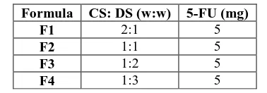
Table 1: The composition of different formulas of 5-FU loaded CS-DS nanoparticles.
Formula CS: DS (w:w) 5-FU (mg)
F1 2:1 5
F2 1:1 5
F3 1:2 5
F4 1:3 5
Particle size analysis and zeta potential
The particle size, polydispersity index and zeta potential of 5-FU loaded CS-DS nanoparticles were determined by photon correlation spectroscopy using a Malvern Zetasizer Nanoseries (Nano-ZS, Malvern Instruments, Malvern, UK). Prior to measurement, nanoparticles dispersions was diluted 20-fold with deionized water and shaked to faint opalescence, All measurements were carried out at 25°C. All the measurements were performed in triplicate. The poly dispersity index value gives an indication for the homogeneity of the preparations.
Morphology of 5-FU loaded CS-DS nanoparticles The morphology of 5-FU loaded CS-DS nanoparticles (F2) was examined using a transmission electron microscopy (Jeol Jem Dos electron microscopy, Japan). After dilution with deionized water, the samples were deposited on a microscope slide and stained with 3% (w/v) phosphotungstic acid and the stained nanoparticles were imaged.
Entrapment efficiency percent (EE %)
5-FU entrapment efficiency, which corresponds to the amount of 5-FU that can be incorporated in the nanoparticles was determined indirectly by measuring the concentration of the free 5-FU in the aqueous phase of the nanoparticle dispersion using centrifugation method.[17,18] The EE % was determined using the following equation:
100
drug
W
)
suspension
in
drug
W
-drug
W
(
%
initial free
initial
x
EE
Where:
Winitial drug: is the amount of 5-FU initially used for the assay.
Wfree drug in suspension: is the amount of free 5-FU determined in the aqueous phase after separation of the nanoparticles.
In-vitro release
In-vitro release of 5-FU from CS- DS nanoparticles were determined by dispersing an amount of nanoparticles equivalent to 5 mg of 5-FU in 2 ml phosphate buffer (PB) pH 7.4 then placed into a dialysis membrane bag (MWCO 10 kDa, pore size 2.4 nm, Spectra/Pro) and placed into 50.0 ml PB (pH 7.4) with continuous oscillation frequency.[20,21] Also the dialysis of free 5-FU was done in the same way to compare it with the release of 5-FU from CS- DS nanoparticles. At different times, 1 ml release medium was removed at each time interval and 1 ml of fresh medium was added into the system. The amount of 5-FU in the release medium was evaluated spectrophotometerlically at λmax = 266 nm against PB (pH 7.4) as a blank. All measurements were performed in triplicate.
In-vivo antitumor effect Study design
The experimental protocols involving animal study were approved by the animal care and use committee of the college of pharmacy, Helwan University. Eighteen healthy adult female Swiss Albino mice of average weight (20-25 g) were supplied from National Cancer Institute (NCI), Egypt. All mice were kept in Makrolon IV polycarbonate cage and allowed to a free access of normal tap water and regular Purina rodent diet pellet chow all over the period of the experiment. They were maintained in a light controlled room at a temperature of 22o C and relative humidity of 55%. The mice were acclimatized to the animal house condition for two days prior to the experiment.
Solid Ehrlich carcinoma (SEC) induction
Establishment of tumors in the Swiss Albino mice was attained by subcutaneously injection of Ehrlich carcinoma cell line through 23-G needle into the right thigh of each mouse (0.2 ml/ 2- 2.5x l06 cells/ mouse).[22] Ehrlich carcinoma cell line was obtained by intra-peritoneal aspiration of ascetic fluid of Ehrlich ascetic carcinoma (EAC) mouse, followed by dilution with an appropriate volume of isotonic saline. The palpable solid tumors were developed approximately after 13 days and the average tumor solid mass volume is measurable.
Treatment protocol
After the mice developed palpable solid tumors; they were divided randomly into 3 groups (6 mice each). The first group was left as untreated control group and only received an inta-tumor injection of an isotonic saline. The second and third groups received an intra tumor injection of plain formula and 5-FU loaded CS-DS nanoparticles (F2) respectively. 5-FU was given at a dose 20 mg/kg body weight since the intra-tumor delivery of anticancer was considered a valid therapeutic strategy for treatment of cancer.[23,24,25] Each mouse received 4 doses, the first dose was received after l3 days post SEC induction (The first day of treatment) while the second, third and forth dose was received at 20, 28 and 36 days post SEC induction , respectively. Table 16 illustrates and summarizes the treatment protocols. Such multiple intra-tumor doses were reported before.[26, 27]
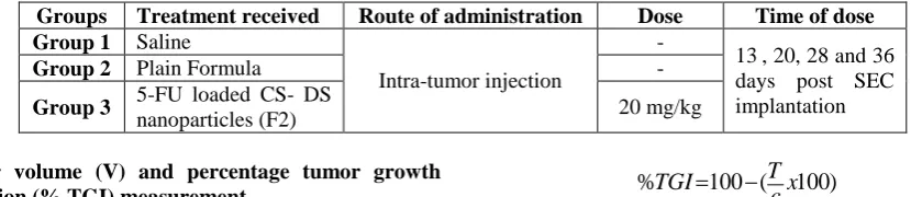
Table 2: Treatment protocol of the in-vivo study design.
Groups Treatment received Route of administration Dose Time of dose Group 1 Saline
Intra-tumor injection
-
13, 20, 28 and 36 days post SEC implantation
Group 2 Plain Formula -
Group 3 5-FU loaded CS- DS
nanoparticles (F2) 20 mg/kg
Tumor volume (V) and percentage tumor growth inhibition (% TGI) measurement
Tumor volumes were recorded twice weekly from the first day of injection till the day of mice scarification. This was attained by recording the two perpendicular diameter of the tumor mass; length (b) (large diameter in mm) and width (a) (small diameter in mm) of the developed solid tumor mass using digital caliber. Tumor volume was calculated as follow:[28]
Tumor volume =
Where: a and b are the width and length of tumor, respectively.
Drug efficiency in reducing tumor volume is expressed as percentage tumor growth inhibition (TGI) which is calculated as:[29]
)
100
(
100
%
x
c
T
TGI
Where T is the mean relative tumor volume (RTV) of the treated group and C is the RTV of control group. RTV of any group is calculated by the following equation:[29]
i
v
x
v
RTV
Where Vx is the tumor volume at the end of the experiment (day of scarification) and Vi is the tumor volume at the start day of treatment.
RESULTS AND DISCUSSION Particle size analysis and zeta potential
presented in Table 3. The results revealed that all 5-FU loaded CS-DS nanoparticles showed a mean particle size less than 318.47± 25.86 nm and ranged from 165.65 ± 17.23 to 318.47± 25.86 nm and PDI values ranged from 0.135 ±0.04 to 0.48 ±0.06.
Theses results revealed that, the mean particles size (table 3) is clearly affected by the amount of DS in the formulations. Increasing the amount of DS in the formulations resulted in a decrease in particle size.
Zeta potential
A higher electric charge on the surface of the nanoparticles will prevent aggregation of the nanoparticles because of the strong repellent forces among particles.[30,31]
Zeta potential values are presented in table 3. The values are found to fall between 35.57 ± 3.05 to 48.86 ± 3.57 mV. These values suggest that the Nanoparticles formulation have good stability.[32,33]
The zeta potential values of the nanoparticles prepared from different ratios of CS: DS decrease as the weight ratio of CS decrease. The 2:1 CS: DS ratio has the highest zeta potential values followed by that of 1:1
ratio. 1:3 ratio has the lowest values. The lower zeta potential with increasing DS ratio might be caused by an increased masking of free positively changed amino groups of CS.
Nanoparticles with positive charge can translocated by the tumor cells through either fluid phase endocytosis or charge interactions and ligand receptor docking.
Entrapment efficiency percent (EE %)
Table 3 shows that the EE % of the prepared 5-FU loaded CS- DS nanoparticles were in the range of 14.54 ±1.35 to 22.43 ±2.12 %.The effect of CS to DS ratio on the EE % was studied at constant 5-FU concentration. Decreasing the CS concentration in the nanoparticles resulted in a decrease in EE %. The highest EE % is observed with the 2:1 CS: DS ratio while the 1:3 ratio has the lowest EE %. This might be due to that the higher ratio of CS: DS (2:1) contain more concentration of CS in the preparation medium which binds more amount of 5-FU. In addition more CS- DS nanoparticles formed as the amount of CS increased, and the amount of 5-FU entrapped in the nanoparticles was also increased. The increase in EE % with increase of CS polymer was reported within many research articles.[34,35]
Table 3: Physicochemical characters of 5-FU nanoparticles prepared from different ratios of CS: DS.
Parameter F1 F2 F3 F4
Mean particle size (nm) ±SD 318.47 ±25.86 250.32 ±24.35 190.83±18.87 165.65 ±17.23 PDI ±SD 0.48 ±0.06 0.287 ±0.04 0.135 ±0.04 0.142 ±0.03 Zeta potential (mV) ±SD 48.86 ±3.57 42.87 ±3.88 38.66 ±2.76 35.57 ±3.05 EE% ± SD 22.43 ±2.12 20.88 ±1.85 16.67 ±1.24 14.54 ±1.35
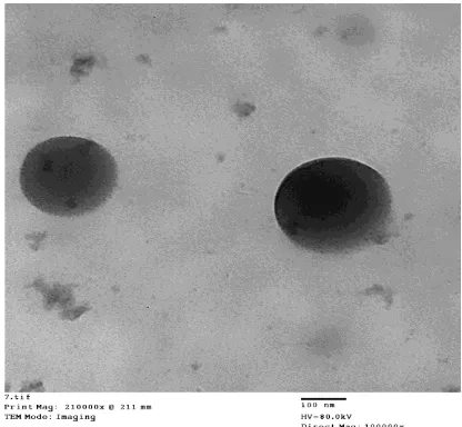
Transmission electron microscope (TEM)
The morphological examination of representative 5-FU loaded CS- DS nanoparticles (F 2) was performed using TEM. Photographs of TEM are illustrated in (Fig. 1). The photographs revealed the spherical shape without aggregation of the nanoparticles.
Fig 1: TEM image of 1:1 5-FU loaded CS: DS nanoparticles (F2).
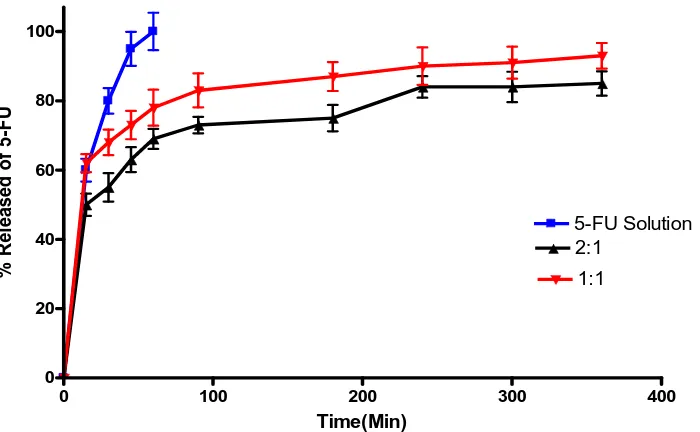
In-Vitro release study of 5-FU from CS- DS nanoparticles
The in-vitro release of free 5-FU and 5-FU loaded CS- DS nanoparticles in PB (pH 7.4) are shown in (Fig. 2). The release of 5-FU from 2:1 CS: DS nanoparticles (F1) are slower than that of 1:1 CS: DS nanoparticles (F2). The release of 5-FU from both of the studied ratios showed an initial rapid burst release of the drug from the surface of nanoparticles. The burst release of 5-FU is about 50% and 62% from F1 and F2, respectively. The observed burst effect was due to dissociation of 5-FU molecules that were loosely bound to the surface of CS: DS nanoparticles.[36] This initial fast release is followed by prolonged sustained release of 5-FU over a period of 6 hours. It has been reported that with lower concentration of CS, a lower viscosity of the gelation medium is observed, which results in a decrease in the liquid phase resistance against dispersion.[37]
0 100 200 300 400 0
20 40 60 80 100
5-FU Solution 2:1
1:1
Time(Min)
%
Re
le
a
s
e
d
o
f
5
-F
U
Figure 2: Percentage released of 5-FU in PB (pH 7.4) from 2:1 (F1) and 1:1 (F2) CS: DS nanoparticles. In-vivo anti-tumor effect
Tumor volume (V) and tumor growth inhibition percentage (% TGI)
Following the treatment protocol as illustrated in Table 2, the anti- tumor activity of 5-FU loaded CS- DS nanoparticles (F2) was evaluated comparatively to control and plain formula using solid ehrlich carcinoma (SEC) tumor bearing Swiss albino mice. Tumor volume and tumor growth inhibition were used to measure the relative antitumor efficacy. The average tumor volume was calculated of all groups (Fig. 3).
The results revealed that the volume of the tumor increased rapidly for the mice treated with isotonic saline (group 1). However 5-FU loaded CS- DS nanoparticles (group 3) showed significantly (p<0.05) lower tumor
volume compared to control group. Lower tumor growth rates were observed in case of mice treated with 5-FU loaded CS- DS nanoparticles.
The results also show that a little difference was found between groups treated with saline (control group) and plain formula (group 2). The tumor volume values of these groups were close to each other.
Meantime, at the ends of the experiment the calculated percentage tumor growth inhibition (TGI %) for plain formula was -21.18 %. These results might indicate that the therapeutic efficacy and anti-tumor activity of plain formula is not present or negligible. In contrast, the estimated tumor growth inhibition (TGI %) for 5-FU loaded CS- DS nanoparticles was 86.27 %.
10 15 20 25 30 35 40 45
0 1000 2000 3000 4000 5000
Control Plain
5 FU formula
Time (day)
Tu
m
or
v
ol
um
e
(m
m
3)
CONCLUSION
5-FU was successfully incorporated into CS-DS nanoparticles. The prepared nanoparticles exhibited nanometer-size particles with positive zeta potential and released the loaded 5-FU in a sustained release manner over 6 hrs. 5-FU nanoparticles showed significantly (p<0.05) lower tumor volume compared to control group. The present work demonstrated that 5-FU -loaded CS-DS nanoparticles can be considered as good candidates for anticancer delivery system.
REFERENCES
1. Ueno H, Okada S, Okusaka T, Ikeda M, Kuriyama H. Phase I and pharmacokinetic study of 5-fluorouracil administered by 5-day continuous infusion in patients with hepatocellular carcinoma. Cancer Chemother Pharmacol, 2002; 49(2): 155-160.
2. Zhang N, Yin Y, Xu S-J, Chen W-S. 5-Fluorouracil: mechanisms of resistance and reversal strategies. Molecules, 2008; 13(8): 1551-1569.
3. Arias JL, Ruiz MA, López-Viota M, Delgado ÁV. Poly (alkylcyanoacrylate) colloidal particles as vehicles for antitumour drug delivery: a comparative study. Colloids Surf B Biointerfaces, 2008; 62(1): 64-70.
4. Devalapally H, Chakilam A, Amiji MM. Role of nanotechnology in pharmaceutical product development. J Pharm Sci, 2007; 96(10): 2547-2565.
5. Yu B, Zhang Y, Zheng W, Fan C, Chen T. Positive surface charge enhances selective cellular uptake and anticancer efficacy of selenium nanoparticles. Inorg Chem, 2012; 51(16): 8956-8963.
6. Acharya S, Sahoo SK. PLGA nanoparticles containing various anticancer agents and tumour delivery by EPR effect. Adv Drug Deliv Rev, 2011; 63(3): 170-183.
7. Sun L, Chen Y, Zhou Y, et al. Preparation of 5-fluorouracil-loaded chitosan nanoparticles and study of the sustained release in vitro and in vivo. Asian J Pharm Sci,2017; 12(5): 418-423.
8. Nasr M, Ghorab MK, Abdelazem A. In vitro and in vivo evaluation of cubosomes containing 5-fluorouracil for liver targeting. Acta Pharm Sin B, 2015; 5(1): 79-88.
9. Cho Y-W, Jang J, Park CR, Ko S-W. Preparation and solubility in acid and water of partially deacetylated chitins. Biomacromolecules, 2000; 1(4): 609-614.
10. Kean T, Thanou M. Biodegradation, biodistribution and toxicity of chitosan. Adv Drug Deliv Rev, 2010; 62(1): 3-11.
11. Amidi M, Mastrobattista E, Jiskoot W, Hennink WE. Chitosan-based delivery systems for protein therapeutics and antigens. Adv Drug Deliv Rev, 2010; 62(1): 59-82.
12. Dash M, Chiellini F, Ottenbrite RM, Chiellini E. Chitosan—A versatile semi-synthetic polymer in
biomedical applications. Prog Polym Sci, 2011; 36(8): 981-1014.
13. Al-Qadi S, Grenha A, Carrión-Recio D, Seijo B, Remuñán-López C. Microencapsulated chitosan nanoparticles for pulmonary protein delivery: in vivo evaluation of insulin-loaded formulations. J Control Release, 2012; 157(3): 383-390.
14. Prego C, Torres D, Alonso MJ. The potential of chitosan for the oral administration of peptides. Expert Opin Drug Deliv, 2005; 2(5): 843-854. 15. Nicolazzo JA, Reed BL, Finnin BC. Buccal
penetration enhancers—how do they really work? J Control Release, 2005; 105(1-2): 1-15.
16. Cadete A, Figueiredo L, Lopes R, Calado C, Almeida A, Gonçalves L. Development and characterization of a new plasmid delivery system based on chitosan–sodium deoxycholate nanoparticles. Eur J Pharm Sci, 2012; 45(4): 451-458.
17. Ammar HO, El-Nahhas S, Ghorab M, Salama A. Chitosan/cyclodextrin nanoparticles as drug delivery system. J Incl Phenom Macrocycl Chem, 2012; 72(1-2): 127-136.
18. Raj CA, Kumar PS, Kumar KS. Kinetics and drug release studies of isoniazid encapsulated with PLA-co-PEG/gold nanoparticles. Int J Pharm Pharm Sci, 2012; 4(4): 1-7.
19. Barbas C, Burton D, Scott J, Silverman G. Quantitation of DNA and RNA. CSH Protocols, 2007; 11: pdb. ip47.
20. Ling G, Zhang P, Zhang W, et al. Development of novel self-assembled DS-PLGA hybrid nanoparticles for improving oral bioavailability of vincristine sulfate by P-gp inhibition. J Control Release, 2010; 148(2): 241-248.
21. Kheradmandnia S, Vasheghani-Farahani E, Nosrati M, Atyabi F. Preparation and characterization of ketoprofen-loaded solid lipid nanoparticles made from beeswax and carnauba wax. Nanomedicine, 2010; 6(6): 753-759.
22. Silva LA, Nascimento KA, Maciel MC, et al. Sunflower seed oil-enriched product can inhibit Ehrlich solid tumor growth in mice. Chemotherapy, 2006; 52(2): 91-94.
23. Le UM, Shaker DS, Sloat BR, Cui Z. A thermo-sensitive polymeric gel containing a gadolinium (Gd) compound encapsulated into liposomes significantly extended the retention of the Gd in tumors. Drug Dev Ind Pharm, 2008; 34(4): 413-418. 24. Arai T, Benny O, Joki T, et al. Novel local drug
delivery system using thermoreversible gel in combination with polymeric microspheres or liposomes. Anticancer Res, 2010; 30(4): 1057-1064. 25. Xing J, Qi X, Jiang Y, et al. Topotecan
hydrochloride liposomes incorporated into thermosensitive hydrogel for sustained and efficient in situ therapy of H22 tumor in Kunming mice. Pharm Dev Technol, 2015; 20(7): 812-819.
carbon particles: antitumor effectiveness against human colon carcinoma xenografts and acute toxicity in mice. J Pharmacol Exp Ther, 2004; 311(1): 382-387.
27. Yang RK, Kalogriopoulos NA, Rakhmilevich AL, et al. Intratumoral hu14. 18–IL-2 (IC) induces local and systemic antitumor effects that involve both activated T and NK cells as well as enhanced IC retention. J Immunol, 2012; 1200934.
28. Abdin AA, Soliman NA, Saied EM. Effect of propranolol on IL-10, visfatin, Hsp70, iNOS, TLR2, and survivin in amelioration of tumor progression and survival in Solid Ehrlich Carcinoma-bearing mice. Pharmacol Rep, 2014; 66(6): 1114-1121. 29. Sanceau J, Poupon M-F, Delattre O, Sastre-Garau X,
Wietzerbin J. Strong inhibition of Ewing tumor xenograft growth by combination of human interferon-alpha or interferon-beta with ifosfamide. Oncogene, 2002; 21(50): 7700.
30. Sawant KK, Dodiya SS. Recent advances and patents on solid lipid nanoparticles. Recent Pat Drug Deliv Formul, 2008; 2(2): 120-135.
31. Kedar U, Phutane P, Shidhaye S, Kadam V. Advances in polymeric micelles for drug delivery and tumor targeting. Nanomedicine, 2010; 6(6): 714-729.
32. Honary S, Zahir F. Effect of zeta potential on the properties of nano-drug delivery systems-a review (Part 1). Trop J Pharm Res, 2013; 12(2): 255-264. 33. Honary S, Zahir F. Effect of zeta potential on the
properties of nano-drug delivery systems-a review (Part 2). Trop J Pharm Res, 2013; 12(2): 265-273. 34. Deng Q-y, Zhou C-r, Luo B-h. Preparation and
characterization of chitosan nanoparticles containing lysozyme. Pharm Biol, 2006; 44(5): 336-342. 35. Nesalin JAJ, Smith AA. Preparation and evaluation
of chitosan nanoparticles containing zidovudine. Asian J Pharm Sci, 2012; 7(1): 80-4.
36. Amidi M, Mastrobattista E, Jiskoot W, Hennink WE. Chitosan-based delivery systems for protein therapeutics and antigens. Adv Drug Deliv Rev, 2010; 62(1): 59-82.