Int. J. Nanosci. Nanotechnol., Vol. 13, No. 1, March. 2017, pp. 11-18
11
Effect of Functionalization Process on
Thermal Conductivity of Graphene Nanofluids
Ahmad Ghozatloo1*, Mojtaba Shariaty Niassar2 and Alimorad Rashidi1
1Faculty member of Research Institute of Petroleum Industry (RIPI), West Blvd. Azadi Sport
Complex, P.O. Box: 14665-137, Tehran, Iran
2Transport Phenomena & Nanotech. Lab (TPNT), School of Engineering, University of
Tehran, P.O. Box: 11155-4563, Tehran, Iran
(*)Corresponding author: [email protected]
(Received: 18 January 2015 and Accepted: 05 September 2016)
Abstract
In this research, Graphene was synthesized by chemical vapor deposition (CVD) method in atmosphere pressure (14.7 psi). Different functionalization method was used for oxidizing of graphene such as acid and alkaline treatments. The Functionalized graphene (FG) was characterized by FTIR and Raman spectroscopy. Nanofluid with water and different concentration (0.05, 0.15 and 0.25 wt %) of FG were prepared. Thermal conductivity of nanofluids was measured by transient hot wire method. The acid functionalization introduces significant defects in graphene structure, degrading its unique properties such as superior carrier mobility, mechanical strength and chemical stability. In alkali functionalization method, the graphene is not effectively defected. Therefore, the transport properties of graphene maintained and this method showed enhancement in thermal conductivity more than acid fictionalization in same conditions. In optimum condition (0.25 wt % graphene of alkaline method in water), thermal conductivity ratio were increased (24.4% at 20°C and 33.9% at 60°C).
Keywords: Graphene, Functionalization, Nanofluid Thermal conductivity, Alkaline, Acid
1. INTRODUCTION
Nanofluids (NFs) were called nano sized particles dissolve in fluids which are novel fluids having excellent heat transfer behavior. They can exhibit thermal conductivity values of about 20150% higher than fluids [1]. Therefore NFs are going to have an important role in heat exchangers designs. Further advances are thought to be brought about if the coolant flowing in the micro channels in an enhancement in heat transfer [2].
Many experimental studies on thermal transport of NFs focused on changes in properties created by metal oxides nanoparticles. For example thermal conductivity of water was increased 30% with the addition of 4.3 vol % Alumina nanoparticles [3]. Such an enhancement
phenomenon was reported for CuO/water, Al2O3/water and Cu/Oil NFs [4]. Also many studies on the thermal conductivity characteristics of various NFs by Ag, Cu and TiO2 NFs are carried out [5–7]. Carbon materials have attracted great interest because of their large intrinsic thermal conductivity and low density [8]. Therefore carbon nanotubes (CNT), graphite, nano fibers, diamond and graphene were used in NFs [9-13].
higher than other nano carbon structures [14]. Then graphene has been receiving especially close attention. For example thermal conductivity of graphene/ethylene glycol (EG) nanofluid was increased 86% in a 5 vol % [15]. The thermal conductivity of graphene nanofluid with 0.056 vol % was increased 64% at 50°C relative to base fluid [16]. When the nanosheets of graphene oxide (GO) loading is 5 vol % in EG, the enhancement ratio is up to 61% [8]. Additionally, thermal conductivity of graphene NFs was measured according to temperature, and concluded that, the thermal conductivity depended on the temperature [17]. Thermal conductivity of alkaline functionalized graphene with 0.05 wt % increased 14.1% relative to water at 25 °C and increased 17% at 50°C [18]. One of the critical steps in preparing graphene NFs is dispersing graphene nanosheets in fluids. Very large specific surface areas and high aspect ratio of graphene nanosheets cause dispersion of graphene in aqueous medium can be challenging [19]. In nature, graphene is hydrophobic and thus has a non-homogeneous form in fluids and create unstable NFs under normal conditions [20]. Many methods are to disperse graphene in base fluids. But chemical methods involves surface modification of graphene with treating in acid or alkaline media is good [21]. Hydrophilic functional groups made by treating in oxidants in acid or alkaline media [22]. Therefore addition of hydrophilic groups at defect sites on
graphene surface, making more
hydrophilic structure. However, chemical treatment in strong acid makes defects in structure of graphene. Therefore, proper care has to be taken during processing to minimize adverse effects [23]. Alkaline oxidation media cause fewer defects to structure of graphene relative to acid [18]. Thus, oxidation process has effect on degradation of graphene structure. However, study of the oxidation process effect on thermal conductivity of graphene nanofluid is not carried out. In the present
study, two kinds of oxidation process for synthesis hydrophilic graphene structure were made and thermal behavior was investigated.
2. EXPERIMENTAL
2.1. Chemical Materials
In the present work, all reagents (H2SO4, HNO3, potassium per sulfate) used were of analytical grade and obtained from Sigma-Aldrich. Methane cylinder (100 bar) containing 99.99% methane was purchased from Air Product Company and used as the feed. Deionized water was purchased from Bahre-e Zolal-e Tehran Company.
2.2. Synthesis and Characterization of Graphene nanosheets
Graphene was synthesized by chemical vapor deposition (CVD) method using methane as a carbon source and hydrogen as a carrier gas in atmosphere pressure at 1050°C for 15 min [18]. The CVD Graphene was characterized with XRD and TEM. The crystal structure of graphene nanosheets were investigated by XRD diffractometer (X-pert Philips, λ=1.54) and was shown in fig.1.
Figure1. XRD of CVD Graphene.
As shown in Fig.1, high-intensity broad
peak was appear about 2θ=26.5
International Journal of Nanoscience and Nanotechnology 13 was used. For Preparation of the sample of
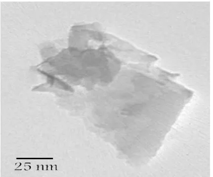
TEM, the Graphene were dispersed in absolute ethanol and sonicated at mild condition. Then the solution was casted onto carbon coated Cu grids. The TEM image of CVD Graphene was shown in fig.2.
Fig. 2 exhibits a high magnification TEM image of CVD Graphene, showing a completely sheet structure and ordered with few layers [26].
Figure2. TEM image of CVD Graphene.
2.3. Oxidation Process of Graphene
The structure and properties of functionalized graphene depend on particular synthesis method and degree of oxidation. Graphene nanosheets have chemically reactive oxygen functionality, such as carboxylic acid, groups at their edges during oxidation process. Therefore FG has been prepared by oxidation of Graphene in acid or alkaline methods. It should be noted that FG demonstrate considerable variations of properties depending on degree of oxidation and synthesis method. The oxidants we have tried include a mixture of H2SO4 and HNO3 and potassium per sulfate (KPS) in alkali solution. These methods are effective in hydrophilic the graphene structure but are very different in oxidation ability. The oxidation processes were as follows:
2.3.1. Acid Treatment of Graphene
Nitric and sulfuric acids are common oxidizing agents and react strongly with aromatic carbon surfaces, including graphene nanosheets [27]. In a typical example, 120 mg of graphene was suspended in 100 mL of a 3:1 mixture of concentrated H2SO4 (98 wt %) /HNO3 (16M) and sonicated in a water bath at 60°C for 120min. The resultant suspension was then diluted with 1000 mL of distillated water, and the graphene were collected on a 100nm pore membrane filter and washed with deionized water as follows [28]. The samples formed from Graphene treated with acid mixture were designated as AGNS. The reaction scheme for the acid treatment of Graphene shows in fig. 3.
Figure 3. Schematic of Graphene oxidation
by acid treatment.
According to fig 3, carboxylic groups were present at the periphery of the edge graphene sheets.
2.3.2. Alkaline Treatment of Graphene
Figure4. Schematic of Graphene oxidation
by KPS treatment.
3. RESULTS AND DISCUSSION 3.1. FTIR Analysis
All of oxidation processes are effective on graphene structure but are very different in oxidation ability. First the oxidized graphene are sonicated, centrifuged, dried, and then analyzed to conform of presence of variety functional groups. Therefore in order to study the structural changes in the Graphene after oxidation treatment, FTIR
analysis was performed. FTIR
spectrometer (Thermo Scientific, Nicolet 6700) was recorded typically 100 scans over the range 4504000cm-1 were taken from each sample with a resolution of 2cm -1 and summed to provide the spectra. Fig. 5
shows FTIR spectra for prepared of the functionalized Graphene and compared to CVD graphene.
Figure5. FTIR spectra for a) CVD
graphene b) acid oxidation c) alkaline oxidation.
According to fig 5 (a), the peak at 1574 cm-1 corresponds to the active phonon mode of the Graphene. This peak is assigned to the (C=C) stretching mode which it can be attributed aromatic structures [30]. The FTIR spectra were very different from this after oxidation. A
typical FTIR spectrum of Graphene treated with the acid mixture is shown in Fig 5 (b), in which a new peak appears around 1718 cm-1. It is normally assigned to the (C=O) strength vibration in the carboxylic groups (-COOH) [31] and the broad peak at 3400 cm-1 corresponds to the presence of the oxygenated groups [32]. These peaks indicate successful generation of (-COOH) groups on graphene.
In the case of the oxidized Graphene by alkaline treatment with KPS, the two main peaks observed. A peak in the 810 cm-1 due to the symmetrical stretching of the (C–S) [32], and peaks of 1233 cm-1 , 1578 cm-1 are associated with resonance of the (C-O) and stretching of the (C-O) respectively related to carboxylate groups (-COO) [33]. Therefore (-COO) group was formed in KGNS structure.
3.2. Raman Spectroscopy
Raman spectroscopy is a powerful tool to characterize the degree of functionalizing of nano structures. Raman spectrum of Graphene has a D band in 1350 cm-1 and a G band in 1600 cm-1[27]. The ratio between D and G band is a good indicator of the quality on bulk samples and is very important factor in the way that allows distinguishing between the functional samples after treatment with different agents. Increased ID/IG ratio corresponds to the increased degree of functionalizing [34]. Therefore Raman spectroscopy (RENISHAW RM1000-Invia) was used to investigate graphene structural changes during the oxidation process. Fig. 6 compares the Raman of graphene with functionalized structures.
International Journal of Nanoscience and Nanotechnology 15
Figure 6. Raman spectrum of Graphene,
AGNS & KGNS.
graphene structure than alkaline method. Concentrations of the functional groups were determined by Boehm titration method [35]. Theresults indicated that the concentrations of the total functional groups are 2.4 and 3.6 mmol/gr for graphene alkaline oxide and graphene acid oxide respectively. The obvious increase on ID/IG ratio of acid oxidation and result of Boehm titration, suggests that the high degree of functionalizing of Graphene is obtained in acid oxidation processes. But KPS is very good at generating few defects on graphene sheets, which can be seen from the Raman results of long time treatment, whereas concentrated H2SO4 and HNO3 needs short time to have more effect on graphene.
3.3. Nanofluid Preparation
3.3.1. Preparation of Functionalized Graphene Nano Fluids
The required amount of FG were determined to prepare 0.05, 0.15 and 0.25 wt% NFs using water as base fluid. The mixture was placed in ultrasonic bath for 30 min in order to attain the complete dispersion of FG. The properties of FG/NFs were summarized in table 1. The NFs made in this way were found to be very stable for weeks without visually observable sedimentation.
Table1. Properties of the graphene oxide
nanofluids.
Sample name
Graphene oxide (wt %)
Water (wt %)
Oxidation media
AGNS-1 0.05 99.95
Acid AGNS-2 0.15 99.85
AGNS-3 0.25 99.75 KGNS-1 0.05 99.95
Alkaline KGNS-2 0.15 99.85
KGNS-3 0.25 99.75
3.3.2. Stability of Functionalized Graphene Nano Fluids
UV-Spectral absorbency analysis is efficient way to evaluate the stability of nanofluids. In order to ensure the purity of graphene NFs, UV-visible absorption spectrum analysis is carried out (Fig. 7). Absorption spectrum measurements are taken after the preparation of FG/NFs.
Figure7. UV-vis absorption spectrum of
the graphene oxide nanofluid.
3.3.3. Thermal conductivity of FG nano fluids
The thermal conductivity was measured using a KD2 Pro thermal properties analyzer (Decagon devices, Inc., USA) and meets the standard of ASTM D5334. The thermal conductivity measurements were carried out three times for each sample at temperatures between 2560°C in steps of 10°C, which ensured the repeatability of the response. The average value of measured thermal conductivity is curved in fig 8.
Figure8. Thermal conductivity of the
graphene oxide nanofluids.
Fig. 8 depicts the thermal conductivity enhancements of FG/NFs as a function of FG concentration. Also in the range from 10 to 60°C, thermal conductivity of the NFs increases.
For 0.25 wt % of alkaline FG/NFs (KGNS-3), the enhancement ratio is 24.4% at 20°C and 33.9% at 60°C. There is a linear relationship between the enhancement ratios and FG concentrations. The NFs containing FG show different thermal conductivity enhancement behaviors, and the thermal conductivity enhancement ratios remain constant when temperatures differ. This indicates that many factors affect the thermal conductivity enhancement.
One of the factors is the structure of graphene that oxidize during oxidation process. This fact indicates that oxidation process in alkaline media have a less defect on graphene structure therefore thermal
conductivity of alkaline FG/NFs is more than acid FG/NFs in same concentration. Alkaline FG/NFs have better thermal transport than acid FG/NFs because it consists of potassium as a metallic agent in its structure.
4. CONCLUSION
Systematic investigation of the effect of different functionalization method on the structure of graphene was studied. The results reveal that different functional groups can be introduced when the graphene is treated with different oxidants. The FTIR and Raman results clearly show that the hydrophilic groups such as hydroxyls and carboxylic have been introduced onto the treated graphene surface. Therefore the functionalized graphene can be dispersed directly in polar solvents without the addition of any surfactant.
However, the acid mixture is strong enough to generate an abundance of functional groups. As the treatment time increases, we get more COOH) and (-OH) with the acid mixture, which lowers the frequency of the (C=O) stretch.
The thermal conductivity of NFs of two different FG structures which were prepared through oxidation process was investigated. Clear thermal conductivity enhancements were observed in all types of FG/NFs.
International Journal of Nanoscience and Nanotechnology 17
REFERENCES
1. Li J., Kleinstreuer C., (2008). “Thermal performance of nanofluid flow in micro channels”, International Journal of Heat and Fluid Flow, 29(4): 1221–1232.
2. Safi M.A., Ghozatloo A., Hamidi A.A., Shariaty-Niassar M., (2014).“Calculation of Heat Transfer Coefficient of MWCNT-TiO2 Nanofluid in Plate Heat Exchanger”, Int. J. Nanosci. Nanotechnol., 10(3): 153-162.
3. Masuda H., Ebata A., Teramae K., Hishinuma N., (1993). “Alteration of thermal conductivity and viscosity of liquid by dispersing ultra-fine particles (dispersion of Al2O3, SiO2 and TiO2 ultra-fine particles)”, Netsu Bussei, 7(4): 227–233.
4. Eastman J.A., Choi U.S., Li S., Thompson L.J., Lee S., (1997). “Enhanced thermal conductivity through the
development of nanofluids”, In Nanophase and Nanocomposite Materials II. Materials Research Society. 5. Teng T.P., Hung Y.H., Teng T.C., Mo H.E., Hsu H.G., (2010). “The Effect of Alumina/Water Nanofuid
Particle Size on Thermal Conductivity”, Applied Thermal Engineering, 30: 2213-2218.
6. Nasiri A., Shariaty-Niasar M., Morad Rashidi A., Amrollahi A., Khodafarin R., (2012). “Effect of CNT structures on thermal conductivity and stability of nanofluid”, International Journal of Heat and Mass Transfer, 55(5): 1529-1535.
7. Li Y., Zhou J., Tung S., Schneider E., Xi S., (2009). “A review on development of nanofluid preparation and characterization”, Power Technology, 196(2): 89-101.
8. Wei Y., Huaqing X., Dan B., (2010). “Enhanced thermal conductivities of nanofluids containing graphene oxide Nanosheets”, Nanotechnology, 21(5): 055705-1-8.
9. Shaikh S., Lafdi K., Ponnappan R., (2007). “Thermal conductivity improvement in carbon nanoparticle doped PAO oil: An experimental study”, Appl. Phys, 101: 064302-7.
10. Ding Y., Alias H., Wen D., Williams R.A., (2006). “Heat transfer of aqueous suspensions of carbon nanotubes (CNT nanofluids)”, International Journal of Heat and Mass Transfer, 49(1-2): 240-250
11. HB M., Wilson C., Borgmeyer B., Park K., Yu Q., Choi S., Tirumala M., (2006). “Effect of nanofluid on the heat transport capability in an oscillating heat pipe”, Applied Physics Letters, 88: 143116-3.
12. Yang Y., Zhang Z.G., Grulke E.A., Anderson W.B., Wu G., (2005). “Heat transfer properties of nanoparticle-in-fluid dispersions (nanofluids) in laminar flow”, International Journal of Heat and Mass Transfer, 48(6): 1107-1116.
13. Zhu H.T., Zhang C.Y., Tang Y.M., Wang J.X., Ren B., Yin Y., (2007). “Preparation and thermal conductivity of suspensions of graphite nanoparticles”, Carbon, 45(1): 226-228.
14. Ghozatloo A., Shariaty-Niasar M., Rashidi A.M., (2014). “Investigation of Heat Transfer Coefficient of Ethylene Glycol/ Graphenenanofluid in Turbulent Flow Regime”, Int. J. Nanosci. Nanotechnol., 10(4): 237-244.
15. Wei Y., Huaqing X., Xiaoping W., Xinwei W., (2011). “Significant thermal conductivity enhancement for nanofluids containing grapheme Nanosheets”, Physics Letters A, 375(10): 1323–1328.
16. Tessy T.B., Ramaprabhu S., (2010). “Investigation of thermal and electrical conductivity of graphene based nanofluids”, Applied Physics, 108: 124308-11.
17. Gupta S.S., Siva V.M., Krishnan S., Sreeprasad T.S., Singh P.K., Pradeep T., (2011). “Thermal conductivity enhancement of nanofluids containing graphene nanosheets”, Applied Physics, 110: 084302-15.
18. Ghozatloo A., Shariaty-Niasar M., Rashidi A.M., (2013). “Preparation of nanofluids from functionalized Graphene by new alkaline method and study on the thermal conductivity and stability”, International Communications in Heat and Mass Transfer, 42(1): 89–94
19. Nasiri A., Shariaty-Niasar M., Rashidi A.M., Amrollahi A., Khodafarin R., (2011). “Effect of dispersion method on thermal conductivity and stability of nanofluid”, Experimental Thermal and Fluid Science, 35(4): 717–723.
20. Hwang Y.J., Ahn Y.C., Shin H.S., Lee C.G., Kim G.T., Park H.S., Lee J.K., (2006). “Investigation on characteristics of thermal conductivity enhancement of nanofluids”, Curr. Appl. Phys., 6:, 1068–1071. 21. Chen L.F., Xie H.Q., Li Y., Yu W., (2009). “Carbon nanotubes with hydrophilic surfaces produced by a
22. Tchoul M.N., Ford W.T., Lolli G., Resasco D.E., Arepalli S., (2007). “Effect of mild nitric acid oxidation on dispersability, size and structure of single-walled carbon nanotubes”, Chem. Mater, 19(23): 5765–5772. 23. Assael M.J., Chen C.F., Metaxa I., Wakeham W.A., (2004). “Thermal conductivity of suspensions of carbon
nanotubes in water”, Thermophys, 25(4): 971–985
24. Guoxiu W., Juan Y., Park J., Xinglong G., Wang B., Liu H., Yao J., (2008). “Facile Synthesis and Characterization of Graphene Nano sheets”, Phys. Chem. C, 112(22): 8192–8195.
25. Hae M.J., Sung H.C., Seung H.H., (2010). “X-ray Diffraction Patterns of Thermally-reduced Graphenes”,
the Korean Physical Society, 57(61): 1649-1652.
26. Zheng D., Cai Z.B., Shen M.X., Li Z.Y., Zhu M.H., (2016). “Investigation of the tribology behaviour of the graphene nanosheets as oil additives on textured alloy cast iron surface”, Applied Surface Science, 387: 66-75.
27. Lakshminarayanan P.V., Toghiani H., Jr C.U., (2004). “Nitric acid oxidation of vapor grown carbon nanofibers, Carbon”, Carbon, 42(12-13): 2433–2442.
28. Pei S., Zhao J., Du J., Ren W., Cheng H., (2010). “Direct reduction of graphene oxide films into highly conductive and flexible graphene films by hydrohalic acids”, Carbon, 48(15): 4466–4474.
29. Zhang J., Hongling Z., Quan Q., Yanlian Y., Qingwen L., Zhongfan L., Xinyong G., Zuliang D., (2003). “Effect of Chemical Oxidation on the Structure of Single-Walled Carbon Nanotubes”, Phys. Chem. B, 107(16): 3712-3718.
30. Rike Y., Holia O., Sudirman Y., Sait O., Tadahisa I., Jun I.A., (2011). “Analysis of functional group sited on multi-wall carbon nanotube surface”, The Open Materials Science Journal, 5: 242–247.
31. kyung O.P., Jeevananda T., Nam H.K., Seong K., Joong H.L., (2009). “Effect of surface modification on the dispersion and electrical conductivity of carbon nanotube/polyaniline composite”, Scripta Materialia, 60(7): 551–554.
32. Pan D., Wang S., Zhao B., Wu M., Zhang H., Wang Y., Jiao Z., (2009). “Li Storage properties of disordered graphene nanosheets”, Chemistry of Materials, 21(14): 3136–3142.
33. Ghozatloo A., Rashidi A.M., Shariaty-Niassar M., (2014). “Convective heat transfer enhancement of graphene nanofluids in shell and tube heat exchanger”, Experimental Thermal and Fluid Science, 53: 136– 141.
34. Zhang L., Qing Q.N., Yaqin F., Toshiaki N., (2009). “One-step preparation of water-soluble single-walled carbon nanotubes”, Applied Surface Science, 255(15): 7095–7099.
35. Hung L.C., Kuo H.L., Shih Y.C., Ching G.C., San D.P., (2007). “Dye adsorption on biosolid adsorbents and commercially activated carbon”, Dyes and Pigments, 75(1): 52-59.
36. Wei Y., Huaqing X., (2012). “Review Article, A Review on Nanofluids: Preparation, Stability Mechanisms, and Applications”, J. Nanomaterials, 2012: 1-17.