Effects of Electrocauterization Smoke on Nasal Mucosa in
Rats
Introduction
The improvements and simplification of treatment methods, especially the surgery, are due to the advances in medical procedures and equipment. However, some of these tools are problematic regardless of being useful. Electrosurgical unit (electrocautery) is one of such devices that is used in the operating room. In addition, electrocautery is the most common heat-producing device that utilizes the high-frequency electrical current to cut or coagulate the targeted tissue in the surgery. This heat causes the boiling of the cellular contents thus releases the plume into the air that is called “surgical smoke” (1,2).
The Occupational Safety and Health Administration in the United States estimates that 500 000 workers in the
US are exposed to surgical smoke and many health care professionals such as surgeons and the operating room technicians are exposed to it for several hours a day for years (1).
Further, surgical smoke contains 95% water and 5% toxic and mutagenic components such as acetaldehyde, formaldehyde, toluene, ethylene, phenol, xylene, and benzene (2-4). Unfortunately, standard surgical masks do not adequately filter surgical smoke particles because these masks generally filter particles of about 5 µm in size and about 77% of the particulate matter in the smoke is 1.1 µm and smaller. In addition, surgical masks are a relatively effective filter for the exhaled air instead of the inhaled air (5-7). Hence, plume inhalation is a workplace hazard in Abstract
Objectives: Electrocautery unite is used for vascular homeostasis. Despite the advantage of electrocautery in surgical interventions, it has several disadvantages such as pulmonary respiratory disorders, emphysema, bronchitis, and asthma. Given the above-mentioned explanation, the present study investigated the effects of electrocauterization smoke on rat nasal mucosa.
Materials and Methods: Fifteen Wistar rats were divided into 3 groups (n=5) for three-time phases. Smoke was produced by the electrocauterization of an anesthetized rat within the smoke chamber and then smoke was entered into the test chamber containing the experimental rats. Finally, rats were anesthetized and the nasal mucosa was dissected under a stereomicroscope. After Hematoxylin and Eosin staining, the samples were impartially evaluated by a pathologist, followed by comparing the frequency of complications between different groups. Eventually, the TUNEL (The terminal deoxynucleotidyl transferase dUTP nick end labeling) technique was utilized to investigate the apoptosis.
Results: Pathological studies showed complications such as vascular congestion, epithelial vacuolation, acute inflammation, and the presence of necrotic cells. In addition, statistical analysis indicated that the rate of vascular congestion, acute inflammation, and inflammation in glands in the second and third phases of the study significantly increased compared to the control group. Further, the difference between the experimental groups was only significant when the first phase was compared with the second (P<0.05). However, the rate of epithelial vacuolation and the presence of necrotic cells demonstrated no significant increase in comparison to the control group. Contrarily, the numerical density of apoptotic cells significantly increased within the respiratory epithelium and submucosal glands of experimental rats (P < 0.05).
Conclusions: The results of this study indicated that short-term exposure to electrocauterization smoke has no specific effect, but longer duration of smoke exposure can damage the rat nasal mucosa.
Keywords: Electrocauterization smoke, Complication, Nasal mucosa, Rat
Hoda Khoshdel Sarkarizi1, Ramin Salimnejad2, Amir Hosein Jafarian3, Masoumeh Fani1, Nasim Khajavian4,
Esmail Nourmohamadi5, Ghasem Sazegar1*
Open Access Original Article
Crescent Journal of Medical and Biological Sciences
Received 17 February 2018, Accepted 19 July 2018, Available online 3 August 2018
1Department of Anatomical Sciences and Cell Biology, Faculty of Medicine, Mashhad University of Medical Sciences, Mashhad, Iran. 2Research Laboratory for Embryology and Stem Cells, Department of Anatomical Sciences and Pathology, School of Medicine, Ardabil
University of Medical Sciences, Ardabil, Iran. 3Department of Pathology, Ghaem Hospital, Mashhad University of Medical Sciences, Mashhad,
Iran. 4Department of Biostatistics and Epidemiology, Faculty of Health, Mashhad University of Medical Sciences, Mashhad, Iran. 5Department eISSN 2148-9696
the operating room because of its health consequences on the surgical teams, including respiratory disorders (8,9), dizziness, coughing, nausea and vomiting, headache, and tearing (10,11). Furthermore, this smoke is a vehicle for the transition of malignant cells such as hepatitis, human immunodeficiency virus, (12) and human papillomavirus (13, 14).
Respiratory disorders encompass asthma, pulmonary congestion, chronic bronchitis and emphysema, inflammatory responses, pneumonia, alveolar congestion, hypertrophy, and the hyperplasia of the respiratory epithelium (8,9). The nasal mucosa is the first region of the respiratory system that is exposed to the electrocauterization smoke, and then, the smoke enters the lung after passing through the ventilation system of the nose. Therefore, a high volume of particles could become trapped in the nose and fail to reach the lungs, causing the health risks associated with the plume which was exposed to the nasal mucosa. Accordingly, this study was performed to evaluate the effects of electrocauterization smoke on the nasal mucosa of the rat.
Materials and Methods Animals and Study Design
Fifteen adult Wistar rats were obtained from the Animal House of Mashhad University of Medical Sciences and were kept in the same place under the controlled conditions (22-24°C and 12 hours light/darkness cycle with natural light), followed by receiving the standard pellet with tap water except for the experiment time. Then, the rats were randomly categorized into three groups (n=5) for three phases, one rat as control and 4 rats as experimental in each group. The experimental rats were exposed to the electrocauterization smoke while the control rats were exposed to distilled water steam under a similar condition.
Time Phases
Phase 1: Plume exposure for two minutes, followed by 2-minute exposure for the rest. This procedure was repeated for 4 days and 4 times daily.
Phase 2: Plume exposure for 4 minutes, followed by 2-minute exposure for the rest, which was repeated for seven days and 4 times daily.
Phase 3: Plume exposure for 4 minutes and then 2 minutes for the rest, which was repeated for 14 days and 4 times daily.
During the rest, the animal remained within the test chamber while no smoke or steam was entered into the chamber (8, 9).
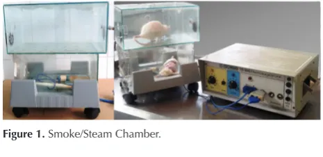
Smoke/Steam Applying
In our study, smoke or steam was applied from a glass device that included the test (the upper one) and smoke/ steam (the lower one) chambers. Then, the experimental rat and one anesthetized cauterizing rat were placed in the
test and within the smoke chambers, respectively. For the control group, generated distilled water steam was entered into the steam chamber. Moreover, the separating wall, which was the floor of the test chamber and the roof of the smoke chamber, between the chambers had several small holes for transferring smoke or steam from the smoke/ steam chamber to the test chamber based on the upward flowing of the smoke or steam (Figure 1).
Test Chamber
Each of 2 lateral walls of the test chamber had one hole for air entrance and another for smoke/steam exhaust by suctioning (i.e., suction machine, MEDASA, and CV2). The anterior wall had a door to pass the experimental rat as well (Figure 1).
Smoke/Steam Chamber
The posterior wall had an opening to enter the cauterizing hands and the anterior wall, similar to the test chamber, had a door for the passage of the anesthetized rat. Electrocautery plaque was also fixed to the floor of this chamber.
Electrocauterization was conducted using an electrocautery machine (ESCHMAN TD 300, Britain) to observe the visible smoke in the test chamber, followed by anesthetizing the cauterizing rat. Next, the subcutaneous tissue was cauterized after skin incision. At the end of the daily experiment, the anesthesia of the cauterizing rat was continued to die and created suctioning continued smoke/steam circulation within the test chamber. Finally, behavioral changes in the rats were investigated during the test.
Preparation of Tissues
At the end of the experiment, the rats were anesthetized with ketamine (90 mg/kg, i.p.). Then, the animals were decapitated after intracardiac perfusion and their heads were fixed in %10 Formalin. Next, nasal mucosa was dissected under a stereomicroscope by the sagittal incision of the nose. Tissue processing and embedding in the paraffin was performed and each nasal mucosa was exhaustively sectioned into 5 μm thickness. Eventually, to assess the pathological changes, the samples were stained with Hematoxylin and Eosin, and the terminal deoxynucleotidyl transferase dUTP nick end labeling (TUNEL) assay was conducted to investigate the apoptosis.
TUNEL Assay
The tissue sections were treated with 3% H2O2 in methanol in darkness at ambient temperature for 10 minutes after deparaffinization, rehydration, and rinsing in 0.1 M phosphate buffered saline (PBS) for 15 minutes. Then, they were rinsed in PBS (for 15 minutes) and treated with proteinase K (Roche, Germany) at the room temperature for 15 minutes. The sections were also incubated in the labeling reaction mixture of the TUNEL kit (Roche, Germany) at 4°C overnight. Next, they were incubated in Convertor-POD for 1.5 hours. Finally, the sections were treated with 0.03% Diaminobenzidine (DAB; Sigma-Aldrich, USA) solution for 10 minutes and washed with running water. Eventually, the sections were mounted after counterstaining with Hematoxylin, dehydration, and clearing. Apoptotic nuclei are dark brown in this method (15).
Quantification Analysis
Micrographs were captured by a light microscope (BX51, Japan) connected to a camera (DP12, Japan). Then, the sections, stained with Hematoxylin and Eosin, were impartially evaluated by a pathologist, followed by reporting the observed pathological changes. Additionally, the frequency of the reported changes was measured and compared between different groups and the number of TUNEL-positive cells in the images of the TUNEL technique was counted using a 1000 μm2 counting frame and the following formula:
/ .
A N
a fQQ
Σ =
Σ
NA and ΣQ are the number of apoptotic cells per unit area and the sum of the counted particles in the sections, respectively. In addition, a/f and ΣP denote the area associated with each frame and the sum of the counted frames in the sections.
Statistical Analysis
The obtained data were analyzed by SPSS, version 20. The frequency of each of the reported pathological changes in the groups was determined and the results were compared by the chi-square and Man-Whitney tests. Further, the data from the TUNEL technique were analyzed using one-way ANOVA and post hoc Tukey test was utilized to compare the differences between the groups. The level of significance was estimated at P<0.05.
Results
Behavioral Changes
During the smoke exposure, the rats became sluggish, their movements ceased, and they went into a squatting position (Figure 2a). Smoke exposure was discontinued in the rest periods and the animals resumed their activities (Figure 2b), but re-exposure with smoke caused their activities to stop once more. No cyanotic symptom or
death was observed in the subjects.
Pathological Assessment
The samples showed pathological changes such as vascular congestion, epithelial vacuolation, and the existence of necrotic cells, as well as acute inflammation, and inflammation in the glands based on the presence of inflammatory cells. The results related to the comparison of the frequency of reported pathological changes between the different groups are as follows:
Vascular Congestion
The comparison between the control and first phase group represented no significant difference, but there was a significant increase in the frequency of the reported vascular congestion in the second and third phase groups when compared to the control group (P < 0.05). Furthermore, as shown in Figures 3a and 4, the only difference between the first and second phase groups was significant based on the comparison between the three experimental groups (P < 0.05).
Epithelial Vacuolation
Statistical analysis revealed that the frequency of the reported epithelial vacuolation failed to significantly increase in the experimental groups when compared to the control group and as compared together (Figures 3b & 4).
Necrotic Cell
Based on the results, necrotic cells were observed in the samples of the second and third phase groups, but no significant increase was found when compared with the control group (Figures 3c & 4).
Acute Inflammation
Exposure to electrocauterization smoke within the two groups in the second and third phases caused acute inflammation and significantly increased the signs of inflammation in comparison with the control rats (P < 0.05). However, the difference between the first phase and the control group was not significant. Comparing the experimental groups, the second phase group revealed
Figure 2. (a) During the Smoke Exposure; (b) During the Rest Period.
a significant increase in the frequency of reported acute inflammation when compared to the first phase group (P < 0.05) while the other group demonstrated no significant change in this regard (Figures 3d & 4).
Inflammation of Glands
Similarly, the signs of inflammation were observed in the glands of the samples of the second and third phase groups and a significant increase was detected when compared to the control group (P < 0.05) although there was no significant difference between the first phase and control. On the other hand, the difference between the experimental groups was only significant between the first and second phase groups (Figures 3d & 4).
Apoptosis Assay
The results of the terminal deoxynucleotidyl transferase dUTP nick end labeling (TUNEL) assay indicated that the exposure of nasal mucosa to electrocauterization smoke increased the apoptotic cell number in the respiratory
Figure 3. The Images of Rat Nasal Mucosa Demonstrating the Reported-pathological Changes. Note.The black arrows display necrotic cells.
Figure 4. The Percent of Reported-Pathological Changes in Different Groups. Note.* P<0.05 compared to the control group; **P<0.05 compared to the experimental group.
Figure 5. The Images of Rat Nasal Mucosa Showing Apoptotic Cells in Different Groups. Note. The yellow arrows show the terminal deoxynucleotidyl transferase dUTP nick end labeling-positive cells. epithelium and submucosal glands. As shown in Figure 5 of the respiratory epithelium, the numerical density of apoptotic cells in all three time phases considerably increased compared to the control group (P < 0.05). Moreover, there was a significant difference (P < 0.05) between the first and second phase groups (Figures 5-7). Based on the data in Figures 6 and 7, the comparison between TUNEL-positive cell number in submucosal glands showed an obvious increase in the experimental groups when compared to the control (P < 0.05).
Discussion
The purpose of the current study was to evaluate the effects of electrocauterization smoke on the nasal mucosa. To the best of our knowledge, this is the first study that investigates the effects of electrocautery smoke with the histopathological methods and behavioral changes. The
results of this study confirmed that smoke or plume from electrocauterization has negative impacts on the nasal mucosa.
This study examined behavioral changes in the rats which were exposed to electrocautery smoke or plume such that they became lethargic during the exposure and returned to the normal state in the resting period. Our results are consistent with the findings of previous studies which addressed the effects of laser smoke on the lungs of the rats and observed several complications such as asthma, pulmonary congestion, chronic bronchitis and emphysema, as well as inflammatory responses, pneumonia, alveolar congestion, hypertrophy, and the hyperplasia of the respiratory epithelium (8, 12).
Those rats that were exposed to electrocauterization smoke showed the signs of inflammation, which was significantly higher when compared to the control group. There was no significant difference in vacuolation and necrotic cells between the experimental and control groups. The findings of a similar study, investigating the effects of smoke produced by electrocautery on the laryngeal mucosa, revealed a significant difference between the experimental and control groups regarding the inflammation (16).
Likewise, Navarro-Meza et al. designed a study on physicians and reported that the rate of exposure to smoke in neurosurgeons had the highest and electrocautery smoke caused respiratory symptoms such as a sore throat, the sensation of a lump in the throat, and nasal congestion in all physicians (17). Similarly, Choi et al concluded that the toxic substances in the plume of electrocauterization smoke increased the risk of cancer (18).
In addition, the results of this study indicated that electrocautery smoke could increase apoptotic cells in the nasal mucosa. Other studies mainly focused on hazardous chemicals and their concentration in electrocautery smoke. For instance, Al Sahaf et al (19) found the concentration of chemical substances in the smoke generated by electrocautery was similar to cigarette smoke (i.e., toluene
Figure 7. The Mean of TUNEL-positive Cell Number in the Respiratory Epithelium and Submucosal Glands in Different Groups. Note.* P<0.05 compared to the control group; **P<0.05 compared to the experimental group.
and ethylbenzene). The thermal destruction of 1 g of the tissue by electrocautery was estimated to produce the levels of the harmful chemical similar to that produced by 6 cigarettes (20). In addition, the researchers showed that ethylene, toluene, xylene, and benzene were among the most hazardous substances found in electrocautery smoke that could cause cell damage, which is in line with the findings of our study indicating that the effects of the above substances could probably increase apoptotic cells.
Conclusions
Overall, the results of this study indicated that short-term exposure to electrocauterization smoke has no specific effect while the longer duration of smoke exposure can damage the rat nasal mucosa. Further, there is growing evidence that electrocautery smoke is harmful to human health. Thus, surgeons and operating room staff should be informed about the harmful substances of electrocauterization smoke and measures should be taken to reduce this potential risk. It is noteworthy that the use of standard surgical masks alone provides no adequate protection from electrocauterization smoke. Therefore, filter masks with higher quality and/or double masking may reduce this serious occupational risk.
Conflict of Interests
The authors declare that they have no conflicts of interest.
Ethical Issues
The procedures involving the animals were in accordance with the Guide for the Care.
Financial Support
The present study was supported by the Deputy Research of Mashhad University of Medical Sciences by a grant No. 922174).
Acknowledgments
We would like to thank the Vice-chancellor of Research of Mashhad University of Medical Sciences for financial support. In addition, the authors gratefully thank Mrs. Motejaded and other depended personals who helped us in this study.
References
1. Ulmer BC. Report of OSHA’s draft: information for health care workers exposed to laser and electrosurgery smoke. Todays Surg Nurse. 1999;21(2):18-19.
2. Ulmer BC. The hazards of surgical smoke. AORN J. 2008;87(4):721-734; quiz 735-728. doi:10.1016/j. aorn.2007.10.012
3. Weld KJ, Dryer S, Ames CD, et al. Analysis of surgical smoke produced by various energy-based instruments and effect on laparoscopic visibility. J Endourol. 2007;21(3):347-351. doi:10.1089/end.2006.9994
composition of smoke produced by high-frequency electrosurgery in a closed gaseous environment. An in vitro study. Surg Endosc. 1998;12(8):1017-1019.
5. Chen CC, Willeke K. Aerosol penetration through surgical masks. Am J Infect Control. 1992;20(4):177-184. doi:10.1016/S0196-6553(05)80143-9
6. Kunachak S, Sobhon P. The potential alveolar hazard of carbon dioxide laser-induced smoke. J Med Assoc Thai. 1998;81(4):278-282.
7. Weber A, Willeke K, Marchioni R, et al. Aerosol penetration and leakage characteristics of masks used in the health care industry. Am J Infect Control. 1993;21(4):167-173. doi:10.1016/0196-6553(93)90027-2
8. Baggish MS, Elbakry M. The effects of laser smoke on the lungs of rats. Am J Obstet Gynecol. 1987;156(5):1260-1265. doi:10.1016/0002-9378(87)90158-x
9. Wenig BL, Stenson KM, Wenig BM, Tracey D. Effects of plume produced by the Nd:YAG laser and electrocautery on the respiratory system. Lasers Surg Med. 1993;13(2):242-245. doi:10.1002/lsm.1900130213
10. Garden JM, O’Banion MK, Bakus AD, Olson C. Viral disease transmitted by laser-generated plume (aerosol). Arch Dermatol. 2002;138(10):1303-1307. doi:10.1001/ archderm.138.10.1303
11. Alp E, Bijl D, Bleichrodt RP, Hansson B, Voss A. Surgical smoke and infection control. J Hosp Infect. 2006;62(1):1-5. doi:10.1016/j.jhin.2005.01.014
12. Baggish MS, Poiesz BJ, Joret D, Williamson P, Refai A. Presence of human immunodeficiency virus DNA in laser smoke. Lasers Surg Med. 1991;11(3):197-203. doi:10.1002/ lsm.1900110302
13. Ferenczy A, Bergeron C, Richart RM. Human papillomavirus DNA in CO2 laser-generated plume of smoke and its consequences to the surgeon. Obstet Gynecol.
1990;75(1):114-118.
14. Sawchuk WS, Weber PJ, Lowy DR, Dzubow LM. Infectious papillomavirus in the vapor of warts treated with carbon dioxide laser or electrocoagulation: detection and protection. J Am Acad Dermatol. 1989;21(1):41-49. doi:10.1016/s0190-9622(89)70146-8
15. Hazrati A, Salimnejad R, Alipour MR, Mirzaei Bavil F, Alihemmati A. Protective effect of ghrelin on testicular damages caused by chronic hypoxia in rats: A histopathological study. Andrologia. 2018. doi:10.1111/ and.12989
16. Fitzgerald JE, Malik M, Ahmed I. A single-blind controlled study of electrocautery and ultrasonic scalpel smoke plumes in laparoscopic surgery. Surg Endosc. 2012;26(2):337-342. doi:10.1007/s00464-011-1872-1
17. Navarro-Meza MC, González-Baltazar R, Aldrete-Rodríguez MG, Carmona-Navarro DE, López-Cardona MG. [Respiratory symptoms caused by the use of electrocautery in physicians being trained in surgery in a Mexican hospital]. Rev Peru Med Exp Salud Publica. 2013;30(1):41-44. doi:10.1590/S1726-46342013000100008 18. Choi SH, Kwon TG, Chung SK, Kim TH. Surgical smoke
may be a biohazard to surgeons performing laparoscopic surgery. Surg Endosc. 2014;28(8):2374-2380. doi:10.1007/ s00464-014-3472-3
19. Al Sahaf OS, Vega-Carrascal I, Cunningham FO, McGrath JP, Bloomfield FJ. Chemical composition of smoke produced by high-frequency electrosurgery. Ir J Med Sci. 2007;176(3):229-232. doi:10.1007/s11845-007-0068-0 20. Tomita Y, Mihashi S, Nagata K, et al. Mutagenicity of
smoke condensates induced by CO2-laser irradiation and electrocauterization. Mutat Res. 1981;89(2):145-149. doi:10.1016/0165-1218(81)90120-8