R E S E A R C H A R T I C L E
Open Access
Initial and delayed thyroid-stimulating
hormone elevation in extremely
low-birth-weight infants
Shin Ae Yoon
1†, Yun Sil Chang
2†, So Yoon Ahn
2, Se In Sung
2and Won Soon Park
2*Abstract
Background:To determine the incidence, etiology, and outcomes of thyroid-stimulating hormone (TSH) elevation
in extremely low-birth-weight infants (ELBWIs).
Methods:Newborn thyroid screening data of 584 ELBWIs (birth weight, < 1000 g; gestational age,≥23 weeks) were retrospectively analyzed to identify initial (≤2 postnatal weeks) and delayed (> 2 weeks) TSH elevations. Growth and neurodevelopmental outcomes at 2 years’corrected age (CA) were assessed according to levothyroxine replacement.
Results:Initial and delayed TSH elevations were detected at CAs of 27 and 30 weeks, respectively, with incidence rates of 0.9 and 7.2%, respectively. All infants with initial TSH elevations had perinatal asphyxia, and 95% of those with delayed TSH elevation were exposed to various stressors, including respiratory support, drugs, and surgery within 2 weeks before diagnosis of TSH elevation. Free thyroxine (T4) levels were simultaneously reduced in 80 and 57% of infants with initial and delayed TSH elevations, respectively. Both initial and delayed TSH elevations were transient, regardless of levothyroxine replacement. Infants receiving levothyroxine replacement therapy had significantly higher TSH elevations, significantly lower free T4 levels, and significantly reduced mortality, compared to untreated infants. However, levothyroxine replacement had no significant effect on long-term growth and neurodevelopmental outcomes.
Conclusions:The timing of insult superimposition on hypothalamic–pituitary–thyroid axis maturation is a major determinant of initial or delayed TSH elevation in ELBWIs. Levothyroxine replacement did not affect growth or neurodevelopmental outcomes in this population.
Keywords:Thyroid function tests, Thyroxine supplementation, Preterm infants, Hypothalamic–pituitary–thyroid axis, Transient hypothyroxinemia of prematurity
Background
Thyroid-stimulating hormone (TSH) level is the preferred screening marker for the identification of infants with con-genital hypothyroidism. Hypothyroidism in term newborn infants can easily be identified by an elevated TSH level to-gether with a reduced thyroxine (T4) level after the second postnatal day [1]. However, it remains difficult to interpret TSH levels in extremely low-birth-weight infants (ELBWIs),
with birth weights < 1000 g. Transient hypothyroxinemia of prematurity (THOP), defined as a temporarily low T4 level with a normal or low TSH level, is the most common thy-roid dysfunction affecting preterm infants [2]. The etiology of THOP is considered to be multifactorial [2] and may in-clude the postnatal cessation of maternal placental T4 transfer [3,4], hypothalamic pituitary thyroid axis immatur-ity [5,6], and non-thyroidal illness (NTI) [7–10]. Nonethe-less, initial and delayed TSH elevations occur more frequently in preterm infants, especially ELBWIs, than in term infants [11,12].
A recent study suggested that, besides primary congeni-tal hypothyroidism, elevated TSH levels in preterm infants
© The Author(s). 2019Open AccessThis article is distributed under the terms of the Creative Commons Attribution 4.0 International License (http://creativecommons.org/licenses/by/4.0/), which permits unrestricted use, distribution, and reproduction in any medium, provided you give appropriate credit to the original author(s) and the source, provide a link to the Creative Commons license, and indicate if changes were made. The Creative Commons Public Domain Dedication waiver (http://creativecommons.org/publicdomain/zero/1.0/) applies to the data made available in this article, unless otherwise stated. * Correspondence:[email protected];[email protected]
†Shin Ae Yoon and Yun Sil Chang contributed equally to this work.
2Department of Pediatrics, Samsung Medical Center, Sungkyunkwan
University School of Medicine, 81 Irwon-Ro, Gangnam-gu, Seoul 06351, South Korea
(< 28 weeks of gestation) might be associated with previ-ous or concomitant inflammation, similarly to the non-thyroidal inflammation syndrome [13]. However, given the paucity of relevant clinical information, many factors remain unclear, including the true incidence of initial and delayed TSH elevation among ELBWIs, the clinical associ-ation of this condition with NTI, and the ability of thyrox-ine supplementation to improve outcomes in these cases. Therefore, the present retrospective observational study primarily aimed to determine the incidence of initial and delayed TSH elevation in ELBWIs. Secondarily, this study aimed to assess the clinical association of TSH elevation with NTI and to examine the effect of thyroxine supple-mentation on the growth, neurodevelopmental, and endo-crine outcomes of ELBWIs at the corrected age (CA) of 2 years.
Methods
We retrospectively reviewed the medical records of 584 ELBWIs (birth weights < 1000 g) with gestational ages (GAs) of ≥23 weeks who were born at and admitted to the Samsung Medical Center neonatal intensive care unit between January 2000 and July 2013, and for whom the results of initial thyroid function tests (TFTs) per-formed within the first 2 postnatal weeks were available. The data collection procedure was approved by the In-stitutional Review Board of Samsung Medical Center (2015–01-088), which waived the requirement for in-formed consent in this retrospective chart review.
Assays for TSH, T4, and free T4 (fT4) were performed using the Siemens ADVIA Centaur® XP chemilumines-cent competitive immunoassay kits (Tarrytown, NY, USA). In this study, initial TSH elevation was arbitrarily defined as a TSH level > 20.0μIU/ml [14,15] during the initial TFT screening within the first 2 postnatal weeks of life. Delayed TSH elevation was defined as a TSH level≤20.0μIU/ml, regardless of the T4 level, during the same screening and a later TSH level increase > 20.0 μIU/ml [15] on the subsequent follow-up TFT. TFT normalization was defined as a decrease in the TSH level to 0.7–7.0 μIU/ml, the T4 level to 4.5–12.5 ng/dl, and the fT4 level to 0.9–1.8 ng/dl. Follow-up TFT screening tests were repeated every 2–6 weeks if the TFT results were abnormal and/or the infant was critically ill (in-cluding the requirement for mechanical ventilation or surgery) until a follow-up TFT yielded normalized re-sults and the infant’s clinical condition stabilized. Levothyroxine supplementation for an initially relatively high (arbitrarily ≥40μIU/ml) and/or sustained TSH ele-vation was initiated at a dosage of 10–15μg/kg/d, based on recommendations from the attending pediatric endocrinologists.
Clinical characteristics, including GA, birth weight, Apgar scores at 1 and 5 min, sex, delivery mode, small
for GA (birth weight below the 10th percentile), pregnancy-induced hypertension, gestational diabetes mellitus, and antenatal steroid use, were analyzed. GA was determined based on the maternal last menstrual period and the modified Ballard test. To identify the clinical risk factors associated with the development of TSH elevation, we investigated various confounding var-iables, including exposure to drugs such as dopamine and steroids, and surgery, especially 2 weeks prior to the detection of TSH elevation.
Outcome measures, including death before discharge, bronchopulmonary dysplasia (BPD) (≥ moderate) [16], intraventricular hemorrhage (IVH) (≥grade 3) [17], peri-ventricular leukomalacia (PVL), necrotizing enterocolitis (NEC) (≥Bell’s stage 2b) [18], and retinopathy of prema-turity (ROP) [19] requiring laser treatment, were also analyzed.
Upon follow-up at a CA of 2 years, each infant’s head circumference, height, and body weight were measured for the growth assessment. These values were converted to sex- and age-specific z scores of weight, height, and head circumference using the lambda, mu, and sigma method and the 2007 Korean National Growth Charts database [20]. Catch-up growth was defined as a weight, height, or head circumference exceeding the 10th per-centile, according to the 2007 Korean National Growth Charts [20]. Neurodevelopmental factors such as cere-bral palsy, hearing impairment, blindness, and Bayley scores were assessed. Cerebral palsy was defined as a Palisano gross motor function score≥2. Blindness was defined as a visual acuity of < 20/200. Hearing loss was defined as bilateral impairment requiring hearing aids. Neurodevelopmental delay was defined as a mental de-velopmental index or psychomotor dede-velopmental index score < 70, according to the Bayley Scales of Infant De-velopment, Second Edition.
Statistical analysis
(SPSS Inc., Chicago, IL, USA) software was used for all statistical analyses, and a P-value< 0.05 was con-sidered statistically significant.
Results
In our study group, the mean GA and birth weight were 26 (range: 23–34) weeks and 769 ± 148 (370– 999) g, respectively. The initial screening test was done at a mean of 7 ± 3 (1–14) postnatal days, and the second test was performed at a mean of 21 ± 17 (7–101) days. TFT screening was performed a mean of 3.7 ± 2.9 (1–10) times at mean intervals of 25 ± 8 (15–41) days during a mean hospital stay of 103 ± 52 (15–547) days. Of the 584 ELBWIs, 5 had initial TSH elevation. Of the 328 infants with initially lower T4or
fT4 levels and normal TSH levels, 39 developed
de-layed TSH elevation according to the subsequent TFT results. On the initial TFT, 251 patients had normal T4 and TSH levels; of these, 3 had delayed TSH
ele-vations on the follow-up TFTs.
Incidence of initial and delayed TSH elevation
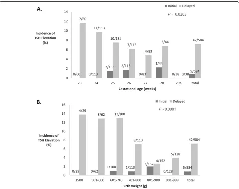
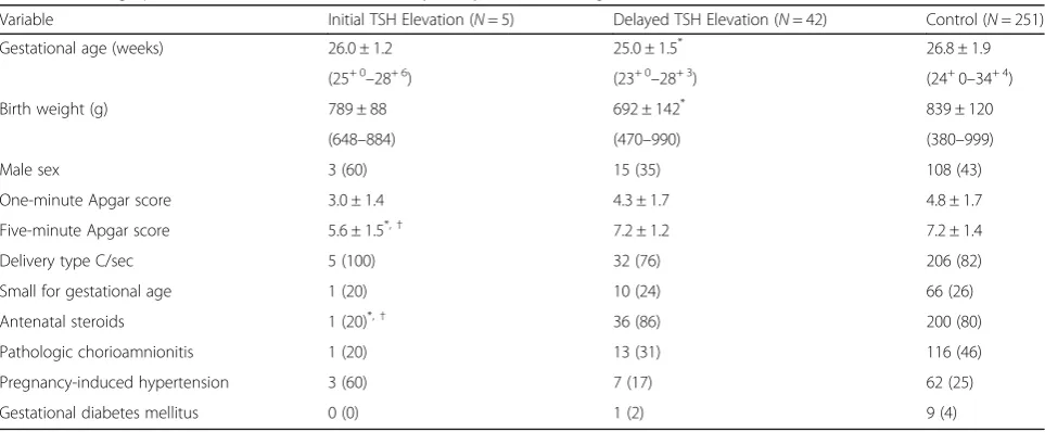
The overall incidence rates of initial and delayed TSH elevation among ELBWIs were 0.9% (5/584) and 7.2% (42/584), respectively. The incidence rates of initial and delayed TSH elevation among ELBWIs according to GA and birth weight are shown in Fig.1. Initial TSH eleva-tion was observed only in infants with a GA of 25–28 weeks’ (1.3%, 5/373), and the highest incidence rate of 2.0% (3/152) was observed among infants with a birth weight in the 801–900 g range. However, the incidence of delayed TSH elevation exhibited significant inverse correlations with GA and birth weight, with the highest incidence rates of 11.7% (7/60) among infants at a GA of 23 weeks and 10.9% (33/304) among infants with birth weights≤800 g.
Clinical characteristics of infants with initial and delayed TSH elevations
The demographic and clinical characteristics of in-fants with initial and delayed TSH elevation are
shown in Table 1. The Apgar score at 5 min and the frequency of antenatal steroid use were significantly lower among infants with initial TSH elevation, compared to those with delayed TSH elevation and normal controls. Statistically significant lower GA and birth weight were observed among infants with delayed TSH elevation, compared to those with initial TSH elevation and normal controls. No maternal thyroid disease or major congenital anomalies were observed.
Clinical outcomes of infants with initial and delayed TSH elevations
The adverse clinical outcomes of infants with initial and delayed TSH elevations are shown in Table 2. The mortality rates of infants with initial TSH eleva-tions tended to be higher than those of infants with delayed TSH elevations (P= 0.05) and normal controls (P= 0.028) after adjustment for GA, birth weight, Apgar score at 5 min and antenatal steroid use. How-ever, there were no significant differences in the inci-dence rates of other morbidities such as BPD (≥ moderate), IVH (≥ 3), and PVL after adjusting for all the confounding variables.
Thyroid function tests of infants with initial and delayed TSH elevation
The TFT screening results are described in Table 3. The initial mean TSH level, measured at an average CA of 27.0 weeks, increased to a mean of 61.0 μIU/ ml among infants with initial TSH elevation. Among infants with delayed TSH elevation, however, the ini-tial TSH measured at an average CA of 26.0 weeks
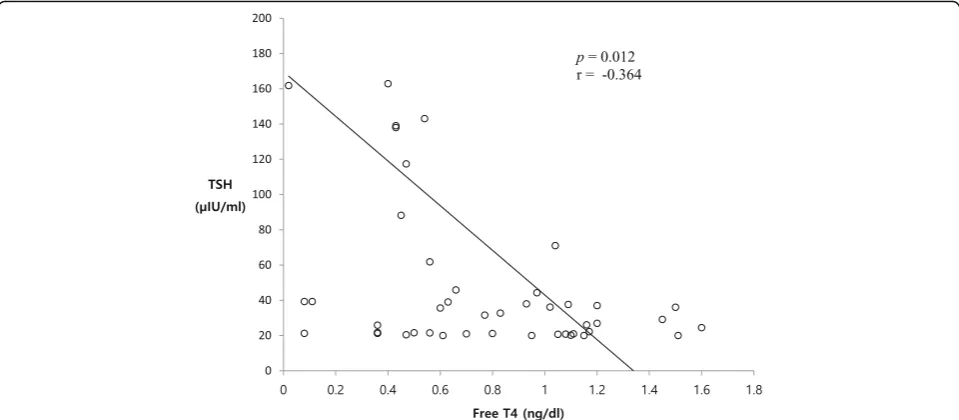
was normal, followed by a delayed increase to 78.3 μIU/ml at an average CA of 30.1 weeks. Whereas 80% (4/5) of infants with initial TSH elevation exhibited simultaneous reductions in T4 and/or fT4 levels, 93% (39/42) of infants with delayed TSH elevation exhib-ited decreased T4 and/or fT4 levels along with non-elevated TSH levels on the initial TFT. Moreover, the incidence of decreased T4 and/or fT4 levels was re-duced to 57% (24/42) at the time of delayed TSH ele-vation detection. A Spearman correlation analysis revealed a significant inverse correlation of the extent of TSH elevation with the fT4 level (r=−0.4, P= 0.012) (Fig. 2).
Thyroid function tests with or without levothyroxine supplementation
Of the 47 infants with TSH elevation (5 initial, 42 de-layed), 25 (1 initial, 24 delayed) received levothyroxine supplementation; the remaining 22 (4 initial, 18 delayed) received no treatment. In the non-treated group, one in-fant with an initial TSH elevation was declared NPO (nothing per oral) because of critical illness. The remaining 21 infants had relatively low TSH elevations (< 40 μIU/ml), with decreases in TSH levels at subse-quent TFTs. At the initial TSH elevation, 1 infant had a very high TSH level of 207.1μIU/ml and a low fT4 level and was treated with levothyroxine. Although the initial TFT results of infants with delayed TSH elevation did not differ significantly according to later levothyroxine supplementation, the TSH levels were significantly higher and fT4 levels significantly lower among infants with levothyroxine supplementation than among non-treated infants.
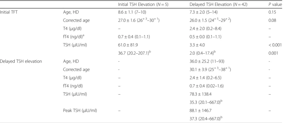
Table 1Demographics of infants with initial and delayed thyroid-stimulating hormone elevations and control
Variable Initial TSH Elevation (N= 5) Delayed TSH Elevation (N= 42) Control (N= 251)
Gestational age (weeks) 26.0 ± 1.2 25.0 ± 1.5* 26.8 ± 1.9
(25+ 0–28+ 6) (23+ 0–28+ 3) (24+0–34+ 4)
Birth weight (g) 789 ± 88 692 ± 142* 839 ± 120
(648–884) (470–990) (380–999)
Male sex 3 (60) 15 (35) 108 (43)
One-minute Apgar score 3.0 ± 1.4 4.3 ± 1.7 4.8 ± 1.7
Five-minute Apgar score 5.6 ± 1.5*,† 7.2 ± 1.2 7.2 ± 1.4
Delivery type C/sec 5 (100) 32 (76) 206 (82)
Small for gestational age 1 (20) 10 (24) 66 (26)
Antenatal steroids 1 (20)*,† 36 (86) 200 (80)
Pathologic chorioamnionitis 1 (20) 13 (31) 116 (46)
Pregnancy-induced hypertension 3 (60) 7 (17) 62 (25)
Gestational diabetes mellitus 0 (0) 1 (2) 9 (4)
Values are presented as means ± SD (range) or n (%) TSHThyroid-stimulating hormone,HDHospital day
*
P< 0.05, vs Control
Predisposing factors for the development of initial and delayed TSH elevation
All infants with an initial TSH elevation had perinatal asphyxia and remained intubated and ventilated, and 80% (4/5) were exposed to dopamine prior to the initial TFT performed at an average of 8.6 postnatal days. Among infants with delayed TSH elevation, 95% (40/42) had been exposed to more than one stressor, including respiratory support (37/42, among them, 28 were requir-ing ventilator support) and exposure to drugs (antibi-otics: 32/42, dopamine 17/42, postnatal steroids: 13/42) and surgery (10/42), within 2 weeks prior to the detec-tion of the delayed TSH elevadetec-tion.
Natural history and outcome of TSH elevation
Infants with TSH elevations were regularly followed up and either received no treatment or discontinued levothyroxine supplementation according to the decisions of the attending
pediatric endocrinologists. TSH levels in the non-treated group normalized within a mean of 30.5 ± 21.3 days and were transient, except for 1 infant who died before the follow-up TFT evaluation. TSH levels in the treated group normalized within 21.8 ± 11.9 days and were transient as well. There was no significant difference in the time to normalize between the two groups. Infants in the treated group discontinued the levothyroxine supplementation with an average treatment duration of 635 ± 427 days. Their thyroid scan and sonogram revealed normal results, and the follow-up TFT remained normal up to 1 year after the discontinuation of the levothyroxine treatment.
Short- and long-term outcomes according to levothyroxine supplementation
Regarding short-term outcomes, the incidence of morbid-ities such as BPD (≥moderate), IVH (≥3), or PVL did not differ significantly by levothyroxine supplementation status.
Table 3Thyroid function test data of infants with initial and delayed thyroid-stimulating hormone elevations
Initial TSH Elevation (N= 5) Delayed TSH Elevation (N= 42) Pvalue
Initial TFT Age, HD 8.6 ± 1.1 (7–10) 7.3 ± 2.0 (5–14) 0.15
Corrected age 27.0 ± 1.6 (26+ 3–30+ 1) 26.0 ± 1.5 (24+ 5–29+ 2) 0.08
T4 (μg/dl) – 2.4 ± 2.0 (0.2–8.4) –
fT4 (ng/dl)a 0.7 ± 0.4 (0.1–1.1) 0.5 ± 0.0 (0.1–1.1) –
TSH (μIU/ml) 61.0 ± 81.9 3.3 ± 4.0 < 0.001
36.7 (20.2–207.1)b 2.0 (0.4–17.4)b 0.001
Delayed TSH elevation Age, HD - 36.0 ± 25.2 (11–93)
-Corrected age - 30.1 ± 3.9 (25+ 3–38+ 1)
-T4 (μg/dl) – 2.4 ± 1.4 (0.2–6.5) –
fT4 (ng/dl) – 0.7 ± 0.4 (0.02–1.6) –
TSH (μIU/ml) – 78.3 ± 138.4 –
35.3 (20.1–667.0)b
Peak TSH (μIU/ml) – 88.1 ± 146.7 –
37.3 (20.4–667.0)b
TSHThyroid-stimulating hormone,TFTThyroid function test,HDHospital day,T4Thyroxine,fT4free thyroxine
a
The number of subjects with delayed TSH elevation was insufficient for comparison
b
Geometric mean (range)
Table 2Clinical outcomes of infants with initial and delayed thyroid-stimulating hormone elevations and control
Outcome Initial TSH Elevation (N= 5) Delayed TSH Elevation (N= 42) Control (N= 251)
Mortality 2 (40)* 2 (5) 9 (4)
BPD (≥moderate) 2/3 (67) 22/40 (55) 95/250 (38)
IVH (≥3) 1 (20) 7 (17) 13 (5)
PVL 1 (20) 3 (7) 15 (6)
NEC (≥Stage 2b) 0 (0) 2 (5) 18 (7)
ROP requiring laser treatment 1/4 (25) 20 (48) 42/250 (17)
Composite morbidity 4 (80) 35 (83) 131 (52)
Values are presented as n (%). Values are adjusted by gestational age, birth weight, Apgar score at 5 min and antenatal steroid use
BPDbronchopulmonary dysplasia,IVHintraventricular hemorrhage,PVLperiventricular leukomalacia,NECnecrotizing enterocolitis,ROPretinopathy of prematurity
*
Although the incidence of mortality was significantly higher among non-treated infants than among levothyroxine-supplemented infants, this was attributed to underlying disease, rather than a lack of thyroid supplementation. A comparison of the long-term growth and neurodevelop-mental outcomes of 17 out of 22 non-treated infants and 22 out of 25 levothyroxine-supplemented infants at a CA of 2 years revealed no significant differences in body weight, height, head circumference z scores, catch-up growth, the incidence of cerebral palsy, and neurodevelopmental delays.
Discussion
In the present study, TSH elevation was detected in only 0.9% of ELBWIs during the initial TFT performed within 2 postnatal weeks, whereas delayed TSH elevation was de-tected in a significantly higher number of infants (7.2%). This relatively high incidence of initial TSH elevation among ELBWIs within 2 postnatal weeks is inconsistent with the current belief that initial TSH elevation is very rare [21–23]. Moreover, the finding of a 7.2% incidence of delayed TSH elevation in our cohort was much higher than the previously reported incidence of 1.7% among ELBWIs [11]. In the present study, the incidence of de-layed TSH elevation correlated inversely with the GA and birth weight, even among these ELBWIs. Therefore, in addition to more diligent follow-up TFT screening, this wide discrepancy in the incidence of delayed TSH eleva-tion might be attributable to disparities in the composieleva-tion of the most immature population within these ELBWIs, characterized by GAs of 23–24 weeks and birth weights
≤800 g. These findings suggest that a modification to the TFT screening protocol, such as the establishment of a new routine of regular follow-up TFT screening program
at 4 weeks intervals or until the patient becomes clinically stable, might be needed to detect hypothyroidism in ex-tremely preterm infants near the limit of viability (i.e., GA of 23–24 weeks), who have the highest risk of developing delayed TSH elevation [24].
The etiology of initial and delayed TSH elevation in ELBWIs remains unclear. ELBWIs are more suscep-tible to morbidities, including perinatal asphyxia, in-fection, surgery, and exposure to thyroid function-inhibiting drugs, which could result in THOP [25, 26]. Moreover, as the hypothalamic–pituitary–thyroid axis has not been established in extremely preterm in-fants, the thyroid gland cannot generate sufficient levels of thyroid hormones in response to these stressors [27–29]. Therefore, the observation of THOP in critically ill ELBWIs might represent an epi-phenomenon of these morbidities and could thus be considered an NTI [25, 30, 31]. In the present study, all infants with initial TSH elevations had perinatal asphyxia and remained intubated and ventilated, and 80% were exposed to dopamine despite its known suppression of TSH secretion [32] prior to the initial TFT (performed at a mean of 8.6 postnatal days). Furthermore, the frequency of antenatal steroid use and Apgar scores at 5 min were significantly lower among infants with initial TSH elevation, compared to those with delayed TSH elevation and normal con-trols. Although the initial TSH levels measured at a mean age of 7.3 postnatal days were normal, delayed TSH elevation was detected via follow-up TFTs per-formed at a mean of 36.0 postnatal days. Moreover, 95% of infants with delayed TSH elevation were ex-posed to multiple predisposing factors for TSH
elevation, including respiratory support (88%), expos-ure to drugs such as dopamine (41%) and steroids (31%) despite their known inhibition of TSH secretion [32, 33], and surgery (24%), within 2 weeks prior to the detection of the delayed TSH elevation. In infants with delayed TSH elevation, the initial 93% incidence of decreased T4 and/or fT4 levels along with non-elevated TSH levels decreased to 57% when the de-layed TSH elevation was detected. Overall, these find-ings suggest that the initial timing of insult determines whether TSH elevation is initial or de-layed, and that both initial and delayed TSH eleva-tions are representative of hypothyroidism caused by NTI during the early recovery phase and signal relief from the severity of the stressors [9, 34].
Critical changes in therapeutic strategy might have af-fected the clinical parameters, including the thyroidal function test results. During the study period, we experi-enced a decreased mortality and morbidity rate, as a re-sult of better perinatal and neonatal intensive care, including the administration of more antenatal steroid use, active resuscitation at the delivery room, and appli-cation of a less-invasive ventilator management policy [35–37]. In parallel with the increased intact survival of ELBWIs, we observed significantly reduced incidences of THOP and subsequent TSH elevation in ELBWIs at 25– 28 weeks’ gestation [38]. These findings support the as-sumption that the decrease in critical morbidities among ELBWIs due to the recent advances in neonatal intensive care medicine has decreased the incidence of TSH eleva-tion in these infants.
Regarding the time of TSH elevation onset, initial TSH elevation was detected only in ELBWIs at a GA of 25– 28 weeks, and was measured at a mean CA of 27 weeks. Although the highest incidence of delayed TSH elevation was observed in ELBWIs with a GA of 23 weeks, the ini-tial TSH levels measured at a mean CA of 26 weeks were normal, whereas delayed elevation was indicated by a follow-up TFT performed at a mean CA of 30 weeks. These findings suggest that maturation of the hypothal-amic–pituitary–thyroid axis [6, 27], when superimposed on the timing of insult, is the primary determinant of initial or delayed TSH elevation in an ELBWI.
The natural history of TSH elevation in ELBWI has not yet been delineated. In the present study, elevated TSH levels normalized at a mean age of 31 days, regard-less of the levothyroxine supplementation status, and levothyroxine replacement therapy was discontinued at a mean age of 635 days. These findings support the idea that both the initial and delayed TSH elevations
ob-served in ELBWIs represent epiphenomena of
hypothyroidism due to NTI during the early recovery phase [13] and are therefore transient, in contrast to congenital hypothyroidism [39].
The indication for levothyroxine replacement therapy is another critical issue, requiring attention. The extent of thyroid dysfunction during NTI was found to correl-ate inversely to the severity of morbidity [25,31,40,41]. In the present study, the extent of TSH elevation was found to exhibit a significant inverse correlation with the fT4 level. Furthermore, infants receiving levothyrox-ine replacement therapy had significantly higher TSH levels and significantly lower fT4 levels, compared with untreated infants. In our separate study [38], the initial fT4 level was the best predictor of mortality and com-posite morbidities compared with the Apgar and CRIB-II scores in ELBWIs. These findings suggest that al-though the levothyroxine replacement therapy was arbi-trarily determined by the attending endocrinologist’s decision without clear treatment criteria, the initial crit-ical morbidities experienced by infants receiving levothyroxine replacement therapy might have been more severe than those of untreated infants. Neverthe-less, our data showing significantly improved mortality and no significant differences in growth and neurodeve-lopmental outcomes at a CA of 2 years, suggest that it might be prudent to administer levothyroxine to infants with high TSH and low fT4 levels. However, a high TSH level in the context of NTI is thought to signal relief from the severity of a stressor and has been associated with an improved prognosis in adults [42,43]. Addition-ally, many clinical trials of premature infants failed to demonstrate the beneficial effects of levothyroxine re-placement therapy [44–47]. Moreover, although our en-docrinologists preferred longer treatment in the present study because thyroid hormones are critical for brain de-velopment during the first 2 to 3 years of life [48], the in-fants in a study by Woo et al. [11] received only a short-term levothyroxine replacement therapy. By contrast, the shortest duration of levothyroxine supplementation in the present study was 103 days. Additional well-designed studies are needed to determine the optimal indication, timing, and duration of levothyroxine replacement therapy.
study period. As only 24% of patients with delayed TSH elevation had undergone surgical procedures, the effect of iodine exposure on the incidence of TSH elevation might be insignificant.
Conclusion
In conclusion, transient TSH elevation occurs frequently in ELBWIs and might result from NTI during the early recovery phase. The superimposition of the insult timing of stressors on the hypothalamic–pituitary–thyroid axis maturation is the primary determinant of initial or de-layed TSH elevation. Additional well-designed prospect-ive controlled studies are needed to clarify the benefits of levothyroxine replacement.
Abbreviations
BPD:Bronchopulmonary dysplasia; CA: Corrected age; ELBWI: Extremely low birth weight infant; fT4: Free thyroxine; GA: Gestational age;
IVH: Intraventricular hemorrhage; MDI: Mental developmental index; NEC: Necrotizing enterocolitis; NTI: Non-thyroidal illness; PDI: Psychomotor developmental index; PVL: Periventricular leukomalacia; ROP: Retinopathy of prematurity; TFT: Thyroid function test; THOP: transient hypothyroxinemia of prematurity; TSH: thyroid-stimulating hormone
Acknowledgements
We would like to thank Sang-Yong Eom for statistical consulting and support in this study.
Authors’contributions
SAY conceptualized and designed the study, performed the initial analyses, and drafted the initial manuscript. YSC conceptualized and designed the study and reviewed and revised the manuscript. SYA and SIS collected data and reviewed and revised the manuscript. YSC and WSP coordinated and supervised data collection and critically reviewed the manuscript. All authors approved the final manuscript as submitted.
Funding Not applicable.
Availability of data and materials
The data that support the findings of this study are available from the corresponding author ([email protected]) upon reasonable request.
Ethics approval and consent to participate
Data collection was approved by the Institutional Review Board of Samsung Medical Center.
The informed consent requirements for this retrospective chart review were waived by the Institutional Review Board.
Consent for publication Not applicable.
Competing interests
The authors declare that they have no competing interests.
Author details
1
Department of Pediatrics, Chungbuk National University Hospital, 1 Sunhwan-ro 776, Seowon-gu, Cheongju 28644, South Korea.2Department of
Pediatrics, Samsung Medical Center, Sungkyunkwan University School of Medicine, 81 Irwon-Ro, Gangnam-gu, Seoul 06351, South Korea.
Received: 6 November 2018 Accepted: 20 September 2019
References
1. Leger J, Olivieri A, Donaldson M, Torresani T, Krude H, van Vliet G, et al. European Society for Paediatric Endocrinology consensus guidelines on
screening, diagnosis, and management of congenital hypothyroidism. Horm Res Paediatr. 2014;81(2):80–103.
2. Williams FL, Visser TJ, Hume R. Transient hypothyroxinaemia in preterm infants. Early Hum Dev. 2006;82(12):797–802.
3. Calvo RM, Jauniaux E, Gulbis B, Asuncion M, Gervy C, Contempre B, et al. Fetal tissues are exposed to biologically relevant free thyroxine concentrations during early phases of development. J Clin Endocrinol Metab. 2002;87(4):1768–77. 4. Vulsma T, Gons MH, de Vijlder JJ. Maternal-fetal transfer of thyroxine in
congenital hypothyroidism due to a total organification defect or thyroid agenesis. N Engl J Med. 1989;321(1):13–6.
5. Fisher DA, Dussault JH, Sack J, Chopra IJ. Ontogenesis of hypothalamic--pituitary--thyroid function and metabolism in man, sheep, and rat. Recent Prog Horm Res. 1976;33:59–116.
6. Murphy N, Hume R, van Toor H, Matthews TG, Ogston SA, Wu SY, et al. The hypothalamic-pituitary-thyroid axis in preterm infants; changes in the first 24 hours of postnatal life. J Clin Endocrinol Metab. 2004;89(6):2824–31. 7. Pavelka S, Kopecky P, Bendlova B, Stolba P, Vitkova I, Vobruba V, et al. Tissue
metabolism and plasma levels of thyroid hormones in critically ill very premature infants. Pediatr Res. 1997;42(6):812–8.
8. van Wassenaer AG, Kok JH, Dekker FW, de Vijlder JJ. Thyroid function in very preterm infants: influences of gestational age and disease. Pediatr Res. 1997; 42(5):604–9.
9. Simpson J, Williams FL, Delahunty C, van Toor H, Wu SY, Ogston SA, et al. Serum thyroid hormones in preterm infants and relationships to indices of severity of intercurrent illness. J Clin Endocrinol Metab. 2005;90(3):1271–9. 10. Klein AH, Foley B, Kenny FM, Fisher DA. Thyroid hormone and thyrotropin
responses to parturition in premature infants with and without the respiratory distress syndrome. Pediatrics. 1979;63(3):380–5.
11. Woo HC, Lizarda A, Tucker R, Mitchell ML, Vohr B, Oh W, et al. Congenital hypothyroidism with a delayed thyroid-stimulating hormone elevation in very premature infants. Incidence and growth and developmental outcomes. J Pediatr. 2011;158(4):538–42.
12. Larson C, Hermos R, Delaney A, Daley D, Mitchell M. Risk factors associated with delayed thyrotropin elevations in congenital hypothyroidism. J Pediatr. 2003;143(5):587–91.
13. Soto-Rivera CL, Fichorova RN, Allred EN, Van Marter LJ, Shah B, Martin CR, et al. The relationship between TSH and systemic inflammation in extremely preterm newborns. Endocrine. 2015;48(2):595–602.
14. Ford G, LaFranchi SH. Screening for congenital hypothyroidism: a worldwide view of strategies. Best Pract Res Clin Endocrinol Metab. 2014;28(2):175–87. 15. Mitchell ML, Hsu HW, Sahai I, Massachusetts Pediatric Endocrine Work G. The increased incidence of congenital hypothyroidism: fact or fancy? Clin Endocrinol. 2011;75(6):806–10.
16. Ehrenkranz RA, Walsh MC, Vohr BR, Jobe AH, Wright LL, Fanaroff AA, et al. Validation of the National Institutes of Health consensus definition of bronchopulmonary dysplasia. Pediatrics. 2005;116(6):1353–60. 17. Papile LA, Burstein J, Burstein R, Koffler H. Incidence and evolution of
subependymal and intraventricular hemorrhage: a study of infants with birth weights less than 1,500 gm. J Pediatr. 1978;92(4):529–34. 18. Walsh MC, Kliegman RM, Fanaroff AA. Necrotizing enterocolitis: a
practitioner’s perspective. Pediatr Rev. 1988;9(7):219–26.
19. Fierson WM. Screening examination of premature infants for retinopathy of prematurity. Pediatrics. 2013;131(1):189–95.
20. Moon JS, Lee SY, Nam CM, Choi JM, Choe BK, Seo JW, et al. 2007 Korean National Growth Charts: review of developmental process and an outlook. Korean J Pediatr. 2008;51(1):1–25.
21. Buyukgebiz A. Newborn screening for congenital hypothyroidism. JPEM. 2006;19(11):1291–8.
22. Kugelman A, Riskin A, Bader D, Koren I. Pitfalls in screening programs for congenital hypothyroidism in premature newborns. Am J Perinatol. 2009; 26(5):383–5.
23. Hallett A, Evans C, Moat S, Barton J, Warner J, Gregory JW. Hypothyroidism in preterm infants following normal screening. Ann Clin Biochem. 2011; 48(Pt 6):572–4.
24. Kaplowitz PB. Neonatal thyroid disease: testing and management. Pediatr Clin N Am. 2019;66(2):343–52.
25. Pereira DN, Procianoy RS. Effect of perinatal asphyxia on thyroid-stimulating hormone and thyroid hormone levels. Acta Paediatr. 2003;92(3):339–45. 26. Rooman RP, Du Caju MV, De Beeck LO, Docx M, Van Reempts P, Van Acker
27. Thorpe-Beeston JG, Nicolaides KH, Felton CV, Butler J, McGregor AM. Maturation of the secretion of thyroid hormone and thyroid-stimulating hormone in the fetus. N Engl J Med. 1991;324(8):532–6.
28. LaFranchi S. Thyroid function in the preterm infant. Thyroid. 1999;9(1):71–8. 29. Schwarze CPS-WK, Wollmann HA. Thyroid function in healthy and sick
preterm Infant’s: changes in TSH, T4, fT4 and T3 from day 1 to 12. JPEM. 2007;20(Suppl 1):135–41.
30. Marsh TD, Freeman D, McKeown RE, Bowyer FP. Increased mortality in neonates with low thyroxine values. J Perinatol. 1993;13(3):201–4. 31. Kratzsch J, Pulzer F. Thyroid gland development and defects. Best Pract Res
Clin Endocrinol Metab. 2008;22(1):57–75.
32. de Zegher F, Van den Bershe G, Dumoulin M, Gewillig M, Daenen W, Devlieger H. Dopamine suppresses thyroid-stimulating hormone secretion in neonatal hypothyroidism. Acta Paediatr. 1995;84(2):213–4.
33. Brabant G, Brabant A, Ranft U, Ocran K, Kohrle J, Hesch RD, et al. Circadian and pulsatile thyrotropin secretion in euthyroid man under the influence of thyroid hormone and glucocorticoid administration. J Clin Endocrinol Metab. 1987;65(1):83–8.
34. Warner MH, Beckett GJ. Mechanisms behind the non-thyroidal illness syndrome: an update. J Endocrinol. 2010;205(1):1–13.
35. Kim JK, Chang YS, Sung S, Ahn SY, Yoo HS, Park WS. Trends in survival and incidence of bronchopulmonary dysplasia in extremely preterm infants at 23-26 weeks gestation. J Korean Med Sci. 2016;31(3):423–9.
36. Park JH, Chang YS, Sung S, Ahn SY, Park WS. Trends in overall mortality, and timing and cause of death among extremely preterm infants near the limit of viability. PLoS One. 2017;12(1):e0170220.
37. Kim JK, Chang YS, Sung S, Ahn SY, Park WS. Trends in the incidence and associated factors of late-onset sepsis associated with improved survival in extremely preterm infants born at 23-26 weeks’gestation: a retrospective study. BMC Pediatr. 2018;18(1):172.
38. Yoon SA, Chang YS, Ahn SY, Sung SI, Park WS. Incidence and severity of transient hypothyroxinaemia of prematurity associated with survival without composite morbidities in extremely low birth weight infants. Sci Rep. 2019; 9(1):9628.
39. Vigone MC, Caiulo S, Di Frenna M, Ghirardello S, Corbetta C, Mosca F, et al. Evolution of thyroid function in preterm infants detected by screening for congenital hypothyroidism. J Pediatr. 2014;164(6):1296–302.
40. Williams FL, Hume R. Perinatal factors affecting thyroid hormone status in extreme preterm infants. Semin Perinatol. 2008;32(6):398–402.
41. Lafranchi SH. Congenital hypothyroidism: delayed detection after birth and monitoring treatment in the first year of life. J Pediatr. 2011;158(4):525–7. 42. O’Keefe LM, Conway SE, Czap A, Malchoff CD, Benashski S, Fortunato G,
et al. Thyroid hormones and functional outcomes after ischemic stroke. Thyroid Res. 2015;8:9.
43. Chopra IJ. Clinical review 86: Euthyroid sick syndrome: is it a misnomer? J Clin Endocrinol Metab. 1997;82(2):329–34.
44. van Wassenaer AG, Kok JH, de Vijlder JJM, Briët JM, Smit BJ, Tamminga P, et al. Effects of thyroxine supplementation on neurologic development in infants born at less than 30 weeks’gestation. N Engl J Med. 1997;336(1):21–6. 45. Beeram MR, Wilson DP. Hypothyroxinemia of prematurity: rite of passage or
therapeutic necessity? Tex Med. 2000;96(11):60–3.
46. Suzumura H, Nitta A, Tsuboi Y, Watabe Y, Kuribayashi R, Arisaka O. Thyroxine for transient hypothyroxinemia and cerebral palsy in extremely preterm infants. Pediatr Int. 2011;53(4):463–7.
47. Dilli D, Eras Z, Andiran N, Dilmen U, Sakrucu ED. Neurodevelopmental evaluation of very low birth weight infants with transient hypothyroxinemia at corrected age of 18-24 months. Indian Pediatr. 2012;49(9):711–5. 48. Patel J, Landers K, Li H, Mortimer RH, Richard K. Thyroid hormones and fetal
neurological development. J Endocrinol. 2011;209(1):1–8.
Publisher’s Note