Onset of Jaundice in Glucose-6-Phosphate Dehydrogenase–Deficient
Neonates
Michael Kaplan, MB ChB*§; Nurit Algur, MSc‡; and Cathy Hammerman, MD*§
ABSTRACT. Objective. We asked whether neonatal
jaundice associated with glucose-6-phosphate dehydro-genase (G-6-PD) deficiency commences either in utero or in the immediate postnatal period and whether this peri-natal bilirubinemia is the precursor of the subsequent neonatal jaundice and hyperbilirubinemia.
Methods. Mandatory serum total bilirubin (STB) de-terminations were performed within 3 hours of birth, to reflect the in utero state (first STB), and on the third day of life (second STB), with additional determinations as clinically necessary, on healthy, term male neonates at high risk for G-6-PD deficiency. G-6-PD Mediterranean mutation was determined by molecular means. G-6-PD– deficient neonates were compared with control partici-pants. The relationship of first STB values to second STB and subsequent hyperbilirubinemia (defined as STB
>256mol/L [15.0 mg/dL]) was determined.
Results. Both first and second STB values were sig-nificantly higher in the G-6-PD– deficient neonates (nⴝ
52) than in control participants (nⴝ166; 50ⴞ12mol/L vs 44ⴞ10 mol/L [2.9ⴞ0.7 mg/dL vs 2.6ⴞ0.6 mg/dL] and 174 ⴞ 52 mol/L vs 152 ⴞ 52 mol/L [10.2 ⴞ 3.1 mg/dL vs 8.9ⴞ3.0 mg/dL] for the first and second STB values, respectively). The rate of rise between these 2 points was greater in the G-6-PD– deficient neonates (2.6 ⴞ 0.9 mol/L/h vs 2.2 ⴞ 0.9 mol/L/h [0.15 ⴞ 0.05 mg/dL/h vs 0.13ⴞ0.05 mg/dL/h). Sixteen (30.8%) of the G-6-PD– deficient neonates developed hyperbiliru-binemia compared with 10 (6%) of control participants (relative risk: 5.11; 95% confidence interval: 2.47–10.56). In both G-6-PD– deficient and normal populations, first STB values correlated significantly with both second STB values and with those who subsequently developed hyperbilirubinemia. Significantly more G-6-PD– defi-cient neonates with a first STB value greater than or equal to the mean developed hyperbilirubinemia com-pared with those with first STB less than the mean: 13 of 28 neonates versus 3 of 24 (relative risk: 3.7; 95% confi-dence interval: 1.20 –11.51). This difference did not reach statistical significance in the control group.
Conclusions. Higher first STB values, an increased risk of hyperbilirubinemia in G-6-PD– deficient neonates with first STB value greater than or equal to the mean, and significant correlation between first STB values and second STB values and hyperbilirubinemia suggest that jaundice in G-6-PD– deficient neonates commences in
the immediate perinatal period, most likely in utero.
Pediatrics 2001;108:956 –959; bilirubin, jaundice, hyper-bilirubinemia, glucose-6-phosphate dehydrogenase defi-ciency, perinatal, in utero.
ABBREVIATIONS. STB, serum total bilirubin; G-6-PD, glucose-6-phosphate dehydrogenase; CI, confidence interval.
A
n association between umbilical cord blood bilirubin levels, reflecting the in utero state, and serum total bilirubin (STB) values in early neonatal life has been recognized in some ne-onates for many years.1This concept has been usedreliably in the prediction of hyperbilirubinemia at-tributable to Rh isoimmunization.2Although the
suc-cess in identifying infants who are at risk for subse-quent development of hyperbilirubinemia has not been universal,3,4in some series of ABO
incompati-bility5–7and also in nonhemolytic conditions,8 –11use
of umbilical cord blood STB determination has shown some success in the prediction of severe dice. These observations suggest that neonatal jaun-dice frequently— but not consistently— has its ori-gins in utero.
Glucose-6-phosphate dehydrogenase (G-6-PD) de-ficiency is a commonly occurring enzyme defect that is associated with a high incidence of severe neonatal hyperbilirubinemia with the potential of irreversible bilirubin encephalopathy if not treated in time.12The
pathogenesis of this hyperbilirubinemia is different from that in G-6-PD–normal neonates: decreased bil-irubin conjugation, the result of an interaction be-tween G-6-PD deficiency and promoter polymor-phism of the gene that controls the bilirubin conjugating enzyme UDP glucuronosyltransferase, is a crucial factor.13,14 To elucidate further the
patho-genesis of the associated bilirubinemia, we therefore asked whether the jaundice associated with the G-6-PD Mediterranean mutation, as in some other conditions, commences in the perinatal period and whether this in utero or very early bilirubinemia is the precursor of subsequent jaundice or hyperbiliru-binemia in these neonates.
METHODS Study Protocol
The study was approved by the Institutional Review Board of the Shaare Zedek Medical Center. A cohort of consecutively born healthy boys who were born atⱖ37 weeks’ gestation at the Shaare Zedek Medical Center to Sephardic Jewish mothers whose fami-lies originated in Asia Minor were studied. This subgroup of the Israeli population has been shown to have an exceptionally high
From the *Department of Neonatology and ‡Clinical Biochemistry Labora-tory, Shaare Zedek Medical Center; and §the Faculty of Medicine of the Hebrew University, Jerusalem, Israel.
Received for publication May 17, 2000; accepted Feb 7, 2001.
Presented in part at the Pediatric Academic Societies–Society for Pediatric Research Annual Meeting, San Francisco, CA, May 1– 4, 1999.
incidence of G-6-PD deficiency.15,16Blood was drawn for
manda-tory STB determinations within the first 3 hours after birth, to reflect the in utero status (first STB), and again at the time of routine metabolic screening on the third day of life (second STB). Simultaneously with 1 of these determinations, blood was col-lected for DNA extraction.
Routine medical care for these neonates included screening for G-6-PD deficiency on the first day of life, blood group determina-tion, and direct Coombs’ testing for infants born to Rh-negative or O blood group mothers. Infants were monitored visually as inpa-tients by our medical and nursing staff for the development of jaundice with additional STB determinations if warranted clini-cally. Those with a second STB valueⱖ50th percentile for hour of life17and therefore at high risk for subsequent hyperbilirubinemia
were scheduled to be followed as outpatients, whereas those with lower predischarge STB values, at low risk for hyperbilirubinemia, were evaluated at well-infant clinics or by family pediatricians or ritual circumcisers (mohel) and referred for evaluation if deemed necessary. Finally, any parent who had any doubt as to their infant’s jaundice status was able to return for an STB at any time during the first week of life. The patient compliance in our pop-ulation was excellent, and we are confident that we were aware of virtually all infants with an STB ⱖ256 mol/L (15.0 mg/dL). These neonates were followed by us until stabilization of the STB values. When phototherapy became necessary after discharge, the infants were readmitted to our unit. Phototherapy was com-menced in G-6-PD– deficient newborns when STB values exceeded 15.0 mg/dL. Breastfeeding was encouraged, although mothers were warned of the dangers of eating fava beans or taking drugs known to be triggers of hemolysis in G-6-PD– deficient individuals while nursing. Infants with any other condition that was likely to exacerbate hyperbilirubinemia, such as cephalhematoma, direct Coombs’-positive isoimmunization, maternal diabetes, sepsis, or Down’s syndrome, were excluded from the study.
Laboratory Methods
DNA was extracted from peripheral blood leukocytes using a high-salt extraction procedure.18 For G-6-PD genotyping, DNA
was shipped to The Scripps Research Institute (La Jolla, CA) for molecular classification. Polymerase chain reaction followed by allele-specific oligonucleotide hybridization was used to deter-mine the presence or absence of nt 563, the nucleotide mutated in G-6-PD Mediterranean.19Details of the procedure have been
pub-lished elsewhere.13
STB values were determined by reflectance spectrophotometry using an Ektachem analyzer (Vitros 700c/750XRC Chemistry Sys-tem; Johnson and Johnson Clinical Diagnostics, Rochester, NY). Blood group determinations and direct Coombs’ testing were performed by routine laboratory techniques.
Data Analysis
The G-6-PD genotype was used to classify the infants into G-6-PD– deficient hemizygote (study) and normal hemizygote (control) groups. Hyperbilirubinemia was defined as a serum total bilirubinⱖ256mol/L (15.0 mg/dL) in the first week of life, for standardization with our previous studies.13,17Results were
com-pared using Studentttest,2analysis, or linear correlation, as
appropriate. Significance of these tests was determined asP⬍.05. Evaluation of the effect of either G-6-PD deficiency or first STB greater than or equal to the mean value on the subsequent devel-opment of hyperbilirubinemia was determined by calculating the relative risk and 95% confidence intervals (CI). Significance in these cases was defined as a 95% CI range that did not include the digit 1. Rate of rise of STB was calculated as the difference between the first and second STB values divided by the number of hours between these tests.
RESULTS
A total of 225 infants were enrolled in the study. Seven (6 Coombs’ positive, 1 maternal diabetes) were recognized not to meet study criteria after enroll-ment. Thus, the cohort that met study criteria com-prised 218 infants, 52 of whom were hemizygotes for G-6-PD Mediterranean 563T and 166 of whom did not have this mutation. Demographic data for these
infants are summarized in Table 1. Despite that sig-nificantly fewer G-6-PD– deficient neonates were nursed (Table 1), 16 (30.8%) of the G-6-PD– deficient neonates developed hyperbilirubinemia, compared with 10 (6%) of the control participants (relative risk: 5.11; 95% CI: 2.47–10.56;P ⬍.0001).
Results of the first and second STB tests (mean ⫾ SD) and the age at sampling are summarized in Table 2. Despite the similarity in times of sampling between study and control groups, both first and second STB values and the rate of rise between these 2 determinations were significantly higher in the G-6-PD– deficient neonates than in the control group. In both G-6-PD– deficient and control groups, first STB values correlated with second STB values (r ⫽ 0.6,P⬍.0001 andr⫽0.5,P⬍.0001 for the G-6-PD– deficient and control infants, respectively), with those who developed serum bilirubin values ⱖ256
mol/L (15.0 mg/dL;r⫽0.34,P⫽.01 andr⫽0.21, P⫽ .01, respectively), and with the rate of rise (r⫽ 0.56,P⬍.0001 andr⫽0.39,P⬍.0001, respectively). In the G-6-PD– deficient cohort, a significantly greater number of neonates among those with a first STB value greater than or equal to the mean devel-oped hyperbilirubinemia compared with those with a first STB less than the mean: 13 (46%) of 28 neo-nates versus 3 (12.5%) of 24 neoneo-nates (relative risk: 3.7; 95% CI: 1.20 –11.51;P⫽.02). This difference did not reach statistical significance in the control group: 9 (9.6%) of 94 versus 1 (1.4%) of 72, respectively (relative risk: 6.89; 95% CI: 0.89 –53.18;P⫽ .06).
DISCUSSION
Some studies of umbilical cord blood bilirubin values have shown that both nonhemolytic jaun-dice8 –11and hemolysis attributable to Rh
isoimmu-nization2 or ABO incompatibility5–7 commence in
utero. In these conditions, the cord blood bilirubin values correlate with subsequent hyperbilirubinemia and have been used to predict its severity. To under-stand further the pathophysiology of G-6-PD defi-ciency–associated neonatal jaundice, we studied its onset to determine whether it commences perina-tally, ie, either during fetal life or in the immediate postnatal period, as in some hemolytic conditions noted above, or well into the neonatal period, as with other nonhemolytic bilirubinemias. In the current study, already immediately after birth, most likely reflecting the in utero status, the G-6-PD– deficient neonates had significantly higher serum bilirubin values than control participants. Significantly higher STB values were evident again on the third day. The
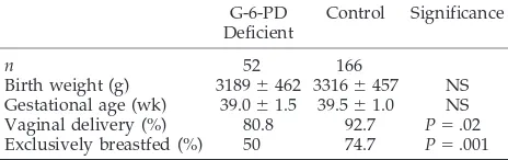
TABLE 1. Demographic Data of the Infants Studied G-6-PD
Deficient
Control Significance
n 52 166
Birth weight (g) 3189⫾462 3316⫾457 NS
Gestational age (wk) 39.0⫾1.5 39.5⫾1.0 NS
Vaginal delivery (%) 80.8 92.7 P⫽.02
Exclusively breastfed (%) 50 74.7 P⫽.001
Values are mean⫾standard deviation or percentages, as appro-priate.
NS indicates not significant.
ARTICLES 957
at Viet Nam:AAP Sponsored on August 30, 2020 www.aappublications.org/news
rate of rise of STB and the incidence of hyperbiliru-binemia were similarly increased in the G-6-PD– de-ficient neonates compared with control participants. Higher umbilical cord blood STB levels20,21 or
in-creased STB values on the first day of life22,23 have
been shown in previous studies of G-6-PD– deficient neonates, suggesting either in utero or a very early postnatal onset of jaundice. However, as there was no attempt at correlation between this early biliru-binemia and subsequent jaundice in these studies, it cannot be concluded that the former was the fore-runner of hyperbilirubinemia. Our demonstration of a significant correlation between very early STB val-ues and later bilirubin valval-ues and the significantly higher relative risk of first STB result greater than or equal to the mean in the subsequent development of hyperbilirubinemia now demonstrates that this peri-natal bilirubinemia is the precursor of subsequent jaundice and hyperbilirubinemia.
Although umbilical cord blood sampling undoubt-edly would have offered a more accurate represen-tation of the in utero status, for logistical reasons and because of difficulties in coordinating the study with the delivery room staff, we chose to obtain samples from the infants within 3 hours of delivery to reflect the in utero situation. Although we cannot exclude categorically a very early postnatal onset of the bil-irubinemia, we are confident that this method of sampling reliably reflected the in utero state. Pre-suming that the hourly rates of rise during the first days of life reflected the rise during the first 3 hours of life as well, the first STB values should have been only marginally elevated over the actual cord blood values and should not have affected the comparisons between the study and control groups to any major degree. The slightly higher but clinically insignificant mean first STB values for the control groups than those reported by others for cord blood samples may represent a combination of the timing of the sample and interlaboratory variation between our laborato-ry’s STB determination and those that have been used by other laboratories in the past.24
Although the current and previous studies showed that umbilical cord or first-day STB determi-nations25 may have some predictive value for the
subsequent development of hyperbilirubinemia, the aim of this study primarily was to shed light on the pathophysiology of G-6-PD deficiency–associated neonatal jaundice. It was not designed as a study of prediction of subsequent hyperbilirubinemia and should not be interpreted as such. Accurate predic-tion can be accomplished by predischarge STB
test-ing at the time of metabolic screentest-ing, as recently described both for normal26 and G-6-PD-deficient17
newborn populations.
G-6-PD deficiency is estimated to affect hundreds of millions of people not only in areas in which the condition is indigenous but also with a potential for serious complications in North America.27
Aware-ness of the condition and its dangers and an under-standing of the differing pathophysiology of the as-sociated jaundice compared with that of G-6-PD– normal individuals are essential if the potential of bilirubin encephalopathy is to be limited.
ACKNOWLEDGMENTS
This study was supported at Shaare Zedek Medical Center by grants for neonatal jaundice research from The Golden Charitable Trust, London, United Kingdom, and the Mirsky Research Fund. We thank Ernest Beutler, MD, for the genotype analysis of G-6-PD Mediterranean mutation and Chana Amsalem for techni-cal assistance.
REFERENCES
1. Davidson LT, Merrit KK, Weech AA. Hyperbilirubinemia in the new-born.Am J Dis Child. 1941;61:958 –980
2. Allen FH, Diamond LK. Erythroblastosis Fetalis. Boston, MA: Little, Brown; 1958
3. Haque KN. Value of measuring cord blood bilirubin concentration in ABO incompatibility.Br Med J. 1978;2:1604 –1605
4. Jacobson M, Bernstein H. Limited diagnostic value of routine cord blood bilirubin determinations.Clin Pediatr. 1982;21:610 – 612 5. Risemberg HM, Mazzi E, MacDonald MG, Peralta M, Heldrich F.
Cor-relation of cord bilirubin levels with hyperbilirubinemia in ABO incom-patibility.Arch Dis Child. 1977;57:219 –222
6. Desjardins L, Blajchman MA, Chintu C, Gent M, Zippursky A. The spectrum of ABO hemolytic disease of the newborn infant.J Pediatr. 1979;95:447– 449
7. Peevy KJ, Wiseman HJ. ABO hemolytic disease of the newborn: evalu-ation of management and identificevalu-ation of racial and antigenic factors. Pediatrics. 1978;61:475– 478
8. Knudsen A. Prediction of the development of neonatal jaundice by increased umbilical cord blood bilirubin.Acta Paediatr Scand. 1989;78: 217–221
9. Knudsen A, Lebech M. Maternal bilirubin, cord bilirubin, and placenta function at delivery and the development of jaundice in mature new-borns.Acta Obstet Gynecol Scand. 1989;68:719 –724
10. Rosenfeld JA. Umbilical cord bilirubin levels as a predictor of subse-quent hyperbilirubinemia.J Fam Pract. 1986;23:556 –558
11. Law L-K, Pang C-P, Cheung K-L, Fok T-F. Cord blood biochemistry and idiopathic neonatal jaundice.Clin Chem. 1996;42:1716 –1717
12. Beutler E. G6PD deficiency.Blood. 1994;84:3613–3636
13. Kaplan M, Renbaum P, Levy-Lahad E, Hammerman C, Lahad A, Beu-tler E. Gilbert syndrome and glucose-6-phosphate dehydrogenase deficiency: a dose dependent genetic interaction crucial to neonatal hyperbilirubinemia.Proc Natl Acad Sci U S A. 1997;94:12128 –12132 14. Kaplan M, Hammerman C. Severe neonatal hyperbilirubinemia: a
po-tential complication of glucose-6-phosphate dehydrogenase deficiency. Clin Perinatol. 1998;25:575–590
15. Sheba C, Szeinberg A, Ramot B, Adam A, Ashkenazi I. Epidemiologic surveys of deleterious genes in different population groups in Israel. Am J Public Health. 1962;52:1101–1105
TABLE 2. Values for First and Second STB, Age at Sampling, Rate of Rise of Serum Bilirubin, and the Incidence of Hyperbilirubinemia G-6-PD Deficient
(n⫽52)
Control (n⫽166)
Significance
Age at 1st sample (h) 1.7⫾1.0 1.9⫾0.7 NS
1st STB (mol/L [mg/dL]) 50⫾12* [2.9⫾0.7*] 44⫾10 [2.6⫾0.6] P⫽.003
Age at 2nd sample 53⫾10 52⫾8 NS
2nd STB (mol/L [mg/dL]) 174⫾54* [10.2⫾3.1*] 152⫾52 [8.9⫾3.0] P⫽.007
Rate of bilirubin rise (mol/L/h) [mg/dL/h]) 2.6⫾0.9* [0.15⫾0.05*] 2.2⫾0.9 [0.13⫾0.05] P⫽.01
Hyperbilirubinemia (n) 16 (30.8%) 10 (6.0%) P⬍.0001
16. Kaplan M, Hammerman C, Kvit R, Rudensky B, Abramov A. Neonatal screening for glucose-6-phosphate dehydrogenase deficiency: sex dis-tribution.Arch Dis Child. 1994;71:F59 –F60
17. Kaplan M, Hammerman C, Feldman R, Brisk R. Predischarge bilirubin screening in glucose-6-phosphate dehydrogenase-deficient neonates. Pediatrics. 2000;105:533–537
18. Miller SA, Dykes DD, Polesky HF. A simple salting out procedure for extracting DNA from human nucleated cells.Nucleic Acids Res. 1988;16: 1215
19. Vives-Corrons JL, Kuhl W, Pujades MA, Beutler E. Molecular genetics of G6PD Mediterranean variant and description of a new G6PD mutant, G6PD Andalus1361A.Am J Hum Genet. 1990;47:575–579
20. Valaes T, Karaklis A, Stravrakakis D, Bavela-Stravrakakis K, Perakis A, Doxiadis SA. Incidence and mechanism of neonatal jaundice related to glucose-6-phosphate dehydrogenase deficiency. Pediatr Res. 1969;3: 448 – 458
21. Brown WR, Boon WH. Hyperbilirubinemia and kernicterus in glucose-6-phosphate dehydrogenase deficient infants in Singapore.Pediatrics. 1968;41:1055–1062
22. Lu T-C, Wei H, Blackwell RQ. Increased incidence of severe hyperbil-irubinemia among newborn Chinese infants with G-6-PD deficiency. Pediatrics. 1966;37:994 –999
23. Tan KL. Glucose-6-phosphate dehydrogenase status and neonatal jaun-dice.Arch Dis Child. 1981;56:874 – 877
24. Vreman HJ, Verter J, Stevenson DK, et al. Interlaboratory variability of bilirubin measurements.Clin Chem. 1996;42:869 – 873
25. Alpay F, Sarici SU, Tosuncuk D, Serdar MA, Inanc N, Gokcay E. The value of first-day bilirubin measurement in predicting the development of significant hyperbilirubinemia in healthy term newborns.Pediatrics. 2000;106(2). Available at: http://www.pediatrics.org/cgi.content/full/ 2000/106/e2
26. Bhutani VK, Johnson L, Sivieri EM. Predictive ability of a predischarge hour-specific serum bilirubin for subsequent significant hyperbiliru-binemia in healthy term and near-term newborns.Pediatrics. 1999;103: 6 –14
27. Kaplan M, Hammerman C. Glucose-6-phosphate dehydrogenase-deficient neonates: a potential cause for concern in North America. Pediatrics. 2000;106:1478 –1479
THE GREAT TOOTH ROBBERY
“The night of 18 June 1815 was one to remember. After 23 years of war in Europe, Napoleon faced the combined might of England, Holland, and Prussia at Waterloo. By 10 pm, the battle was over. The French were defeated and 50,000 men lay dead or wounded on the battlefield. The casualties were high, but for one group of people that was reason to celebrate. They were the dentists who were about to benefit from the great tooth bonanza.
In the early part of the 19th century, patients with plenty of money, but few teeth were prepared to pay enormous sums for a good set of dentures. The best were made with real human teeth at the front. Most of the time demand for second-hand incisors far outstripped supply, but wars helped make up the shortfall. The windfall from Waterloo provided enough to ship supplies all round Europe and even across the Atlantic.“
Pain S.New Scientist. June 16, 2001
Noted by JFL, MD
ARTICLES 959
at Viet Nam:AAP Sponsored on August 30, 2020 www.aappublications.org/news
DOI: 10.1542/peds.108.4.956
2001;108;956
Pediatrics
Michael Kaplan, Nurit Algur and Cathy Hammerman
Deficient Neonates
−
Onset of Jaundice in Glucose-6-Phosphate Dehydrogenase
Services
Updated Information &
http://pediatrics.aappublications.org/content/108/4/956
including high resolution figures, can be found at:
References
http://pediatrics.aappublications.org/content/108/4/956#BIBL
This article cites 25 articles, 12 of which you can access for free at:
Subspecialty Collections
http://www.aappublications.org/cgi/collection/gastroenterology_sub Gastroenterology
following collection(s):
This article, along with others on similar topics, appears in the
Permissions & Licensing
http://www.aappublications.org/site/misc/Permissions.xhtml
in its entirety can be found online at:
Information about reproducing this article in parts (figures, tables) or
Reprints
http://www.aappublications.org/site/misc/reprints.xhtml
DOI: 10.1542/peds.108.4.956
2001;108;956
Pediatrics
Michael Kaplan, Nurit Algur and Cathy Hammerman
Deficient Neonates
−
Onset of Jaundice in Glucose-6-Phosphate Dehydrogenase
http://pediatrics.aappublications.org/content/108/4/956
located on the World Wide Web at:
The online version of this article, along with updated information and services, is
by the American Academy of Pediatrics. All rights reserved. Print ISSN: 1073-0397.
the American Academy of Pediatrics, 345 Park Avenue, Itasca, Illinois, 60143. Copyright © 2001 has been published continuously since 1948. Pediatrics is owned, published, and trademarked by Pediatrics is the official journal of the American Academy of Pediatrics. A monthly publication, it
at Viet Nam:AAP Sponsored on August 30, 2020 www.aappublications.org/news