Coccygeal Pits
Bradley E. Weprin, MD*‡§, and W. Jerry Oakes, MD储¶
ABSTRACT. Background. Congenital dermal sinuses represent cutaneous depressions or tracts that are lined by stratified squamous epithelium. They communicate between the surface of the skin and deeper structures and may occur anywhere along the craniospinal axis. These sinuses are thought to result from abnormal sep-aration of the cutaneous and neural ectoderm between the third and fifth week of intrauterine life. They may be often accompanied by other cutaneous stigmata, various dysraphic abnormalities, or intraspinal tumors.
In the sacrococcygeal area, cutaneous congenital abnor-malities are relatively common. It is estimated that 2% to 4% of children harbor intergluteal dorsal dermal sinuses. These intergluteal sinuses in the perianal region are fre-quently referred to as pits or dimples. Their cause is considered similar to other congenital dermal sinuses and appears unrelated to acquired pilonidal conditions observed in adults. They may become susceptible to local recurrent infection from trauma or hirsutism.
Controversy regarding the evaluation and manage-ment of cutaneous defects in the coccygeal region exists. Methods. Both a literature review and a career review of clinical material were performed. Databases for arti-cles published in English were surveyed for key words relating to coccygeal sinuses using standard computer-ized search techniques. The medical records of children presenting to our neurosurgical clinic for evaluation of dorsal dermal sinuses were reviewed to identify those with intergluteal sinuses.
Results. In the evaluation of reported cases and of our own, we were unable to identify any children with coc-cygeal sinuses without other cutaneous markers other than hair with findings suggestive of intraspinal commu-nication.
Conclusions. Intergluteal dorsal dermal sinuses are relatively common lesions that frequently come to neu-rosurgical attention. They do not seem to be associated with significant risk of spinal cord and intraspinal anom-alies. Simple intergluteal dorsal dermal sinuses without other cutaneous findings do not require radiographic or surgical evaluation and treatment. If other markers or neurologic symptoms are present, however, radiographic evaluation may be indicated.Pediatrics2000;105(5). URL: http://www.pediatrics.org/cgi/content/full/105/5/e69; oc-cult spinal dysraphism, spina bifida ococ-culta, dermal sinus, pilonidal sinus.
ABBREVIATION. OSD, occult spinal dysraphism.
C
utaneous abnormalities of the back mayrep-resent underlying malformations of the spine. One such anomaly, the congenital dermal si-nus, is a superficial depression or tract in the skin that is lined by stratified squamous epithelium. Its appearance can signify the presence of an abnormal connection between the skin surface and subarach-noid space and/or an occult dysraphic state. This potential communication places the child at addi-tional neurologic risk from meningitis, which can sometimes be recurrent. These congenital dermal si-nuses are frequently associated with other cutaneous signatures, occult dysraphic lesions, or intraspinal tumors. The natural history of such occult spinal dysraphic abnormalities is variable and often unpre-dictable. Although some individuals remain asymp-tomatic throughout adulthood, others may develop progressive dysfunction of the lower limbs and blad-der. The insidious fashion in which such complica-tions develop may lead to irreversible damage before any symptomatic manifestation. The risk of neuro-logic deterioration exists at all ages and increases with time and is frequently progressive.1– 8 The
de-tection of such a subtle cutaneous anomaly in a child may be crucial to future neurologic, urologic, and orthopedic development.
Congenital dermal sinuses may be difficult to identify. They can be located anywhere along the craniospinal axis. Embryologically, the lesions are thought to develop from faulty neurulation. The neu-ral ectoderm incompletely separates from the cuta-neous surface ectoderm, a term referred to as incom-plete dysjunction.9 Histologically, the sinus tract is
lined by statified squamous epithelium with sur-rounding dermal tissue. The majority of these lesions occur in the lumbar or lumbosacral region followed by the occipital and thoracic regions, respectively. They may extend rostral a considerable distance to terminate several spinal segments above the cutane-ous ostium.10 The dermal sinus tract may actually
end blindly in the subcutaneous tissue or it may extend into the spinal canal, as it does in nearly one half of cases. They are infrequently associated with complex vertebral abnormalities unless other forms of occult spinal dysraphism (OSD) are present.
In the coccygeal region cutaneous, congenital ab-normalities are relatively common (Fig 1). They are frequently referred to by multiple names (Table 1). It has been determined that ⬃2% to 4% of children harbor intergluteal dorsal dermal sinuses.11–14These
From *Children’s Medical Center of Dallas, Dallas, Texas; ‡Neurosurgeons for Children, Dallas, Texas; §Department of Neurological Surgery, Univer-sity of Texas-Southwestern Medical School, Dallas, Texas;储Department of Surgery, University of Alabama; and ¶Children’s Hospital of Alabama, Birmingham, Alabama.
Received for publication Jul 28, 1999; accepted Dec 20, 1999.
Reprint requests to (W.J.O.) Pediatric Neurosurgery, 1600 7th Ave S, ACC 400, Birmingham, AL 35233. E-mail: jerry.oakes@ccc.uab.edu
sinuses that occur below the natal cleft in the peri-anal region are frequently referred to as pits or dim-ples. They may become susceptible to local recurrent infection from trauma or hirsuitism. They are not related to acquired pilonidal conditions observed in adults.12Their cause is not entirely understood.
Controversy regarding an association between coccygeal pits and spina bifida or any communica-tion with the subarachnoid space exists in the litera-ture. Some authors argue that the presence of any cutaneous abnormality in the gluteal region warrants radiographic and/or surgical evaluation because of a suspected association with abnormal communica-tions with or various abnormalities of the contents of the intraspinal cavity.13,15–18 Some clinicians suggest
that the respective appearance of the lesion should determine its further work-up. Lesions are inspected for the ability to discern the cutaneous base or for the presence of hair.13 Others, however, believe that all
coccygeal dimples or sinuses are innocent and war-rant no additional evaluation other than physical examination.19 –23 Hence, the proper evaluation and
management of these isolated cutaneous defects in the coccygeal region are relatively uncertain.
Given the common occurrence of these cutaneous abnormalities in children, any statement requiring diagnostic evaluation and/or surgical exploration for all coccygeal pits is of a public health concern. Do patients with coccygeal pits warrant investigation or treatment for possible intraspinal anomalies or infec-tion? In an attempt to determine the appropriate therapeutic assessment of these intergluteal abnor-malities, 2 tasks were performed.
METHODS
We searched the medical literature for published studies con-cerning the association between coccygeal pits and spinal dysra-phism and/or infection. Using standard computer search tech-niques, articles written in English containing the following key words were reviewed: dermal sinus, pilonidal sinus, spina bifida occulta, OSD, congenital dermal sinus, and sacrococcygeal dermal sinus. Original and review abstracts and articles were evaluated. The bibliographies of the relevant articles were examined to iden-tify additional studies of association. The 2 investigators reviewed all reports.
In addition, the medical records of all children presenting to our neurosurgical clinic for evaluation of dorsal dermal sinuses between July 1978 and July 1998 were reviewed. The clinic
spanned 2 academic institutions during the study. The clinical presentation, radiographic evaluation, and subsequent manage-ment of patients were studied to identify appropriate individuals for inclusion. The clinical evaluation consisted of a detailed neu-rological and general physical examination in all patients.
A uniform definition was applied to the diagnosis of an isolated coccygeal pit: a cutaneous pit, dimple, or sinus located below the level of a symmetric intergluteal crease that is without the asso-ciated presence of any additional cutaneous anomaly. Children were excluded from additional review if hemangiomas, abnormal tufts of hair, areas of cutaneous hypo- or hyper-pigmentation, sinuses, dimples, or subcutaneous masses were identified any-where on the back in addition to the presumed coccygeal lesion (Fig 2). Children were also excluded by the presence of an asym-metric gluteal cleft.
RESULTS
After an extensive and critical review of the En-glish literature, only 7 cases of cutaneous, coccygeal abnormalities associated with abnormalities of or ab-normal communications with intraspinal contents were identified (Table 1). These 7 individuals formed the basis of 5 reports.13,15,24 –26Their clinical
presenta-tion varied. Six individuals presented with a neuro-logic infection, bacterial meningitis affected 5, and a spinal epidural abscess occurred in another. The final patient was neurologically normal and without his-tory of antecedent infection but underwent prophy-lactic surgical exploration. An intradural dermoid tumor was identified.
Our literature review suggests that the relative risk of associated neurologic infection or deficit is exceed-ingly rare. Only 7 individuals have been reported in the English literature to exhibit findings suggestive of coccygeal pit in association with an intraspinal abnormality or neurologic infection. Careful inspec-tion of these published reports may reduce this small number even further. In 5 cases, the coccygeal abnor-mality was not in isolation.13,15,24Additional dimples
and/or sinuses above the intergluteal crease and hemangiomas were documented. The risk of associ-ated OSD and neurologic infection has been clearly demonstrated for such cutaneous abnormalities. The presence of coccygeal pit, shown to be quite com-mon, may have been incidentally present in these patients. The serendipitous presence of the coccygeal anomaly may have had nothing to do with the asso-ciated neurologic abnormality.
Similarly, the description of exact location is incon-clusive in the reports of 2 additional patients.25,26The
terminology used for location description is incon-sistent and photographic documentation is lacking with these respective reports. The sinuses described in the reports by Ripley and Thompson25 and by
Stammers26 may actually be located above the natal
cleft of the buttocks representative of well-character-ized cutaneous signatures of OSD.
After a comprehensive review of the medical records of individuals evaluated in our neurosurgi-cal clinic during a 20-year interval and exclusion of those who exhibited additional cutaneous abnormal-ities, a total of 1000 patients with simple coccygeal pits were identified. Nearly all patients were below 6 months of age. Evaluation was limited to clinical examination and history. Radiographic imaging studies were not routinely obtained unless
formed before referral. The patient ages ranged be-tween 1 week and 20 years. No patient was found to exhibit any history of neurologic infection or neuro-logic deficit on either their initial evaluation or fol-low-up.
DISCUSSION
The general terms spina bifida and spinal dysra-phism refer to those malformations involving any or all the tissues on the midline of the back. They are used to designate those spinal anomalies that possess an incomplete or an inadequate fusion of dorsal mid-line structures of the developing embryo.27They
rep-resent a spectrum of deformities that include abnor-malities of the skin, vertebral column, meninges, or neural elements that may occur alone or in combina-tion.28 –30The extent of the malformations may be of
mild, moderate, or severe degree. Vertebral column abnormalites are invaribly present with involvement of the spinal cord and meninges. Abnormalities of the skin are also common in such instances.29Hence,
the detection of a subtle cutaneous anomaly in a child may be crucial to future neurologic, urologic, and orthopedic development.
OSD refers to lesions that are concealed without exposure of neural tissue or cystic masses. The loca-tion and nature of the neural malformaloca-tion is less obvious on physical examination than overt forms of open spina bifida. They are a heterogeneous group of conditions that are categorized together because of their common embryological origin and the ten-dency for multiple pathologic entities to be ex-pressed simultaneously in a single individual. Exam-ples include the tight filum terminale, intraspinal lipoma, split cord malformation, dermal sinus and inclusion tumor/cyst, neurenteric cyst, menigocele manque, and myelocystocele. The exact incidence of OSD in the general population is not entirely clear. Many defects remain undiscovered and persist with-out evidence suggestive of neurologic, musculoskel-etal, or urologic impairment into adult life. These occult forms of spinal bifida are much more common than are those that are open. The natural history of OSD is variable and often unpredictable. Although some individuals remain asymptomatic throughout adulthood, others may develop progressive dysfunc-tion of the lower limbs and bladder. The insidious fashion in which such complications develop may lead to irreversible damage before any symptomatic manifestation. The risk of neurologic deterioration exists at all ages. It increases with time and is fre-quently progressive.1– 8 Neurosurgical intervention
has been demonstrated to halt progression of neuro-logic deficits.
The optimal management for the multiple abnor-malities of OSD includes early diagnosis, neurosur-gical referral, and surneurosur-gical intervention. The primary problem with these conditions is not the risk of in-tervention, but actually the identification of which individuals are at risk for neurologic compromise and the recognition of the earliest possible clinical manifestations that will provide their detection. Clin-ical abnormalities may vary according to age. They may bear no obvious relationship to the nervous system. In addition, monitoring the bowel and blad-der function in a young child is difficult and too often postponed until an age consistent with urinary continence is reached and irreversible deficits are already present.31
Cutaneous signatures are often the initial marker of congenital spine abnormalities and are the most common finding leading to investigation.1,32 It is
es-timated that over one half of individuals with OSD exhibit such stigmata at presentation.8,20,31,33,34 They
tend to occur in the midline of the back and are often located at the level of the intraspinal abnormality. They are most commonly identified in the lumbosa-cral region. Numerous cutaneous lesions have been described that may occur singularly or in combina-tion.20 Superficial lesions include areas of abnormal
or unusual patterns of hair growth, hemangiomas, paraspinal telangiectasias, areas of hyper- or hypo-pigmentation, lobulated fatty subcutaneous masses, skin tags or tails, asymmetrical gluteal creases, and dermal sinuses or dimples.
Congenital dermal sinuses are cutaneous depres-sions or tracts that are lined by stratified squamous epithelium. They can signify both the occult dys-raphic state and the presence of a connection be-tween the skin surface and subarachnoid space. They may be difficult to identify and can be located any-where along the craniospinal axis. They are thought to develop in response to an abnormal separation of the cutaneous and the neural ectoderm between the third and fifth weeks of intrauterine life. They are frequently associated with other cutaneous abnor-malities, various dysraphic lesions, or intraspinal tu-mors.
In the sacrococcygeal region, cutaneous congenital abnormalities are common. In a prospective search for congenital dermal abnormalities of the craniospi-nal axis, Powell et al11 examined 1997 consecutive
newborns delivered at a single institution during a 1-year period. Approximately 3% of the neonates exhibited significant paraspinal abnormalities above the intergluteal crease, while 4.3% of children exhib-ited coccygeal pits. 11 Hence, these intergluteal
ab-normalities are not infrequent.
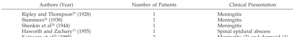
TABLE 1. Literature Search of Patients With a Presumed Association Between a Coccygeal Pit and Either Neurologic Infection or Intradural Pathology13,15,24 –26
Authors (Year) Number of Patients Clinical Presentation
Ripley and Thompson25(1928) 1 Meningitis
Stammers26(1938) 1 Meningitis
Shenkin et al24(1944) 1 Meningitis
Haworth and Zachary13(1955) 1 Spinal epidural abscess
They may become susceptible to local recurrent infection from trauma or hirsuitism. They are not related to acquired pilonidal conditions observed in adults.12Their cause is not clear. Controversy
regard-ing an association with OSD and the proper evalua-tion and management of isolated cutaneous defects in the coccygeal region exists.13,15–23Given the relative
frequency of these cutaneous abnormalities, any statement requiring diagnostic evaluation is of pub-lic health concern. Based on our studies, it becomes difficult to recommend surgical treatment or even radiographic evaluation for isolated coccygeal pits.
Retrospective review of our own patient data sup-ports the innocence of coccygeal pits. Although such data can be criticized for lacking radiographic docu-mentation, others have already demonstrated
evi-dence of radiographic benignity. Herman et al35
per-formed spinal ultrasound on 53 infants with coccygeal pits. The average age of those studied was 24 days. The location of the conus medullaris was found to be between T12 and L1 in 13%, behind the L1 vertebral body in 20%, and behind the L2 verte-bral body in 67%. No intraspinal anomalies were identified. 35 Gibson et al36 prospectively examined
95 neonates harboring cutaneous abnormalities of the back with ultrasound. Seventy-five of the 95 chil-dren had isolated cocygeal pits. No abnormality of the spinal axis was identified in those with coccygeal pits.36The radiographic data appear to correlate and
follow our clinical impressions that isolated cocygeal pits are benign. Hence, the burden of proof is not with us to radiographically demonstrate that simple
coccygeal pits are benign. The burden of proof must rest with those who mandate radiographic or even surgical investigation. To this, there seems to be no justification.
CONCLUSIONS
Coccygeal pits are very common abnormalities of the skin. Lesions in isolation are associated with a small incidence for associated neurologic infection or neurologic deterioration. Therapeutic evaluation may be limited to physical examination. Lesions in association with other well-defined cutaneous stig-mata of OSD warrant further radiographic and/or surgical inspection.
REFERENCES
1. Pierre-Kahn A, Lacombe J, Pichon J, et al. Intraspinal lipomas with spina bifida: prognosis and treatment in 73 cases.J Neurosurg. 1986;65:756 –761 2. Bruce DA, Schut L. Spinal lipomas in infancy and childhood.Childs
Brain. 1979;5:192–203
3. Chapman PH. Congenital intraspinal lipomas: anatomic considerations and surgical treatment.Childs Brain. 1982;9:37– 47
4. Lapras CL, Patet JD, Huppert J, et al. The tethered cord syndrome: experience of 58 cases.J Pediatr Neurosci. 1985;1:39 –50
5. Hoffman HJ, Taecholara C, Hendrick EB, Humphreys RP. Management of lipomyelomeningoceles: experience at the Hospital for Sick Children, Toronto.J Neurosurg. 1985;62:1– 8
6. Hendrick EB. On diastematomyelia.Prog Neurol Surg. 1974;4:277–288 7. Hendrick EB, Hoffman HJ, Humphreys RP. The tethered spinal cord.
Clin Neurosurg. 1982;30:457– 463
8. Guthkelch AN. Diastematomyelia with median septum.Brain. 1974;97: 729 –742
9. McLone DG, Dias MS. Normal and abnormal early development of the nervous system. In: Cheek WR, Marlin AE, McLone DG, et al, eds. Pediatric Neurosurgery: Surgery of the Developing Nervous Sytem. Philadel-phia, PA: WB Saunders; 1994:1–39
10. Barkovich AJ, Edwards MSB, Cogen PH. MR evaluation of spinal der-mal tracts in children.AJNR Am J Neuroradiol. 1990;12:123–129 11. Powell KR, Cherry JD, Hougen TJ, Blinderman EE, Dunn MC. A
pro-spective search for congenital dermal abnormalities of the craniospinal axis.J Pediatr. 1975;87:744 –750
12. Chamberlain JW, Vawter GF. The congenital origin of pilonidal sinus. J Pediatr Surg. 1974;9:441– 444
13. Haworth JC, Zachary RB. Congenital dermal sinuses in children-their relation to pilonidal sinus.Lancet. 1955;2:10 –14
14. Solla JA, Rothenberger DA. Chronic pilonidal disease: an assessment of 150 cases.Dis Colon Rectum. 1990;33:758 –761
15. Kajiwara H, Matsukado Y, Hiraki T, Yokata A. Intraspinal
communi-cation of sacrococcygeal dermal sinuses. Childs Nerv Syst. 1985;1: 264 –267
16. Calvit MF, Aranda G. Timing of surgery in patients with infected spinal dermal sinuses: report of two cases.Childs Nerv Syst. 1995;11:29 –32 17. Carrillo R, Carreiera LM, Prada JJ, Rosas C. Lateral congenital spinal
dermal sinus: a new clinical entity.Childs Nerv Syst. 1985;1:238 –240 18. Mount LA. Congenital dermal sinuses: a cause of meningitis, intraspinal
abscess and intracranial abscess.JAMA. 1949;139:263–269
19. Kanev PM, Park TS. Dermoids and dermal sinus tracts of the spine. Neurosurg Clin North Am. 1995;6:359 –366
20. Humphreys RP. Clinical evaluation of cutaneous lesions of the back: spinal signatures that do not go away.Clin Neurosurg. 1995;43:175–187 21. Peter JC, Sinclair-Smith C, deVilliers JC. Midline dermal sinuses and cysts and their relationship to the central nervous system.Eur J Pediatr Surg. 1991;1:73–79
22. Sherburn EW, Park TS. Occult spinal dysraphism.Contemp Neurosurg. 1997;19:1– 8
23. McComb JG. Congenital dermal sinus. In: Pang D, ed.Disorders of the Pediatric Spine. New York, NY: Raven Press; 1995:359
24. Shenkin HA, Hunt AD, Horn RC. Sacrococcygeal sinus (pilonidal sinus) in direct continuity with the central canal of the spinal cord. Surg Gynecol Obstet. 1944;79:655– 659
25. Ripley W, Thompson DC. Pilonidal sinus as a route of infection in a case of staphylococcus meningitis.Am J Dis Child. 1928;36 –785-788 26. Stammers FAR. Spinal epidural suppuration, with special referance to
osteomyelitis of the vertebrae.Br J Surg. 1938;26:366 –374
27. Lichtenstein BW. Spinal dysraphism: spina bifida and myelodysplasia. Arch Neurosurg Psychiatry. 1940;44:792– 809
28. Lemire RJ, Leoser JD, Leech RW, et al, eds. Normal and Abnormal Development of the Human Nervous System. Hagerstown, MD: Harper and Row; 1975
29. James CCM, Lassman LP, eds.Spina Bifida Occulta: Orthopedic, Radiolog-ical, and Neurosurgical Aspects. London, UK: Academic Press; 1981 30. Till K. Spinal dysraphism: a study of congenital malformations of the
lower back.J Bone Joint Surg (Br). 1969;51:415– 422
31. O’Neill P, Singh J. Occult spinal dysraphism in children: need for early neurosurgical referral.Childs Nerv Syst. 1991;7:309 –311
32. Kanev PM, Lemire RJ, Loeser JD, Berger MS. Management and long-term follow-up review of children with lipomyelomeningocele, 1952–1987.J Neurosurg. 1990;73:48 –52
33. Oakes WJ. The borderlands of the primary tethered cord syndrome.Clin Neurosurg. 1995;43:188 –202
34. Tavafoghi V, Ghandchi A, Hambrick GW. Cutaneous signs of spinal dysraphism.Arch Dermatol. 1978;114:573–577
35. Herman TE, Oser RF, Shackleford GD. Intergluteal dorsal dermal sinuse: the role of neonatal spinal sonography. Clin Pediatr. 1993; 627– 628
DOI: 10.1542/peds.105.5.e69
2000;105;e69
Pediatrics
Bradley E. Weprin and W. Jerry Oakes
Coccygeal Pits
Services
Updated Information &
http://pediatrics.aappublications.org/content/105/5/e69
including high resolution figures, can be found at:
References
http://pediatrics.aappublications.org/content/105/5/e69#BIBL
This article cites 30 articles, 0 of which you can access for free at:
Subspecialty Collections
http://www.aappublications.org/cgi/collection/surgery_sub
Surgery
following collection(s):
This article, along with others on similar topics, appears in the
Permissions & Licensing
http://www.aappublications.org/site/misc/Permissions.xhtml
in its entirety can be found online at:
Information about reproducing this article in parts (figures, tables) or
Reprints
http://www.aappublications.org/site/misc/reprints.xhtml
DOI: 10.1542/peds.105.5.e69
2000;105;e69
Pediatrics
Bradley E. Weprin and W. Jerry Oakes
Coccygeal Pits
http://pediatrics.aappublications.org/content/105/5/e69
located on the World Wide Web at:
The online version of this article, along with updated information and services, is
by the American Academy of Pediatrics. All rights reserved. Print ISSN: 1073-0397.