THE EFFECT OF CHLORHEXIDINE AND OTHER
ANTIMICROBIAL AGENTS ON THE FORMATION AND
VIABILITY OF ORAL BACTERIAL BIOFILMS
Thesis submitted by
Jonathan Pratten BSc (Hons)
for the degree of
DOCTOR OF PHILOSOPHY
in the
Faculty of Medicine
University of London
Department of Microbiology
Eastman Dental Institute
for Oral Health Care Sciences
256 Gray’s Inn Road
London, W C1X 8LD
and
SmithKline Beecham
Oral Care Research
St. George’s Avenue
Weybridge, Surrey
KT13 0 D E
1998-ProQuest Number: U643340
All rights reserved
INFORMATION TO ALL USERS
The quality of this reproduction is dependent upon the quality of the copy submitted.
In the unlikely event that the author did not send a complete manuscript
and there are missing pages, these will be noted. Also, if material had to be removed, a note will indicate the deletion.
uest.
ProQuest U643340
Published by ProQuest LLC(2016). Copyright of the Dissertation is held by the Author.
All rights reserved.
This work is protected against unauthorized copying under Title 17, United States Code. Microform Edition © ProQuest LLC.
ProQuest LLC
789 East Eisenhower Parkway P.O. Box 1346
Abstract
Caries and periodontal diseases have been shown to stem from an
imbalance in the normal oral microflora present on tooth surfaces as biofilms
(plaque). There is considerable interest in developing chemical agents to
supplement mechanical means (i.e. toothbrushing) of controlling these
diseases. Although many studies have shown that planktonic oral bacteria
can be easily killed by a range of available agents, such studies are largely
irrelevant to the situation in vivo as the sessile (biofilm) forms of these
bacteria are less susceptible to the action of such agents. The aims of this
study, therefore, were to develop and test a laboratory model suitable for
evaluating the effectiveness of antimicrobial agents against oral bacterial
biofilms. A constant depth film fermentor was used to grow the biofilms. In
order to mimic the situation in vivo, the biofilms were grown in an aerobic
environment on a substratum similar to human enamel, with nutrients
supplied by an artificial saliva.
Initial studies were carried out using a mono-species (Streptococcus
sanguis) biofilm grown on a variety of substrata and these were found to
influence the susceptibility of the biofilms to various agents including
chlorhexidine (CH), cetylpyridinium chloride and triclosan. The effectiveness
of mouthwashes (containing different antimicrobial agents) against S.
sanguis biofilms was then determined. The effect of CH on multi-species
biofilms and microcosm plaques was also investigated. The reproducibility of
the multi-species biofilms was poor, with varying proportions of species
present in each run. The microcosm plaques were far more reproducible and
found in supra-gingival plaque. The effects of a number of treatment regimes
(involving CH) against these biofilms were investigated. When challenged
with CH pulses there was an initial 2 logio reduction in the number of viable
organisms, however, although pulsing continued, the biofilms recovered in
terms of both the viability and relative proportions of the constituent species.
Cryosectioning of the microcosm plaques was carried out to determine the
viability and proportions of species present at various depths throughout the
biofilm.
The CDFF proved to be a good method for reproducibly generating large
numbers of bacterial biofilms comparable to supra-gingival plaque and
enabled the evaluation of the activity of antimicrobial agents in a model
Declaration
I hereby certify that the work embodied in this thesis is the result of my own
investigations, except where otherwise stated. The electron microscopy
sample preparation was carried out by Mrs Nikki Morgan of the Electron
Microscopy unit at the Eastman Dental Institute. The scanning confocal laser
microscopy work was carried out in collaboration with Dr. John Warrack,
Microscopy and Flow Cytometry Analytical Sciences Department, SmithKline
Acknowledgements
I would like to sincerely thank my supervisor Prof. Mike Wilson and my
industrial sponsors Dr. Paul Barnett and Dr. Andy Smith for their support and
guidance throughout this project.
I would also like to thank all of my colleagues at the Eastman Dental Institute
for all their help and encouragement throughout this thesis. I would
particularly like to thank Dr. Tracy Burns, Dr. Dave Spratt, Ms. Zoey Jackson,
Mr. Alun Kirby, Mr. Warwick May and Ms. Julie Fletcher.
I am very thankful to my Mum and Dad for their continued support over my
student years, and to my brother, David, for his friendship and assistance,
Index of contents
Title... 1
Abstract... 2
Declaration... 4
Acknowledgements... 5
Dedication... 6
Index of contents... 7
List of figures... 12
List of tables... 16
Abbreviations... 17
Publications as a result of this thesis... 18
Chapter One
19
Introduction... 1.1. The State vs. Copley Pharmaceuticals... 201.2. Biofilms... 21
1.2.1. Biofilm structure... 23
1.2.2. Extracellular Matrix... 27
1.2.3. Bacterial Adherence... 28
1.2.4. Bacterial Accumulation... 30
1.2.5. Biofilm Nutrition... 32
1.2.6. Biofilm Resistance... 33
1.3. Dental Plaque... 36
1.3.1. The Development of Dental Plaque... 38
1.4. Caries... 41
1.4.1. Microbiology of Caries... 44
1.4.2. Oral Streptococci... 45
1.5. Control and Prevention of Caries... 47
1.5.2. Mechanical Methods... 51
1.5.3. Fluoride... 52
1.5.4. Antimicrobials... 53
1.5.4.1. Chlorhexidine... 56
1.5.4.2. Cetyl-pyridinium chloride... 58
1.5.4.3. Triclosan... 59
1.6. Biofilm Models... 60
1.6.1. The Robbins Device... 62
1.6.2. The Perfused Biofilm Fermentor... 63
1.6.3. Constant Depth Film Fermentor... 64
1.6.4. Continuous culture chemostat... 64
1.7. Aims of the study... 65
Chapter Two
Materials and Methods... 2.1. Bacteriological media... 672.1.1. Cadmium fluoride acriflavin tellurite agar... 67
2.1.2. Artificial saliva... 67
2.2. The Constant Depth Film Fermentor... 68
2.3. Organisms... 73
2.4. Inoculation of the fermentor... 73
2.4.1. Inoculation of single species... 74
2.4.2. Inoculation of multi-species... 74
2.4.3. Inoculation of microcosm plaque... 75
2.5. Continuous flow... 76
2.6. Culture methods... 78
2.7. Hydroxyapatite and bovine enamel preservation methods 79 2.7.1. Mineralising solution... 79
2.7.2. Cleaning discs which have been exposed to agents.... 79
2.8. Viability staining assays... 80
2.9. Cryosectioning... 82
2.10. Electron microscopy... 82
2.10.1. Transmission electron microscopy... 83
2.10.2. Scanning electron microscopy... 83
2.11. Statistical analysis... 84
Chapter Three
Effect of mouthwashes on the formation and growth of Streptococcus sanguis biofilms on various substrata 85 3.1. Aims... 863.2. Materials and Methods... 86
3.2.1. Growth of Biofilms... 86
3.2.2. Effect of mouthwashes on the viability of Streptococcus sanguis biofilms... 86
3.2.3. Effect of mouthwashes on biofilm formation... 87
3.3. Results... 88
3.4. Discussion... 94
Chapter Four
The susceptibility of Streptococcus sanguis biofilms grown on bovine enamel to chlorhexidine... 1004.1. Aims... 101
4.2. Materials and Methods... 101
4.3. Results... 101
Chapter Five
Growth and susceptibility of a 6 membered biofilm
community to chlorhexidine... 111
5.1. Aims... 112
5.2. Materials and Methods... 112
5.3. Results... 113
5.4. Discussion... 125
Chapter Six
Growth and structure of a microcosm community biofilm and susceptibility to chlorhexidine... 1326.1. Aims... 133
6.2. Materials and Methods... 133
6.2.1. Inoculation and sampling... 133
6.2.2. Chlorhexidine pulsing... 133
6.2.3. Chlorhexidine penetration through the biofilm... 134
6.2.4. Susceptibility of varying thicknesses of microcosm plaque... 134
6.2.5. Scanning confocal laser microscopy... 134
6.3. Results... 136
6.3.1. Growth... 136
6.3.2. Susceptibility... 136
6.3.3. Structure... 139
Chapter Seven
Growth and susceptibility of microcosm plaques grown
in the presence of sucrose... 166
7.1. Aims... 167
7.2. Materials and Methods... 167
7.2.1. Growth of sucrose supplemented biofilms... 167
7.2.2. Determination of pH... 167
7.3. Results... 168
7.4. Discussion... 175
Chapter Eight
Summary and Conclusions... 182List of figures
1.1 The archetypal biofilm model 24
1.2 Heterogeneous mosaic biofilm model according to Keevil et 25
al.
1.3 Water channel model according to Costerton at ai. 26
1.4 Chronic plaque build-up on the teeth 36
1.5 A diagrammatic representation of the attachment of bacteria 39
from saliva to pellicle-coated enamel
1.6 Tooth structure and sites of carious infections 42
1.7 Structure of Chlorhexidine 56
1.8 The modified Robbins device showing the sample ports and 59
the stud design
2.1 Constant Depth Film Fermentor 69
2.2a Schematic vertical section through the fermentor 71
2.2b Schematic horizontal section through the fermentor 72
2.3a Schematic diagram of fermentor showing recirculation of 74
inoculum
2.3b Schematic diagram of fermentor with through-flow of 77
medium
2.4 Structures of fluorescent probes 80/
81
2.5 Diagrammatic cross-section of embedded sample for
cryosectioning 82
3.1 Scanning electron micrograph of Streptococcus sanguis 90
biofilm at 24 h
3.2 The growth of Streptococcus sanguis biofilms on various 91
substrata
3.3 Effect of mouthwashes on the viability of Streptococcus 92
sanguis biofilms grown on hydroxyapatite discs
3.4 Effect of mouthwashes on the viability of S. sanguis biofilms
grown on bovine enamel discs 92
hydroxyapatite discs treated with mouthwashes 93
3.6 Formation of S. sanguis biofilms on bovine enamel discs 93
treated with mouthwashes
4.1 Scanning electron micrograph of Streptococcus sanguis 103
biofilm at 120 h.
4.2 Effect of pulsing with 0.1 % chlorhexidine on the viability of
S. sanguis biofilms 104
4.3 Effect of pulsing with 0.2 % chlorhexidine on the viability of
S. sanga/s biofilms 104
4.4 Live / dead staining results following pulsing of S. sanguis
biofilms with 0.1 % chlorhexidine 105
4.5 Live / dead staining results following pulsing of S. sanguis
biofilms with 0.2 % chlorhexidine 106
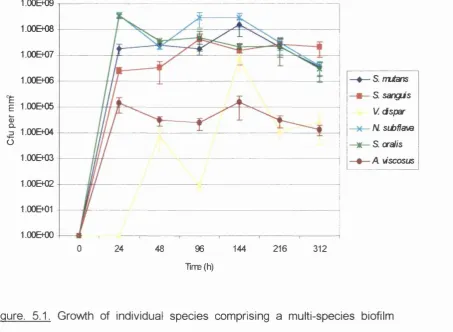
5.1 Growth of individual species comprising a multi-species
biofilm formed on bovine enamel discs with artificial saliva
as the sole nutrient source 117
5.2 Viable counts of constituent bacteria in 30 pm thick sections
of a 300 pm thick biofilm 119
5.3 Transmission electron micrograph of a transverse section of
biofilm (216 h) at the biofilm/air interface 120
5.4 Transmission electron micrograph of a transverse section of
biofilm (216 h) at the biofilm/substratum interface 121
5.5 The relative proportions of live and dead bacteria in 30 pm
thick sections of a 216 h old, 300 pm thick biofilm 122
5.6a/b Response of individual species comprising multi-species
biofilms to various periods of exposure to 0.2 %
chlorhexidine 123
5.7 Growth of 6 membered biofilm community inoculated with S.
sanguis for 24 h prior to inoculation with the remaining 5
species 124
5.8 Composition of nine-membered community biofilms - from
Kinniment at a! (1996). 126
Confocal laser scanning microscope 135
6.2 Percentage proportions of species comprising polled saliva
from 5 separate inocula 141
6.3a The total aerobic viable counts (120 h) from 5 pans of the
same run 142
6.3b The total aerobic viable counts (120 h) from 5 pans of the
same run 142
6.3c The total Actinomyces species viable count (120 h) from 5
pans of the same run 143
6.3d The total Streptococcus species viable count (120 h) from 5
pans of the same run 143
6.3e The total Veillonella species viable counts from 5 pans of
the same run 144
6.4 Graph showing the anaerobic counts from 3 separate runs
over 1 9 2 h 144
6.5 Effect of pulsing 0.2 % chlorhexidine on the viability of
microcosm plaques (168 h) 145
6.6 Growth of a microcosm plaque community on bovine
enamel discs treated with 0.2 % chlorhexidine and then
pulsed with chlorhexidine after 8 h 146
6.7a Susceptibility of 100 pm thick microcosm plaques to cetyl
pyridinium chloride and chlorhexidine for 1 and 5 mins. 147
6.7b Susceptibility of 300 pm thick microcosm plaques to cetyl
pyridinium chloride and chlorhexidine for 1 and 5 mins. 148
6.8a Absorbance readings of constituents used in cryosectioning
microcosm plaque biofilms over a range of wavelengths 149
6.8b Absorbance readings taken from sections through a
microcosm plaque community 150
6.9 Viable counts from 30 pm thick sections through a
microcosm plaque 151
6.10 Viable counts from 30 pm thick sections through a
microcosm plaque which had been exposed to 0.2 %
6.11a Confocal laser scanning microscope image of a microcosm
plaque grown on bovine enamel viewed with live/dead stain 153
6.11b Confocal laser scanning microscope image of a microcosm
plaque grown on bovine enamel exposed to 0.2 %
chlorhexidine for 1 h viewed with live/dead stain 153
6.12 Transmission electron micrograph of a section through a
microcosm plaque (120 h) grown in the CDFF 154
6.13 Transmission electron micrograph of in vivo dental plaque 162
7.1 Growth of various groups of bacteria comprising a
microcosm plaque community pulsed thrice daily with
sucrose 170
7.2 Viable counts of sucrose pulsed microcosm plaque
additionally pulsed twice daily with 0.2 % chlorhexidine at
1 2 0 h 171
7.3 Comparisons between the pH of three different types of
biofilms 172
7.4 Viable counts from 30 pm thick sections through microcosm
plaque grown in the presence of sucrose 173
7.5 Viable counts of 30 pm thick sections through microcosm
plaque grown in the presence of sucrose following exposure
List of tables
1.1 Currently recognised S. mufans species 46
1.2 Specific and non-specific host defences in the mouth 48
1.3 Composition of synthetic saliva from Shellis (1978) 50
2.1 CFAT agar composition (g/L in dH2Û) 67
2.2 Composition of artificial saliva in g/L 68
2.3 Composition of suspending medium 75
2.4 Composition of mineralising solution 79
5.1 Total viable counts and percentage proportions of each
species comprising 216 h biofilms formed during three
separate runs 118
6.1 Comparison of the bacterial composition of approximal dental
plaque (data from Newman and Nisengard, 1988), microcosm
plaques and the pooled human saliva used as the inoculum 141
7.1 Percentage of genera (relative to the total anaerobic count)
Abbreviations
02°c
AnÛ2 CDFF Gfu CH CPC CLSM h L LogM9
MBC MIC ml MRD PBF PTFE SEM T TEM Aerobicdegrees Celsius
Anaerobic
constant depth film fermentor
colony forming units
Chlorhexidine digluconate
Cetyl pyridinium chloride
confocal laser scanning microscopy
Hours
Litre
Logarithmic
Microgram
minimum bactericidal concentration
minimum inhibitory concentration
Millilitre
Modified Robbins device
perfused biofilm fermentor
Polytetrafluoroethylene
scanning electron microscopy
Triclosan
Publications resuiting from this thesis
Pratten, J. and Wilson, M. 1996. Effect of repeated chlorhexidine exposure
on Streptococcus sanguis biofilms. Journal of Dental Research. Vol. 75 (5) p.1137
Pratten, J. and Wilson, M. 1997. Effect of Antiseptics on Formation and
Viability of Streptococcal Biofilms. Advances in Dental Research. Vol. 11 (1) p.190
Pratten, J. Barnett, P. and Wilson, M. 1997. The susceptibility of a 6
membered biofilm community to chlorhexidine. Journal of Dental Research.
Vol. 76 (5) p. 136
Pratten, J. Barnett, P. and Wilson, M. 1997. The susceptibility of a
microcosm plaque to chlorhexidine pulsing in vitro. Biofilms: Community
Interactions and Control. BioLine., U.K. p.245 - 250
Pratten, J., Wills, K., Barnett, P. and Wilson. M. 1998. \n vitro Studies of the Effect of Antiseptic-containing Mouthwashes on the Formation and Viability
of Streptococcus sanguis Biofilms. Journal of Applied Microbiology. In Press.
Pratten, J., Smith, A, W. and Wilson, M. 1998. Response of single species
biofilms and microcosm dental plaques to pulsing with chlorhexidine. Journal of Antimicrobial Chemotherapy. In Press.
Pratten, J., Barnett, P. and Wilson. M. 1998. Composition and Susceptibility
to Chlorhexidine of Multi-species Biofilms of Oral Bacteria. Applied and
1.1. The State vs. Copley Pharmaceuticals
100 people died in late 1993 and early 1994 due to a mysterious bacterial
infection that struck hundreds of asthmatics throughout the United States.
The antibiotics administered to the patients in hospital apparently failed to
subdue the virulent infection.
All the patients had been using a generic albuterol inhalant, produced by
Copley Pharmaceuticals, to treat their asthma. The infection was traced back
to the manufacturer’s albuterol processing tank. Nothing illegal had been
carried out, the tank had been treated with chemical disinfectants, the
standard treatment according to the health and safety laws.
Many court cases arose from the incident, one in Cheyenne, Wyoming in the
summer of 1995. The lawyer for the plaintiffs called to the stand a
microbiologist. Dr. Costerton, who examined the records submitted to the
court by the Food and Drug Administration. He noted the presence of a
particular species of bacteria. Pseudomonas aeruginosa, floating freely in
the tank. Not only does this species cause pneumonia, but P. aeruginosa is
notorious for forming biofilms, a matt of bacteria covered in ‘slime’ which
forms on surfaces. These biofilms are resistant to chemical disinfectants,
antibiotics, and the immune system.
It was not the first time that biofilms had played a role in high-profile cases,
Costerton has testified in court about the presence of biofilms on intrauterine
devices, but the asthmatics tragic experience highlighted a gap in the way
1.2. Biofilms
The consequences, both beneficial and harmful, of the association between
microbes and surfaces have long been recognised. As far back as the 14**^
Century, Guy de Chauliac, a French surgeon, recorded the relationship
between foreign bodies and delayed wound healing (Voorhees,1985), while
just over a century ago the symbiosis between Rhizobium and the roots of
leguminous plants was first recorded (Beijerinck,1888). Although the first
detailed description of microbial attachment to surfaces appeared more than
50 years ago (Zobell,1943), it was not until the late 1970's that the term
‘biofilm’ made its first appearance in the scientific literature.
The almost universal association between micro-organisms and surfaces is
now widely accepted. Mature biofilms may contain as many as 10^° cells/ml,
considerably more than usually arise in suspension. Indeed many regard
surfaces as the preferred site for microbial growth.
Costerton et a! (1995) have now observed that adhesion of a bacterium to a
surface triggers the expression of a sigma factor that depresses a large
number of genes so that biofilm cells are clearly phenotypically distinct from
their planktonic counterparts. Each biofilm bacterium lives in a customised
microniche in a complex microbial community that has primitive
homeostasis, a primitive circulatory system, and co-operates metabolically,
and each of these sessile cells reacts to its special environment so that it
differs fundamentally from a planktonic cell of the same species.
Biofilms usually become thick enough that certain solutes, in particular
oxygen, become exhausted before the base of the biofilm is reached. This
as nutrients are available. The close association between aerobic and
anaerobic species causes many interactions to take place. For example,
where anaerobic corrosion, due to sulphate-reducing bacteria takes place,
sulphide formed in the anaerobic regions can be reoxidised in the aerobic
surface layers by sulphide-oxidising bacteria (Hamilton, 1985).
Microbial films have an important economic role besides their obvious
interest ecologically. Examples of the problems that biofilms can cause
include the colonisation of boat hulls, leading to fouling by larger organisms;
films causing the erosion of marine steel or concrete installations; growth in
water pipes causing reduced flow rates and infection. Moreover,
considerable morbidity and mortality results from the microbial biofilms that
may arise on implanted medical devices, for example, catheters and artificial
heart valves.
The effects of biofilms are not always adverse, most effluent treatment
plants encourage the growth of microbial films, for example, on aerobic or
anaerobic filter systems and on rotating disc aerators which are all used to
recycle organic pollutants (Characklis,1980).
Biofilms are also associated with numerous other surfaces. Most solid-liquid
interfaces can become coated with microbes that tend to attach to a thin
layer of adsorbed macromolecules, which quickly bind to any ‘clean’ surface
immersed in natural aquatic systems. Films are found on the gastric mucosa
and internal epithelial linings of many animals. While many oral organisms
attach to dental enamel, others ‘prefer’ the cheek and tongue epithelial cells.
Films are formed on most surfaces immersed in any of the natural water
also the surfaces of plants and aquatic animals. Microbes develop on
terrestrial surfaces, too. The phylloplane is a habitat on the surface of
leaves, which shows a succession of organisms throughout the growing
season. Sometimes these proliferate enough to form a coherent film. A
corresponding region around plant roots, the rhizoplane, leads to a
cylindrical film-like proliferation of microbes using root exudates as nutrients.
There is even a region around germinating seeds called the spermosphere
that has some of the characteristics of a biofilm.
Research that has been carried out on biofilms has tended to focus on two
main aspects. Firstly, the elimination of the biofilm from an infected area
which would involve both killing and removal of the film, and secondly, the
control of their formation and activity where they perform some useful
function. Microbiologists have become increasingly interested not so much in
the film itself but in the mechanisms involved in the attachment of microbes
to surfaces. Very little is known about the structure and physiological
functioning of biofilms. In nature a biofilm is rarely composed of just a single
species, but a group of different genotypes each having some part to play in
the overall behaviour of the community.
1.2.1. Biofilm structure.
The use of new microscopic and computer-aided techniques has been
instrumental in the development of models which assist in explaining the
complex structures of biofilm communities.
Scanning and transmission electron microscopy have been used for several
as a dense biofilm which is complex in structure and is associated with a
large number of different bacteria. Observations from several studies
(Marsh, 1995; Listgarten,1976) have shown that there is structural
organisation present in these dense biofilms. These structures include
microcolonies of similar-shaped bacteria, parallel orientated bacteria and
specific associations between bacteria, described as ‘corn cobs’. A
generalised biofilm structure is shown diagrammatically below (Fig. 1.1), it
consists of a base film attached to the conditioning film with bulk liquid and
gas space above.
gas space
bulk liquid
surface film
base film conditioning film
substratum
biofilm volume elem ent
Figure 1.1. The archetypal biofilm model.
Differential interference contrast microscopes have been used by Keevil et al
to study biofilms in water distribution systems (Walker et a!., 1995). Using
this technique the structure of the biofilm as a whole can be examined.
According to their observations, such biofilms consist of a thin film (approx.
5|jm) of attached cells over the surface with microcolonies attaching to the
substratum forming ‘stacks’. These stacks' are well separated allowing a
flow of nutrients through the biofilm (Fig. 1.2).
W a ter flow
y Raised layer ( (up to 100|jm ) Grazing
protozoon Microcolonies
Basal layer (c. 5pm)
%---
GOFigure 1.2. Heterogeneous mosaic biofilm model according to Keevil et al.
The use of confocal scanning laser microscopes (CSLM) has now enabled
the examination of living, fully hydrated, biofilms and the use of this
technique has provided valuable structural information. Costerton et al
(1994) have studied many pure cultures and natural bacterial populations
using CSLM, the studies concluded that biofilm bacteria grow predominantly
in microcolonies of similar morphotypes and that these colonies are
interspersed between water channels (Lawrence et a!., 1991). The channels
contained few bacterial cells and appeared to contain a more permeable
matrix material. The ‘idealised’ structure is shown in Figure 1.3 (Costerton et
Figure 1.3. Water channel model according to Costerton et a / (1994).
The cellular automaton model (Wimpenny and Colasanti, 1997) suggests
that the structure of a biofilm depends on the substrate concentration. The
model indicates that the highest concentration of substrates lead to the
formation of dense films while, contrary to this, the lowest concentrations
form stacked structures. The model also implies a layer of individual cells at
the surface in a low nutrient environment from which stacks develop. The
dense biofilms of which dental plaque is an example can also be simulated
and, using slightly lower substrate concentrations, channels which are often
observed using electron microscopy can be demonstrated.
1.2.2. Extracellular Matrix.
One of the most notable features of biofilms is their high content (50-90 %)
of exopolysaccharide or EPS (Characklis and Cooksey, 1983). The
terminology for the extracellular material associated with cell aggregates or
biofilms varies in the literature, being referred to as slime, capsule, sheath,
EPS and glycocalyx. Zobell (1943) suggested the involvement of
extracellular ‘cementing’ substances in the adhesion of cells to the
substratum. The last stage of cell attachment to a surface, involving specific
interactions, is associated with the production of adhesive materials such as
exopolysaccharides (Lappin-Scott and Costerton, 1989). Corpe (1970)
demonstrated the involvement of acidic polysaccharides in bacterial
adhesion, and Fletcher and Floodgate (1973) observed this by means of
electron microscopy.
The matrix is usually assumed to be constructed of long-chain or polymeric
materials (lipids, proteins, polysaccharides, polyphenols, nucleic acids)
although the proportions of these materials are not known (Palenik, 1989).
The majority, however, are polysaccharides. Common sugars such as
glucose, galactose, mannose, fructose, rhamnose, A/-acetylglucosamine,
glucuronic acid, galacturonic acid, mannuronic acid and guluronic acid, are
all typical constituents of bacterial polysaccharides (Christensen, 1989).
Pseudomonas aeruginosa synthesises an exopolysaccharide called alginate
in response to environmental conditions (Boyd and Chakrabarty, 1995).
Alginate serves to protect the bacteria from adversity in its surroundings and
also enhances adhesion to solid surfaces. Transcription of the alginate
genes is induced upon attachment to the substratum and this leads to
advantageous to the survival and growth of the bacteria. In certain
circumstances, P. aeruginosa produces an alginate lyase which cleaves the
polymer into short oligosaccharides. This negates the anchoring properties
of the alginate and results in increased detachment of the bacteria from the
surface, allowing them to spread and colonise new sites. Thus, both alginate
biosynthetic and degradative enzymes are important for the development,
maintenance and spread of P. aeruginosa biofilms.
The extracellular matrix may also contain particulate materials: clays, organic
debris, phages, lysed cells and precipitated minerals. Understanding the
physical and chemical characteristics of the matrix and its relationship to the
resident organisms may influence the understanding of the structure and
function of biofilms.
1.2.3. Bacterial Adherence.
Several stages are involved in the adhesion of a microbe to a surface
whether inanimate or another living cell. If we imagine a microbe
approaching a surface then at a distance of tens of nanometres the two
objects are influenced by two types of forces - van der Waals and
electrostatic. At a distance of >50 nm Van der Waals interactions occur and
these are the result of the mutual induction of dipoles in the two objects
resulting in their mutual attraction. As the distance between the objects
decreases (10-20 nm), electrostatic forces become significant and, as most
microbes and surfaces have a net negative charge, the net effect is
repulsion. However, these repulsive forces decrease with increasing ionic
reduce or overcome this repulsion.
As the bacterium approaches more closely, intervening water molecules will
act as a barrier to attachment. However, hydrophobic molecules on the
surface of either the bacterium or host cell (or both) can exclude these other
molecules. Hydrophobic interactions between the bacterium and the host cell
can then result in adhesion or can enable a close enough approach (<1.0
nm) for other adhesive interactions to occur. The latter include hydrogen
bonding, cation bridging and receptor-ligand interactions i.e. the specific
binding of a molecule (ligand) on the bacterial surface to a complementary
substrate molecule (receptor) on the host cell surface (Fletcher, 1996),
Bacterial adhesion to host cells is thought to be mediated primarily by
hydrophobic interactions, cation bridging and receptor-ligand binding. While
hydrophobic interactions are recognised as being important in the adhesion
of bacteria to host cells and to inanimate substrata, more is known about the
role of receptor-ligand interactions. The specific molecules on the bacterial
surface responsible for adhesion are known as adhesins.
Collectively, bacteria elaborate a number of structures which may be
involved in adhesion to cell surfaces. These include fimbriae and
proteinaceous fibrils whose primary function appears to be that of adhesion,
as well as capsules and flagella which have other functional roles namely
protection and locomotion respectively. All of these structures contain
adhesins although the chemical identity of many of these has not yet been
determined. As well as these structures, the cell walls of many species
contain macromolecules which function as adhesins. A particular species
either concurrently or consecutively. In the case of the latter, this may enable
the organism to adhere to the different cell types it encounters during the
course of the infectious process in which it partakes.
1.2.4. Biofilm Accumulation.
The initial events of biofilm accumulation or colonisation at a substratum are
the net result of transport, adsorption and desorption and growth processes.
Diffusive or advective transport processes carry the cell to a point adjacent
to the substratum. Colonisation processes can be expressed in terms of two
variables: colony forming units (CPU) and cells. This distinction is important
as cells can adsorb in groups or as single cells. Thus a single cell is a CPU,
but an aggregate of five cells is also a CPU. It must also be taken into
account that not all cells accumulate at the substratum through transport, but
cells also form at the substratum through growth. The following four
processes have been distinguished by Escher (1986).
1. Diffusive or advective transport carries the cfu to a point adjacent to the
substratum. In laminar flow, only diffusive transport is involved. In
turbulent flow, advective transport generally dominates.
2. Adsorption is the linking of the cfu with the substratum. The cell is
adsorbed to the substratum if it has a linkage to it and hence becomes
immobilised for a certain time period.
3. Desorption is the breaking of the substratum-cfu linkage and the complete
removal of the cfu from the substratum, and is therefore the opposite of
4. Cfu separation, although not related to adsorption or desorption,
contributes to the accumulation of cfu at the substratum by changing the
number of cfu adsorbed. A cfu with more than one cell can separate into
two independent cfu as a result of a fluid shear or even cell motility. Cfu
separation does not influence cell numbers on the substratum.
The processes can be described in terms of cells by determining the number
of cells in each cfu. In addition to advective transport, adsorption, and
desorption, two additional processes need to be considered when the
concentration of cells at the surface is the variable:
1. Multiplication is related to cellular growth. In contrast to growth, which
includes the entire growth cycle of the cell, multiplication represents the
singular event of cell division. Cells within a cfu multiply, and the number
of cells within this cfu increases. This does not change the accumulated
number of cfu, but does change the accumulated number of cells.
2. Cfu erosion arises from the fact that cells within a cfu can detach and
hence reduce the cell number of the cfu. This process is the reverse of
multiplication in the sense that it is a non-selective ‘death’ rate in the cfu.
Erosion is distinct from desorption, which is the detachment of an entire
cfu from the substratum.
There are therefore many factors which contribute to the accumulation of
bacteria to form a biofilm over time and its subsequent detachment to
1.2.5. Biofilm Nutrition.
Naturally-occurring biofilms generally exist under conditions of extremely low
nutrient concentration (Peters and Wimpenny, 1988) and growth is generally
substrate limited (Walsh, 1989). In fact adhesion of bacteria to solid surfaces
is said to encourage growth of bacteria when the organic nutrient
concentration is very low (Fletcher and Floodgate, 1973). It has been
suggested that the exopolysaccharide in a biofilm may bind nutrients that are
essential for growth, thereby creating a nutrient-rich micro-environment in an
otherwise nutrient-poor micro-environment (Allison, 1993). Bacterial
polysaccharides also have the ability to bind cations, with ion-uptake being
selectively influenced by the level of exopolysaccharide acétylation. Hence,
the glycocalyx matrix modifies the environment of the adherent cells by
concentrating nutrients (Prosser et al., 1987). In fact, the glycocalyx
performs a homeostatic function and minimises the consequences of
fluctuations in the macro-environment. In this way, sessile biofilm
populations have many important properties which are distinct from their
planktonic counterparts and which contribute towards their survival (Brown et
al., 1988).
Within a biofilm, microbial cell-cell interactions between primary colonisers
and other micro-organisms with different nutritional requirements can occur
(Allison, 1993). Micro-colonies of cells capable of primary production of
nutrients are often surrounded by heterotrophic organisms that are
stimulated by the exudate to grow and to produce adjacent colonies. Death
biofilm traps and recycles cellular components. The formation of biofilms on
surfaces can be regarded as a universal bacterial strategy for survival and
for optimum positioning with regard to available nutrients. Even so, bacteria
in biofilms grow extremely slowly due to the depletion of organic nutrients,
inorganic ions and oxygen (Nichols, 1991).
1.2.6. Biofilm Resistance.
It is well documented that when cells grow in the form of a biofilm they are
less susceptible to attack by antibiotic or biocide treatments than are freely
suspended cells of the same strains (Costerton et a!., 1994). The biofilm
structure can also provide protection against host defence systems in the
case of pathogenic bacteria (Sheth eta!., 1983).
An antibacterial molecule must interact with the surface components of the
bacterial cell to gain access to its targets located inside the cell. The
interactions of the cells with each other and their interaction with the
antibiotic molecules obviously play important roles in this resistance.
Protection may arise from the numbers of cells present and their close
proximity to each other, thus preventing passage of antibacterial molecules
through to the core mass of cells. Biofilms vary greatly and although some
may only comprise a monolayer of bacteria other cells are enmeshed within
a thick matrix of fibrous glycocalyx. Exopolysaccharide slimes could make up
to 90 % of the dry weight of a biofilm with microbial cells comprising just a
small percentage, it may be these other components of the biofilm which
cause the resistance by protecting the cells from direct contact or altering the
antibacterial agents, changes in the physiology of the cells in response to
their different environment within the biofilm also contribute to this
recalcitrance. Any changes in the surface composition of the bacterium may
cause dramatic alterations in the ability of antibacterial molecules to cross
the cell envelope. Studies from a number of laboratories have concluded
that the surface composition of micro-organisms is remarkably flexible and is
regulated by the nature of the environment (Gibbons and Etherden, 1983;
Busscher et al., 1992). The protein, phospholipid and divalent metal cation
components of the outer membrane have been shown to be influenced by
the conditions used in the cultivation of micro-organisms (Marshall, 1994). It
is of immense importance for the cell to maintain a certain degree of
plasticity in the composition of its envelope to respond to the frequent
changes in its growth environment.
Some studies suggest that growth rate plays a role in mediating resistance
of biofilms to antibiotics. Several classes of antibiotics were assessed by
(Ashby at a!., 1994) for activity against non-growing Escherichia coli and
cells grown as a biofilm. Antibiotics which had activity against non-growing
cells also showed some activity against biofilms. Cephamycins were more
active than other cephalosporins, but the most effective antibiotics were
imipenem and ciprofloxacin, which were also active against steady state
biofilms. However, none of the antibiotics studied was capable of completely
eradicating a biofilm. The effect of growth rate on the antimicrobial
susceptibility of Staphylococcus epidermidis has been determined using a
culture device developed by Duguid et al (1992).
populations grown in a chemostat, and also for newly-formed daughter cells
shed from the biofilm during its growth and development. Susceptibility
increased for intact and resuspended biofilms, and also for planktonic
cultures, with increases in growth rate. The dependence of susceptibility
upon growth rate was greatest for slow-growing cells. At any particular
growth rate, biofilms appeared more susceptible than their planktonic
counterparts. Newly-formed daughter cells were relatively tolerant to the
agent at all rates of growth. Lack of growth rate dependency for the
newly-formed cells suggested a role for the cell-division cycle in determining
resistance. This was confirmed by examining the susceptibility of S.
epidermidis throughout batch cultures with cell division synchronized.
Perfusion of various steady-state biofilms with ciprofloxacin demonstrated
killing of the adherent population even at much reduced rates of growth.
The retention of antibiotic-inactivating enzymes (such as yg-lactamases) by
the matrix, resulting in high local concentrations has also been suggested as
a reason for reduced susceptibility by Lambert et ai (1994). They showed
that P. aeruginosa, when in a biofilm is highly resistant to piperacillin and
imipinem but is susceptible to these agents when the biofilms were
disrupted. High levels of y^lactamase were detected in the matrix of the
intact biofilm.
As already mentioned, the induction or repression of gene expression in
organisms constituting a biofilm could result in a phenotypic change that
1.3. Dental plaque
Dental plaque is the diverse microbial community found on the tooth surface
embedded in a matrix of polymers of bacterial and salivary origin (Fig. 1.4).
Figure 1.4 Chronic plaque build-up on the teeth (A) causing inflammation of
the gums (B).
Once a tooth surface is cleaned, a conditioning film of proteins and
glycoproteins is adsorbed rapidly to the tooth surface. Plaque formation
involves the interaction between early bacterial colonisers and this film (the
acquired enamel pellicle). To facilitate colonisation of the tooth surface,
some receptors on salivary molecules are only exposed to bacteria once the
molecule is adsorbed to a surface. Subsequently, secondary colonisers
adhere to the already-attached early colonisers (co-aggregation) through
specific molecular interactions. These can involve protein-protein or
carbohydrate-protein (lectin) interactions, and this process contributes to
determining the pattern of bacterial succession. As the biofilm develops,
gradients in biologically-significant factors (e.g. oxygen levels, redox
potential, pH) develop, and these permit the co-existence of species that
would be incompatible with each other in a homogeneous environment
(Marsh and Martin, 1992).
Dental plaque is found on healthy enamel but when the balance of this
community becomes altered it is implicated in the aetiology of two of the
most prevalent diseases in industrial societies: caries and periodontal
disease (McKee et ai., 1985). Plaque is a heterogeneous system which is
composed of a liquid phase containing salivary components, bacteria and
their products: principally polysaccharides which retain the acidic products
arising from the fermentation of carbohydrates adjacent to the enamel and
cause its demineralisation (resulting in caries) (Glenister et al., 1988). The
bacterial composition of plaque varies widely depending on its location within
the oral cavity but the largest differences are between plaques formed above
(supra-gingival) and below (sub-gingival) the gingival margin (Marsh and
Martin, 1992).
Supra-gingival plaque exists in an aerobic atmosphere and receives its
nutrients mainly from saliva. Sub-gingival plaque, however, is bathed in
gingival crevicular fluid and is in an anaerobic environment.
1.3.1. The Development of Dental Plaque.
Bacteria rarely come into contact with clean enamel. As soon as the tooth
surface is cleaned, salivary glycoproteins are adsorbed, forming the acquired
enamel pellicle (Marsh, 1995).
Bacteria attach to teeth and oral mucosal surfaces in a surprisingly selective
Attachment is thought to involve lectin-like and/or hydrophobic ligands,
called adhesins, often present on bacterial surface appendages which
interact with receptors on oral tissues. A variety of factors can influence
bacterial attachment, and therefore have the potential to affect host-parasite
interactions in the mouth (Gibbons, 1984).
The pioneer species include Neisseria and streptococci, predominantly
Streptococcus sanguis (McBride and Gislow, 1977 and Shibata et a!., 1980).
These pioneer populations multiply, forming micro-colonies and secrete the
extracellular matrix. Salivary polymers will continue to be adsorbed on to
bacteria already on the tooth surface and so contribute to the extracellular
matrix (Fig. 1.5).
Coaggregation is one of the most important mechanisms by which plaque is
built-up and encourages species diversity (Kolenbrander and London, 1992).
For example, bacterial accumulation will be accelerated by intrageneric
coaggregation among streptococci and among actinomyces as well as
intergeneric coaggregation between these. The subsequent development of
dental plaque will involve intergeneric coaggregation between other genera
and the primary colonisers.
Several salivary components have been shown to aggregate micro
organisms. This aggregating ability has been taken to support a role of
certain salivary components in microbial adhesion to the pellicle-covered
tooth surface and to confer specificity to the adhesive process of the early
colonisers (Ericson and Magnusson, 1976). For instance, salivary
oligosaccharide-containing glycoproteins may serve as receptors for oral
salivary proline-rich protein 1 and statherin have been implicated as
receptors for type 1 fimbriae of A. viscosus (Gibbons et al., 1988).
Pellicle
ISI
Enamel
Figure 1.5. A diagrammatic representation of the attachment of bacteria from
saliva to pellicle-coated enamel. From Marsh and Martin (1992). The pattern
of attachment is influenced by the composition of the pellicle (ligands) and
the surface components of the bacteria (adhesins). The diagram illustrates
the role of specific molecular interactions both between the enamel pellicle
and cells A and D, and in the co-aggregation of cell Bi to cell A. Salivary
components can both promote plaque formation by, for example, facilitating
adhesion between cells B2 and Bi, and also prevent plaque attachment by
saturating receptors on the bacterial surface, as with cell C. Salivary
components can also prevent adhesion by causing aggregation; large
aggregates of cells are more easily lost from the mouth by swallowing.
confluent film. As the plaque develops, the metabolism of the pioneer
species creates an environment suitable for further colonisation by bacteria
with different atmospheric and nutritional requirements. Oxygen is consumed
by the aerobic and facultatively anaerobic species and replaced with carbon
dioxide. This allows the colonisation of anaerobic rods and filaments, which if
the plaque is allowed to accumulate, can dominate the climax community.
Metabolic end-products of primary colonisers can serve as nutrients for other
organisms e.g. some strains of S. mutans require p-aminobenzoic acid for
growth, and this could be supplied by S. sanguis (Marsh and Martin, 1992).
This process of plaque formation leads to an increase in the diversity of the
microflora. Variations occur in the plaque microflora at the same site both
between and within mouths; variations can also occur at the same site over
relatively small distances (Marsh, 1995). The balance of this microflora
remains stable unless influenced by an environmental stress, this is
therefore able to prevent colonisation by exogenous species. The stability or
homeostasis is due, in part, to a dynamic balance of microbial interactions,
including synergism and antagonism. Synergistic interactions include
coaggregation, the development of food-chains, and the degradation of
complex host and bacterial polymers (Grenier and Mayrand, 1986).
Antagonism can be due to the production of enzymes, organic acids and low
pH (Perrons and Donoghue, 1990). The heterogeneity of a biofilm such as
plaque can lead to the co-existence of species that would be incompatible
1.4. Caries
Dental caries, or tooth decay, can be defined as a pathological process
involving localised destruction of the tissues of the teeth by micro-organisms
(Bowden, 1996). For caries to occur there must be a susceptible host, a
cariogenic oral micro-flora and a suitable metabolisable substrate which
must be present for an adequate length of time.
In industrialised societies, enamel caries affects the majority of individuals,
particularly up to the age of 20 years, after which time its incidence is
reduced (Brunelle et al., 1982 and Easley, 1995). Root-surface caries is
becoming a problem in the elderly due to gingival recession exposing the
vulnerable cementum to destruction by bacterial action (Powell et ai., 1981
E n a m e l caries D entin al caries
Dentine
Infected pulp
Pulp Gum
Root surface
caries Cementum
Periodontal pocket
Infected root canal L ateral canal
A n ach oresis
following b ac te ra e m ia
Figure 1.6. Tooth structure and sites of carious infections.
Cavities begin as small demineralised areas on the surface of the enamel,
and can progress through the dentine and into the pulp. Demineralisation of
the enamel is caused by acids, and in particular lactic acid, produced from
the microbial fermentation of dietary carbohydrates (Hazen, 1973). Lesion
formation involves the dissolution of the enamel and the transport of the
calcium and phosphate ions into the surrounding environment. This initial
stage is reversible and remineralisation can occur, particularly in the
presence of fluoride (Sognnaes, 1965).
In the last century, Leber and Rottenstein in 1867 and Miller in 1890
deduced the fundamental principles involved in dental caries. Miller
suggested the Chemico-Parasitic Theory which stated that oral bacteria
converted dietary carbohydrates into acid which solubilised the calcium
phosphate of the enamel to produce a caries lesion. Clarke (1924) isolated
an organism (which he called Streptococcus mutans) from a human caries
lesion, however, proof of the causative role of bacteria came only in the
1950s and 1960s following experiments with germ-free animals (Ellen,
1961).
Pioneer experiments showed that germ-free rats developed caries when
infected with bacteria described as enterococci. Evidence for the
transmissibility of caries came from studies on hamsters. Caries-inactive
animals had no caries even when fed a sucrose-rich diet. Caries only
developed in these animals when they were caged with or ate the faecal
pellets of a group of caries-active hamsters. Further proof came when
streptococci, isolated from caries lesions in rodents, caused rampant decay
when inoculated into the oral cavity of previously caries-inactive hamsters
(Ellen, 1961).
The importance of diet became apparent when the colonisation and
production of caries by most streptococcal populations occurred only in the
presence of sucrose (Holbrook at al., 1995). Subsequent research has
shown that some oral streptococci not only produce acid from sucrose (i.e.
they are acidogenic), but also they can tolerate the low pH produced, and
synthesise extracellular polysaccharides that are important in oral
colonisation and plaque development (Marsh, 1995).
Mutans streptococci can cause caries of smooth surfaces (as well as in pits
and fissures) in hamsters, gerbils, rats and monkeys fed on cariogenic diets,
and are the most cariogenic group of bacteria found (Thott at a!., 1974).
groups, E. faecalis, A. naeslundii, A. viscosus and lactobacilli can also
produce caries under conducive conditions in some animals, although the
lesions are usually restricted to fissures. Evidence for the significance of the
role of mutans streptococci in dental caries has also come from vaccination
studies. Immunisation of rodents or monkeys with whole cells or specific
antigens of S. mutans and S. sobrinus leads to a reduction in the number of
these organisms in plaque and a decrease in the number of caries lesions
(Caldwell and Lehner etal., 1982).
Unlike animal studies, any relationship between particular oral bacteria and
caries in humans must be derived by indirect means. Evidence for bacterial
involvement has come from several sources. Patients on long-term
broad-spectrum antibiotic therapy frequently exhibit a reduced caries experience.
Similar results are found with experimental animals kept on diets
supplemented with antibiotics active against Gram-positive species. A variety
of epidemiological surveys of different human populations have found a
strong association between mutans streptococci and caries (Leverett, 1982).
Much research effort over the past two decades has been focused on
determining the precise bacterial aetiology of caries so that effective
preventive measures can be devised.
1.4.1. Microbiology of Caries.
Superimposed on the problems of study design are those associated with
the microbiological analysis of plaque. The plaque microflora is diverse, and
disease is not due to exogenous species, which would be relatively easy to
identify, but to changes in the relative proportions of members of the resident