ATIBACTERIAL ACTIVITY OF LEAF METHAOLIC EXTRACTS OF SOME IMPORTAT MEDICIAL PLATS
Y. L. Ramachandra1*, H. V. Sudeep1, S. Padmalatha Rai2 and and K. Ramadas1
1
Department of P.G. Studies and Research in Biotechnology, Kuvempu University, Jnana Sahyadri, Shankaraghatta - 577 451, Karnataka, India.
2
Department of Biotechnology, Manipal Life Sciences Center, Manipal University, Manipal-576104, India.
Corresponding author: * ylrpub@gmail.com
Summary
The present study aimed at evaluating the in vitro antimicrobial activity of methanolic extracts of some medicinal plants against Pseudomonas aeruginosa, Klebsiella pneumonia, Staphylococcus aureus and Salmonella Typhi. The selection of methanolic extracts was based on the phytochemical screening for presence of secondary metabolites. The methanolic extract of Alstonia scholaris presented the highest anti-S. aureus activity and was effective against all bacterial strains tested.
Key words: Medicinal plants, antibacterial activity, Agar well diffusion
Introduction
Many higher plants accumulate extractable organic approaches, substances in quantities sufficient to be economically management of disease. Plants have been a rich source of medicines because they produce wide array of bioactive molecules, most of which probably evolved as chemical defense against predation or infection. In recent times, the rapid development of multiresistant bacterial and fungal strains of clinically important pathogens fetches the interest of scientist to develop newer broad spectrum antimicrobial agents [1]. Antibiotic resistance has become a global concern [2]. The clinical efficiency of many antibiotics, in existence is being treated by the emergence of multi drug-resistant pathogen [3]. Throughout the history of mankind, many infectious diseases have been known to be treated with herbal remedies. The researchers are increasingly turning their attention to folk medicine. Continuous search leads into developing better drugs against microbial infections [4].
Methods
Plant material and preparation of the extract
Fresh leaves of six different plants viz., Leucas aspera, Tabernaemontana coronariae, Adhatoda vasica, Mimosa pudica, Alstonia scholaris and Asparagus racemosus free from disease were collected from local areas in Davanagere district, Karnataka. The plant material was shade dried and then powdered using a mechanical grinder. 100 grams of pulverized plant part was extracted successively in 500 ml of petroleum ether, ethyl acetate and methanol (LR grade, Merck, India) using Soxhlet apparatus. At the end of extraction, extracts were filtered under vacuum through a Whatman No. 1 filter paper and the process repeated until all soluble compounds had been extracted. The filtrate obtained was concentrated in vacuo using a Rotavapor (Buchi Flawil, Switzerland). The extracts were stored at 4 °C in air tight bottle until further use.
Phytochemical screening
The preliminary phytochemical analysis of petroleum ether, ethyl acetate, methanol extracts was carried out using the methods as described in Harborne (1984) [5]; Trease and Evans (1989) [6]; Kokate et al. (1998) [7]; Khandelwal (2005) [8].
The dry weight of the methanol extracts was obtained by allowing the solvent to evaporate and was used to determine concentration in mg/mL. (Methodology based on Betoni et al. [9]; Table 1). The extracts of the plants were screened for the antibacterial activity by the agar well diffusion method [10].
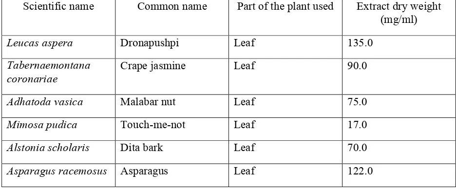
Table 1. Characteristics of the plant extracts
Scientific name Common name Part of the plant used Extract dry weight (mg/ml)
Leucas aspera Dronapushpi Leaf 135.0
Tabernaemontana coronariae
Crape jasmine Leaf 90.0
Adhatoda vasica Malabar nut Leaf 75.0
Mimosa pudica Touch-me-not Leaf 17.0
Alstonia scholaris Dita bark Leaf 70.0
Microorganisms used:
The bacterial strains used in this study clinical isolates from different infection status of patients presenting symptoms of Pseudomonas aeruginosa, Klebsiella pneumoniae, Staphylococcus aureus and Salmonella typhi - associated diseases. The isolates were identified by a standard method [11]. The standard strains used were P. aeruginosa ATCC-20852, K. pneumoniae MTCC-618, S. aureus ATCC- 29737, S. typhi MTCC- 3214, S. paratyphi MTCC- 735, P. vulgaris ATCC- 13315 and C. albicans ATCC-2091. The bacteria were maintained on agar slope at 4 °C andsub-cultured into nutrient broth by a picking-off technique [12] for 24 hrs before use.
Preparation of culture medium and inoculation
Nutrient agar (Hi Media, India) was used as the bacteriological medium. The media were sterilized by autoclaving at 120 °C for 20 minutes. Under aseptic conditions, in the laminar air flow, 15 ml of culture medium was dispensed into pre-sterilized petridishes to yield a uniform depth of 4 mm. After solidification of the medium, the microbial cultures were inoculated by spread plating technique.
Agar well diffusion
The extracts were dissolved in 10 % DMSO to a final concentration of 100 mg/ml. Pure DMSO was taken as the negative control and 50 mg/ml Ciprofloxacin as the positive control. Wells were prepared in the agar plates using a sterile cork borer of 6.0 mm diameter. 10 mg /100µL of each extract were loaded in the corresponding wells. The plates were allowed to stand at room temperature for 1 h for extract to diffuse into the agar and then they were incubated at 37 °C for 18 h. Subsequently, the plates were examined for microbial growth inhibition and the inhibition zone diameter (IZD) measured to the nearest millimeter.
Statistical analysis
The results of the antibacterial study are expressed as mean ± SEM of three replicates in each test. The data were evaluated by one-way Analysis of Variance (ANOVA) followed by Tukey’s multiple pairwise comparison tests to assess the statistical significance. The data were considered significant at P< 0.001.
Results
Table 2. Phytochemical screening of different plant extracts
Plant constituent
Plant Extract Alkaloids Flavanoids Triterpenoids Sterols Tannins Saponins Glycosides
PE + +
EA + + +
L. aspara ME + + + + +
PE + +
EA + + +
T. coronariae ME + + + + +
PE + +
EA + + +
A. vasica ME + + + + +
PE + +
EA + +
M. pudica ME + + + +
PE + +
EA + + + +
A. scholaris ME + + + + + +
PE + +
EA + + +
A. racimosus ME + + + + +
Abbreviations: PE, Petroleum ether extract; EA, ethyl acetate extract; ME, Methanol extract
Phytochemical test: - negative and + positive
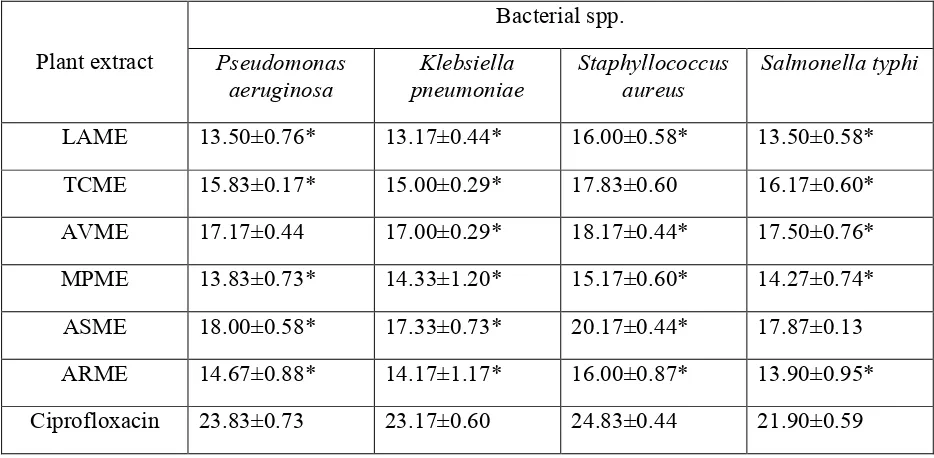
Table 3. Antibacterial activity of selected medicinal plants against clinically isolated strains (Inhibition zone in mm)
Bacterial spp.
Plant extract Pseudomonas aeruginosa
Klebsiella pneumoniae
Staphyllococcus aureus
Salmonella typhi
LAME 13.50±0.76* 13.17±0.44* 16.00±0.58* 13.50±0.58*
TCME 15.83±0.17* 15.00±0.29* 17.83±0.60 16.17±0.60*
AVME 17.17±0.44 17.00±0.29* 18.17±0.44* 17.50±0.76*
MPME 13.83±0.73* 14.33±1.20* 15.17±0.60* 14.27±0.74*
ASME 18.00±0.58* 17.33±0.73* 20.17±0.44* 17.87±0.13
ARME 14.67±0.88* 14.17±1.17* 16.00±0.87* 13.90±0.95*
Ciprofloxacin 23.83±0.73 23.17±0.60 24.83±0.44 21.90±0.59
The values are the mean of three experiments ± S.E.
*p< 0.001 vs. Standard antibiotic (Tukey’s pairwise comparison test)
Abbreviations: LAME, L. aspara methanol extract; TCME, T.coronariae methanol extract; AVME, A. vasica
methanol extract; MPME, M.pudica methanol extract; ASME, A. scholaris methanol extract; ARME, A.racimosus.
Among all the bacterial strains used, S. aureus (Gram positive bacteria), was observed to be more susceptible to the plant extracts with the inhibition zone ranging from 16.00±0.58 to 20.17±0.44. K. pneumoniae was least susceptible to the extracts except for M. pudica which was less effective against P. aeruginosa (13.83±0.73).
Among the plants screened, A. scholaris extract showed very good zone of inhibition with respect to the test strains. The values were significant except for S. typhi (17.87±0.13). L. aspara and M. pudica leaf extracts were less effective against both Gram positive and Gram negative bacteria (13.17±0.44 to 16.00±0.58 and 13.83±0.73 to 15.17±0.60). The values were moderate in case of T. coronariae, A. vasica and A. racimosus.
Discussion
properties [16]. In the present work, all the plant extracts used were extracted from polar sovent and hence had exhibited appreciable activity against clinical strains. Our findings are in agreement with reports showing that polar extracts inhibited the growth of both Gram-positive and Gram-negative bacteria [17, 18]. The stronger extraction capacity of methanol could have produced a greater number of active constituents responsible for antimicrobial activity.
The results were more promising against the gram positive bacteria S. aureus. This could be attributed to the fact that the cell wall in Gram-positive bacteria has a single layer, whereas the Gram-negative cell wall is a multi-layered structure [13, 14], acting as a barrier to many environmental substances, including antibiotics [15].
Conclusion
Plant extracts have shown inhibitory effect on the growth of the bacteria studied, although of distinct forms. However, the activity level of the extracts may be more accurately evaluated in terms of MIC values as the zone of inhibition might be influenced by solubility and diffusion rate of the phytocompounds. In addition, in vivo studies are necessary to determine the toxicity of the active constituents, their side effects, circulating levels, pharmacokinetic properties and diffusion in different body sites. The antibacterial activities could be enhanced if the active components are purified and adequate dosage determined for proper administration. It is therefore recommended that the nature and the number of the active antibacterial principles involved in each plant extract be studied in detail.
References
[1] Rojas R. Bustamante B. Bauer Fernandez I. Alban J. Lock O. Antimicrobial activity of selected Peruvian medicinal plants. J Ethnopharmacol 2003; 88: 199-204.
[2] Westh H. Zinn C.S. Rosdahl V.T. Sarisa Study Group. An international multicenter study of antimicrobial consumption and resistance in Staphylococcus aureus isolates from 15 hospitals in 14 countries. Microbiol Drug Resist 2004; 10(2): 169-176.
[3] Bandow J.E. Brotz H. Leichert L.I.O. Labischinski H. Hecker M. Proteomic approach to understanding antibiotic action. Antimicro Agents Chemotherap 2003; 47: 948-955.
[4] Benkeblia N. Antimicrobial activity of essential oil extracts of various onions (Allium cepa) and garlic (Allium sativum). Lebensm Wiss u Technol 2004; 37: 263–268
[5] Harborne J.J. Phytochemical Methods: A guide to modern techniques of plant analysis (2nd Edn), Chapman and Hall, New York, 1984; pp 85
[6] Trease G.E. Evans W.C. Pharmacognosy (13th Edn), ELBS Publication, Delhi, 1989; pp 171
[8] Khandelwal K.R. Practical Pharmacognosy: Techniques and Experiments (13th Edn), Nirali Prakashan, Pune, 2005; pp 149-156.
[9] Betoni J.E.C. Mantovani R.P. Barbosa L.N. Di-Stasi L.C. Fernandes A. Synergism between plant extract and antimicrobial drugs used on Staphylococcus aureus diseases. Mem Inst Oswaldo Cruz 2006; 101: 387-90
[10] Nair R. Kalariya T. Chanda S. Antibacterial activity of some selected Indian medicinal flora. Turk J Biol 2005; 29: 41-47
[11] Cowan S.T. Steel S. Manual for the Identification of Medica Bacteria, Barrow GI, Feltham RKA (Eds), Cambridge University Press 1993; pp 32.
[12] Aneja K.R. Experiments in Microbiology, Plant Pathology and Biotechnology (4th Edn), New Age International Ltd., New Delhi, India, 2003; pp 196-197.
[13] Yao J. Moellering R. Antibacterial agents. In: Murray P. Baron E. Pfaller M. Tenover F. Yolken R. (Eds) Manual of Clinical Microbiology, ASM, Washington DC 1995; pp 1281-1290.
[14] Ozcelik S. Genel Mikrobiyoloji, Suleyman Demirel Universitesi Isparta Turkey 1998; pp 259 (in Turkish)
[15] Tortora G.J. Funke B.R. Case C.L. Microbiology: An Introduction, Benjamin Cummings San Francisco 2001; 88 pp
[16] Shibata H. Kondo K. Katsuyama R. Kawazoe K. Sato Y. Murakami K. Takaishi Y. Arakaki N. Higuti T. Alkyl Gallates, Intensifiers of ß-Lactam Susceptibility in Methicillin-Resistant Staphylococcus aureus Antimicrob. Agents Chemother 2005; 49(2): 549-555. [17] Marquez B. Neuville L. Moreau N.J. Genet J.P. Santos A.F. Andrade M.C.C. Sant Ana AEG Multidrug resistance reversal agent from Jatropha elliptica Phytochem 2005; 66: 1804-1811.
[18] Smith E.C.J. Williamson E.M. Wareham N. Kaatz G.W. Gibbons S. Antibacterials and modulators of bacterial resistance from the immature cones of Chamaecyparis lawsoniana. Phytochem 2007; 68(2): 210-217.
[19] Lewis K. Ausubel F.M. Prospects for plant-derived antibacterials. Nat Biotechnol 2006; 24(12): 1504-1507.
[20] Karaman I. Sahin F. Gulluce M. Ogutcu H. Sngul M. Adiguzel A. Antimicrobial activity of aqueous and methanol extracts of Juniperus oxycedrus L. J Ethnopharmacol 2003; 85: 231-235.