Managing Passengers with Respiratory Disease Planning Air Travel
British Thoracic Society Recommendations, British Thoracic Society Standards of Care Committee
The Air Travel Working Party was chaired by Dr RK Coker. The committee
members were Dr DAR Boldy, Dr R Buchdahl, Mr D Cramer, Professor D Denison, Wing Commander DP Gradwell, Professor JMB Hughes, Dr JA Innes, Dr AOC Johnson, Dr KP McKinlay and Professor MR Partridge.
Contents
Introduction
Need for recommendations for managing passengers with respiratory disease planning
air travel
Purpose of recommendations
Methods of production
Summary of key points and recommendations The flight environment and effects of altitude
Pre-flight assessment for adults
Pre-flight assessment for children
Summary of practical recommendations
Logistics of travel with oxygen
Background literature review
The flight environment
Physiological effects of exposure to altitude
Clinical pre-flight assessment
Fitness to fly in childhood
Respiratory disorders with potential complications for air travellers
Research questions
Appendices (A 1-7) A 1 Reviewers
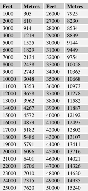
A 2 AHCPR grading scheme for recommendations A 3 Referral centres with decompression chambers A 4 Major destinations exceeding 2438 m (8000 ft) A 5 Sample MEDIF form
A 6 Figures 1-4
A 7 Examples of equations for predicting hypoxaemia
Introduction
Need for recommendations on managing passengers with lung disease planning air travel
Air travel is now a common mode of travel for millions, with a single UK airline carrying over 33 million passengers annually. It is estimated that over one billion passengers travel by air world-wide each year, and for the vast majority this is safe. Despite current security concerns, air travel is likely to remain a convenient form of transport for many, and in the longer term passenger numbers may increase further. Given the rising age of Western populations, the age of air travellers is also likely to increase, with greater propensity for medical impairment. Over 25 years ago it was already estimated that 5% of commercial airline passengers were ambulatory patients with some illness including chronic obstructive pulmonary disease (COPD).[1]
With the introduction of the Airbus 380, passengers will be exposed to a cabin altitude of up to 8000 ft for up to and in some cases exceeding 20 hours. Aside from the potential for inter-current medical incidents to occur with increasing frequency, since longer flights increase the odds of such an event, the associated physiological disturbances associated with moderate but prolonged hypoxia, prolonged immobility and protracted exposure to reduced barometric pressure remain unknown. Recent data do however suggest that longer flights are associated with an increased risk of oxygen desaturation, which may partly reflect a progressive fall in cabin PO2. [2] It is also recognised that acute mountain sickness can occur, albeit rarely, after just 16-18 hours’ exposure to altitudes of 7-8000 ft.
There are still no established methods for quantifying the risk of in-flight medical problems. However, a North American service offering expert assistance by radio link for in-flight medical emergencies logged 8,450 calls in 2001, of which 10.2% were respiratory in nature.[3] Physicians should therefore be aware of the potential effects of the flight environment in passengers with lung disease. One million residents of Denver, Colorado live at 5280 ft (1609 m), and coaches crossing high Alpine passes reach 10,000 ft (3048 m), indicating that moderate hypoxaemia is not
generally hazardous. We nevertheless consider that greater awareness of the risks of air travel enables physicians to encourage patients to fly safely wherever possible, and increases the comfort of fellow air passengers.
While pilots are subject to regular medical examination, passengers are not. For potential passengers with lung disease it is valuable for their physician to have recommendations for assessing their patients’ fitness for flight. A previous national survey of respiratory physicians indicated many would welcome advice.[4] Sources of available information include British and European,[5][6][7] North American and Canadian [8][9] COPD guidelines, aviation medicine textbooks,[10] supplements to the Aviation, Space & Environmental Medicine Journal [11][12][13] and other publications on air travel.[14] However, these references are not always readily accessible to physicians and do not all provide consistent, practical or comprehensive coverage. In particular, there is disparity between European and North American guidelines, uncertainty about assessment methods, and failure to consider other respiratory causes of hypoxaemia such as pulmonary fibrosis.
To meet the need for consistent, practical and comprehensive advice, the British Thoracic Society (BTS) Standards of Care Committee set up a Working Party to formulate national recommendations for managing patients with lung disease planning air travel. There is currently insufficient evidence to produce formal guidelines. The following recommendations are derived from literature reviews and aim to provide practical advice for respiratory specialists in secondary care. A leaflet for general practitioners is available from the BTS website (www.brit-thoracic.org.uk). They apply to commercial flights only (including scheduled repatriation with a medical or nurse escort) and exclude emergency aeromedical evacuation situations.
Purpose of recommendations
1. Enhance safety for passengers with lung disease travelling by air and reduce the number of in-flight medical incidents due to respiratory disease
2. Increase recognition amongst healthcare professionals that patients with respiratory disease may require clinical assessment and advice before air travel
3. Provide an authoritative up-to-date literature review of available evidence 4. Provide consistent, practical and comprehensive advice for healthcare
professionals managing such patients
5. Formulate key research questions to provoke further investigation. This should produce a strengthened, high quality evidence base from which clearer evidence-based guidelines can be developed
6. Promote the development of methods for monitoring the size of the problem
Methods of production
The Working Party defined the target and purpose of the recommendations. Independent literature searches were performed by Working Party members. From this literature a draft document was produced summarising current evidence and containing recommendations regarding (1) the flight environment, (2) physiological effects of exposure to altitude, (3) clinical assessment, (4) respiratory disorders presenting a possible risk for potential air travellers, (5) oxygen supplementation. The document was reviewed by the Working Party and re-drafted. It was then circulated to the BTS Standards of Care Committee and reviewers listed in Appendix 1 before being made available to BTS members on the members-only section of the BTS website. A final draft was then produced incorporating feedback after discussion and further review by the BTS Standards of Care Committee.
The current version was published on the BTS website (www.brit-thoracic.org.uk) in 2004 following a second literature review. The most significant change since the original document’s publication in Thorax in 2002 is the emergence of Severe Acute Respiratory Syndrome (SARS), and a new section covering this topic has been added.
The search engines were Medline (English language) 1966-2003 and the Cochrane Library Database. The word titles were:
accidents, altitude, anoxia, aeroplane, aerospace medicine, asthma, aircraft, aircraft emergencies, air travel, aviation, bronchiectasis, bronchitis, cabin pressure, child, COPD, cross infection, cystic fibrosis,
decompression chamber, desaturation, diffuse parenchymal lung disease, emergencies, emphysema, fibrosing alveolitis, fitness for air travel, fitness to fly, flight, hypoxic challenge, hypoxia inhalation simulation test, hypoxia/c inhalation test,
infection, interstitial lung disease
kyphoscoliosis, lung diseases (restrictive), mycobacterium
tuberculosis, neuromuscular disease, obstructive sleep apnoea syndrome, opportunistic infections, passenger, pneumothorax, rehabilitation, pre-flight test, pre-pre-flight assessment, pulmonary fibrosis, respiratory failure, respiratory tract disease, respiratory tract infections,
saturation, thoracic surgery, travel, traveller,
venous thromboembolism, walking test
Conflicts of interest
Members of the Air Travel Working Party have submitted a written record of possible conflicts of interest to the Standards of Care Committee of the BTS. These are available for inspection on request from the Chairman of this Committee. Preparation and publication of the document was paid for entirely by the British Thoracic Society and no external funding was received.
Summary of key points and recommendations with AHCPR grading
The flight environment and effects of altitude
Commercial aircraft are pressurised to cabin altitudes up to 2438 m (8000 ft). In practice, actual cabin altitude is lower, since 8000ft is the regulatory maximum except in emergencies. At 2438 m (8000 ft) the partial pressure of oxygen falls to the equivalent of breathing 15.1% oxygen at sea level. In a healthy passenger, PaO2 at 2438 m (8000 ft) will be influenced by age and minute ventilation, but will fall to between 7.0 and 8.5 kPa (53-64 mmHg, SpO2 85-91%). Altitude exposure may therefore exacerbate hypoxaemia in patients with lung disease, and particular caution seems justified in those who are hypoxaemic at sea level. The physiological compensations for acute hypoxaemia at rest are mild to moderate hyperventilation (moderated by the fall in PaCO2) and moderate tachycardia.
Pre-flight assessment for adults
The following groups should be assessed • severe COPD or asthma (B)
• severe restrictive disease (including chest wall and respiratory muscle disease), especially with hypoxaemia and/or hypercapnia (C)
• patients with cystic fibrosis (C)
• history of air travel intolerance with respiratory symptoms (dyspnoea, chest pain, confusion or syncope) (C)
• co-morbidity with other conditions worsened by hypoxaemia (cerebrovascular disease, coronary artery disease, heart failure) (C)
• pulmonary tuberculosis (C)
• passengers from an area with recent local transmission of Severe Acute Respiratory Syndrome (C)
• contacts of probable or confirmed Severe Acute Respiratory Syndrome (C) • within six weeks of hospital discharge for acute respiratory illness (C) • recent pneumothorax (B)
• risk of or previous venous thromboembolism (B)
The following assessment is recommended
• history and examination with particular reference to cardio-respiratory disease, dyspnoea and previous flying experience (C)
• spirometry (in non-tuberculous patients only) (C)
• measurement of SpO2 by pulse oximetry. Readings should be taken from a warm ear or finger after sufficient delay for the oximeter to display a stable reading. Blood gases are preferred if hypercapnia is known or suspected (C)
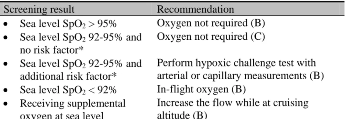
In those who are screened who have resting sea level oximetry between 92% and 95% and who have additional risk factors (table 1) we would recommend hypoxic challenge testing (C)
Table 1 Results of initial assessment
Screening result Recommendation
• Sea level SpO2 > 95% Oxygen not required (B) • Sea level SpO2 92-95% and
no risk factor*
Oxygen not required (C) • Sea level SpO2 92-95% and
additional risk factor*
Perform hypoxic challenge test with arterial or capillary measurements (B) • Sea level SpO2 < 92% In-flight oxygen (B)
• Receiving supplemental oxygen at sea level
Increase the flow while at cruising altitude (B)
*Additional risk factors: hypercapnia, FEV1 <50% predicted, lung cancer, restrictive lung disease involving the parenchyma (fibrosis,) chest wall (kyphoscoliosis) or respiratory muscles, ventilator support, cerebrovascular or cardiac disease, within six weeks of discharge for an exacerbation of chronic lung or cardiac disease.
In those who undergo hypoxic challenge testing, we would recommend the following (table 2):
Table 2 Results of hypoxic challenge test (15% FiO2 for 15 minutes) with
AHCPR grading (Appendix 2)
Hypoxic challenge result Recommendation • PaO2 > 7.4 kPa (> 55
mmHg)
Oxygen not required (B) • PaO2 6.6-7.4 kPa (50-55
mmHg)
Borderline. A walk test may be helpful (C)
• PaO2 < 6.6 kPa (< 50 mmHg)
Notes
1. The following groups should not fly
• patients with infectious tuberculosis (TB) must not travel by public air transportation until rendered non-infectious. HIV negative patients in whom drug resistant TB is not suspected and who have completed two weeks of effective antituberculous treatment are usually considered non-infectious. For HIV positive patients three smear negative sputum examinations on separate days, or a single negative sputum culture result, are required while on effective anti-tuberculous treatment (B)
• passengers from an area with recent local transmission of Severe Acute Respiratory Syndrome (SARS) and with symptoms compatible with SARS should postpone their flight until fully recovered (C)
• contacts of probable or confirmed SARS should not undertake travel for ten days after exposure (C)
• those with a current closed pneumothorax should avoid commercial air travel (C) 2. patients who have undergone major thoracic surgery should ideally delay flying
for two weeks after an uncomplicated procedure (C). Patients should only fly if essential, and formal medical assessment is required before departure.
3. Lung cancer per se is not a contra-indication to flying. However, associated respiratory disease should be considered in its own right (C).
4. Additional precautions for all passengers
• excess alcohol should be avoided before and during the flight, particularly in those with obstructive sleep apnoea and those at risk of VTE (C)
• individuals not receiving oxygen should remain mobile during the flight (C)
• exercise without supplemental oxygen may worsen hypoxaemia. It may be prudent for the most compromised to use oxygen while walking on the plane if possible, and to let a flight attendant know how long they expect to be away from their seat (C)
• the risk of thromboembolic disease should initiate prophylactic measures as detailed in the following summary (B)
• patients should carry well-filled reliever and preventer inhalers (as prescribed by their doctor) in their hand luggage (C)
• portable battery-operated nebulisers may be used at the discretion of the cabin crew, but passengers must notify the airline in advance. Spacers are as effective as nebulisers in treating asthma (A)
• patients should check with their local or hospital pharmacists whether any unusual or trial medications may be adversely affected by the extreme temperature in the hold baggage compartment (C). A full supply of all medication should be taken as hand luggage, preferably in the original packaging with pharmacy labels. A doctor’s note is recommended if carrying unusual or trial medication
• dry cell battery-powered CPAP machines may be required by patients with obstructive sleep apnoea on long haul flights, but they must be switched off before landing (C)
• ventilator-dependent patients should inform the airline of their requirements at the time of reservation, and a doctor’s letter is required outlining diagnosis, necessary equipment, recent blood gas results and ventilator settings. A medical escort is required as the ventilator may have to be switched off for take-off and landing, and the patient manually ventilated. Arrangements must be made for proceeding through air terminals before and after the flight (C)
5. Logistics of air travel with oxygen
Supplementary in-flight oxygen is usually prescribed at a rate of 2L/min and should be given by nasal cannulae. In-flight oxygen need not be switched on until the plane is at cruising altitude, and may be switched off at the start of descent. For patients on oxygen at sea level, the rate should only be increased while at cruising altitude. (B) Some airlines do not permit use of supplemental oxygen during take-off or landing, and such patients should therefore always be discussed first with the airline
6. In complex circumstances, patients can be referred for testing in a hypobaric chamber. Centres are listed in Appendix 3.
Even with in-flight oxygen, travel cannot be guaranteed to be safe. Air travel is almost always possible with appropriate medical support, but the logistics and economic costs may outweigh the benefits in individual cases.
Pre-flight assessment for children with AHCPR grading
• it is prudent to wait for one week after birth before allowing infants to fly to ensure they are healthy (C)
• if the infant has had any neonatal respiratory problems, the proposed journey should be discussed with a paediatrician and hypoxic challenge considered (B) • for children with cystic fibrosis (CF) or other chronic lung disease and FEV1
<50% predicted, hypoxic challenge testing is recommended as described below • for oxygen dependent children including ex-premature infants with chronic lung
disease (broncho-pulmonary dysplasia) where flying is imperative, oxygen requirements should be titrated in a body box (B) as follows:
The infant or young child, receiving oxygen via nasal cannulae, is placed in the body box with a parent or carer, and SpO2 monitored. The air in the body-box is then diluted to 15% oxygen with nitrogen. If SpO2 falls below 90%, supplementary in-flight oxygen is recommended. The flow required is determined by the oxygen flow which restores SpO2 to the original value. The flow-rate available on-board will then need to be discussed with the airline
In older children, hypoxic challenge testing is performed using a mouthpiece rather than in the body box
Summary of disease-specific key points and recommendations
Asthma
• assessment is recommended as described above
• reliever and preventer inhalers, as prescribed, should always be carried in the hand luggage
• from April 2004, bronchodilator inhalers are included as part of the mandatory medical kit carried by aircraft on flights to and from the USA. Requirements on flights to other destinations vary
• portable battery-operated nebulisers may be used at the discretion of cabin crew, but passengers must notify the airline in advance. Nebulisers may be connected to the aircraft electrical supply on some but not all airlines. This must be checked in advance since availability, voltage and power outputs can all differ. Some airlines can provide nebulisers for in-flight use, but passengers must check when booking. Spacers are as effective as nebulisers
COPD
• assessment is recommended as described above
• passengers should travel on a non-smoking flight. Most airlines now have a smoking ban and a summary of policies is available online [15]
• reliever and preventer inhalers, as prescribed, should always be carried in the hand luggage
• portable battery-operated nebulisers may be used at the discretion of cabin crew, but passengers must notify the airline in advance. Nebulisers may be connected to the aircraft electrical supply on some but not all airlines. This must be checked in advance since availability, voltage and power outputs can all differ. Some airlines can provide nebulisers for in-flight use, but passengers must check when booking. • patients prescribed in-flight oxygen should receive oxygen while visiting high
altitude destinations (see Appendix 4)
Cystic fibrosis
• assessment by the CF physician is advised as described above
• a full supply of all medication should be carried in the hand luggage to allow for delays and stopovers, preferably in the original packaging with pharmacy labels. A doctor’s note is recommended if carrying unusual or trial medication. Passengers should also check with their pharmacist whether any unusual or trial medications may be adversely affected by extreme temperatures in the hold baggage compartment
• portable battery-operated nebulisers may be used at the discretion of cabin crew, but passengers should notify the airline in advance. Nebulisers may be connected to the aircraft electrical supply on some but not all airlines. This must be checked in advance, since availability, voltage and power outputs can all differ. Some airlines can provide nebulisers for in-flight use, but passengers must check when booking. Spacers are as effective as nebulisers for relieving bronchospasm. • passengers should undertake physiotherapy during stopovers
• in-flight nebulised antibiotics and DNase should not be necessary
• many airports can provide wheelchairs for transport to and from the aircraft Infections
• assessment is recommended as described above
• aircraft boarding should be denied to those known to have infectious TB, those from an area with recent local transmission of SARS and symptoms compatible with SARS, and contacts of probable or confirmed SARS cases within the preceding ten days
• patients with infectious TB must not travel by public air transportation until rendered non-infectious. WHO guidelines state that three smear negative sputum examinations on separate days in a person on effective anti-tuberculous treatment indicate an extremely low potential for transmission, and a negative sputum culture result virtually precludes potential for transmission.[16] This may be over-cautious. While this remains the policy for HIV positive patients, HIV negative patients in whom drug resistant TB is not suspected and who have
completed two weeks of effective anti-tuberculous treatment are in practice generally considered non-infectious.[17]
Fibrosing alveolitis
• assessment is recommended as described above Neuromuscular disease and kyphoscoliosis
• assessment is recommended as described above Ventilator-dependent patients
For all patients
• the airline must be consulted before reservation
• a doctor’s letter is required outlining the medical diagnosis, necessary equipment, recent blood gas results and ventilator settings. It should state that the ventilator must travel in the cabin as extra hand luggage
• a dual 110 / 240 volt function is recommended so that the ventilator is compatible with the voltage at the intended destination
• a dry cell battery pack is essential for back-up, and for proceeding through air terminals before and after the flight
For patients on permanent (24 hour) ventilation • ventilator-dependent patients need a medical escort
• an electrical supply may be provided on the flight if arranged in advance • wet acid batteries are prohibited
• the medical escort must be competent to change the tube, operate suction, and ventilate the patient by hand for up to an hour if electrical power fails or if the ventilator has to be switched off for take-off and landing
• a spare tracheostomy tube and battery powered suction must be taken
• owing to reduced barometric pressure at altitude, patients with a tracheostomy should have the cuff pressure monitored on ascent (when a little air will need to be released) and on descent (when a little air will need to be added)
• airline experience indicates that the logistics of airport transfers often pose more challenges than the flight itself, and attention must therefore be paid to these details when planning the journey
Obstructive sleep apnoea syndrome (OSAS)
• assessment is recommended as described above • the airline must be consulted before reservation
• a doctor’s letter is required outlining the medical diagnosis and necessary equipment. It should state that the CPAP machine should travel in the cabin as extra hand luggage. A fact sheet for passengers to show to airport security personnel is available from the American Sleep Apnea Association [18]
• a dual 110 / 240 volt function is recommended so that the CPAP machine is compatible with the voltage at the intended destination
• dry cell battery-powered CPAP can be used during the flight but must be switched off before landing
• patients should avoid alcohol immediately before and during the flight
• patients with mild snoring and hypersomnolence are unlikely to require CPAP during the flight
• patients with significant desaturation planning to sleep during the flight should consider using their CPAP machine
• patients with significant desaturation should use CPAP during sleep while visiting high altitude destinations (see Appendix 4)
Previous pneumothorax
• patients with a current closed pneumothorax should not travel on commercial flights
• patients who have had a pneumothorax must have had a chest radiograph confirming resolution before flight. Many would regard it as prudent for a further seven days to elapse before embarking upon flight. There is insufficient evidence to support the previous recommendation of a six week delay after resolution before travel
• in the case of a traumatic pneumothorax the time period following full radiographic resolution should be two weeks
• a definitive surgical intervention designed to reduce the risk of further pneumothorax is likely to be successful and patients should be allowed to fly once they have recovered from the effects of their surgery
• although recurrence is unlikely during flight, the consequences of a pneumothorax at altitude may be significant given the absence of prompt medical care. The risk of recurrence is higher in those with co-existing lung disease. This risk does not decline significantly for at least a year, and those not undergoing definitive surgical procedures may wish to consider alternative forms of transport within one year of the initial event
Venous thromboembolic disease (VTE)
• all passengers should avoid excess alcohol and caffeine-containing drinks, and preferably remain mobile and/or exercise their legs during the flight
•
•
passengers at slightly increased risk of VTE include those aged over 40, those who are obese or who have extensive varicose veins, polycythaemia, and those within 72 hours of undergoing minor surgery. In addition to the above precautions they should avoid taking sleeping pills and/or sleeping for prolonged periods in abnormal postures. Physicians may wish to recommend support tights or non-elasticated long socks
passengers at moderately increased risk of VTE include those with a family history of VTE, recent myocardial infarction, pregnancy or oestrogen therapy (including hormone replacement therapy and some types of oral contraception), post-natal patients within two weeks of delivery and those with lower limb paralysis, recent lower limb trauma or recent surgery. In addition to the above precautions, physicians may wish to recommend graduating compression stockings and/or pre-flight aspirin
• passengers at high risk of VTE include those with previous VTE, thrombophilia, those within six weeks of major surgery, with a history of previous stroke, or current known malignancy. If flying cannot be avoided or delayed, then as an alternative to low dose aspirin it may be prudent to recommend either low molecular weight heparin or formal anticoagulation (with INR 2-3) before departure. Depending on the length of stay abroad, passengers may need to remain anticoagulated until the homeward journey
Thoracic surgery
• assessment is recommended as described above
• air travel should be delayed for at least two weeks after uncomplicated chest surgery, and CXR confirmation of resolution of any pneumothorax or collected air is recommended. Careful medical assessment is required before travel
For all patients
• the need for oxygen should be disclosed when the patient books with the airline • the airline medical department will issue a MEDIF form (Appendix 5) or their
own medical form. This requires completion by both the patient and the GP or hospital specialist and requests information about the patient’s condition and oxygen requirements. The airline’s Medical Officer then evaluates the patient’s needs
• the need for oxygen on the ground and while changing flights must be considered • the airline should be consulted in advance if the patient wishes to use
humidification equipment
• airlines do not provide oxygen for use at the airport. Some airports restrict oxygen use in the airport because of the risk of explosion
• airlines will provide nasal cannulae for passenger use • in-flight oxygen flow is usually limited to 2L/min or 4L/min
• international regulations permit passengers to use their own oxygen on board aircraft and to carry small, full oxygen cylinders (for medical purposes) with them as hand luggage, provided they have the approval of the airline concerned. Patients must check with the airline first. A charge may be made for this service, in addition to a charge for in-flight oxygen
• patients are advised to check charges with several airlines before reservation as considerable variation exists in fees and services
For totally oxygen-dependent patients
• special arrangements must be made with the airline and airport authorities. Transport to the aircraft by ambulance is possible, and some airports have a specially designated medical unit
• some airlines do not permit use of supplemental oxygen during take-off or during landing; passengers or carers must therefore check with the carrier in advance • the patient should have a supply of all their usual medication, a copy of their
medical form and be accompanied
• a direct flight is preferable. If connecting flights are unavoidable, separate arrangements must be made for oxygen whilst on the ground during stopovers. The main oxygen distributors have their own international distribution network
and can supply oxygen at intended destinations if active in those areas. A charge is likely to be made for this service
• patients normally using LTOT should ensure they have LTOT throughout their stay. Oxygen supplies can be provided as emergency health care in all EEA countries under the E111 arrangements. Prior arrangement with the destination country is essential to ensure availability of supplies, and a charge is likely. Tour operators should be able to help; limited information on services available may also be obtained by calling the International Division of the Department of Health on (44) 207 210 5318, but this office is unable to assist in making travel arrangements. The British Lung Foundation may also be able to give advice on (44) 207 688 5555 (www.britishlungfoundation.org)
• attention should be drawn to the need to make prior arrangements for the return as well as outward journey
The Frequent Traveller’s Medical Card (FREMEC)
Patients who travel frequently and have particular medical needs can obtain a Frequent Traveller’s Medical Card. This contains important medical information for care, replacing forms otherwise necessary for every flight. Once registered, the reservations office keeps details of requirements on record so that special assistance can be arranged whenever the patient flies. The period of validity is dependent on the nature of the condition. FREMEC is issued by many airlines, but if a patient chooses to fly with an airline other than that which issued the FREMEC card they should check its validity with the new airline.
Medical insurance
Passengers should not only travel with an E111 form (where relevant) but also with travel insurance, having fully disclosed their medical history beforehand.
Background literature review
The flight environment
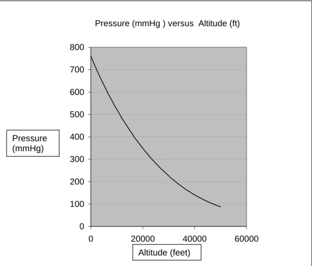
To understand how the flight environment influences physiology and occasionally pathology, it is useful to consider the physical properties of the atmosphere and changes that occur on ascent to altitude. The atmosphere consists of several concentric “shells” around the Earth. The innermost shell is the troposphere, which extends from ground level to 9144 m (30000 ft) at the poles and 18288 m (60000 ft) at the Equator. Conventional aircraft operate in this region. It is characterised by a relatively constant decline in temperature with increasing altitude, at a rate of 1.98oC/305 m (1000 ft) ascent. Owing to gravity, air has weight. Atmospheric pressure is therefore greatest at sea-level and declines logarithmically with ascent (Figure 1). Small changes in height at low altitude thus cause a much greater pressure change than the same change in height at high altitude.
The troposphere has a constant composition, containing 21% oxygen, 78% nitrogen and 1% other gases. Other gases include argon and carbon dioxide, the latter present at a concentration of 0.03%. It is the fall in the partial pressure of oxygen as total pressure declines on ascent that can give rise to hypobaric hypoxia, not a change in its percentage in air. Changes in pressure and temperature have other physical effects as described by the gas laws. Boyle’s law predicts that as pressure falls on ascent there will be an inversely proportional increase in gas volume. Since gas in body cavities is fully saturated with water vapour, gas expansion with altitude is significantly greater than predicted by Boyle’s law as applied to dry gas alone. This affects body parts where gases are trapped, including the middle and inner ear, sinuses and intestines. The same effect occurs in the lungs although gas in free communication with ambient air equilibrates easily. Gas trapped in bullae or a closed pneumothorax is unlikely to equilibrate as rapidly, if at all. The volume of a gas is also related to temperature, but the temperature of gases trapped in the body stays constant at 37oC. Relative expansion of humidified gas is expressed as follows:
(initial pressure of the gas in the cavity at sea level – 47) (final pressure of gas in cavity – 47)
This becomes (760 – 47) ÷(566 –47) = (713 ÷518) = 1.376
where 760 is atmospheric pressure in and 565 is atmospheric pressure at 8000 ft. The volume of gas in a non-communicating bulla will thus increase by 37.6% on ascent from sea level to 2438m (8000ft).
Cabin pressurisation in modern aircraft ensures that the effective altitude to which occupants are exposed is much lower than that at which the aircraft is flying. Commercial aircraft are not pressurised to sea level, but to a relatively modest intermediate cabin altitude. This allows the aircraft to fly at much higher altitudes, which is fuel efficient for jet engines and more comfortable since it avoids much turbulence. Aircraft cabin altitude can thus approach 2438 m (8000 ft) while the aircraft is flying at 11582 m (38000 ft).
Consequently a pressure differential exists across the cabin wall, commonly of up to 9 pounds per square inch (psi). International aviation regulations [19] stipulate that at a plane’s maximum cruising altitude the cabin pressure should not exceed 2438 m (8000 ft). This may be exceeded in emergencies. One study of in-flight cabin altitude on 204 scheduled commercial aircraft flights revealed significant variations in cabin altitude.[20]
In the event of failure of the cabin pressurisation system at high altitude, all occupants would require supplemental oxygen to prevent an unacceptable degree of hypoxaemia. Commercial aircraft are thus equipped with an emergency oxygen system for passengers, demonstrated before each flight in accordance with civil aviation regulations. However, some passengers with impaired respiratory function may be unusually susceptible to the effects of ascent even to normal cabin altitudes. It is these problems which are addressed here. These recommendations apply only to larger commercial aircraft. They do not apply to small, private or un-pressurised aircraft operating under General Aviation regulations.[21]
Physiological effects of exposure to altitude
Breathing air at 2438 m (8000 ft) is equivalent to breathing 15.1% oxygen at sea level. In healthy subjects exposed to these conditions, their PaO2 will be influenced by their age and minute ventilation, but the PaO2 is likely to fall to between 7.0 and 8.5 kPa (53-64 mmHg, SpO2 85-91%).[22][23] However, healthy passengers do not generally experience symptoms.
Clinical pre-flight assessment
A recent audit of 109 applications for in-flight oxygen conducted by a major UK airline revealed that they are rarely provided with objective information to assess risk, only 61% of requests including simple data such as oximetry or spirometry results (M Popplestone, personal communication). In the absence of such information, airlines traditionally favour the 50 metre walk test. Other procedures used to assess whether patients are fit to fly are predicting hypoxaemia from equations, and the hypoxic challenge test.
The 50 metre walk
The ability to walk 50 metres without distress has the merit of being simple, but is often the only subject of enquiry and is not verified. There is no evidence validating this test. Although it may seem a crude assessment, the ability to increase minute ventilation and cardiac output in response to an exercise load is a good test of cardiorespiratory reserve. It is also a common-sense approach to simulating the stress of the additional hypoxaemia patients will experience at rest during a flight. Respiratory physicians have experience of the value of walk tests in other contexts, including the six or 12 minute walk and the shuttle walk test [24][25][26]. Such tests are increasingly being used as part of the assessment of patients for lung volume reduction surgery and lung transplantation.
The walk test should be that in use in the laboratory where the assessment is being performed. Failure to complete the task (in terms of distance or time) or moderate to severe respiratory distress (recorded on a Visual Analogue Scale) will alert the
physician and the patient to the possible need for in-flight oxygen. Walk tests are obviously not suitable for those with significantly impaired mobility.
Predicting hypoxaemia from equations
Some centres use one of several equations predicting PaO2 or SpO2 from measurements at sea level [27][28][29][30][31]. The equations have been derived almost exclusively from patients with COPD who have had measurements of PaO2 in a hypobaric chamber, or before and during exposure to simulated altitude while breathing 15% inspired oxygen from a reservoir bag. Measuring FEV1 may improve the accuracy of predicted values[28][29]. One weakness is that the 90% confidence limits are ± 1 kPa (~ ±2-4% SpO2). However, the predictions are usually reliable enough to establish upper and lower thresholds for ‘no in-flight oxygen required’ (SpO2 > 95%) or ‘in-flight oxygen needed’ (SpO2 < 92%) [see table 1]. Flight duration and cabin conditions are not reproduced.
Hypoxic challenge test
The ideal test, which is to expose a subject to hypoxia in a hypobaric chamber, is not widely available. The hypoxic challenge test described by Gong is therefore often used.[30] It assumes that breathing hypoxic gas mixtures at sea level (normobaric hypoxia) equates to the hypobaric hypoxia of altitude.[32] The maximum cabin altitude of 2438 m (8000 ft) can be simulated at sea level with a gas mixture containing 15% oxygen in nitrogen. Subjects are usually asked to breathe the hypoxic gas mixture for 20 minutes or until equilibration. Saturation is monitored throughout, and blood gases measured before and on completion.
Fifteen percent oxygen can be administered in several ways. Oxygen and nitrogen can be mixed in the appropriate proportions in a Douglas bag or cylinders of 15% oxygen in nitrogen can be bought from British Oxygen Corporation. The gas mixture can be given with a non-rebreathing valve with a mouth-piece or tight-fitting face mask. It is also possible to fill a body box with 15% oxygen to provide the hypoxic environment without using a face mask or mouth piece.[33] This allows oxygen requirements to be titrated accurately using nasal prongs to supply oxygen within the body box. A similar but unpublished suggestion is to use a hood over the subject’s
head which is filled with 15% oxygen. Finally, similar levels of hypoxic gas mixtures can be given with a commercial 40% venturi mask if nitrogen is used as the driving gas. The entrained air dilutes the nitrogen producing a 14-15% oxygen mixture under experimental conditions in subjects with COPD.[34] Using a 35% Venturi mask will yield a 15-16% oxygen mixture.
A subject is usually judged to require in-flight oxygen if the PaO2 falls below 6.6 kPa (50 mmHg) or SpO2 falls below 85%.[33] These figures appear arbitrary with no supporting evidence, but many physicians have adopted them as a reasonable compromise. Hypoxic challenge testing is the pre-flight test of choice for patients with hypercapnia. As with equations, flight duration and cabin conditions are not reproduced.
Fitness to fly in childhood
Childrens' lung physiology differs from that of adults. In particular, during early life compliance is lower while residual volume and airway resistance are higher.[35] In the neonatal period regional lung perfusion may remain labile with estimates of a 10% persistent right to left pulmonary shunt in healthy infants at one week of age.[36] Foetal haemoglobin is present in significant amounts up to three months of age. Its effect on the oxygen dissociation curve is to enhance oxygen loading in a hypoxic environment but possibly to decrease unloading in peripheral tissues.[37] Some of these factors may explain why the response to a hypoxic environment is less predictable in infants than it is in adults.
There are few data on oxygen saturation in normal healthy infants and children exposed to cabin altitudes. A study by Lee et al [2] examined oxygen saturation in 80 children (43 boys) during prolonged commercial air travel. Oxygen saturation declined significantly during flight. Average sea level SpO2 was 98.4%, falling to 95.7% after three hours and to 94.4% after seven hours. This was associated with reduced cabin partial pressure of oxygen (159 mmHg at sea level, 126 mmHg after three hours and 124 mmHg after seven hours), but the marked difference between SpO2 at three and seven hours suggests that flight duration may also contribute to worsened oxygen desaturation. This study provides valuable control paediatric data
from which it may eventually be possible to derive cut-off values for the hypoxic challenge test in children. However, more data are required on normal children and particularly infants. In an otherwise normal term infant we have chosen to recommend a delay of one week after birth to be sure the infant is otherwise healthy.
Should infants and children with lung disease undergo tests of fitness to fly? There is very little documented evidence of what happens to such children during flight. The spectrum of disease is wide. Infants, especially those born premature less than 32 weeks gestation, who develop an acute viral respiratory infection are known to be at risk of apnoea because they appear to revert to a more immature pattern of breathing.[38][39] Exposure to a hypoxic environment at this time may increase the risk of apnoea. Ex-premature infants who develop respiratory infection should therefore probably not fly under the age of six months post-expected date of delivery.
Children with chronic lung disease such as cystic fibrosis (CF) may be better adapted to a hypoxic environment possibly though changes in haemoglobin oxygen dissociation characteristics. A study of 87 children with CF suggested that, in children old enough to do spirometry, an FEV1 < 50% predicted is a better predictor of desaturation below 90% while flying than hypoxic challenge.[40] These authors now recommend that if a child with CF (or other chronic lung disease) has an FEV1 <50% predicted they should undergo hypoxic challenge, and that if SpO2 falls below 90% during the test, in-flight oxygen should be made available. The hypoxic challenge test allows the physician to determine the flow rate of oxygen required. A recent study (research letter in press) of pre-flight body box measurements in 20 infants and young children with structural lung disease showed that 6 children with SpO2 ≥ 95% at sea level desaturated below 90% when breathing 15% oxygen (R Buchdahl, personal communication).
On the basis of current evidence we recommend that infants with a history of neonatal respiratory illness, and children with hypoxia due to chronic lung disease such as cystic fibrosis, who must fly, should undergo pre-flight assessment including hypoxic challenge testing. The most practical and non-invasive way of performing a hypoxic challenge test is to titrate the extra oxygen requirement of the infant or young child in
a body box as described in the summary section. Young children with chronic respiratory disease whose saturations fall below 90% on hypoxic challenge testing should have in-flight oxygen.
Respiratory disorders with potential complications for air travellers
Asthma
The flight environment experienced by commercial passengers should not pose a problem for most patients with asthma. Low cabin humidity may theoretically predispose to bronchospasm as a result of water loss from bronchial mucosa. In-flight asthma is however uncommon in practice. Patients with severe chronic asthma should be assessed as above prior to arranging a flight.
Surveys report [41-43] that around 10% of in-flight medical emergencies are respiratory, of which perhaps one third are ascribed to asthma. It is not possible from these surveys to distinguish patients with genuine asthma from those reporting breathlessness due to hyperventilation or panic. Severe asthma appears rare although fatalities have been reported.[44]
The key recommendation is for patients to carry their own well-filled reliever and preventer inhalers, as prescribed by their doctor, in their cabin luggage so that they are available at all times. From April 2004, FAA regulations have been changed to mandate inclusion of a bronchodilator inhaler in the aircraft emergency kit [45] on all flights to and from the USA. Regulations for other flights and destinations vary.
For in-flight acute asthma, the patient’s own bronchodilator inhaler (or if unavailable the emergency kit inhaler) should be administered, and the dose repeated until symptomatic relief is achieved.
Many airlines permit use of dry cell battery operated nebulisers (except during take-off and landing), but passengers must check in advance.[46] Nebulisers are not routinely carried as part of aircraft emergency kit owing to the weight and bulk of compressors. For emergency use, there is good evidence that spacers are as effective
as nebulisers in treating acute asthma.[47] If asthma is severe, of it unusual or trial medications are being carried, a doctor’s letter describing the patient’s condition and listing medications is recommended.[48]
Cardiac disease
Cardiac disease is considered here briefly because it often co-exists with lung disease and may give rise to symptoms attributable to respiratory disease. Co-morbidity may present more of a risk to the passenger than the respiratory disease alone, but no data exist to support or refute this view.
Patients with cardiac disease alone seem to be remarkably tolerant of the hypoxaemia induced by moderate altitude exposure. Patients with stable congestive heart failure were exercised to their maximum at simulated altitudes up to 3000m (FiO2 14%) [49]. No angina, arrythmias or ECG evidence of ischaemia were reported. At peak exercise at 3000m, those with the severest disability had the greatest fall in SaO2 (to 88%) and in exercise capacity (by 33%), but would have coped easily with the demands of air travel. Two studies [50, 51] showed that a flight (in one case long-haul) within two to three weeks of an acute coronary event or myocardial infarction was not associated with any risk of death and that supplemental oxygen need not be prescribed routinely.
In congenital heart disease, the picture is not quite so straightforward. One study measured SpO2 at simulated altitudes and on commercial flights in 12 patients with cyanotic congenital heart disease (CCHD) and acquired pulmonary hypertension, and in 27 control subjects.[52] At the simulated altitude (equivalent to FiO2 15%), mean SpO2 fell from 86% (range 69-98%) to 78% (range 56-90%) in patients. In controls it fell from 98% to 90%. During air travel, the mean in-flight SpO2 was higher at 83% (range 78-94%). There were no changes in lactic acid, pH or PaCO2, and no clinical problems.
Another study [53] found that children with Down’s syndrome (aged three to six years) were at risk from high altitude pulmonary oedema within 24 hours of coming to reside at altitudes of 1740-3250 m, especially if they had a preceding or concurrent
upper respiratory tract infection. Careful assessment of children with Down’s syndrome before long-haul flights would seem to be prudent.
The tolerance of patients with cardiorespiratory disease in a stable clinical condition to a moderate increase in hypoxaemia is unremarkable since they are effectively ‘acclimatised’ to hypoxia. From the point of view of oxygen delivery to the tissues, a fall in SpO2 of 10% is easily overcome by a similar percentage increase in cardiac output. Hypoxaemia is a cardiac stimulant, and even patients in severe but stable heart failure can increase their cardiac output by 50% on mild exercise.
COPD
Data on patients with COPD are limited, and existing guidelines contain largely empirical advice based on relatively small studies. In addition to the risk of hypoxaemia, patients with severe COPD may be put at risk from high levels of carboxyhaemoglobin resulting from smoking. They may experience expansion of emphysematous bullae and abdominal gases which could further compromise lung function.
Gong et al [30] studied 22 patients (13 men) with stable mild COPD (FEV1 < 80% predicted), 17 of whom reported variable discomfort (chest tightness or exertional dyspnoea) on previous flights. They inhaled sequential gas mixtures of 20.9% (sea level baseline), 17.1 (simulating 1524 m), 15.1 (simulating 2438 m), 13.9 (simulating 3048 m) and 20.9% O2 (sea level recovery). With 15.1% inspired oxygen there was a mean fall in SpO2 of 11% (94% to 83%). The lowest recordings were 87% on 21% inspired oxygen and 74% on 15.1% inspired oxygen. Progressive hypoxia induced mild hyperventilation resulting in small but significant falls in PaCO2.
Supplemental oxygen was given during inhalation of 15.1% O2 in five subjects and 13.9% O2 in 16. Supplemental oxygen significantly increased PaO2. PaCO2 returned to baseline with oxygen or, in eight subjects, rose modestly above baseline. Heart rate rose and asymptomatic cardiac dysrrythmias occurred in 10 subjects. Blood pressure was unchanged. Eleven subjects had no symptoms. Eleven reported mild symptoms,
which did not correlate with hypoxia or hypoxaemia. Variable sleepiness noted by the investigators was partly reversed by supplemental oxygen.
Over 28 months, Dillard et al [54] examined 100 patients (retired military personnel and dependents) with severe COPD. Forty-four travelled on commercial flights, of whom eight reported transient symptoms during air travel but reached their destination apparently without complications. The group that did not travel by air had a lower mean FEV1 and greater use of domiciliary oxygen, suggesting that many COPD patients choose not to fly.
Kramer et al [55] reported on 21 patients with advanced lung disease flown to remote specialist centres. Three patients with emphysema (FEV1 13-20% predicted; resting sea level PaO2 6.0–7.1 kPa) who insisted on walking to the bathroom without supplemental oxygen developed severe cyanosis and near syncope, with oximetry of 65%-80%. Christensen et al studied 15 patients with COPD with FEV1 < 50% predicted and sea level SpO2 > 94%, PaO2 > 9.3 kPa.[56] Arterial blood gases were measured at sea level, 2438 m (8000 ft) and 3,048 m (10000 ft) in an altitude chamber at rest and during light exercise (20-30 watts). At 2438 m (8000 ft), PaO2 fell below 6.7 kPa in three patients at rest, and in 13 during exercise. None developed symptoms, probably because of existing acclimatisation. Resting arterial PaO2 > 9.3 or SpO2 > 94% do not therefore exclude significant hypoxaemia at altitude in patients with severe COPD, and light exercise, equivalent to slow walking along the aisle, may worsen hypoxaemia.
The risk of recurrent pneumothorax is discussed separately, but it should be noted here that COPD patients with large bullae are theoretically at increased risk of pneumothorax as a result of volume expansion at reduced cabin pressures. As described previously, the volume of gas in a non-communicating bulla will increase by nearly 38% on ascent from sea level to 2438 (8000 ft). There is one case report of fatal air embolism in a patient with a giant intrapulmonary bronchogenic cyst.[57] However, there are no data to state with any confidence what the maximum volume of a bulla should be before it entails an unacceptable level of risk of rupture leading to tension pneumothorax, pneumomediastinum or air embolism.
UK guidelines on oxygen prescribing [58] quote evidence from two studies [27][59] suggesting that the best predictor of arterial PaO2 at altitude is pre-flight PaO2 on the ground. In one study, the authors measured arterial PaO2 and PaCO2 in 13 patients with COPD at 1650 m and 2250 m. No symptoms attributable to hypoxia were recorded although arterial PaO2 fell from 68.2 mmHg (9.1 kPa) at sea level to 51 mmHg (6.6 kPa) at 1650 m and 44.7 mmHg (6 0 kPa) at 2250 m. Arterial PO2 on air at sea level measured some weeks before did not correlate with that measured at altitude, but arterial PaO2 measured within two hours of flight time did. In the second study, 18 retired servicemen with severe COPD were exposed to an altitude of 2438 m (8,000 ft) in a hypobaric chamber. Mean PaO2 fell from 9.6 kPa to 6.3 kPa after 45 minutes at steady state. The authors describe a predictive equation and recommend using the patient’s pre-flight FEV1 to limit variation in the altitude PaO2 result.
In a review of acute responses of cardiopulmonary patients to altitude, Gong [60] recommends in-flight oxygen if the pre-flight PaO2 breathing 15% O2 at sea level is < 6.6 kPa. He concludes that equations do not accurately predict altitude PaO2 and favours the hypoxia-altitude test.
A study of eight patients with mild to moderate COPD (FEV1 25-78% predicted) at sea level and after ascent to 1920 m (6298 ft) revealed no significant complications at altitude and 2,3-diphosphoglycerate levels remained unchanged.[61] This was despite levels of hypoxaemia similar to those observed in healthy mountaineers at altitudes of between 4000 and 5000m (13000 to 16000 ft). The authors suggest that pre-existing hypoxaemia resulting from disease may facilitate patients’ adaptation to hypoxia and prevent symptoms of acute mountain sickness.
One study has examined the vasopressor responses to hypoxia in 18 men with severe COPD (FEV1 0.97 l +/- 0.32 l) at sea level, at 2438 m in a hypobaric chamber and after oxygen supplementation at 2438 m.[62] Mean arterial pressure, systolic and diastolic blood pressure and pulsus paradoxicus were unchanged at simulated altitude. Oxygen reduced systolic blood pressure, pulsus paradoxicus and pulse pressure. In one subject who developed increased cardiac ectopy, it was reduced by supplemental
oxygen. The authors conclude that vasopressor responses to hypoxia do not increase the risk of flying in this group, but that in-flight oxygen may be beneficial.
In summary, the clinical significance of temporary altitude-induced hypoxaemia in COPD is unclear. The available controlled studies involve relatively small numbers of patients with stable disease and no coexisting medical problems. Simulated altitude exposure did not generally exceed one hour. These studies also largely excluded additional stressors such as exercise, dehydration, sleep and active smoking. The only two reports to study the effects of exercise suggest that FEV1 <50% predicted is a risk factor for desaturation. We therefore recommend that patients with severe COPD are assessed before flying. Although there are no data to support this view, we also recommend that patients who require in-flight oxygen should receive oxygen when visiting high altitude destinations. Major high altitude destinations are listed in Appendix 4.
Cystic fibrosis
There are limited data on the risks of air travel to patients with cystic fibrosis (CF). In 1994, a study of 22 children with CF aged 11 to 16 years examined the value of hypoxic challenge testing.[63] . The children were assessed in the laboratory, in the Alps and on commercial aircraft, and all desaturated at altitude. Hypoxic challenge was found to be the best predictor of hypoxia. However, a later study [40] of 87 children with CF aged 7-19 who travelled on flights lasting between eight and 13 hours suggested that spirometry was a better predictor of desaturation. We now recommend that children with CF and FEV1 < 50% predicted should undergo hypoxic challenge testing, and receive in-flight oxygen if SpO2 falls below 90% during testing. Low cabin humidity may increase the risk of acute bronchospasm and retention of secretions with possible lobar or segmental collapse, but there are no data to quantify this risk.
Diffuse parenchymal lung disease
Data remain scarce. Kramer and colleagues reported on six patients with pulmonary fibrosis flown to specialist centres for single-lung transplantation [55]. Resting sea level PaO2 ranged from 5.3 to 7.3 kPa and FEV1 from 23 to 68% predicted. All
patients flew with in-flight oxygen (4-8L/min), four had a medical escort and flight duration ranged from 4.5 to 20.5 hours. All arrived safely without complications. During a more recent study of hypobaric hypoxia in patients with restrictive lung disease, Christensen et al [64] examined 10 patients with lung fibrosis (three with sarcoidosis, two with fibrosing alveolitis and the remainder unspecified fibrosis). All had FEV1 ~ 50% predicted and TLC <80% predicted. At simulated altitude PaO2 fell significantly and fell further during light (20W) exercise, equivalent to slow walking along the aircraft aisle. Supplementary oxygen restored PaO2 to acceptable levels.
Infections
There is concern about the potential for transmission of infectious disease to other passengers on board commercial aircraft. There is also concern about the effect of travel after recent respiratory tract infections. Despite the recent emergence of SARS, the most important consideration world-wide remains that of transmission of pulmonary tuberculosis, especially that of multiple drug-resistant (MDR) TB. TB is considered first, followed by a review of the current literature on SARS.
There are seven reports by the Center for Disease Control and Prevention (CDC), Atlanta, Georgia, USA, into possible transmission of Mycobacterium tuberculosis on aircraft. [65-70]. In all seven, the index patient was considered highly infectious and sputum specimens were heavily positive for acid-fast bacilli. All were culture positive and had extensive pulmonary disease on chest x-ray. Laryngeal TB [70] is the most infectious form. In two instances, the M. tuberculosis strain isolated was resistant to at least isoniazid and rifampicin [66][69]. Despite the highly infectious nature of all seven index cases, only two reports yielded evidence of tuberculin skin test (TST) conversion.[65][69].
In the first, evidence of transmission was limited to crew members exposed to the index case for over 11 hours. In the second, transmission was demonstrated only in a few passengers seated in close proximity to the index case, and only on a flight lasting more than eight hours. Although pulmonary TB does therefore appear to be transmissible during the course of air travel, none of the passengers with documented TST conversion have since developed active tuberculosis. The World Health
Organisation (WHO) concludes that air travel does not carry a greater risk of infection with M. tuberculosis than other situations in which contact with infectious individuals may occur, such as travelling by rail, bus or attending conferences.[71]
A coronavirus has recently been confirmed as the infectious agent responsible for Severe Acute Respiratory Syndrome (SARS) [74]. Between February and July 2003, SARS spread from Asia to Europe, North and South America; 8098 cases were reported, with 774 deaths (9.6% mortality). [73] As there was (and still is) no diagnostic test available to diagnose the illness immediately, the WHO developed a clinical definition of SARS which could be applied globally [74]. The following case definitions (probable and confirmed) are designed for use during an outbreak of SARS once its re-emergence has been verified by the WHO. They are outlined in the boxes below (see also hyperlink to WHO page).
Probable SARS
An individual with a respiratory illness requiring hospitalisation on clinical grounds and characterised by:
fever of >38oC and
cough or breathing difficulty and
radiographic evidence consistent with SARS, ie. infiltrates consistent with pneumonia or respiratory distress syndrome (RDS)
or
autopsy findings consistent with pneumonia or RDS with no identifiable cause
AND
a potential epidemiological link ie. in the 10 days before the onset of illness:
travel to an area classified by WHO as having recent local transmission or
a history of exposure to laboratories or institutes which have retained SARS virus isolates and/or diagnostic specimens from SARS patients
or
close contact* with a probable or confirmed SARS case
AND
no alternative diagnosis to fully explain their illness
* close contact means health care worker or persons having cared for, lived with or had face-to-face (within 1 metre) contact with, or having had direct contact with respiratory secretions and/or body fluids of a person with SARS
Confirmed SARS
An individual with symptoms and signs clinically suggestive of SARS
AND
laboratory evidence of SARS-CoV infection based on one or more of the following:
a) PCR positive for SARS-CoV using a validated method from:
• at least two different clinical specimens (eg. respiratory and stool) or
• the same clinical specimen collected on two or more occasions during the course of the illness or
• two different assays or repeat PCR using a new RNA extract from the original clinical sample on each occasion of testing
b) seroconversion by ELISA or IFA:
• negative antibody test on acute serum followed by positive antibody test on convalescent phase serum tested in parallel or
• four-fold or greater rise in antibody titre between the acute and convalescent phase sera tested in parallel
c) virus isolation:
• isolation in cell culture of SARS-CoV from any specimen; plus PCR confirmation using a validated method
Soon after SARS was described, it was apparent that it had travelled from continent to continent along air travel routes. Epidemiological evidence suggested that close contact with an infected person is the major route of transmission, mainly through large droplets from respiratory secretions. Consequently, the risk of transmission during air travel was a major concern to all national infection control agencies. However, the WHO estimate that the risk of transmission in aircraft is low, with only five flights being associated with possible onboard transmission.[75]
In one instance, a flight attendant contracted SARS following a flight from New York to Singapore in March 2003.[76] . A doctor who had attended two SARS patients from the original Singapore outbreak fell ill with a high fever and a dry cough, but was cleared to fly. The airline was alerted to the possibility of infection and the passenger isolated at the back of the aircraft before being transferred to hospital on arrival at Frankfurt, Germany. He was attended by several flight attendants, only one of whom developed SARS, four days after arriving back in Singapore. Three other flights were associated with probable transmission to three passengers and one flight attendant.[75]
Recently, a “super spreading event” has been reported.[77] On a flight from Hong Kong to Beijing in March 2003 there were 120 individuals, one of whom had a four day history of fever. Laboratory-confirmed SARS developed in 16 persons and six others were diagnosed as probable SARS, with five fatalities (excluding the index case). Passengers in the three rows in front of the index patient were more likely to be infected (relative risk, 3.1; 95% CI 1.4-6.9). All these flights preceded introduction of WHO recommended screening measures.[78] (see also hyperlink to WHO page)
These simple measures are: Prior to departure
• If a passenger or crew member develops symptoms compatible with SARS, they should postpone their flight until fully recovered
• Contacts of probable SARS cases should not undertake travel for a 10 day period after exposure
• Public health authorities in areas with recent local transmission should introduce exit screening measures, for example, temperature checks on all passengers and crews
In-flight precautions
• All passengers and crew should observe good personal hygiene
• If a passenger or crew member on a flight from an area with recent local transmission of SARS develops symptoms compatible the illness, they should wear a protective face mask (N95 or equivalent), be isolated from other passengers, and be given access to a toilet not shared with anyone else on board • The designated crew member caring for the person with symptoms compatible
with SARS should wear a protective face mask (N95 or equivalent), gloves and eye protection
• All contacts of the ill passenger should be identified during the flight (passengers in the same seat row or within at least three rows in front or behind, all flight attendants, same household passengers, and anyone with intimate contact)
• If a flight attendant is considered as a suspect or probable SARS case, then all passengers should be regarded as contacts
On arrival
• Prior to arrival, the destination airport should be notified that a potential SARS case is on board
• The person with symptoms should be assessed by the airport medical officer • Disembarkation should be delayed, and all crew and passengers should provide
contact details for the next fortnight
• All passengers and crew should be given information about SARS, and advised to seek immediate medical attention if they develop any symptoms suggestive of SARS within 10 days of the flight. They should ensure that the possibility of SARS is immediately brought to the attention of any attending medical staff
• Healthy individuals should continue their onward journey
• Contact tracing procedures should be undertaken if the ill individual is confirmed as a probable case of SARS
• The aircraft should be properly cleaned, with particular attention being paid to the zone of greatest risk. The coronavirus loses infectivity when exposed to standard disinfectants
It must be recognised that these measures will cause both concern and inconvenience to passengers and aircrew involved in a potential SARS outbreak or flight. A well-rehearsed disaster management plan will minimise the physical and psychological effects on passengers [79], and these simple methods helped to curtail the SARS outbreak by July 2003.
There are other studies of potential transmission of airborne infectious diseases on aircraft. An influenza outbreak occurred in 1979 among passengers on a flight with three hours’ ground delay before take-off.[80] Seventy two percent of the 54 passengers developed symptoms. A similar virus was isolated from eight out of 31 cultures, and 20 out of 22 patients had serological evidence of infection with the same virus. The high attack rate was attributed to the ventilation system being switched off during the ground delay. Measles may be transmitted during international flights.[81][82] In a study of patients with recent lower respiratory tract infections, Richards reports that 23 patients travelling by air after acute respiratory infection suffered no adverse effects.[83] There are no other data specifically relating to patients travelling after infection.
There is currently no evidence that air re-circulation facilitates transmission of infectious agents on commercial aircraft. A prospective questionnaire-based study of 1100 passengers flying from San Francisco to Denver showed that passengers on aircraft that did (53%) and did not (47%) re-circulate air had similar rates of post-flight respiratory symptoms.[84] Although passengers seated next to a person with meningococcal disease on a long flight may theoretically be at high risk for developing meningococcal disease, no cases of secondary disease among air travel contacts of persons with meningococcal infection have been reported.[85]
![Figure 2 Cumulative freedom from pneumothorax recurrence in relation to pre- pre-existing lung disease (adapted with permission from Lippert et al [101])](https://thumb-us.123doks.com/thumbv2/123dok_us/1416806.2689642/53.892.195.686.280.664/figure-cumulative-pneumothorax-recurrence-relation-existing-permission-lippert.webp)