POSITION
DOCUMENT
Topical negative
pressure
in wound
management
Understanding topical negative pressure
therapy
Health economics and topical negative
pressure therapy
Selecting topical negative pressure
therapy in practice
Managing the open abdomen using
topical negative pressure therapy
Christine Moffatt
Professor and Co-director, Centre for Research and Implementation of Clinical Practice, Faculty of Health and Social Sciences, Thames Valley University, London, UK
CONSULTANT EDITORS Paul Banwell
Consultant Plastic Surgeon, Queen Victoria Hospital, East Grinstead, West Sussex, UK
Peter Vowden
Visiting Professor of Wound Healing, University of Bradford, and Consultant Vascular Surgeon, Bradford Teaching Hospitals NHS Foundation Trust, Bradford, UK
EDITORIAL ADVISORS Peter Franks
Professor of Health Sciences and Co-director, Centre for Research and Implementation of Clinical Practice, Faculty of Health and Social Sciences, Thames Valley University, London, UK
Finn Gottrup
Professor of Surgery, University of Southern Denmark, The University Centre of Wound Healing, Department of Plastic Surgery, Odense Hospital, Denmark
Raymund E Horch
Professor of Plastic and Hand Surgery and Chairman, Department of Plastic and Hand Surgery, University Hospital Erlangen, Friedrich-Alexander-University Erlangen-Nuremberg, Erlangen, Germany
Zena Moore
Lecturer, Faculty of Nursing and Midwifery, Royal College of Surgeons of Ireland, Dublin, Ireland
Marco Romanelli
Director, Wound Healing Research Unit, University of Pisa, Italy
J Javier Soldevilla Ágreda
Professor of Geriatric Care, EUE University of La Rioja, Logroño, Spain
Luc Téot
Assistant Professor of Surgery, University Hospital, Montpellier, France
Kathryn Vowden
Nurse Consultant, Bradford Teaching Hospitals NHS Foundation Trust, Bradford, UK DESIGNER
Jane Walker PRINTED BY
Viking Print Services, UK
FOREIGN EDITION TRANSLATIONS
RWS Group, Medical Translation Division, London, UK DEPUTY EDITOR
Rachel Wheeler
EDITORIAL PROJECT MANAGER Kathy Day
PUBLISHING DIRECTOR Jane Jones
PUBLISHED BY MEDICAL EDUCATION PARTNERSHIP LTD
53 Hargrave Road, London N19 5SH, UK
Tel: +44(0)20 7561 5400 Email: info@mepltd.co.uk EUROPEAN WOUND MANAGEMENT ASSOCIATION Secretariat: PO BOX 864, London SE1 8TT, UK
Tel: +44 (0)20 7848 3496 www.ewma.org © MEDICAL EDUCATION
PARTNERSHIP LTD, 2007
All rights reserved. No reproduction, copy or transmission of this publication may be made without written permission. No paragraph of this publication may be reproduced, copied or transmitted save with written permission or in accordance with the provisions of the Copyright, Designs & Patents Act 1988 or under the terms of any license permitting limited copying issued by the Copyright Licensing Agency, 90 Tottenham Court Road, London W1P 0LP.
To reference this document cite the following:
European Wound Management Association (EWMA). Position Document: Topical negative pressure in wound management.
London: MEP Ltd, 2007.
Sponsored by an educational grant from KCI Europe Holding BV.
The comments and views expressed are those of the authors only and do not necessarily reflect those of KCI.
POSITION
DOCUMENT
Topical negative pressure in
wound management
CJ Moffatt
Professor and Co-director, Centre for Research and Implementation of Clinical Practice, Faculty of Health and Social Sciences, Thames Valley University, London, UK and Past President, European Wound Management Association (EWMA).
The challenges of effective wound management are becoming increasingly complex. This EWMA position document on topical negative pressure (TNP) therapy is a welcome resource to aid healthcare professionals in managing situations that require more than conventional, conservative management. This important development in wound care is being shown in a growing number of randomised controlled trials, as well as case studies, to produce dramatic improvements in clinical outcomes. It can impact on healing rates, hospitalisations and, in the case of mediastinitis and the open abdomen, mortality. Evidence is particularly strong for managing trauma, especially open fractures, with TNP therapy.
It should be noted that the majority of clinical trials of TNP therapy have used the
vacuum assisted closure (V.A.C.®Therapy*) system, and it is therefore this specific device
that is described throughout this document. The VAC system incorporates a polyurethane or polyvinyl alcohol foam dressing that maintains porosity under suction and equalises the pressure applied across the wound bed. There are alternative methods of applying TNP to a wound surface, for example one that uses gauze as its contact layer. However, evidence supporting this system is largely limited to case studies1.
The benefits and successes of TNP therapy are well reviewed in the literature. This position document outlines these reviews and indicates areas where TNP therapy should be used with caution or avoided completely, and shows the need for accurate wound
assessment and a precise application technique. It reviews the pathophysiological effects of TNP and presents a European perspective on the practical issues of successfully integrating TNP therapy into clinical practice.
Gustafsson, Sjögren and Ingemansson introduce this position document by outlining the theory and background to the development of TNP therapy and describing the key components of the VAC system. They then discuss the mechanisms by which TNP therapy promotes wound healing. These include increasing local blood flow, reducing oedema and stimulating the formation of granulation tissue.
Despite the clear-cut clinical benefits to using TNP therapy, its perceived expense compared with alternative dressings is sometimes an obstacle to its use, particularly outside of hospital settings. In the second paper, Trueman puts forward an economic case for the intervention. By factoring in cost savings due to faster healing, reduced nurse time and shorter hospital stays, it can be suggested that the higher acquisition costs of VAC dressings can be offset. Additional ongoing research should help rectify some of the limitations of existing data and bolster the economic argument.
In the third paper, Vowden, Téot and Vowden suggest that a generic strategy can be used across all wound types to integrate this technology into practice. They put forward general patient and wound specific issues and use the concept of wound bed preparation to present a straightforward approach to identifying when it is appropriate to use TNP therapy. Building on this, they describe how to incorporate the intervention into an overall management strategy, with particular emphasis on the importance of defining treatment objectives and clinical endpoints.
While Vowden et alsuggest a generic therapeutic strategy that can be applied to all wound types, it is vital that clinicians appreciate that application techniques will be different depending on the type of wound being treated. In the final paper, Wild illustrates this point using the management of the open abdomen as an example. His illustrated guide, for example, demonstrates that by using a special abdominal VAC dressing, intestinal adhesions – a serious complication of this wound type – can be avoided.
It is acknowledged that many questions remain to be answered about TNP therapy. However, TNP therapy is nonetheless a major breakthrough in wound management with the potential to dramatically improve survival and reduce the risk of complications. It is our responsibility as clinicians to ensure that we have the knowledge and practical skills to enable all appropriate patients to benefit maximally from its use both in the hospital and community setting.
1. Gupta S, Bates-Jensen B, Gabriel A, et al. Differentiating negative pressure wound therapy devices: an illustrative case series. Wounds 2007; 19(1 Suppl): 1-9.
INTRODUCTION
HISTORY OF TNP THERAPY
PATHOPHYSIOLOGICAL EFFECTS
1. Consultant in Cardiac Surgery; 2. Specialist in Cardiothoracic Surgery; 3. Associate Professor, Department of Cardiothoracic Surgery, University Hospital, Lund, Sweden.
Understanding topical negative
pressure therapy
R Gustafsson
1, J Sjögren
2, R Ingemansson
3Surgery and other advanced technical interventions are increasingly being carried out on the elderly, compromised patient population and this trend looks set to continue. As a result, healthcare professionals are now more likely to encounter wounds that are difficult to manage with complex healing problems. The recent introduction of a technology that uses topical negative pressure (TNP) via a polyurethane (PU) or polyvinyl alcohol (PVA) foam dressing to enhance wound healing is therefore particularly welcome, and its use has seen a paradigm shift in the management of many different wound types. This paper describes the physiological effects and considers the mechanisms of action of TNP therapy using this system.
Negative pressure is a term used to describe a pressure that is below normal atmospheric pressure. At room temperature and at sea level a defined volume of air contains molecules moving in random directions. These moving molecules exert a force that is equal to the normal atmospheric pressure of 760mmHg. Negative pressure can be achieved by transferring gas molecules away from the area of interest (eg wound site) with, for example, a suction pump.
The clinical application of negative pressure dates back thousands of years. It was first used as an adjunct to acupuncture techniques in Chinese medicine after it was noted to
cause hyperaemia1. Later, in 1841, Junod adopted the method using warmed glass cups
applied to the patient’s skin to “stimulate the circulation”. As the air cooled, a subatmospheric pressure was created within the glass cups, which caused the hyperaemia1.
Numerous versions of TNP therapy have since been reported1. In 1993, Fleischmann
et alapplied TNP to wounds via a foam dressing for an extended period to promote granulation and healing in 15 patients with open fractures2. They noted an efficient
cleaning of the wound with no bone infection (although one patient sustained a soft tissue infection). In their early studies, negative pressure within the wound was achieved with an unsophisticated wall suction apparatus or surgical vacuum bottles. These were associated with practical problems in terms of the delivery, control and maintenance of negative pressure levels.
Pioneer investigators, Morykwas and Argenta, set up a series of animal studies using TNP therapy with a PU foam dressing acting as an interface between the wound surface
and the vacuum source3. This foam was a crucial element, and prompted the
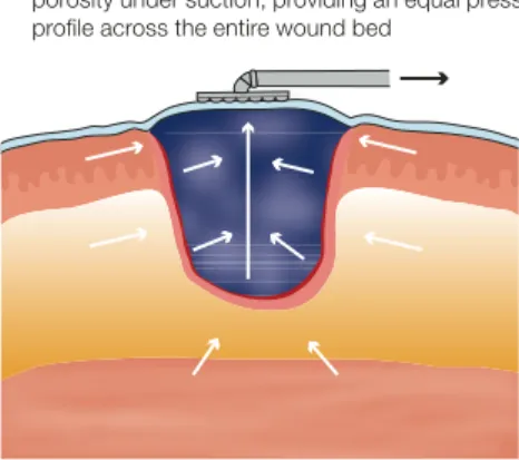
development by Kinetic Concepts Inc of a commercial system (the vacuum assisted closure [VAC] system) (Figure 1, Table 1). The regular structure of large open pores (400–600µm) within the PU foam dressing used as part of the system allows equal distribution of pressure across the entire wound surface. The foam also reduces in volume under pressure, resulting in cell stretch, wound contraction and removal of fluid (Figure 2).
More recently, competitors have developed variations of this system, but these do not use the PU foam.
At a basic level the VAC system provides a sophisticated, sterile, closed dressing with properties allowing a moist healing environment. Various mechanisms have also been confirmed that support healing. These include:
● increases local blood flow
● reduces oedema
● stimulates formation of granulation tissue
● stimulates cell proliferation
● removes soluble healing inhibitors from the wound
● reduces bacterial load
● draws the wound edges closer together.
KEY POINTS
1. The recent introduction of topical negative pressure (TNP) therapy has created new possibilities for the management of many different wound types. 2. Several mechanisms occur
as a result of TNP that support healing. These include stimulation of blood flow, granulation tissue and angiogenesis.
3. In addition, the negative pressure within the pores of the polyurethane or polyvinyl alcohol foam dressing used contracts the wound and draws the wound edges closer together. 4. By understanding these
mechanisms clinicians can consider whether it is appropriate to use this intervention.
TOPICAL NEGATIVE PRESSURE
IN WOUND MANAGEMENT
Morykwas et al studied the effect of TNP therapy on blood flow by producing deep tissue
defects in pigs and dressing them with PU foam3. Their results indicated a maximum
fourfold increase in blood flow with a negative pressure of 125mmHg. At higher pressures the risk is that the capillaries will distort and blood flow lessen. Indeed, blood flow was inhibited by applying negative pressures of 400mmHg and above.
Timmers et alevaluated the effect of TNP therapy on blood flow in the healthy skin of 10
human volunteers4. Blood flow improved by fivefold with PU foam and by threefold with
PVA foam with increasing negative pressure up to 300mmHg. The difference is due to the smaller pore size of PVA foam, which reduces the effect of TNP. Various factors affect the pressure achieved at the wound bed, for example clot formation, bleeding and an interposed dressing layer will reduce the pressure level1. It is proposed that blood flow is increased
directly by the negative pressure and indirectly by removing interstitial fluid.
Using their pig model, Morykwas et alalso determined the rate of granulation tissue
formation during TNP therapy by measuring the reduction in wound volume over time. Compared with control wounds dressed with standard saline-soaked gauze, increased rates of granulation tissue formation were observed with continuous and intermittent
application of negative pressure amounting to 63% and 103% respectively5. Intermittent
Figure 2 |Mechanisms of action of the VAC system
●Oedema is removed from periwound tissues, local blood perfusion increases and angiogenesis is stimulated
●Contracture of the foam draws the wound edges together, stabilises the wound margin and provides an anchor point for muscles and deeper structures
●Exudate, inhibitory substances and fine debris are removed from the wound
The system described throughout this document includes the following components:
A black, hydrophobic PU foam dressing with open pores, which is introduced into the wound. A white, hydrophilic PVA foam dressing with denser, smaller pores may be used instead (choice of foam type depends on wound characteristics and treatment aims)
A transparent, semi-occlusive adhesive drape, which is secured firmly over the foam dressing to the healthy skin around the wound margin. This stops ingress of air and allows a partial vacuum to form within the foam A pad attached to a drainage tube, which is placed over a small hole cut into the drape. The end of the drainage tube is connected to a suction source
A disposal reservoir, into which wound fluid is withdrawn under negative pressure through the foam via the drainage tube
An electrically powered therapy unit, which creates negative pressure by transferring gas molecules continuously from the inlet to the outlet of the unit by a rotating valve
A microprocessor, which computes signals from the system’s components and triggers an alarm if pressure levels are incorrect, there is an air leak etc
The pressure at the wound site is usually lowered by 125mmHg, which is about 10 times lower than the pressure used for normal chest drains in patients following lung surgery
Table 1 |TNP therapy using the VAC system
Figure 1 |The VAC therapy system Increasing blood perfusion and reducing oedema Stimulating formation of granulation tissue
1. The PU foam is placed in the wound 2. Following the application of TNP therapy (125mmHg) the foam loses volume but maintains porosity under suction, providing an equal pressure profile across the entire wound bed
1. Banwell P, Téot L. Topical Negative Pressure (TNP) Therapy. First international topical negative pressure (TNP) therapy focus group meeting proceedings. London: TXP Communications, 2004.
2. Fleischmann W, Strecker W, Bombelli M, et al. [Vacuum sealing as treatment of soft tissue damage in open fractures]. Unfallchirurg1993; 96(9): 488-92. 3. Morykwas MJ, Argenta LC, Shelton-Brown EI, et al. Vacuum-assisted closure:
a new method for wound control and treatment: animal studies and basic foundation. Ann Plast Surg1997; 38(6): 553-62.
4. Timmers MS, Le Cessie S, Banwell P, et al. The effects of varying degrees of pressure delivered by negative-pressure wound therapy on skin perfusion. Ann Plast Surg2005; 55(6): 665-71; discussion 1097-98.
5. Morykwas MJ, Faler BJ, Pearce DJ, et al. Effects of varying levels of subatmospheric pressure on the rate of granulation tissue formation in experimental wounds in swine. Ann Plast Surg2001; 47(5): 547-51. 6. Philbeck TE Jr, Whittington KT, Millsap MH, et al. The clinical and cost
effectiveness of externally applied negative pressure wound therapy in the treatment of wounds in home healthcare Medicare patients. Ostomy Wound Manage1999; 45(11): 41-50.
7. Vowden K. Conservative management of pressure ulcers. In: Banwell PE, Harding K (eds). Vacuum Assisted ClosureTMTherapy: Science and Practice.
London: MEP Ltd, 2006.
8. Sumpio BE, Banes AJ, Link WG, et al. Enhanced collagen production by smooth muscle cells during repetitive mechanical stretching. Arch Surg1988; 123(10): 1233-36.
9. Ilizarov GA. Clinical application of the tension-stress effect for limb lengthening. Clin Orthop Relat Res1990; (250): 8-26.
10. Saxena V, Hwang CW, Huang S, et al. Vacuum-assisted closure:
microdeformations of wounds and cell proliferation. Plast Reconstr Surg2004; 114(5): 1086-96.
11. Greene AK, Puder M, Roy R, et al. Microdeformational wound therapy: effects on angiogenesis and matrix metalloproteinases in chronic wounds of 3 debilitated patients. Ann Plast Surg2006; 56(4): 418-22.
12. Fabian TS, Kaufman HJ, Lett ED, et al. The evaluation of subatmospheric pressure and hyperbaric oxygen in ischemic full-thickness wound healing. Am Surg2000; 66(12): 1136-43.
13. Gustafsson RI, Sjögren J, Ingemansson R. Deep sternal wound infection: a sternal-sparing technique with vacuum-assisted closure therapy. Ann Thorac Surg2003; 76(6): 2048-53; discussion 2053.
14. Stechmiller JK, Kilapadi DV, Childress B, et al. Effect of vacuum-assisted closure therapy on the expression of cytokines and proteases in wound fluid of adults with pressure ulcers (letter to editor). Wound Rep Regen2006; 14: 371-74.
References
treatment is thought to be more effective than continuous treatment because the cells within the wound become accommodated (ie they no longer respond) to the constant physical forces applied with continuous therapy. Proposed mechanisms for the beneficial effects of intermittent therapy include6:
● increases tissue perfusion, by inactivating capillary autoregulation (whereby
capillaries are shut down if high blood flow is not needed)
● allows the proliferating cells time to rest between cycles of cell division, which is
necessary for the production of new cellular components. Constant stimulation with negative pressure may switch off the mitotic (nuclear division) process.
Many clinicians use continuous pressure because it is better tolerated by patients. Some recommend using the continuous setting for the first 48 hours with a target pressure of
125mmHg before switching to the intermittent mode7.
Mechanical stress has long been known to induce cell proliferation and division8. This
effect has been used for many years by plastic and orthopaedic surgeons to expand soft tissues and to lengthen bones9. It is also one of the most important features of TNP
therapy; a computerised model has shown that the negative pressure induces tissue microdeformations within the wound, and this has also been observed in the clinical setting10. This mechanical stretching of the cells stimulates proliferation and accelerates
wound healing. In chronic wounds this mechanism stimulates angiogenesis and
epithelialisation11. Fabian et alalso observed enhanced angiogenesis and a trend towards
increased rates of epithelialisation using TNP therapy in a rabbit model12.
By removing the harmful components (such as cytokines and matrix metalloproteinases) associated with excess exudate in the non-healing wound, TNP therapy may promote an active healing state where delayed primary closure can be achieved11,13,14. It may also help
reduce bacterial load3. For example, the seal created by the foam and drape reduces the risk
of external contamination and the improved blood perfusion may increase resistance to infection. Also, the partial vacuum created by TNP therapy causes the entire foam to shrink and this draws the wound margins to the centre, thereby facilitating wound closure1.
The mechanisms described above have a substantial impact on many of the factors that are known to promote healing. Used in conjunction with conventional treatments and professional wound assessment, TNP therapy, when used appropriately, has become a valuable tool for the clinician and the patient.
Stimulating cell proliferation
Other effects
In the context of chronic wounds topical negative pressure (TNP) therapy using the vacuum assisted closure (VAC) system is sometimes considered an expensive
intervention. For example, the acquisition costs of the dressing, tubing and canister, and the rental costs for home use are considerably more than for alternative
dressings. As a result access to TNP therapy is often restricted, particularly outside hospital settings1. Yet dressing costs typically make up only a small proportion of the
total expense of managing chronic wounds2, with nurse time, hospitalisations and
adverse events accounting for the majority. This paper explores the possibility of developing an economic case for TNP therapy by considering the costs and outcomes associated with the intervention for treating diabetic foot ulcers and pressure ulcers.
Economic evaluation seeks to capture the relative costs and benefits of two or more treatment options, for example advanced wound care dressings versus traditional dressings. Because economic evaluations are usually undertaken to inform healthcare decision making, the majority consider only expenditure directly relevant to the healthcare sector. For a chronic wound such as a diabetic foot ulcer, expenditure might include the costs of wound dressings, nurse time, hospitalisations and adverse events/amputations. While indirect costs, such as productivity losses of patients or informal carers, may be significant, these are not routinely captured in economic evaluations because they do not fall on healthcare budgets.
Most economic evaluations analyse cost-effectiveness, whereby costs are captured in
monetary units and outcomes are captured in clinical units3. For wound care this might
produce outcomes such as cost per additional wound healed or cost per amputation avoided. However, due to demands from health technology assessment agencies (notably in the UK the National Institute for Health and Clinical Excellence) there is an increasing trend towards cost-utility analysis, which captures outcomes in the form of a quality-adjusted life year (QALY). A QALY weights each year of a patient’s life by his or her quality of life during that year. Typically, a year of perfect or full health is given a value of 1.0 while death is usually valued at zero. Therefore, if we assume that a diabetic foot ulcer reduces quality of life by 50%, each remaining year of life with this condition yields 0.5 QALYs.
Values for quality of life can be derived in various ways. Standard gamble and time trade-off techniques can be used to elicit values directly from patients. However, more often values are obtained using standardised questionnaires, such as the EuroQol EQ-5D (see www.euroqol.org) or the Health Utilities Index (see www.fhs.mcmaster.ca/hug).
QALYs can be used to assess and compare the benefits afforded by various healthcare interventions. By factoring in the associated costs we can measure their cost-utility. The cost-utility approach is technically more demanding than other evaluations, but it allows healthcare planners to compare the value of interventions across disease areas (eg a new wound dressing with a new treatment for heart disease).
There are few robust economic evaluations of wound care, mainly because of the dearth of well-designed, longitudinal or clinical studies in this area. Although economists often extrapolate the results of clinical studies, this relies on having a well-defined biological relationship between interim endpoints and longer-term outcomes. For example, there are well-defined relationships between the risk factors for coronary heart disease and
mortality (derived from the Framingham cohort, see www.framingham.com/heart). These allow changes in interim outcomes (eg cholesterol levels) to be extrapolated to longer-term outcomes (eg mortality). Unfortunately, this is not the case for wound care.
Although many routine endpoints used in wound care studies are relevant to clinical decision makers (eg percentage change in wound area) they are relatively meaningless to economists or financial decision makers.
INTRODUCTION COST-EFFECTIVENESS Measuring outcomes Limitations in wound care
Health economics and topical
negative pressure therapy
P Trueman
Director, York Health Economics Consortium, University of York, York, UK.
Research is emerging that links interim endpoints such as wound size and duration to
longer-term outcomes4, but further data on these relationships are needed for economic
studies. In the meantime, clinical studies of wound care should consider how relevant the endpoints are for determining the clinical and cost-effectiveness of interventions. Longer-term measurement of well-defined outcome measures, such as healing, recurrence and amputation, will help in determining the economic value of interventions. The involvement of health economists in the design of clinical protocols will assist in identifying appropriate outcomes that can be incorporated into the protocol.
In examining the relative value of dressings, there is a tendency to focus on the cost of dressings rather than the cost of treatment, which may be influenced by other factors such as time to healing. In the 2003 EWMA position document, Franks and Posnett looked at the cost-effectiveness of compression therapy for venous leg ulcers5. Based on estimated
costs of weekly treatment, using the more expensive individual dressing (ie compression) resulted in a lower total treatment cost over time than the standard cheaper dressing regimen used (1,697 euros per ulcer healed versus 3,558 euros). This was due to the reduced time to healing and the fewer dressing changes with compression therapy.
An analysis by Harding et alhas also illustrated this point and has suggested that
dressings make up between 4% and 29% of the total cost of managing pressure ulcers2.
Furthermore, it found that the dressing with the lowest acquisition cost resulted in the highest total expenditure over the course of wound healing, due to its relative
effectiveness. Therefore, they propose that the factors in the box (left) are considered when analysing the cost of dressings. In some cases, these may be sufficient to offset the acquisition cost of premium priced dressings2.
Exploring these factors and by extrapolating data from the literature, the following sections examine the possibility of developing a cost-effectiveness argument for using TNP therapy in the management of diabetic foot ulcers and pressure ulcers.
TNP therapy using the VAC system offers an effective intervention for managing heavily exudative wounds. From an economic perspective, if dressings can remain in place for longer than other dressings, this allows a reduction in dressing acquisition costs and nurse time. This theory appears to be borne out in randomised controlled trials (RCTs)
comparing TNP therapy with other dressings in patients with diabetic foot ulcers. These studies consistently report that the dressings used with TNP therapy are changed every two days in non-infected wounds in line with the manufacturer’s recommendation,
whereas other dressings (eg saline gauze6, moist wound dressings7and other advanced
dressings8) are changed daily.
Similar comparisons have been made in RCTs in patients with pressure ulcers. These
trials have shown the difference between wet-to-moist9, Healthpoint system10and
wet-to-dry/wet-to-moist11 dressings, which are usually changed two or three times per day, versus
TNP therapy, which should be changed every two days.
Recommendations on wear time are not always adopted in practice (eg because of constraints on nursing time or the potential disruptive impact on the wound), and the high frequency of dressing changes reported in these RCTs may not be the experience of many practising healthcare professionals. With this in mind, it is important to note that two of the RCTs of diabetic foot ulcers included only 10 patients6,7, while the third
included 162 patients8. Sample sizes were also small in the pressure ulcer studies (ranging
from 24 to 34)9-11. This is a major limitation and the results must be interpreted with
caution. One trial is further undermined by the use of saline soaked gauze as the
comparator, which is considered a relatively outdated treatment in most of Europe6. Any
decision to extend wear time should be led clinically rather than by potential economic savings.
Dressing cost analysis
FACTORS AFFECTING THE COST OF
WOUND TREATMENT
The cost of managing chronic wounds can be influenced by: • frequency of dressing
changes and associated nurse time
• healing rates
• impact on hospitalisations and adverse events
ECONOMIC CONSIDERATIONS OF TNP THERAPY
Frequency of dressing changes
TOPICAL NEGATIVE PRESSURE
IN WOUND MANAGEMENT
Healing rates Diabetic foot ulcers
A study by Armstrong et alexamined the use of TNP therapy after partial diabetic foot
amputation8. This multicentre RCT of 162 patients compared TNP therapy using the
VAC system with standard moist wound care. TNP dressings were changed every two days while standard therapy was based on consensus guidelines. Standard therapy was made up of dressings promoting a moist wound environment, ie with alginates,
hydrocolloids, foams or hydrogels, adhering to standard guidelines at the discretion of the attending clinician. Patients were followed for 112 days or until wound healing.
Healing rates at the end of the study were 56% in the TNP therapy arm, compared with 39% in the standard therapy arm (p=0.040). Most patients healed by primary intention and there was no significant difference in the proportion of wounds healed by secondary intention in either arm. The mean time to healing in the TNP therapy arm was 56 days, compared with 77 days in the standard therapy arm. The median time to achieving 76–100% granulation was 42 days in the TNP therapy arm, compared with 84 days in the standard therapy arm (p=0.002).
These findings have important economic implications, suggesting that a higher proportion of patients are likely to heal in a shorter period of time when treated with TNP therapy. These outcomes are directly relevant to economic decision makers in healthcare settings and the impact on budgets can be easily quantified. A full economic analysis of the study findings is expected to be published in 2007.
Pressure ulcers
The only prospective, comparative study of TNP therapy in pressure ulcers that reported
healing rates was conducted by Ford et al10. It compared TNP therapy using the VAC
system with the Healthpoint system, which consists of a papain-urea debridement ointment and a combination of pads and gels containing cadexomer iodine.
Healing rates at six weeks were marginally higher in the Healthpoint system group (13% versus 10%). However, TNP therapy produced a greater (but not statistically significant) percentage change in wound volume (51.8% versus 42.1%, p=0.46) and was better at improving wounds with biopsy-proven osteomyelitis. The authors concluded that TNP therapy has a better rate of wound healing with favourable histological changes in soft tissue and bone compared with the Healthpoint system, although the choice of comparator, the small sample size and the lack of statistical significance of the findings related to healing rates should be borne in mind when considering this evidence.
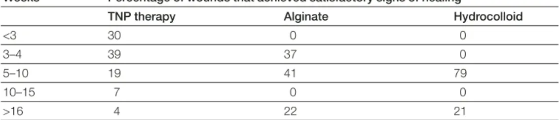
Smith reviewed the literature on the effectiveness of TNP therapy with alginates and
hydrocolloids for treating pressure ulcers12. The study found 93% of wounds treated with
TNP therapy healed, compared with 63% of wounds treated with hydrocolloids
(p<0.002). Table 1 reports the data on time to healing. Most wounds treated with TNP therapy achieved ‘satisfactory’ signs of healing (wound needing little or no further treatment) within four weeks. However, the median time to satisfactory signs of healing
Weeks Percentage of wounds that achieved satisfactory signs of healing
TNP therapy Alginate Hydrocolloid
<3 30 0 0
3–4 39 37 0
5–10 19 41 79
10–15 7 0 0
>16 4 22 21
Hospitalisations
Adverse events
ANALYSIS
with alginates and with hydrocolloids was five to 10 weeks. Again, the improved time to healing has positive economic implications for TNP therapy.
Several studies of TNP therapy have examined the impact on hospitalisation rates. In a retrospective review of the use of the VAC system in a home setting to treat grade 3 or 4
pressure ulcers, Schwein et alcompared a matched group of patients using TNP therapy
(n=60) with a control group who were not treated with VAC (n=2,288)13. The study
found that hospitalisation rates in patients treated with TNP therapy were statistically significantly lower than those treated with standard care (p<0.05). Hospitalisation rates were separated into all-cause hospitalisation, hospitalisations related to wound care problems and emergent care problems related to wound care. In all categories, patients treated with TNP therapy experienced lower hospitalisation rates (Figure 1).
Although the design of the study introduces an increased risk of bias compared with an RCT, this more naturalistic design does provide a true reflection of standard practice and overcomes some of the problems associated with protocol-driven events that are
experienced in trial settings. Some of the potential for bias in the study has been accounted for by matching patient characteristics in the two arms.
In addition to the benefits listed above, several studies have suggested that TNP therapy might reduce adverse events, particularly amputations. However, the data remain
equivocal. Armstrong et alreported a reduction in amputations with TNP therapy
compared with the comparator (3% versus 11%, p=0.06), although this did not reach statistical significance8. It should also be noted that the patients in this study had already
had a prior amputation and this could have affected the likelihood of a further amputation, although the risk is expected to be increased in both arms of the study.
The higher rate of hospitalisations for wound-related problems in the study by Schwein et almay also suggest a lower rate of adverse events, although details of this are not provided in the paper13. Joseph et alalso reported lower adverse event rates for TNP
therapy compared with saline gauze9. The wounds considered were predominantly
pressure ulcers. The reported complication rates were 44% in the saline gauze arm, compared with 17% in the TNP therapy arm. Although this was a statistically significant outcome, the total sample size was 36 so the findings should be considered with caution. Examining the clinical data on TNP therapy highlights a number of economic benefits that may offset the higher acquisition costs of the dressings and rental costs of the TNP therapy unit. A crude analysis of the findings of Armstrong et aldescribed earlier helps to
demonstrate this8. Dressings were changed daily in the standard therapy arm and every
two days in the TNP therapy arm. Let us assume that each dressing change required a
All-cause hospitalisations
Hospitalisations for wound care problems Emergent care problems related to wound care TNP therapy Control 0% 10% 20% 30% 40% 50% 35% 48% 5% 14% 0% 8% Figure 1 |Hospitalisation rates for TNP therapy and standard care13
TOPICAL NEGATIVE PRESSURE
IN WOUND MANAGEMENT
1. Newton H, Benbow M, Hampton S, et al. TNP therapy in the community: findings of a national survey. Wounds UK2006; 2(4): 31-35.
2. Harding K, Cutting K, Price P. The cost-effectiveness of wound management protocols of care. Br J Nurs2000; 9(19 Suppl): S6-S24.
3. Nixon J, Stoykova B, Glanville J, et al. The U.K. NHS economic evaluation database. Economic issues in evaluations of health technology. Int J Technol Assess Health Care2000; 16(3): 731-42.
4. Margolis DJ, Allen-Taylor L, Hoffstad O, et al. Diabetic neuropathic foot ulcers: predicting which ones will not heal. Am J Med2003; 115(8): 627-31. 5. Franks PJ, Posnett J. Cost-effectiveness of compression therapy. In: European
Wound Management Association (EWMA). Position Document: Understanding compression therapy. London: MEP Ltd, 2003: 8-10.
6. McCallon SK, Knight CA, Valiulus JP, et al. Vacuum-assisted closure versus saline-moistened gauze in the healing of postoperative diabetic foot wounds. Ostomy Wound Manage2000; 46(8): 28-32, 34.
7. Eginton MT, Brown KR, Seabrook GR, et al. A prospective randomized evaluation of negative-pressure wound dressings for diabetic foot wounds. Ann Vasc Surg2003; 17(6): 645-9. Epub 2003; Oct 13.
8. Armstrong DG, Lavery LA; Diabetic Foot Study Consortium. Negative pressure wound therapy after partial diabetic foot amputation: a multicentre, randomised controlled trial. Lancet 2005: 366(9498): 1704-10.
9. Joseph E, Hamori CA, Bergman S, et al. A prospective randomized trial of vacuum-assisted closure versus standard therapy of chronic non-healing wounds. Wounds2000; 12(3): 60-67.
10. Ford CN, Reinhard ER, Yeh D, et al. Interim analysis of a prospective, randomized trial of vacuum-assisted closure versus the Healthpoint system in the management of pressure ulcers. Ann Plast Surg2002; 49(1): 55-61. 11. Wanner MB, Schwarzl F, Strub B, et al. Vacuum-assisted wound closure for
cheaper and more comfortable healing of pressure sores: a prospective study. Scand J Plast Reconstr Surg Hand Surg2003; 37(1): 28-33.
12. Smith N. The benefits of VAC therapy in the management of pressure ulcers. Br J Nurs2004; 13(22): 1359-65.
13. Schwein T, Gilbert J, Lang C. Pressure ulcer prevalence and the role of negative pressure wound therapy in home health quality outcomes. Ostomy Wound Manage2005; 51(9): 47-60.
14. Curtis L, Netten A. Unit costs of health and social care 2005. Canterbury: Personal Social Services Research Unit, University of Kent, 2005. Also available at: www.pssru.ac.uk
15. Philbeck TE Jr, Whittington KT, Millsap MH, et al. The clinical and cost effectiveness of externally applied negative pressure wound therapy in the treatment of wounds in home healthcare Medicare patients. Ostomy Wound Manage1999; 45(11): 41-50.
16. Moues CM, van den Bemd GJ, Meerding WJ, et al. An economic evaluation of the use of TNP on full thickness wounds. J Wound Care2005; 14(5):
224-27.
17. Vuerstaek JD, Vainas T, Wuite J, et al. State-of-the-art treatment of chronic leg ulcers: a randomized controlled trial comparing vacuum-assisted closure (V.A.C.) with modern wound dressings. J Vasc Surg2006; 44(5): 1029-37. References
KEY POINTS
1. Emerging research suggests there may be economic as well as clinical advantages to using TNP therapy for chronic wound management. 2. Savings due to faster healing
rates and reduced dressing changes, nurse time and hospital stays with TNP therapy may offset higher acquisition costs.
3. TNP therapy may also reduce costs associated with adverse events. 4. Current economic
evaluations of wound care are limited by difficulties capturing economically useful clinical outcomes, the dearth of robust studies and lack of data from Europe.
CONCLUSION
home nurse visit estimated to cost 35 euros per visit14, and that nurse visits were needed
only while the wound was unhealed. Where a wound was unhealed over the course of the study, dressing changes are assumed to have continued over the entire duration (112 days). In the standard therapy arm 39% of patients healed and are assumed to have had daily visits for 77 days (mean time to healing in this group). The remaining 61% of unhealed patients had daily visits for 112 days. This gives an average nurse cost per patient of 3,443 euros. In the TNP therapy arm, 56% of patients healed and are assumed to have had visits every two days for 56 days (mean time to healing in this group), with the remaining 44% of unhealed patients having visits every two days for 112 days. This gives an average nurse cost per patient of 1,411 euros.
While this is clearly a partial analysis (it takes no account of dressing costs or rental of the TNP therapy unit), it shows that the reduced nurse costs associated with TNP therapy can help to release some ‘headroom’ to account for the other expenses. A more detailed analysis is needed to determine the net impact of TNP therapy on total treatment costs.
Philbeck et alestimated the cost of treating pressure ulcers using estimates of daily wound area reductions for TNP therapy and standard care15. Costs of treatment to healing were
$23,465 (18,155 euros) for standard care and $14,546 (11,256 euros) for TNP therapy. However, the study design is not strong as it uses average wound area reductions derived from published papers and applies these to an average sized wound. It also assumes healing rates are constant over time. As such, the data sources may not be directly comparable. A growing body of evidence suggests there may be economic as well as clinical benefits to using TNP therapy. Findings seem to imply that the savings accrued due to faster healing, reduced nurse time and hospital stay may offset the additional acquisition cost of TNP therapy dressings. However, there are limitations in the evidence. None of the studies mentioned here provides a full cost-effectiveness analysis capturing both costs and outcomes of care. In particular, studies have focused on measures of clinical outcomes (eg wound healing or wound area reduction) rather than the impact on quality of life. With the exception of a study in venous leg ulcers and a study in surgical wounds there is no
economic evidence from European settings16,17. Additional research is underway to
develop a more robust economic evaluation of TNP therapy and alternative regimens for the treatment of diabetic foot ulcers. This should provide a more thorough analysis of the treatment costs and outcomes of care, including the impact on quality of life.
INTRODUCTION EVIDENCE FOR TNP THERAPY WOUND BED PREPARATION AND TNP THERAPY Tissue management Inflammation and infection control
1. Nurse Consultant, Bradford Teaching Hospitals NHS Foundation Trust, Bradford, UK. 2. Assistant Professor of Surgery, University Hospital, Montpellier, France. 3. Visiting Professor of Wound Healing, University of Bradford, and Consultant Vascular Surgeon, Bradford Teaching Hospitals NHS Foundation Trust, Bradford, UK.
Selecting topical negative pressure
therapy in practice
K Vowden
1, L Téot
2, P Vowden
3When considering topical negative pressure (TNP) therapy, the stratification of wound types into acute and chronic is in many ways irrelevant. Acute and chronic wounds of all aetiologies require a holistic assessment of the cause, an understanding of the underlying medical and social conditions that may affect healing and
treatment decisions, and a full evaluation of the wound status. This paper uses the concept of wound bed preparation to suggest a therapeutic strategy to help clinicians identify when to use TNP therapy. This approach will aid the integration of this intervention into the management of many types of complex wounds.
TNP therapy is used for the treatment of acute and chronic wounds in both inpatient and outpatient settings. Recently, the range of indications has been extended on the basis of many scientific publications (over 250 peer-reviewed articles, 330 published abstracts and 42 book chapters). These show that TNP therapy has been used successfully in the management of a wide variety of acute and chronic wounds. Almost all the published evidence relates to the use of the vacuum assisted closure (VAC) system (Kinetic Concepts Inc). Initially much of this evidence was in the form of extended case studies. However, recently the results from a number of randomised controlled trials (RCTs) have been published supporting the findings of the earlier reports1-9(see box, below right).
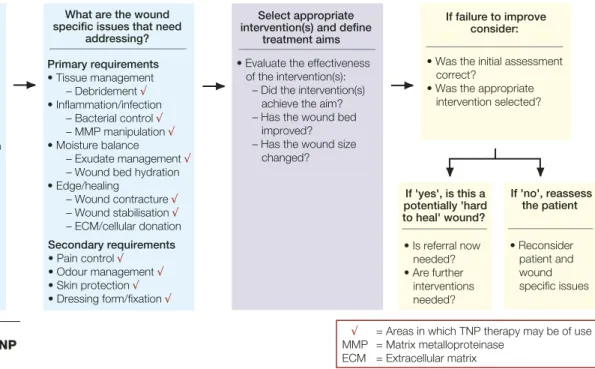
Studies soon to be published support that VAC is highly suited to managing trauma wounds with or without loss of bone substance. The key to choosing an appropriate wound management strategy is to carry out a thorough patient assessment and identify the patient and wound specific issues that need addressing. Figure 1 illustrates this approach. Wound bed preparation focuses on tissue management, inflammation and infection control, moisture balance and epithelial edge advancement10. By exploring these factors, it
is possible to recognise when TNP therapy is an appropriate intervention to promote healing2,3or to prepare the wound bed for surgical closure (see Figure 1).
Wound ischaemia is recognised as one of the main causes of delayed healing or non-healing in both acute and chronic wounds. Research has demonstrated through laboratory-based experimental models and clinical work that TNP therapy using the VAC system increases angiogenesis11,12and has a direct effect on microvascular blood flow that may benefit
healing13,14. This effect may go some way towards explaining the benefit seen with TNP
therapy when it is used in the management of patients with skin grafts, diabetic foot wounds (whether neuropathic or surgical amputation), complex traumatic wounds where bone and/or tendon is exposed, or exposed implanted prostheses where angiogenesis is seen in the form of granulation tissue developing on relatively or totally avascular structures15.
Overt wound infection has been considered a contraindication to TNP therapy. However, evidence suggests that TNP therapy may have a role in reducing bacterial load within a wound and may reduce levels of potentially damaging exotoxins and endotoxins simply by rapidly removing exudate from the wound bed. Because TNP therapy works as a closed system it also decreases wound odour between dressing changes and reduces environmental bacterial contamination.
There have been isolated reports suggesting an adverse alteration to wound flora with TNP therapy16. However, one study found a beneficial effect on bacterial load with VAC17.
Most case reports relating to TNP therapy in this situation have also been favourable18-20.
TNP therapy has been shown to be an efficacious adjunctive method for treating
postoperative wound infection after median sternotomy7,18. Mehbod et alhave reported
similar positive outcomes for infected spinal surgical wounds even in the presence of
implanted material21, and Dosluoglu et alhad encouraging results when using TNP
TOPICAL NEGATIVE PRESSURE
IN WOUND MANAGEMENT
Moisture balance
alalso report benefits in managing complicated open gynaecological oncology surgical
wounds with TNP therapy23. However, these are uncommon complicated wounds and
are not a major indication for TNP therapy.
TNP therapy has been used successfully in the management of some cases of
osteomyelitis, including foot, lower limb and sternal infections18,24. Treatment should be
combined with extensive and thorough wound debridement including excision of avascular or clearly infected bone, and appropriate adjuvant therapy such as antibiotics.
Specific infective organisms, such as MRSA and other resistant strains, are not contra-indications to TNP therapy. The management strategy should follow that suggested in
the 2006 EWMA position document on the management of wound infection25. TNP
therapy has a favourable effect on matrix metalloproteinase (MMP) levels in chronic wounds (see page 4). This may largely be due to the removal of exudate but could reflect
a down-regulation of the inflammatory status of the wound26.
Clinical experience and research evidence have established that TNP therapy is an effective method for managing exudate; the system removes excess fluid while maintaining a moist wound environment and protecting the surrounding tissues from maceration and exudate
damage27. In any wound where exudate management is difficult, TNP therapy should be
considered as an option along with other treatments. The VAC system has, for example, been found to be an effective method of protecting the skin from fistula effluent, although this falls outside of the manufacturer’s suggested range of uses28,29.
Correctly applied, it also has the advantage of preventing exudate pooling in the wound and therefore the build up of bacterial load and potentially damaging protease-rich wound fluid in the deeper recesses of a wound. Fluid pooling and spreading sepsis is a significant problem in cavity wounds and this may explain the value of TNP therapy in
the management of open minor diabetic foot amputations or pressure ulcers3,30.
A similar principle applies when TNP therapy is used in conjunction with skin grafting or a bioengineered skin product, where it has been demonstrated to be of significant value in improving take15,31,32. With skin grafts even a small excess of wound fluid developing in
√ = Areas in which TNP therapy may be of use
MMP = Matrix metalloproteinase ECM = Extracellular matrix What are the wound
specific issues that need addressing? Primary requirements • Tissue management – Debridement √ • Inflammation/infection – Bacterial control √ – MMP manipulation √ • Moisture balance – Exudate management √
– Wound bed hydration • Edge/healing – Wound contracture √ – Wound stabilisation √ – ECM/cellular donation Secondary requirements • Pain control √ • Odour management √ • Skin protection √ • Dressing form/fixation √
What are the patient specific issues that need
addressing? • Medical disease management • Systemic/adjunctive therapy designed to improve the wound environment – Elevation/compression – Antibiotics – Nutritional support • Psychosocial management If failure to improve consider: • Was the initial assessment correct?
• Was the appropriate intervention selected? If 'yes', is this a potentially 'hard to heal' wound? • Is referral now needed? • Are further interventions needed? Select appropriate
intervention(s) and define treatment aims • Evaluate the effectiveness of the intervention(s): – Did the intervention(s) achieve the aim? – Has the wound bed improved? – Has the wound size changed? If 'no', reassess the patient • Reconsider patient and wound specific issues
Figure 1 | Wound specific issues and the role of TNP therapy
STUDIES USING TNP THERAPY IN VARIOUS WOUND TYPES
• Burn wounds1 • Chronic leg ulcers2 • Diabetic foot ulcers3 • Open abdomen including
management of fistulae4 • Pressure ulcers5 • Securing a skin graft6 • Sternal wound infections7 • Surgical, non-healing
wounds8 • Trauma9
the interface between graft and wound bed can lead to the loss of all or part of a graft, as can excessive shear between dressing, graft and wound bed.
TNP therapy has been used to reduce wound size by aiding edge closure, and has been
shown to hasten fasciotomy wound closure33. Using TNP therapy to treat sternotomy
wounds has the advantage of creating wound stability and improving pain control. The negative pressure causes the foam to form a soft but rigid anchor point for the deep and superficial compartments of the wound and forms a fixation point for muscles and fascia around the dehisced wound. The technique has been recommended for managing dehisced abdominal wounds34, including those with enterocutaneous fistulation29.
These effects of TNP therapy with the VAC system (ie wound edge stability and
wound contracture) are an obvious advantage in unstable sternal wounds35. They are
equally as important in the management of chronic cavity wounds such as pressure ulcers and diabetic foot ulcers, especially those involving ray amputation. The fixation/
stabilising effect protects the wound from stress and shear damage.
It has been suggested that TNP therapy can be effective for controlling wound pain,
particularly if there is wound instability and marked edge movement and shear. Butter et
alfound it to be well tolerated in a paediatric population, and that it offered many advantages including fewer dressing changes and an earlier return to daily activities36. If
pain is noted at dressing changes it may be necessary to place a non-adherent or interposed dressing layer between the foam and the wound.
Once the wound specific issues have been identified and the intervention selected, clearly defined treatment objectives must be decided and documented, and progress towards endpoints reviewed frequently. The boxes below provide examples of these.
Epithelial edge advancement Controlling wound pain TREATMENT OBJECTIVES CONTRAINDICATIONS Debridement TREATMENT OBJECTIVES
1. Manage excessive exudate because it is affecting care, skin integrity and quality of life
2. Promote rapid improvement in wound bed, eg before surgical wound closure or application of skin graft/bioengineered skin substitute
3. Improve vascularity of wound bed and/or promote granulation tissue, eg to cover relatively avascular tissue or exposed prosthesis 4. Stabilise wound, graft or flap and aid care and rehabilitation,
eg dehisced surgical wounds, open amputation sites and for graft fixation
5. Promote healing status when healing is not progressing with conventional dressings
ENDPOINTS
1. Fall in exudate levels such that wound can be managed with conventional dressings 2. Stable healthy wound bed
with 100% granulation tissue 3. Wound bed preparation or
healing aims managed more clinically and cost-effectively with alternative dressing Note: Recent work in sternal wounds suggests using a fall in inflammatory markers, such as C-reactive protein levels, to monitor effectiveness of care37.
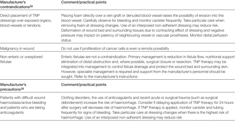
The literature reports on the many successes of TNP therapy. There are, however, occasions when inappropriate choice or application of TNP therapy can result in poor clinical outcomes or adverse events. To promote the safe and effective use of the technique, contraindications and precautions to treatment have been described38. Some are discussed below.
Although TNP therapy may assist in the process of maintenance debridement, it is not suitable for wounds with large volumes of necrotic tissue or eschar. It may also decrease the effectiveness of autolytic debridement by removing the necessary enzymes from the wound bed. All wounds must therefore be adequately debrided before treatment with TNP therapy. The intervention may, however, have a role in the management of adherent
fibrinous exudate on the wound bed. Loree et al, for example, used TNP therapy
TOPICAL NEGATIVE PRESSURE
IN WOUND MANAGEMENT
The clinical needs of the patient mean that on occasions TNP therapy has been used with caution successfully outside some of the manufacturer’s recommended indications. Effective use of TNP therapy has been reported in patients with pyoderma
gangrenosum40even though there is a theoretical risk of ‘pathergy’ (ie an exaggerated
inflammatory response). Ford-Dunn reports good symptom control in a patient with a
malignant wound41, and Kopp et alused TNP therapy as an adjunct to resection and
complex reconstruction and brachytherapy in soft tissue malignancy42. Dosluoglu et al
have reported the use of TNP therapy in patients with exposed vascular grafts even in the presence of infection22. Nonetheless, it is important to note in all these cases careful
observation of the wound and the collected exudate has been key to the effective use of TNP therapy. In a palliative care situation particular vigilance and regular observation is required to protect wounds and individuals from potentially detrimental side effects of TNP therapy, such as bleeding or local tumour stimulation. It is noted that in the presence of overt infection TNP therapy should be used with adequate debridement, effective drainage of all areas of the wound and appropriate additional therapy, such as targeted antibiotic therapy.
The anatomical wound site, clinical setting (eg home care/support), patient’s ability to tolerate treatment and availability of trained staff and equipment may also make the use of TNP therapy inappropriate. Table 1 provides useful comments and practical guidance on some of the manufacturer’s contraindications and precautions.
TNP therapy should be regarded in the same way as other wound care interventions: it should be selected if it provides the most clinically and cost-effective method for achieving defined therapeutic goals. Treatment outcomes should be continually re-evaluated. TNP Precautions
CONCLUSION
Manufacturer’s Comment/practical points contraindications38
Direct placement of TNP Placing foam directly over a vein graft or denuded blood vessel raises the possiblity of erosion into the dressings over exposed organs, blood vessel. Carefully observe for bleeding and monitor canister frequently. Take particular care when blood vessels or tendons removing foam at dressing changes. Use of an interposed non-adherent dressing may reduce risk.
Deformation of wound bed and surrounding tissues due to contracting effect of dressing and negative pressure may impact on patency of neighbouring vessel or vascular prostheses. Monitor distal perfusion status
Malignancy in wound Do not use if proliferation of cancer cells is even a remote possibility
Non-enteric or unexplored Enteric fistulae are not a contraindication. Primary management is reduction in fistula flow, nutritional support, fistulae elimination of distal obstruction and, where possible, surgical closure or resection. TNP therapy may be
integrated into management to control fistula drainage and protect the wound bed and surrounding skin. However, specialist management is required and support from the manufacturer’s personnel should be sought. Refer to the manufacturer’s instructions
Manufacturer’s Comment/practical points precautions38
Patients with difficult wound Clotting disorders, the use of anticoagulants and recent acute or surgical trauma (such as surgical haemostasis/active bleeding debridement) increase the risk of haemorrhage. Consider if delaying application of TNP therapy for 24 hours and patients who are taking after surgery will decrease risk of haemorrhage. If TNP therapy is applied, monitor canister and tubing anticoagulants frequently for signs of bleeding. Take particular care at dressing changes when there is the highest risk of
haemorrhage. Use of an interposed non-adherent dressing may reduce risk
Note: using TNP therapy outside of the manufacturer’s instructions should be done with caution under close clinical supervision, usually in a hospital environment, and will be the responsibility of the lead clinician.
therapy must be regarded as only one important component of overall wound
management, and must be introduced with a defined goal and exit strategy. It should be stopped once the goals have been achieved, if treatment is not meeting defined aims in an acceptable timeframe, or it is unacceptable to the patient or causes complications.
1. Kamolz LP, Andel H, Haslik W, et al. Use of subatmospheric pressure therapy to prevent burn wound progression in human: first experiences. Burns2004; 30(3): 253-58.
2. Vuerstaek JD, Vainas T, Wuite J, et al. State-of-the-art treatment of chronic leg ulcers: a randomized controlled trial comparing vacuum-assisted closure (V.A.C.) with modern wound dressing. J Vasc Surg2006; 44(5): 1029-37.
3. Armstrong DG, Lavery LA; Diabetic Foot Consortium. Negative pressure wound therapy after partial diabetic foot amputation: a multicentre, randomised controlled trial. Lancet 2005; 366(9498): 1704-10.
4. Wild T, Stortecky S, Stremitzer S, et al. [Abdominal dressing - a new standard in therapy of the open abdomen following secondary peritonitis?] Zentralbl Chir 2006; 131(Suppl 1): S111-14.
5. Ford CN, Reinhard ER, Yeh D, et al. Interim analysis of a prospective, randomized trial of vacuum-assisted closure versus the Healthpoint system in the
management of pressure ulcers. Ann Plast Surg2002; 49(1): 55-61; discussion: 61.
6. Jeschke MG, Rose C, Angele P, et al. Development of new reconstructive techniques: use of Integra in combination with fibrin glue and negative-pressure therapy for reconstruction of acute and chronic wounds. Plast Reconstr Surg 2004; 113(2): 525-30.
7. Sjögren J, Gustafsson R, Nilsson J, et al. Clinical outcome after poststernotomy mediastinitis: vacuum-assisted closure versus conventional therapy. Ann Thorac Surg2005; 79(6): 2049-55.
8. Moues CM, Vos MC, van den Bemd GJ, et al. Bacterial load in relation to vacuum-assisted closure wound therapy: a prospective randomized trial. Wound Repair Regen2004; 12(1): 11-17.
9. Stannard JP, Robinson JT, Anderson ER, et al. Negative pressure wound therapy to treat hematomas and surgical incisions following high-energy trauma. J Trauma 2006; 60(6): 1301-06.
10. European Wound Management Association (EWMA). Position Document: Wound bed preparation in practice. London: MEP Ltd, 2004.
11. Saxena V, Hwang CW, Huang S, et al. Vacuum-assisted closure:
microdeformations of wounds and cell proliferation. Plast Reconstr Surg2004; 114(5): 1086-96; discussion 1097-98.
12. Chen SZ, Li J, Li XY, et al. Effects of vacuum-assisted closure on wound microcirculation: an experimental study. Asian J Surg2005; 28(3): 211-17. 13. Wackenfors A, Sjögren J, Gustafsson R. Effects of vacuum-assisted closure
therapy on inguinal wound edge microvascular blood flow. Wound Repair Regen 2004; 12(6): 600-06.
14. Wackenfors A, Gustafsson R, Sjögren J, et al. Blood flow responses in the peristernal thoracic wall during vacuum-assisted closure therapy. Ann Thorac Surg2005; 79(5): 1724-30; discussion 1730-31.
15. Venturi ML, Attinger CE, Mesbahi AN, et al. Mechanisms and clinical applications of the vacuum-assisted closure (VAC) device: a review. Am J Clin Dermatol 2005; 6(3): 185-94.
16. Chester DL, Waters R. Adverse alteration of wound flora with topical negative-pressure therapy: a case report. Br J Plast Surg2002; 55(6): 510-11. 17. Morykwas MJ, Argenta LC, Shelton-Brown EI, et al. Vacuum-assisted closure: a
new method for wound control and treatment: animal studies and basic foundation. Ann Plast Surg1997; 38(6): 553-62.
18. Cowan KN, Teague L, Sue SC, et al. Vacuum-assisted wound closure of deep sternal infections in high-risk patients after cardiac surgery. Ann Thorac Surg 2005; 80(6): 2205-12.
19. Demaria R, Giovannini UM, Teot L, et al. Using VAC to treat a vascular bypass site infection. J Wound Care 2001; 10(2): 12-13.
20. Schuster R, Moradzadeh A, Waxman K. The use of vacuum-assisted closure
therapy for the treatment of a large infected facial wound. Am Surg2006; 72(2): 129-31.
21. Mehbod AA, Ogilvie JW, Pinto MR, et al. Postoperative deep wound infections in adults after spinal fusion: management with vacuum-assisted wound closure. J Spinal Disord Tech2005; 18(1): 14-17.
22. Dosluoglu HH, Schimpf DK, Schultz R, et al. Preservation of infected and exposed vascular grafts using vacuum assisted closure without muscle flap coverage. J Vasc Surg2005; 42(5): 989-92.
23. Schimp VL, Worley C, Brunello S, et al. Vacuum-assisted closure in the treatment of gynecologic oncology wound failures. Gynecol Oncol 2004; 92(2): 586-91. 24. Scholl L, Chang E, Reitz B, et al. Sternal osteomyelitis: use of vacuum-assisted
closure device as an adjunct to definitive closure with sternectomy and muscle flap reconstruction. J Card Surg 2004; 19: 453-61.
25. European Wound Management Association (EWMA). Position Document: Management of wound infection. London: MEP Ltd, 2006.
26. Shi B, Chen SZ, Zhang P, et al. [Effects of vacuum-assisted closure (VAC) on the expressions of MMP-1, 2, 13 in human granulation wound]. Zhonghua Zheng Xing Wai Ke Za Zhi 2003; 19(4): 279-81.
27. Banwell P, Téot L. Topical negative pressure (TNP): the evolution of a novel wound therapy. J Tissue Viability2006; 16(1): 16-24.
28. Cro C, George KJ, Donnelly J, et al. Vacuum assisted closure system in the management of enterocutaneous fistulae. Postgrad Med J 2002; 78: 364-65. 29. Goverman J, Yelon JA, Platz JJ, et al. The "Fistula VAC," a technique for
management of enterocutaneous fistulae arising within the open abdomen: report of 5 cases. J Trauma 2006; 60(2): 428-31; discussion 431.
30. Brem H, Sheehan P, Rosenberg HJ, et al. Evidence-based protocol for diabetic foot ulcers. Plast Reconstr Surg2006; 117(7 Suppl); 193S-209S.
31. Espensen EH, Nixon BP, Lavery LA, et al. Use of subatmospheric (VAC) therapy to improve bioengineered tissue grafting in diabetic foot wounds. J Am Podiatr Med Assoc2002; 92(7): 395-97.
32. Scherer LA, Shiver S, Chang M, et al. The vacuum assisted closure device: a method of securing skin grafts and improving graft survival. Arch Surg2002; 137(8): 930-33; discussion 933-34.
33. Yang CC, Chang DS, Webb LX. Vacuum-assisted closure for fasciotomy wounds following compartment syndrome of the leg. J Surg Orthop Adv2006; 15: 19-23. 34. Heller L, Levin SL, Butler CE. Management of abdominal wound dehiscence
using vacuum assisted closure in patients with compromised healing. Am J Surg 2006; 191(2): 165-72.
35. Hersh RE, Jack JM, Dahman MI, et al. The vacuum-assisted closure device as a bridge to sternal wound closure. Ann Plast Surg2001; 46(3): 250-54. 36. Butter A, Emran M, Al-Jazaeri A, et al. Vacuum-assisted closure for wound
management in the pediatric population. J Pediatr Surg2006; 41: 940-42. 37. Gustafsson R, Johnsson P, Algotsson L, et al. Vacuum-assisted closure therapy
guided by C-reactive protein level in patients with deep sternal wound infection. J Thorac Cardiovasc Surg 2002; 123(5): 895-900.
38. Banwell P. V.A.C.®TherapyTMClinical Guidelines. A reference source for clinicians.
KCI Ltd, September, 2005.
39. Loree S, Dompmartin A, Penven K, et al. Is vacuum assisted closure a valid technique for debriding chronic leg ulcers? J Wound Care2004; 13(6): 249-52. 40. Mandal A, Addison P, Stewart K, et al. Vacuum-assisted closure therapy in
pyoderma gangrenosum. Eur J Plast Surg2006; 28(8): 529-31. 41. Ford-Dunn S. Use of vacuum assisted closure therapy in the palliation of a
malignant wound. Palliat Med2006; 20(40): 477-78.
42. Kopp J, Strnad V, Bach AD, et al. Vacuum application increases therapeutic safety and allows intensified local radiation treatment of malignant soft-tissue tumors. Strahlenther Onkol 2005; 181(2): 124-30.
References
KEY POINTS
1. Identifying patient and wound-specific issues is key to choosing an appropriate wound management strategy. 2. Once TNP therapy has been selected therapeutic goals should be defined and progress carefully and
frequently monitored.