UNIVERSITY OF BERGAMO
Faculty of Education Sciences
PhD Course in Clinical Psychology
MODULATION OF VISUO-SPATIAL ATTENTION
BY MEANS OF EMOTIONAL
FACIAL AND BODILY EXPRESSIONS:
THEORETICAL BASIS AND EMPIRICAL
EVIDENCE
Phd Coordinator:
Prof. Valeria UGAZIO
Tutor:
Prof. Maria Luisa RUSCONI
Phd Candidate: Laura CARELLI
Registration number: 1004369
TABLE OF CONTENTS
Introduction 5
Chapter 1
Visual attention to spatial location under normal and pathological conditions 7
1.1What is (Visual) Attention? 7
1.2Theoretical models of Attention 8
1.3Mechanism of Attentional Control 12
1.4Neural Systems for Spatial Attention 13
1.4.1 Electrophysiology of visual attention: ERP investigations 13 1.4.2 Functional neuroimaging of visual attention: fMRI and PET studies 14
1.4.3 Subcortical modulation of attentional processes 15
1.4.4 The role of parietal cortex in attentional control 17
1.5 Spatial neglect 20
1.5.1 Neuroanatomical correlates and theories about spatial neglect 20
1.5.2 Clinical manifestations of spatial neglect 22
1.5.3 Evaluation and Rehabilitation of spatial neglect 24 1.5.4 Conscious and unconscious detection of controlateral stimuli in
hemispatial neglect 26
Chapter 2
Emotions: Neuroanatomical basis and mechanisms 28
2.1Definition of emotions 28
2.2The main Theories about emotions 29
2.2.1 The psycho-evolutionary theory of emotions 30
2.3Emotion and Brain: neurophysiologycal basis of emotion 32
2.3.1 The limbic lobe of Broca 32
2.3.2 The Papez circuit 33
2.3.3 The amygdala 35
2.3.4 The frontal lobes and the emotions 37
2.4The evaluation of the emotional significance of an event 38
Chapter 3
Interactions between emotion and attention in modulating human behavior
and perceptual processes 45
3.1 Modulation of visual attention by means of meaningful (fearful) emotional
stimuli: evidences in normal subjects and brain damaged patients 45 3.2 Facial expressions as emotional cueing: rational and empirical evidence 48 3.3 Modulation of visual processing by attention and emotion: causal interactions
between different brain regions 54
3.4 Emotional cueing by means of bodily expressions: conceptual basis and
empirical evidence 57
3.5 Emotion and motor programs: correlation between amygdala and motor neuron
activity in emotional experience 63
Chapter 4
Modulation of visuo-spatial attention in a modified line bisection task by means of
emotional cueing in spatial neglect patients and healthy subjects 65
4.1 Introduction 65
4.2 Aims 66
4.3 Methods 66
4.3.1 Sample 67
4.3.2 Assessment 68
4.3.2.1 The Mini Mental State Examination 68
4.3.2.2 The Behavioral Inattention Test (BIT) 68
4.3.2.2 The Ekman 60 faces test 70
4.3.2.4 The Experimental line bisection task 71
4.4 Procedure 72
4.4.1 Experimental Procedure and Task administration 72
4.4.2 Statistical Analysis description 73
4.5 Results 75
4.5.1 Patients group 75
Chapter 5
Discussion and conclusions 81
5.1 Discussion 81 5.2 Conclusions 86 ____________________________________________________________________________ Acknowledgements 89 Appendix 91 References 106
INTRODUCTION
The present research was aimed at investigating how visuo-spatial attention in a line bisection task can be modulated by means of expressions of emotions. In particular, we have investigated these aspects in a sample of unilateral spatial neglect (USN) patients, in order to obtain, according to previous studies, a deeper insight into the implicit detection and recognition of meaningful emotional expressions.
In the first chapter, neural basis of USN have been described, according to the available literature, and the main theories for the interpretation of this syndrome have been shortly discussed. Main clinical manifestations of neglect have been described, together to some neuropsychologycal tests employed in order to evaluate the patient’s impairment in exploring space and some important rehabilitation interventions. Finally, some issues, relevant to this study, concerning the dissociation between implicit and explicit recognition of neglected stimuli have been proposed, according to studies showing that patients with USN are able to process neglected stimuli, up to extract their meaning.
In the second chapter, an overview about the field of emotions and brain has been provided. A short description of the principal theories explaining emotions has been proposed, with particular reference to the psycho-evolutionary theory of emotions. Then, the neurophysiologycal basis of emotion have been presented, according to traditional and recent studies about cerebral regions playing a role in those processes, including both subcortical and cortical areas. Finally, the processes underlying the recognition of emotional significance of an event and the evaluation of the emotional meaning of an expression have been reported.
The third chapter addresses the relationship between emotions and neglect, according to the results of the recent literature available in this field of research.
Main studies has been presented, showing how attention can be captured by means of meaningful emotional stimuli, by providing evidences both in normal subjects and brain damaged patients (mainly represented by unilateral spatial neglect and cortical blind patients). Principal results of studies employing facial expressions as emotional cueing have been reported, followed by the description of causal interactions between different brain regions related to the modulation of visual processing by attention and emotion. Finally, recent insight about similarity and differences between facial and bodily expressions in modulating spatial attention has been discussed, according to de Gelder and colleagues studies.
The fourth chapter describes the research, aimed at investigating how visuo-spatial attention in a line bisection task can be modulated by means of emotional expressions. In particular, we investigated these aspects in a sample of unilateral spatial neglect (USN) patients and a healthy participant’s group. Our objectives were to clarify if a prevalent role is present for negative emotions, with respect to positive ones, in summoning spatial attention, since not unique results have been provided into literature. We also wanted to specifically verify whether facial expressions can more easily involve spatial attention if presented in a peripersonal space and bodily ones in an extrapersonal space, according to the different perceptual characteristics of those stimuli. This aspect is suitable to be studied in spatial neglect, since a dissociation between near and far space can be observed in those patients, with selective impaired performances for one of the two spatial region. In such frame, we aimed to prove that faces and bodies differently modulate spatial attention, according to the specific portion of space involved in spatial neglect (peripersonal or extrapersonal).
In this chapter, the experimental design has been described; it has been developed and implemented, in order to separately study the role played by emotions expressed by faces and bodies, according to the distance from which they are presented (“near” or “peripersonal” and “far” or “extrapersonal” space) and the content of the emotional stimuli, that is happy or fearful versus neutral expressions. Statistical analysis and main results has been presented and discussed.
CHAPTER 1
VISUAL ATTENTION TO SPATIAL LOCATIONS UNDER NORMAL
AND PATHOLOGICAL CONDITIONS
1.1 What is (visual) attention?
The term “attention” apparently refers to something similar to sensation and perception, as in our common language we use this expression to describe experiences like “a children who pays attention to what his/her mother says”. However, a main distinction regards the introspective component of attention, i.e.. is the capability to pay attention to things other than sensory input, such as internal processes.
William James (1890) said that “everybody knows what attention is” and he was interested in the “expectant attention”, or in the attention function of selection. He used terms like “anticipatory thinking”, “ideational preparation” and “anticipatory imagination” in describing his concept of attention (James, 1890). Another description of attention was given by Von Helmholtz in 1894 (Von Helmholtz, Physiologiske Optik (Physiological Optics), p.741, quoted in James, 1890/1950, p.438) who said that “one can concentrate attention”, considering attention as a kind of “force” that can be moved through space in order to enhance perception in the regions of interest. So, attention as a force is viewed as essential for visual perception.
Also Wundt, in 1874, described attention as an “inner activity” determining the degree of presence of ideas in consciousness.
More recently, psychologists have characterized visual attention by using a number of metaphors like “a filter” (Broadbent, 1958), “effort” (Kahnemann, 1973), “resources” (e.g., Shaw & Shaw, 1978), “a control process of short term-memory” (Shifrin & Schneider, 1977), “orienting” (Posner, 1980), “conjoining object features” (Treisman & Gelade, 1980), “a gate” (Reeves & Sperling, 1986), and both “a selective channel” and “a preparatory activity distribution” (LaBerge & Brown, 1989).
These metaphors can be grouped into two main categories: the first one proposes that attention enhances the flow of information in the selected area, while the second category suggests just the opposite, that attention inhibits the information flow in the surround. Well then, what actually is “attention”? For sure, we can think at Attention as not the work of a single centre, but as the result of the coordinated activity of several neuronal networks
(Rizzolatti, Gentilucci & Matelli, 1985).
These networks are composed by several cortical areas working simultaneously, for example in visual attention, as when a person looks at a daisy: the cortical area V4 (or “color area”) is activated, and since the flower location is also encoded, the dorsolateral prefrontal cortex and posterior parietal cortex are also activated. Attending to this particular flower makes the attention to enhance activity in the attended set of pathways relative to the activity in the unattended set of pathways. Besides, an activation of the anterior cortical areas that serve working memory for the location of the flower is also present.
1.2 Theoretical models of attention
When we define attention as a cerebral cognitive mechanism, allowing to process relevant input, thoughts or actions and, at the same time, to ignore irrelevant ones, we describe the principal characteristics of selective attention.
A first main distinction can be made between a passive non-voluntary attention and an active voluntary attention. These two focused modes of attention have also been called automatic allocation of attention and voluntary allocation of attention (Yantis & Jonides, 1984), or exogenous control of attention and endogenous control of attention (Posner, 1980).
The voluntary orientation of attention refers to the capacity to intentionally pay attention to something, while automatic attention refers to those situations when some aspects of sensory experience catch our attention.
In 1894 Helmann von Helmholtz performed an experiment about sensory attention, and discovered another characteristic of this phenomenon, which has been lately named “covert attention”; this is the capacity to focus attention on sensations located in a specific part of the peripheral nervous system, and simultaneously to exclude attention toward other parts, without performing ocular movements or accommodation changes.
The selective function of visual attention is concerned with selecting the relevant stimuli from the visual field relevant to the person, because the visual system in humans has a limited capacity, so that it can not possibly analyse all the visual stimuli at once. This limited capacity of the visual system has been demonstrated by many researchers (see for example Duncan, 1980). Broadbent (1958), who defined attention as a “filter”, developed a model in order to describe the mechanism underlying limitations in attentional capacity: in
this model, a gating process limits the amount of information which can pass to a higher order elaboration, with a “top-down” control mechanism, regulated by voluntary executive processes.
This limited capacity of visual attention has lead to the elaboration of two different hypothesis, with regard to the stage of processing, initial or more advanced, of signals entering the sensory system.
The early selection theory sustains that only individual physical characteristics of the stimulus (eg, orientation and color) can be processed without the involvement of selective attention (preattentively). Consequently, attention would act as a peripheral filter, that excludes from the processing the most information about the stimulus, except those most basic, that are processed without attention (Treisman, 1988).
The second hypothesis states that all the features of the stimulus should be processed without attention, and therefore can be recognized and identified without the aid of selective attention, which instead would act late in the process, i.e.. during the selection of the response (Posner and Snyder, 1975).
The latter hypothesis can explain why some significant stimuli, as our name, are processed even if not attended (see, for example, Mack & Rock, 1998).
Two experimental paradigms have contributed to the understanding of spatial attention: the visual search paradigm and the spatial cueing paradigm.
Visual search tasks require the subject to look for a visual target among distractors (with an ecological example, finding a friend’s face in a crowded room). In a typical visual search task, the number of distractors is varied across trials, and reaction time (RT) is measured as a function of the set size.
A standard demonstration of attentional limited capacity is to increase the number of distractors beside the target in a visual search task. The relationship between the time it takes the subject to report the presence or absence of the target and the number of distractor elements is considered to be the indicator of whether the target stimulus is processed without attention. If the time to report the presence of the target increases as the number of distractors increases, the stimulus is said to require focused attentional processing (see, for example, Treisman & Gelade, 1980). If, in trials where a target is present, an increase in the number of distractor elements does not cause corresponding increase in the time it takes to report the target, it is said that the target stimulus is processed "preattentively" (without attention). In these cases, it is said that the target "pops
out", then it would seem that its perception does not require a serial search through each item in the array.
Targets “pop out” differ from distractors for a single aspect (for example, a letter written in a different colour), so the subject does not need to explore the whole screen in order to find the target. On the contrary, targets processed with a serial search share with the distractors one or more characteristics (for example, a red circle among distractors consisting of both red X and circle or X of different colours) (see Fig.1.1).
Fig. 1.1 A visual search task where the targets differ from the distractors by a single feature, ie colour (A); a
visual search task where the targets differ from the distractors by a conjunction of features, ie color and orientation (B); typical results for a visual search task as showed in A: adding additional distractors doesn’t increase the time to search through the display, showing a parallel search (C); typical results for a visual search task as B: adding distractors increases the time to search through the display, showing a serial search (D).
However, these two systems, attentive and pre-attentive, are so closely coordinated, that in an evolutionary perspective is supposed to help the visual perception of natural environments, by allowing the preattentive visual system to cause attentional shifts to conspicuous elements from the visual field (Miller, 1989; Muller & Rabbitt, 1989).
On the other side, spatial cueing paradigm tipically consists of placing a stimulus or instruction that precedes a target stimulus to be detected. This stimulus or instruction is referred to as the “cue”, and it either predicts the target’s location or does not predict the target’s location. In Posner’s widely used task, depicted in Fig. 1.2, each trial begins with a cue intended to orient an observer’s attention to one of several locations. The cue can be a
presented symbol, such as an arrow (Fig. 1.2b), that points to the location where a target may appear. After a delay, a target is presented and the observer must indicate that he/she has detected the target (for example, by pressing a button as soon as the target appears) or discriminates among several targets.
With the name of “valid” trials, are indicated those trials where the cue correctly predicts the target’s location, while in “invalid” trials the cue is misleading. Some experiments also include neutral trials that provide no information about the target’s location. Observers typically respond fastest to valid trials and slowest to invalid trials, and this difference can be referred to as a ‘‘validity effect’’ (Fig. 1.2c).
After about 300 ms, RT to detect a target is longer when a target appears at the valid location compared with the invalid location; in other words, facilitation is transformed into inhibition. This phenomenon is termed inhibition of return (IOR). It has been suggested that IOR is a mechanism whereby the attentional system favors novel spatial locations by inhibiting already scanned ones (Klein, 2004; Posner, 1985).
Fig. 1.2 Posner’s spatial cueing task. Observers are asked to detect the appearance of a target that has been
validly or invalidly precued. (A) Peripheral precue that automatically summons spatial attention to the cued region. (B) Central, symbolic precue that can be used to voluntarily shift spatial attention to the cued region. (C) Typical results from spatial cuing studies. The graph plots the difference between response times to invalid and valid trials. Nonpredictive peripheral precues, in which valid and invalid trials are equally likely, result in attentional benefits initially, followed by a period of inhibition termed ‘‘inhibition of return’’ (IoR). Predictive central precues require more time to produce an attentional benefit, and these cues may not produce IoR in some circumstances
1.3 Mechanisms of attentional control
Attentional control is influenced by different parameters. Two general classes of control are top-down sources, which arise from the current behavioral goals, and bottom-up sources, which arise from sensory stimuli present in a scene. These two sources can be illustrated with reference to the visual search task. In the previously described visual search task, participants are required to search for a particular target, with specific visual characteristics, that will appear among other distractors (see Fig. 1.1). The target characteristics, its visual description, are temporarily stored in visual memory in order to perform the task. This memory trace, which can be named as a “template”, influences visual search in a top-down manner, since observers actively search for these specific characteristics. On the other side, the actual scene which is presented in the display provides the bottom-up information, indicating where objects are located and which features (eg, color, orientation, shape, and so forth) are present at each location. In order to perform the visual search task, the observer has to find a balance between the top-down and bottom-up information.
These two dimensions of spatial attention control, bottom-up and top-down, can also be examined using spatial cueing tasks, where attention is directed to a location before a target appears. The type of spatial cue can modulate attentional control, by favoring bottom-up factors or top-down factors. For example, peripheral cues that involve an abrupt luminance change (eg, a flicker in the visual periphery) automatically attract attention via bottom-up control parameters, regardless of the observer’s intentions. In contrast, central, symbolic cues orient attention only if the observer voluntarily interprets the cue and shifts attention accordingly. The latter, being dependent on task-related goals and observers’ expectancies, involve top-down control processes. As in visual search, the control of attention in spatial cueing tasks involves a balance between bottom-up and top-down factors. So, bottom-up peripheral cues capture attention, even if they may be influenced by expectations of where the target will appear (top-down processes).
Automatic processes differ from voluntary ones in terms of speed of execution. It has been demonstrated by manipulating the possibility to predict the position of targets: subjects were instructed, in one case, to wait for the target in a specific position, and, in the other case, that the target could appear in a random location in the screen (Wolfe, 2000).
What researchers observed was that the time needed to find the target was inferior when subjects searched for it by using ongoing sensory information, with respect to when voluntarily modulated their focus of attention.
This result suggests that our brain explores the visual world in an automatic way, by using a speed and effective reflector.
1.4 Neural systems for spatial attention
In order to describe the neural mechanism underlying the attentional selection in human beings, researchers employ different methods, such as the electrical cerebral activity recording and neuroimaging techniques.
In particular, ERP (event-related potential) consist of the recording of cerebral waves evoked by a specific event detection, which usually corresponds to a target stimulus. Neuroimaging methods employed mainly consist of fMRI (functional Magnetic Resonance Imaging) and PET (Positron Emission Tomography).
In the next paragraphs, results of some important studies investigating visual attention by mean of ERP, fMRI and PET are described.
1.4.1 Electrophysiology of visual attention: ERP investigation
In visual ERP studies, the electrical activity generated by cerebral neurons, sensitive to visual stimuli, is directly recorded, in order to identify the stages in the elaboration process influenced by visual attention.
Robert Eason and coll. (1969) first showed that during visuo-spatial attention (selective for spatial location), some ERPs showed a change in their amplitude. Other studies proved that this modulation started about 70 ms after the visual stimulus appeared.
In these experiments, participants were verbally instructed to pay covert attention toward a certain spatial location (for example, the right hemispace), and to ignore stimuli coming from other locations (for example, left hemispace). Then, the response to identical stimuli in the two conditions (of covert attention or when stimuli was ignored) was compared: when attention was voluntarily focused, from 70 ms after stimulus appearance an increase in the amplitude was recorded over the posterior controlateral (occipital) region in the scalp. This first big positive wave, named P1, had a larger amplitude with respect to the condition when attention was focused in other regions of space.
These results prove that visuo-spatial attention modulates the elaboration in the visual cortex, in particular in extra-striate areas. An important question is whether similar neural mechanism operates even when attention is automatically attracted, through reflexive processes, toward certain spatial locations. In other words, if when a sensory event captures in an automatic way our attention toward a certain location of the visual field, an increase in the neural elaboration in the visual cortex is observable.
Studies based on the automatic cueing showed that, when a target appears just after a sensory cue located in the same position, the occipital wave P1 is larger with respect to when cue and target stimulus appear in different positions.
These results support the idea that both voluntary (top-down) and automatic (bottom-up) attentions are based on mechanisms which are similar, with regard to the effect observed in the initial stages of cortical elaboration.
1.4.2. Functional neuroimaging of visual attention: fMRI and PET studies
Brain imaging methods allow measuring the physiological correlates of brain activity in human subjects while performing cognitive tasks. In particular, fMRI and PET have been employed in order to investigate the functional anatomy of high order cognitive functions. In PET studies, Corbetta and coll. (1991) found that attention for basic stimuli characteristics, such as colour, shape and movement, tipically causes an increase in blood circulation in the extrastriate visual cortex. These areas were selectively modulated when visual attention was focused (due to a verbal instruction) on the correspondent stimulus characteristics (selective attention for characteristics). In particular, attention to shape caused a bilateral activation in fusiform gyrus, parahippocampal gyrus, collateral sulcus in left hemisphere, the right superior temporal sulcus (STS) and a region between the calcarin scissure and the parietal-occipital sulcus (POS) in right hemisphere. The selective attention to movement (speed) activated the left parahippocampal gyrus, the right superior temporal sulcus (in a region different from that associated to attention for shape), and the left inferior parietal lobule.
Hans-Jochen Heinze and coll. (1997; 2000) performed relevant studies on spatial selective attention, by demonstrating that the attention focused on a certain spatial location lead to activation in the posterior fusiform gyrus.
In addition to cortical areas specific to certain sensory modalities, PET and fMRI studies on selective attention revealed attention-related activities in other regions: thalamus
(probably, only the pulvinar nucleus), basal ganglia, the insular cortex, the frontal cortex, the anterior cingulus gyrus, the parietal posterior cortex and temporal lobe. Some of these regions, such as the frontal cortex, the parietal cortex and the pulvinar, are involved in orienting attention according to the task to be performed.
Corbetta et al. (1993) found that the blood circulation in the parietal posterior cortex increases when attention is moved from a spatial location to another, in order to find a target stimulus. The same cerebral area is activated when attention moves across the visual field during a visual search. These results prove an important role for posterior parietal lobes in spatial attention.
Joseph Hopfinger and coll. (2000) have studied the cerebral circuit which is involved in the control of spatial attention. In order to do this, they employed a spatial cueing paradigm, with bilateral stimuli and a cue, indicating the side where subjects had to pay attention. Thanks to event-related fMRI study during task execution, a top down network of attentional control, composed by the superior frontal cortex, the inferior parietal cortex, the superior temporal cortex and parts of posterior cingulus gyrus and insular cortex, was highlighted; the activations in these areas caused an activation in visual cortex, controlateral to the hemispace where attention was directed. This study demonstrated (as already described with regard to ERP studies) that top-down attentional processes influence the visual cortex, before the appearance of the targets relevant for the task. In fact, activations in visual area involved in the target elaboration have been observed in the temporal interval after the cue presentation and before the target appearance. This kind of “start-up” in sensory cortex due to attentional modulation could serve for preparing the successive selective elaboration of stimuli.
1.4.3. Subcortical modulation of attentional processes
Studies performed on monkeys showed a role for superior colliculus in attentional processes. Robert Wurtz and coll., in the seventies, found that some neurons in the colliculus were activated when animals performed saccadic ocular movements toward target stimuli. These activations were not present when ocular movements where not performed, nor when ocular movements were directed toward other regions of space (and not toward the target stimuli). They concluded that those neurons were not directly involved in voluntary visual selective attention processes, but rather they participate in a system controlling ocular movements; so, they can play a role in overt orienting of visual
attention, but not in covert orienting. Recent findings provide evidences for a role of superior colliculus also in attentional processes not involving ocular movements, even if much investigation is needed to specify the neural mechanisms underlying these processes.
Fig.1.3 The superior colliculus, a midbrain structure that is between the eyes and the occipital lobe along the
optical pathway, also plays a role in spatial attention.
Another subcortical structure which has been studied with regard to attention is the pulvinar, a thalamic nuclei with interconnections to frontal, parietal, occipital and temporal cortical areas. Pulvinar nucleus is also connected to amygdala, suggesting a role in the detection of threat-related stimuli (this aspect will be discussed in chapter 2 and 3).
According to neurophysiological studies performed in monkeys and functional imaging studies in humans, the thalamic pulvinar seems to play a role in attentional processes, such as filtering, selective processing of salient or behaviourally important stimuli and active visual scanning (in Karnath et al., 2001).
Karnath et al. (2001) analyzed the anatomical correlates of spatial neglect (a well studied disorder of spatial attention, which will be described in the next paragraphs) following subcortical damage of the right hemisphere; their results suggest that the pulvinar is the principal site in the right human thalamus associated with spatial neglect, being involved in processes of space exploration and orientation of attention in space.
Fig.1.4 The pulvinar, the prominent medial part of the posterior end of the thalamus, works in a manner that
integrates cortical-subcortical processing (picture taken from Nature Neuroscience).
1.4.4 The role of parietal cortex in attentional control
Parietal cortex is a crucial area for attentional control, contributing to spatial relations representation, as proved by lesional studies of patients with brain damage and brain imaging investigations.
Parietal cortex, situated at the intersection of visual, auditory, and tactile cortices, is considered an “association” cortex. It is highly connected to cortical and subcortical regions and is involved in transforming sensory input into motor output. While performing those activities, a host of cognitive computations are engaged including spatial representation and updating, attention, coordinate transformation, as well as abstract motor planning. In the picture below, anatomical regions of human parietal cortex are displayed.
Fig.1.5. Schematic depiction of relevant anatomical landmarks projected onto the (a) lateral and (b) medial
surface of the human brain. Parietal cortex is located posterior to the postcentral sulcus (PCS), which lies posterior to the central sulcus (CS), and superior to the occipital lobe (OL). It is divided by the intraparietal sulcus (IPS) into the superior parietal lobule (SPL) and the inferior parietal lobule (IPL). The continuation of the SPL on the medial side, anterior to the parietoccipital sulcul (POS) is called “cuneus”.
A cognitive paradigm with a substantial impact on our understanding of how the parietal cortex controls visual attention is the covert orienting of visual attention task developed by Posner and colleagues (Posner, 1978, 1980; Posner, Snyder, & Davidson, 1980). As we already described in the previous paragraphs, in general, RTs are faster for validly cued targets when compared with RTs to invalidly cued targets. Faster RTs on valid trials result from the fact that attention has already been drawn to the location where the target subsequently appears. In contrast, for invalidly cued targets, the participant must first disengage attention from the cued location, and then move, and engage attention at the uncued location (Posner, Walker, Friedrich, & Rafal, 1984). This results in an increase of RT that is typically referred to as the “validity effect” or “cue-effect size (CES)”.
The first systematic investigation of the role of the parietal cortex in covert attention was carried out by Posner and colleagues (1984). In this classic study, Posner and colleagues examined the influence of parietal lesions on covert visual attention in a group of 7 left and
study was that parietal lesions disrupted the ability to disengage attention from ipsilesional stimuli. More specifically, patients were abnormally slow to respond to targets on invalidly cued trials when they were first cued to attend to the ipsilesional field, and the target subsequently appeared in the contralesional field. Interestingly, this “disengage deficit” was larger in patients with right than left parietal lesions, and was positively correlated with the amount of damage to the SPL, suggesting a critical SPL involvement.
The study about the role of parietal region in selective attention have been in part aimed at determining the anatomical locus within the parietal lobe that gives rise to the attentional biasing signal (i.e. the source) that ultimately initiates the sensory enhancement of the selected stimulus (i.e. the effect). In this field, the distinction between the automatic and voluntary attentional engagement have lead to the discovery of different sources for these two attentional mechanisms.
In particular, a number of functional magnetic resonance imaging (fMRI) studies have documented that bottom-up attentional capture, mediated by stimulus salience and/or relevance, is subserved by the temporoparietal junction (TPJ); when the source of the attentional signal is top-down or goal-directed, the superior parietal lobule (SPL) and the precuneus (PC) region are engaged.
Corbetta and Shulman (2002) recently put forward a model to explain the contribution of distinct regions of parietal cortex to the control of visual attention. They argue for two distinct but interacting attention systems within parietal cortex.
The ‘dorsal attention network’, which is bilaterally represented in the SPL/IPS is thought to be important for allocating attention to a specific region in space (i.e.. voluntary attention); the “ventral attention network”, which is lateralized to the right IPL/TPJ, is thought to be important for detecting salient events in the environment, by signaling the dorsal network to reorient attention towards meaningful stimuli or events.
Lesions to these two different networks result in very different clinical syndromes.
Specifically, lesions to the ventral attention network in the right IPL/TPJ commonly lead to spatial neglect, a disorder in which patients are unable to attend to stimuli in left hemispace. In contrast, lesions to the dorsal attention network in the SPL/IPS commonly lead to optic ataxia, a disorder characterized by misreaching to objects in peripheral vision (see for example Karnath & Perenin, 2005).
In the next paragraph the neuropsychological and anatomical correlates of spatial neglect will be described.
1.5 Spatial neglect
1.5.1 Neuroanatomical correlates and theories about spatial neglect
Patients with neglect are unable to attend to or acknowledge stimuli in left (neglected) space (Heilman, Watson, & Valenstein, 1993; Mesulam, 1999). In severe cases patients with neglect may only dress the right side of their body, groom the right side of their face, and eat food from only the right half of their plate (Robertson & Halligan, 1999). So, patients behave as if the left half of their world does not exist (Mesulam, 1981, 1999). Damage to the IPL of the right hemisphere commonly leads to spatial neglect (Karnath, Ferber, & Himmelbach, 2001; Vallar & Perani, 1986).
However, even lesions of other cortical and subcortical regions of the right hemisphere can cause unilateral spatial neglect: frontal premotor regions (BA 44 and BA 6), superior temporal gyrus (BA 22), the subcortical grey nuclei (in particular thalamus and basal ganglia).
Finally, the spatial neglect can be associated with lesions of white matter tracts, especially the fronto-parietal connections and the internal capsule (Vallar, Perani, 1986).
Figure 1.6 shows the main regions and circuits whose injury can lead to spatial neglect. In the case of patient with right hemispatial neglect (consequent to left sided brain lesions), no differences appear about the locations of lesions responsible for the deficits.
Spatial neglect has traditionally been explained in terms of a disorder of attention: neglect patients show a strong rightward attentional bias, and/or with an inability to disengage attention from ipsilesional stimuli in order to reorient attention towards left, neglected space (for a review see Bartolomeo & Chokron, 2002). These deficits are thought to underlie the loss of awareness for left visual field stimuli in spatial neglect. This rightward attentional bias is supported by the observation of the behaviour of some patients with severe neglect, who do not only fail to attend to stimuli on the left side of space, but are seated with their head, body, and eyes deviated rightwards.
So, they seem magnetically attracted to stimuli on the right, since, even though they are capable of making leftward eye and head movements, they fail to explore the left half of space unless prompted to do so.
One of the earliest theories aimed at explaining the rightward attentional bias in neglect was the orientation bias model developed by Kinsbourne (Kinsbourne, 1987).
Kinsbourne proposed that each cerebral hemisphere orients attention to the contralateral side of space, with the left hemisphere orienting attention to right space and the right hemisphere orienting attention to left space. The two hemispheres are considered as mutually inhibitory; however, if the right hemisphere is damaged, as is the case of spatial neglect, then the left hemisphere becomes disinhibited and subsequently biases attention towards the ipsilesional (right) side of space.
This theory predicts that patients with neglect should not only fail to attend to items in contralesional (left) space, but should also display a “hyper-attention” to the right-sided items in ipsilesional (right) space, an hypothesis that remains controversial (see Fig. 1.7).
Fig. 1.7 The picture depicts the hyperorientation of attention shown by patients with unilateral spatial neglect
toward the right hemi-field, namely the side ipsilateral to the cerebral lesion, by ignoring stimuli from the left hemi-field, i.e.. contralateral to cerebral lesion.
Although some studies have found evidence for hyper-attention to the right when comparing neglect patients to healthy controls (e.g. Ladavas, Petronio, & Umiltà, 1990), other researchs have found an overall slowing of RTs in both visual fields in patients with neglect relative to healthy controls, with RTs for stimuli in left space being slower overall in neglect patients (e.g. Bartolomeo & Chokron, 1999b).
In contrast, other theories of neglect have suggested that the left hemisphere directs attention to right space, whereas the right hemisphere directs attention to both left and right space (Heilman & Valenstein, 1979; Mesulam, 1981, 1999). The fact that neglect is much more common following right hemisphere lesions can be easily explained by virtue of the right hemisphere dominance for attention. That is, patients with right brain damage would be unable to attend to left space, while the undamaged left hemisphere would bias attention towards right space. However, if the left hemisphere was damaged, the undamaged right hemisphere would still be able to direct attention to both hemi-spaces, thereby decreasing the probability that the patient will demonstrate neglect for right space. Since the right parietal lobe is thought to control attention in both left and right space, this theory predicts that patients with neglect should also demonstrate deficits, even if less severe, for attention within ipsilesional (right) space. Consistent with this theory, Bartolomeo and colleagues demonstrated that patients with neglect have an overall increase in RT for both visual fields with RTs in the left visual field being slowest overall. Importantly, this overall increase in RT was larger in patients with more severe neglect. This is inconsistent with Kinsbourne’s orientation bias model (1987, 1993) which would predict that RTs for right visual field stimuli should decrease with increasing neglect severity. This suggests that although patients with neglect are biased to attend to items in right space, their ability to attend to this information is impaired, not enhanced (Bartolomeo & Chokron, 1999b). A well known phenomenon, which regards neurologically intact subjects, is named “pseudoneglect”, i.e.. a leftward bias smaller than the rightward bias usually observed in USN patients. It can be present both with regard to representational and perceptual space (Bowers & Heilman, 1980; McGeorge et al., 2007; Jewell G. and McCourt M.E., 2000).
1.5.2 Clinical manifestations of spatial neglect
The spatial neglect is often associated with some deficits in the perception of contralesional stimuli and the monitoring of sensory and motor functions (Vallar, 2007).
The observed deficits are the following:
- Extinction: when two stimuli, right and left sided (competing stimuli), are simultaneously presented, patients declare to perceive only the one which is placed in the ipsilateral side, while being able to identify both stimuli when they are presented individually. Extinction may involve different modalities, visual, tactile and acoustic (Bisiach, 1991; Driver & Vuilleumier, 2001).
- Allochiria (or spatial transposition): the patient seems to correctly perceive the stimulus presented in the controlateral visual field, but he/she places it in the ipsilesional side (Grossi & Trojano, 2004; 2007; Halligan, 1992).
- Anosognosia: the patient does not recognize his/her illness, without signs of psychological distress for his/her condition (Prigatano et al, 1991; Vuilleumier, 2004; Cutting, 1978).
The hemi-inattention is often associated with left hemiparesis/hemiplegia, hemianopia and hemianestesia. Other not frequent manifestations associated with USN can be: anosodiaforia (lack of interest toward the hemiplegic limb); misoplegia (aggression toward the hemiplegic limb); somatoparaphrenia (delusions referred to hemiplegic limb).
When making a diagnosis of spatial neglect, the part of the space involved in the disease should be specified. In fact, there are three types of USN, in relation to the presence of an impairment regarrding mainly the personal, peripersonal and extrapersonal space; these three forms can occur together, or they can be dissociated in some patients (Berti & Smania, 2001; Bisiach et al., 1986).
The neglect of personal space affects the awareness of controlesional hemi-body. Patients with this disorder forget to dress, wash and comb in the controlesional side.
The neglect of the peripersonal space regards the space around the body, bounded by reaching movements. People with this kind of spatial neglect show deficits, for example, in taking items placed before them and delivering them to the examiner, but did not show difficulties when they perform tasks with stimuli placed at greater distances, ie beyond the range of their arm. The neglect of extrapersonal space concers what lies beyond the range of the arm.
There are also patients showing an impairment in their visual imagery (representational neglect): they may show deficitary description of mental images, in a similar way to what happens in the elaboration of visually presented stimuli (Bisiach, 1993; Bisiach & Luzzatti,
1978).
A deficitary exploration of extra-body space can be sometimes due to an impairment different from that regarding the perceptual exploration of the controlesional space; this is the case of the pre-motor neglect, involving the difficulty to start a movement toward the controlesional side with the ipsilesional arm. Pre-motor neglect can be assessed by using a variation of the line bisection task, in which the patient is asked not to manually bisect, but to point out the short end of segments already bisectioned: the patient systematically indicating the left side as the shortest is probably suffering from a perceptual deficit, while those who systematically indicate the right side probably suffer from a premotor deficit (Fischer, 2001).
Another distinction in the spatial neglect syndrome regards the presence of two main spatial frames of reference, which can be equally affected in patients: the ego-centric frame of reference (centered on the subject) and the allo-centric frame of reference (centered on the object). For example, if asked to copy a drawing with two daisies, in the first case, a patient will omit the flower placed in the controlesional side, while in the second case the patient will copy both flowers, sistematically failing petals on the left side of each daisy.
1.5.3 Evaluation and Rehabilitation of spatial neglect
The assessment of the presence of USN can be done through various tests, in which asymmetries in performance with respect to the left and right hemispace are measured and evaluated.
Three common tasks that are used to assess neglect are line bisection, in which the patient is asked to mark the center of a horizontal line placed at their body midline (Schenkenberg et al., 1980), cancellation tests (for example, Albert, 1973), in which the patient has to place a mark through target objects presented on a sheet of paper, and figure copying tasks in which the patient must reproduce line drawings (Marshall & Halligan, 1999). In particular, line bisection tasks have also been employed in the study of pseudoneglect with healthy subjects, showing that neurologically normal subjects systematically misbisect space during visual line-bisection or similar tasks, generally dislocating the centre to the left when bisecting horizontal lines (see for a review Jewell & McCourt, 2000).
Besides, patients may be asked to read some sentences, in order to highlight the presence of neglect dyslexia, with patients omitting the initial part of a phrase or first letters of each word.
Tasks of drawing from memory provide a mean to assess the presence of representational neglect. In fact, since the spontaneous drawing is based on the activation of mental representation of objects, asking a patient to draw a symmetrical object can show if he/she tends to omit or distort the left side of it. By this way, allochiria can also be observed, with some patients putting, in the drawing of a clock, the numbers between 6 and 12 on the right side of the clock.
Another task developed in order to assess representational neglect requires patients to provide a verbal description of a famous square (as the Milan Duomo) from two opposite perspectives (Bisiach & Luzzatti, 1978).
However, patients in chronic stages of the disease sometimes do not obtain pathological performances on such tasks, even if they still present spatial neglect symptoms in everyday activities. For these reasons, more ecological tasks have been developed.
The Behavioural Inattention test (BIT, Wilson et al., 1987) is a battery of six conventional (paper and pencil) tests and nine trials involving behavioural tasks, similar to everyday situations (eg., dialing a number on a phone, reading and setting a watch and using a maps). Besides, another scale involves only ecological tasks (Zoccolotti et al., 1992) and is composed by two parts: in the former, patient behaviour is evaluated during the execution of four activities (serving tea, distributing card for playing a game, describing two figures in black and white, a picture and an environment). In the second part, personal neglect is assessed by means of tasks such as shaving for men, to get the powder for women, combining hair and to put on glasses are administered.
The first attempts to rehabilitate patients with unilateral spatial neglect dating back to the early 60s of last century.
Actually, the rehabilitative procedures for USN can be divided into two main types:
- Behavioral methods, aimed at encouraging the patients to explore the contralesional space through explicit verbal suggestions or calling their attention, using a light or a sound that is presented to the left side; these methods usually require the active partecipation of patients, who are asked to orient their attention toward contralesional stimuli, with the aid of cues able to attract attention (such as a red vertical line) toward the neglected side (Pizzamiglio, 1992).
- Physiological methods, which involve the administration of sensory stimulation, the recruitement of motor activity, asymmetric along the left-right dimensions (Robertson et al., 1993), or which modify the activity of the cerebral hemispheres, for example with TMS (see for example Fierro et al., 2006). Other examples of
these methods are represented by the TENS (transcutaneous electrical nerve stimulation) (Guariglia et al., 2000; Vallar et al., 1996) and the use of adaptation to prism lenses (see for example Rode, 2006).
The main objective of both methods is addressed to the attentional component and in particular the reduction of the pathological ipsilateral orientation, improving management skills and the reintegration, even if only in part, of the damaged representation of the controlesional space.
1.5.4. Conscious and unconscious detection of controlateral stimuli in hemispatial neglect
One problem currently much debated is about the critical role of stimuli presented in the neglected space, that is, those stimuli to which the patient denies the existence. In that regard, two assumptions were made and both require that a stimulus, in order to reach the level of consciousness, must be selected by the mechanism of selective attention. The two theories are the early selection hypothesis and the late selection hypothesis (see paragraph 1.2).
The responses to stimuli in controlesional side may not be guided by attentional processes, and this is demonstrated by the fact that many of them deny that even a stimulus is presented in this field.
A study by Berti and Rizzolatti (1992) has shown that the stimulus on which the patient can not direct the attention, and therefore ignored, is processed at least to the level of category.
These results were confirmed by a study of Làvadas, Paladini and Cubelli (1993) in which it was shown that the neglected stimulus is processed to the level of the attribution of meaning, thus confirming the hypothesis of a late selection.
A surprising dissociation between conscious processing and unconscious processing in patients with hemi-inattention was also highlighted in a series of experiments in which patients with USN, when placed in a position to provide answers that do not require explicit information processing in the neglected part of space, were able to process the neglected stimulus, to extract the meaning. Marshall and Halligan (1988) used pairs of stimuli depicting two houses positioned vertically one above the other. In each pair there were two houses, a house without a fire with flames and the other on the right or left. The patient never counsciously noticed the fire on the left, but, with a forced choice, he/she
chose nine times out of eleven the house without burning by stating that it was bigger. Bisiach and Rusconi confirmed, in part, these results in a larger sample of patients and different stimuli (1990).
Similar striking dissociations between conscious and unconscious processing have been reported in other experimental paradigms leading to the same result also with regard to reading (Làdavas, Paladini & Cubelli, 1993).
One possible interpretation of the observation of an implicit elaboration in neglect is that the encoding of the space is a necessary precondition to be fully aware of stimuli presented in that space (Berti, Rizzolatti 1992; Làvadas, Berti 2000). It would therefore be necessary, to achieve full awareness of a stimulus, the simultaneous and in parallel activation of two representations: the representation of space (related to the activation of occipito-parietal stream or “where” system) and the representation of the object (related to the activation of the occipitotemporal stream, or “what” system).
If the representation of space is compromised, as in the case of neglect patients, the activation of one representation may not be sufficient to represent the object in a conscious way, but it may be sufficient for a processing which does not reach the threshold of consciousness.
The consequence of this mechanism is that the patient remains unaware of the stimulus presented to him, even if it can be demonstrated by indirect evidence that the stimulus is still processed.
CHAPTER 2
EMOTIONS: NEUROANATOMICAL BASIS AND MECHANISMS
2.1 Definition of emotions
Although it is unlikely to find a single definition of emotion, it can be generally defined as a subjective reaction to a saliente event, characterized by physiological, behavioral and experiential modifications (Stroufe, 1996). In addition to this initial label, we are faced with large differences in both the definition of these components, and the weight that each has on the production of emotion. If we want to find a unifying element we have to rely to a very general view, namely that emotions are considered by all the researchers the basic motor of human behavior and that each of the basic emotion has their own specific adaptive functions (Izard, 1991).
Researchers today agree that the emotion does not represent a negative and disorganizing experience, with only a biological basis, as previously believed; rather, it is a complex and multidimensional experience, with a strong function of cognitive-affective organization, mediating the relationship between the body and the environment.
In a more general sense the emotion can be understood as a deviation from the normal state of rest of the body, accompanied by an impulse to action and some specific internal physiological reactions, each of which is expressed in a different manner.
The definition adopted by Stroufe has the advantage of emphasizing the fact that every emotional episode consists of several components:
a. The triggering event is always specific to each emotion. The surprise, for example, is evoked by an unexpected event, the fear by a threatening situation, shame hurts their self-esteem and so on.
b. The physiological components, such as rapid heartbeat, labored breathing, abundant sweating, skin conductivity and other functions controlled by the autonomic nervous system.
c. The experiential components, namely the feelings that the situations arouse in everyone: this is the aspect that we are more familiar with, due to personal experience.
d. Behavioural change, i.e. the overt aspect of which people who observe the emotional state of another individual are aware.
The most overt signs are facial expressions, which have been a topic of great interest for research on emotions espression and recognition; changes in voice and gestures are the signals that allow other people to understand not only the individual is emotionally activated, but also to identify the specific emotion (Shaffer, 2005).
The list of emotions is variable, but we can make a distinction between primary emotions, that are anger, fear, surprise, sadness, disgust and happiness, and secondary or complex emotions, which include compassion, embarassement, shame, guilt, pride, envy, gratitude, indignation and contempt (Benussi, Nichelle, 2007).
2.2. The main Theories about emotions
In the last century, many theories about the origin and expression of emotional experience have been proposed.
In 1884, William James and Carl Lange proposed a theory of emotion, stating that we experience a certain emotion in response to physiological modification in our body.
This conceptualization, defined as the peripheral theory of emotion, assumes the presence of a retroactive mechanism, linking the physiologycal activation or arousal to the interpretation of the variations from the rest state into the central nervous system.
Cannon and Bard, in opposition to the peripheral theory, proposed a central theory (1928-1929) according to which the center responsible for emotions is localized into the hypothalamus. The cortex is not involved in emotional responses, but in conscious experience of emotion; besides, visceral responses are not used to differentiate the emotions or to give rise to awareness, but are used to provide the energy supply needed to implement the emotional responses.
These views of James-Lange and Cannon-Bard represent different perspectives, although both related in some way to an instinctual aspect of emotions, and both partial, since they assigned unilateral correlates to emotional experience, with particular reference to its neurophysiological aspects.
Alongside these historical theories, we find the cognitive-activational theory of Schachter and Singer (Schachter and Singer, 1962), which has effectively contributed to introduce a genuine psychological dimension in the study of emotions.
The emotion is defined as the result of two components: the physiological activation (arousal) and cognitive processes that are the necessary conditions for the occurrence of an emotional state. However, their mere presence is not sufficient to generate an emotion,
since an attribution that establisches the causal connection between the cognitive act itself and the arousal is needed. The interpretation of the physical and social context is a crucial element in the generation of an emotional experience.
Starting from the bifactorial conception of Schachter and Singer, the debate between emotion and cognition started and the so-called Appraisal theories were developed (Schachter, 1962). These have established themselves in the 80’, and according to them the emotions are related to the way in which individuals give meaning to the stimuli of their physical and social environment.
Therefore, as highlighted by Frijda (Frijda, 1988), emotions arise in response to the structure of meaning in a specific situation.
A recent and noteworthy contribution to the theory of emotions is given by Damasio, who proposed an holistic conceptualization, overcoming the dichotomy between mental and bodily aspects of emotional experience (the so-called “Cartesian Error”).
Damasio stated that mind and body should be regarded as integrated and interdependent components of a single organism, able to intelligently and effectively interact with the environment; for the Author, the emotion is the combination of the mental evaluation process of a certain situation and somatic changes.
Continuing the thought of James, Damasio believes that the body represents a kind of resonant surface to the emotional situations. The mind works in synergy with the body to evaluate the various internal and external stimuli. In this perspective is not only the mind to lead the body, but also the body to guide the mind, because its positive or negative, present or stored, resonance is a primary evaluation system of stimuli, giving them different weights and meanings (Gal, 2002).
In the next paragraph, the psycho-evolutionary theory of emotions will be described; a larger part has been dedicated to this theory, since it is particulary coherent with the object of this work and an important tool for the evaluation of facial expression recognition (The Facial Coding Action System) has been developed starting from Ekman’s study.
2.2.1 The psycho-evolutionary theory of emotions
Tomkins and Pluchick, referring directly to the theory of Charles Darwin (Darwin, 1872), believe that emotions are closely associated with the realization of universal purposes, related to the survival of species and individuals, such as the escape for the protection of self in relation to fear.
This position, developed by Ekman (Ekman, 1972) and Izard (Izard, 1978), involves the acceptance of the thesis about the innate facial expressions of emotions (Anolli, 2002). Psychoevolutionistic theories presuppose a categorical conception of the emotions, believed as distinct and separate categories. Emotions are so distinct unites, regulated by specific programs of neural activation and expression.
Darwin, in his classic work, “The Expression of the Emotions in Man and Animals” (1872) held that our emotional experiences resides on a innate and universal basis. Darwin made two interesting hypothesis, arguing that humans possess an innate and universal repertoire of distinct facial expressions, and that those expressions are assigned with the correspondent meanings through an innate mechanism of recognition. As already mentioned, not full agreement is present among researchers on what the primary or basic emotions in humans are, even though most of them are quite unanimous in recognizing those who, according to the categorical conception of Paul Ekman (Ekman, 1984), are the six main emotional dimensions: anger, disgust, happiness, surprise, fear and sadness. The Author also stated that the characteristichs of basic emotions were the following: distinct universal antecedent, universal expressions, different physiological correlates, consistency between the emotional responses, rapid onset, short duration, automatic evaluation, spontanous activation and continuity between the human and animal expressive behavior (Ekman, 1992).
Other researchers, such as Plutchik (Plutchik, 1980) and Frijda (Frijda, 1986), do not rely only on facial expressions, but propose the universality of more global actions involving many parts of the body.
Secondary emotions, such as anger, shame, guilt, pride and envy, would not be easily identifiable as the primary ones (Izard, 1991; D'Urso, Trentin, 1998 ).
According to Plutchik complex secondary emotions were a mixture of emotions; they are also called "complex" because they require a self-assessment in a specific context. Izard (Izard, 1977, 1991), a very representative Author from this current, proposed the theory of differential or discrete emotions, according to which organisms have a repertoire of pre-programmed basic emotions with high adaptive value and functional for survival of individual or species.
Izard theory states that: basic emotions are the primary motivational system; each basic emotion has phenomenological distinct aspects; basic emotion such as joy, sadness, and anger give rise to different behaviours and internal experiences; the basic emotions interact with each other, activating, amplifying and attenuating each other; the emotional process
interact and influence the homeostatis, the cognitive and motor processes (D'Urso, Trentin, 1998).
2.3. Emotion and Brain: neurophysiologycal basis of emotion
The brain activates and regulates the physiological aspects of emotion. Currently, all seem to agree, albeit with small differences, identifying the limbic system and frontal lobes as the neurological substrate of emotions.
The prefrontal cortex performs tasks of behavior organization and anticipation. One of the most important human qualities, which depends on the frontal lobes, is the ability to regulate emotions.
Amygdala and hypothalamus have an essential role in the emotional process. The hypothalamus is the area of the brain that coordinates and regulates the CNS through the complex hormonal activity, regulating different functions within the organism, such as the balance of body temperature.
The amygdala, defined by neuroscientist Joseph LeDoux (LeDoux 1992, 1993) as a sort of "computer of emotionality", records and triggers fast and instinctive emotional reactions. The amygdala acts as a store of emotional memory and is the repository of the very meaning of events; this structure provides an assessment of particular external stimuli, and many studies have shown that damage to the amygdala or its disconnection from other cerebral areas produces serious disturbances in the evaluation of pleasant or threatening stimuli (D’Urso, Trentin, 1998).
The endocrine system comes as part of the emotional function of mediation and integration of the interaction between autonomous nervous system (ANS) and central nervous system (CNS). The CNS control is exercised through the connections between hypothalamus, pituitary and other glands like the thyroid and the gonads. (D'Urso, Trentin, 1998).
In the next paragraphs the main component of the emotional system, i.e. the limbic system, the amygdala and the frontal cortex, will be described.
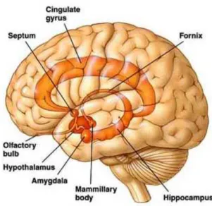
2.3.1 The limbic lobe of Broca
In a study published in 1878 by the French neurologist Paul Broca, it was described that on the medial surface of the brain of all mammals there is a group of cortical area, distinct
from the sorrounding cortex; Broca called this set of cortical areas “limbic lobe”, because they formed a ring around the brain stem (Fig. 2.1).
The limbic lobe is made from the cortex around the corpus callosum, particularly the cingulate gyrus, and the cortex on the surface of the medial temporal lobe, including the hippocampus. However, Broca did not write anything about the importance of these structures for the emotions, and they were long believed to be involved in olfactory perception (Bear, Connors, Paradiso 2002).
Fig. 2.1 The limbic system, composed by primitive areas, including the cingulate cortex, the
parahippocampal cortex and the hippocampus.
According to Papez, the function of the limbic lobe was probably more interesting than what was assumed. His argument was based largely on the neuroanatomy. Papez himself knew that the hypothalamus influences the expression of emotions; he also knew that higher cognitive functions influence emotional behavior. Papez showed that between the cerebral cortex and hypothalamus there are reciprocal connections formed by circuits that were referred to by Papez circuit. (Umiltà, 1999).
2.3.2 The Papez circuit
Papez suggested that the cortex was involved in the experience of emotions. In fact, in the presence of damage to certain areas of the cortex, serious disturbances of emotional behavior were sometimes observed. In addition, tumors located close to the cingulate cortex are associated with emotional disorders including fear, irritability and depression.