UCSF
UC San Francisco Previously Published Works
Title
ARDS Subphenotypes: Understanding a Heterogeneous Syndrome.
Permalink
https://escholarship.org/uc/item/8826s948
Journal
Critical Care, 24(1)
ISSN
1364-8535
Authors
Wilson, Jennifer G
Calfee, Carolyn S
Publication Date
2020-03-24
DOI
10.1186/s13054-020-2778-x
Peer reviewed
eScholarship.org
Powered by the California Digital Library
R E V I E W
Open Access
ARDS Subphenotypes: Understanding a
Heterogeneous Syndrome
Jennifer G. Wilson
1and Carolyn S. Calfee
2,3*Abstract
This article is one of ten reviews selected from the Annual Update in Intensive Care and Emergency Medicine 2020. Other selected articles can be found online athttps://www.biomedcentral.com/collections/annualupdate2020. Further information about the Annual Update in Intensive Care and Emergency Medicine is available fromhttp:// www.springer.com/series/8901.
Introduction
The acute respiratory distress syndrome (ARDS) is a clinical syndrome defined by acute onset hypoxemia (PaO2:FiO2 ratio < 300) and bilateral pulmonary
opaci-ties not fully explained by cardiac failure or volume overload [1]. The Berlin consensus definition of ARDS, like the American-European Consensus definition that preceded it, has enabled clinicians and researchers alike to prospectively identify patients with ARDS, implement lung protective ventilation strategies, and enroll patients in clinical trials. ARDS remains under-recognized clinic-ally, however; therapies are limited, and mortality re-mains high [2]. Under-recognition of ARDS may stem in part from the considerable clinical heterogeneity ob-served among patients who meet standard ARDS cri-teria. The syndrome may be triggered, for example, by pulmonary or extrapulmonary sepsis, aspiration, trauma, blood product transfusion, or pancreatitis. Pulmonary infiltrates can be focal or diffuse. Hypoxemia can range from mild to severe, and duration of respiratory failure can be brief or prolonged. Many of these clinical varia-tions may reflect underlying biological differences be-tween ARDS patients that are now recognized as important drivers of treatment response and ultimate outcomes.
Substantial heterogeneity within the general ARDS population has likely contributed to the failure of
experimental therapies for ARDS in recent large clinical trials, despite promising preclinical data [3]. Identifying subphenotypes of ARDS— more homogeneous groups within the general ARDS population—is one approach to untangling the clinical and biological complexity that many believe is a barrier to discovery of successful new treatments. By identifying meaningful but currently unrecognized subgroups encompassed by the broad con-sensus definition of ARDS, interventions can potentially be tested more efficiently in targeted cohorts. Selecting subphenotypes of patients at higher risk for poor out-comes for enrollment in clinical trials is called prognos-tic enrichment [4]. Selecting for patients more likely to respond to a given therapy due to the mechanism of benefit is called predictive enrichment [4]. Both enrich-ment strategies are recommended by the Food and Drug Administration to increase the efficiency of clinical trials across all fields, either by increasing the rate of the out-come of interest (prognostic enrichment) or by amplify-ing the effect size (predictive enrichment). These approaches may allow researchers to detect treatment effects in smaller cohorts, which is especially important in heterogeneous syndromes like ARDS. Ultimately, however, the discovery of ARDS subphenotypes may en-rich more than clinical trial populations: within the next decade, these innovations could help us move from a one-size-fits-all approach to ARDS treatment to more effective, tailored therapies based on the clinical and bio-logic profile of each patient.
This chapter summarizes the state of the science of subphenotyping of ARDS patients, exploring the physio-logic, clinical, and biologic characteristics that have been found to identify more homogeneous subgroups within
© Wilson and Calfee. 2020
* Correspondence:[email protected]
2
Department of Medicine, University of California, San Francisco, San Francisco, CA, USA
3Department of Anesthesia, University of California, San Francisco, San Francisco, CA, USA
this heterogeneous syndrome (Table 1), and the poten-tial implications of these advances for practicing clini-cians in the intensive care unit (ICU) and the emergency department.
ARDS Subphenotypes and Prognostic Enrichment
Prognostic enrichment in ARDS research involves select-ing patients with a higher likelihood of havselect-ing a particu-lar disease-related endpoint, such as fewer ventilator-free days or higher mortality. Beyond increasing research efficiency, identifying subphenotypes of ARDS patients at highest risk for poor outcomes may also lead to im-proved risk stratification at the bedside, allowing clini-cians to select patients more likely to benefit from inter-facility transfer for higher level of care, or early consider-ation of aggressive therapies such as extracorporeal membrane oxygenation (ECMO).
Physiologic Phenotyping for Prognostic Enrichment Risk stratification of ARDS patients is not a new strat-egy. The Berlin definition itself stratifies ARDS into three subgroups (Table 2) according to the degree of hypoxemia (mild, moderate, and severe), and mortality increases as the PaO2:FiO2ratio decreases [1]. The
ad-vantage of this approach is that the PaO2:FiO2 ratio is
available in all patients with ARDS and does not require expert interpretation or subjective clinical assessment. Multiple large clinical ARDS trials have used the PaO2:
FiO2 ratio for prognostic enrichment. For example, the
ACURASYS trial of early continuous neuromuscular blockade [5], the PROSEVA trial of prone positioning [6], and the ROSE trial reevaluating early continuous neuromuscular blockade [7] all targeted patients with moderate-to-severe ARDS (PaO2:FiO2 ratio < 150
mmHg). All three of these trials had mortality endpoints,
and all three had mortality rates in the control arms that exceeded 40%.
In addition to the PaO2:FiO2 ratio, several other
physiologic variables are known to predict poor out-comes in ARDS. Dead space fraction [8], ventilatory ra-tio (a simple bedside index of impaired ventilara-tion) [9], and driving pressure (a measurement of respiratory sys-tem compliance) are all independently associated with poor outcomes in ARDS [10] and more routine meas-urement of these variables could improve prognostic en-richment in clinical trials and risk prediction in clinical practice.
One limitation of using these physiologic measure-ments, however, is that these variables can rapidly change. The application of higher positive end-expiratory pressure (PEEP), for example, could rapidly move a patient from one subgroup of ARDS severity to another, or a patient who aspirates at the time of intub-ation may develop severe hypoxemia that improves within hours. Perhaps more fundamentally, common physiologic characteristics in most cases do not capture important differences in biology between ARDS patients. A patient with transfusion-associated ARDS may have the same PaO2:FiO2ratio or driving pressure as a patient
with ARDS from H1N1 influenza, but their underlying pathophysiology may be very different, and they do not have the same risk of poor outcomes. Indeed, the Berlin definition of ARDS is far from perfect as a predictor of mortality, with an area under the curve of only 0.577 [1]. Clinical Phenotyping for Prognostic Enrichment
Recognizing the limitations of a purely physiologic ap-proach to subphenotyping ARDS patients, investigators have also examined various clinical variables to enhance prognostic enrichment (Table 1). For example, patients with ARDS following trauma have been found to be at lower risk of death than non-trauma patients with ARDS (odds ratio 0.44) [11]. Luo et al. found that despite over-all similar mortality rates, predictors of mortality differ between direct (pulmonary trigger) and indirect (extra-pulmonary trigger) ARDS [12]. ARDS patients with acute kidney injury (AKI) have been shown to have sig-nificantly higher mortality than patients without AKI in several cohorts [13,14]. Thus, when attempting to iden-tify high-risk ARDS patients, non-trauma patients and patients with significant AKI are a higher-risk subset, but predictors of mortality differ depending on whether the lung injury is direct or indirect.
Beyond baseline clinical characteristics, the time-course of ARDS is another factor that can identify pa-tients at greater risk of poor outcomes. Both time of on-set and duration of disease appear to hold prognostic value. Several studies have shown that ARDS onset >48 h after ICU admission is associated with higher mortality Table 1Examples of factors used for identifying subphenotypes
of the acute respiratory distress syndrome (ARDS)
Physiologic Clinical Biologic PaO2:FiO2 Trauma vs. medical Genomic Dead space fraction Direct vs. indirect Transcriptomic Driving pressure Focal vs. diffuse Proteomic
±Acute kidney injury Metabolomic
Table 2The Berlin Definition of ARDS categorizes patients according to the severity of their oxygenation deficit; increasing severity is associated with increased mortality [1]
Severity PaO2:FiO2ratio (mmHg) Patients (%) Mortality (%)
Mild 201–300 22 27
Moderate 101–200 50 32
Severe ≤100 28 45
[15, 16]. Not surprisingly, rapidly improving ARDS (ARDS that resolves within 1 day) has a better prognosis than persistent ARDS. More interesting, however, is the finding that most (63%) patients with rapidly improving ARDS present with moderate or severe hypoxemia [17], highlighting the limitations of using the PaO2:FiO2ratio
alone to identify patients for enrollment in clinical trials. Recognizing this issue, the PROSEVA trial of prone po-sitioning only enrolled patients if they continued to meet inclusion criteria (PaO2:FiO2 ratio < 150 mmHg) after
12–24 h of stabilization [7].
Radiographic patterns of pulmonary infiltrates have also been used to sort ARDS patients into more homo-geneous subgroups and identify those at highest risk of death, either alone or in combination with other physio-logic and clinical variables. One small prospective study found that ARDS patients with non-focal infiltrates had higher mortality compared to patients with focal radio-graphic findings [18]. The CESAR trial of ECMO for se-vere ARDS required a Murray Lung Score (which incorporates chest radiography) of >3 for eligibility (or a pH < 7.20) [19,20]. More recently, the RALE score— de-veloped to systematically quantify the extent and density of alveolar infiltrates on chest radiograph—has been shown to predict 28-day mortality with an area under the curve of 0.82 [21].
An apparent drawback to relying exclusively on clin-ical characteristics for phenotyping, however, is the po-tential for misclassification. In a recent trial of mechanical ventilation personalized according to the presence of focal vs. diffuse infiltrates in ARDS (dis-cussed further later), 21% of the radiographic subpheno-types assigned at the time of randomization were misclassified [22]. Similarly, investigators have found it difficult to classify patients as having direct or indirect ARDS, with 37% of cases deemed unclassifiable in one trial [23]. Pragmatic and reliable approaches to classifica-tion are needed to overcome the challenges inherent to clinical phenotyping.
Biologic Phenotyping for Prognostic Enrichment
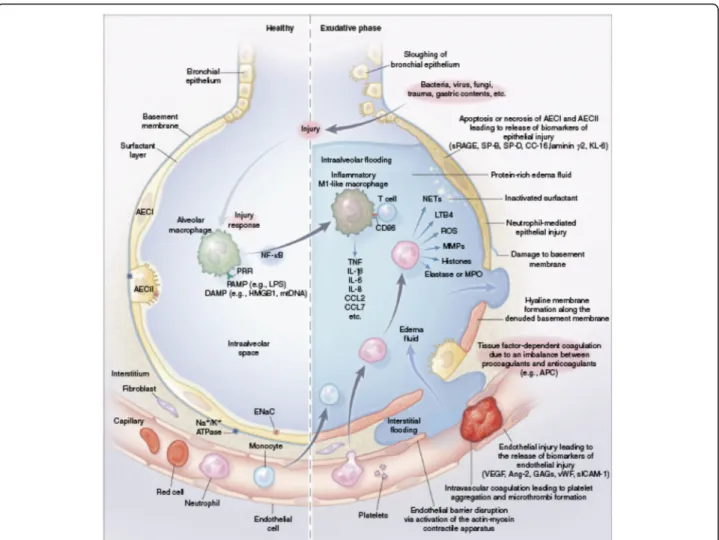
It follows that there has been growing interest in identi-fying biologic subphenotypes of ARDS patients. Biologic markers are considered proximal to the clinical expres-sion of ARDS, and potentially less prone to problems with misclassification that make clinical phenotyping so challenging. Moreover, our understanding of ARDS biol-ogy has advanced greatly in the past decade. We now better understand how an initial insult causes an inflam-matory cascade that results in further injury to the al-veolus and its microvasculature (Fig. 1) [3]. Measuring plasma biomarkers in ARDS can help find subgroups of patients that share important host-response features and/or that have worse clinical outcomes. Numerous
genomic, transcriptomic, proteomic, and metabolomic factors have been studied for this purpose, with the greatest depth of research focused on plasma protein biomarkers of ARDS. These include markers of systemic inflammation (interleukin [IL]-6, IL-8, soluble tumor ne-crosis factor [TNF] receptor-1, IL-18), epithelial injury (angiopoietin-2, intercellular adhesion molecule-1), endothelial injury (soluble receptor for advanced glyca-tion end products [sRAGE], surfactant protein-D), and disordered coagulation (plasminogen activator inhibitor-1, protein C), all of which have been shown to hold prognostic value [24]. Baseline levels of sRAGE, for ex-ample, independently predicted 90-day mortality in one meta-analysis [25]. More recently, Rogers et al. found that elevations in baseline plasma IL-18 levels and rising IL-18 levels were both associated with increased mortal-ity in sepsis-induced ARDS [26].
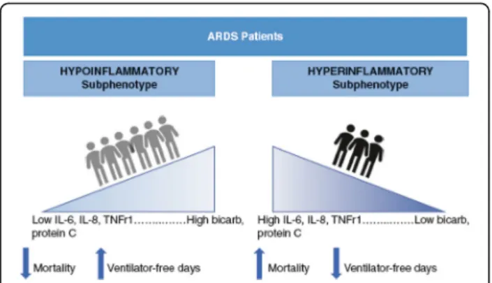
Using an approach to identify subgroups within a het-erogeneous population called latent class analysis (LCA), two distinct subphenotypes of ARDS were identified based on combined clinical and biologic data from pa-tients enrolled in two large clinical trial cohorts [27]. The “hyperinflammatory” subphenotype was character-ized by enhanced inflammation, fewer ventilator-free days, and increased mortality compared to the “ hypoin-flammatory” subphenotype (Fig. 2). These two subphe-notypes have been found in subsequent independent analyses of multiple other ARDS trial cohorts, and the poor prognosis associated with the hyperinflammatory phenotype persists [28, 29]. Using a different approach, called hierarchical clustering, to analyze a panel of plasma biomarkers from ARDS patients, Bos et al. iden-tified two similar subphenotypes: a “reactive” subpheno-type characterized by greater inflammation and increased mortality and an “uninflamed” subphenotype associated with better outcomes [30]. Taken together, these findings support the idea that ARDS patients can be stratified according to markers of inflammation for prognostic enrichment.
The focus on proteomic profiling of ARDS patients has been paralleled by interest in genomic, transcrip-tomic, and metabolomic subphenotypes of ARDS, but less progress has been made in terms of prognostic en-richment with these strategies. Meyer et al. identified an IL-1RN coding variant that increased risk of developing ARDS in sepsis [31], and Zhu et al. found certain micro-RNAs to be risk biomarkers for ARDS among critically ill adults [32], but genomic and transcriptomic subphe-notyping of patients with established ARDS remains largely unexplored. In a small cohort of patients with ARDS, Rogers et al. found a subset of patients with a distinct metabolic profile with higher levels of numerous metabolites in undiluted pulmonary edema fluid [33]. This “high metabolite” subphenotype was associated
with higher mortality but, in part because of the chal-lenges associated with analyzing pulmonary edema fluid, these findings have yet to be reproduced in a larger cohort.
Indeed, measurement of protein biomarkers and “-omics”data in ARDS patients is not currently available outside of the research setting. Furthermore, the impact of identifying biologic subphenotype on downstream outcomes in ARDS patients has not been prospectively evaluated. Investigators have recently identified a “ parsi-monious”model that classifies ARDS patients as“hypo-” or “hyperinflammatory” using only three plasma bio-markers (IL-8, bicarbonate and protein C) [34], and rapid analysis of biomarkers for identification of ARDS
subphenotype at the point of care is now being piloted. Development of rapid assays is a critical step in lever-aging the identification of ARDS subphenotypes for prognostic enrichment in future clinical trials, and ul-timately bringing these discoveries to the bedside. ARDS Subphenotypes and Predictive Enrichment
In parallel with these different strategies for identifying subgroups and phenotypes for prognostic enrichment in ARDS, investigators have also studied how treatment ef-fects vary by subphenotype. By finding a subphenotype-specific treatment response retrospectively or targeting treatment based on mechanism and biologic features, re-searchers can then go on to prospectively test new
Fig. 1Pathobiology of the exudative phase of ARDS. The healthy alveolar-capillary unit (left) and the exudative phase of ARDS (right).AECItype I alveolar epithelial cell,AECIItype II alveolar epithelial cell,Ang-2angiopoietin-2,APCactivated protein C,CC-16club cell (formerly Clara cell) secretory protein 16,CCLchemokine (CC motif) ligand,DAMPdamage-associated molecular pattern,ENaCepithelial sodium channel,GAG glycosaminoglycan,HMGB1high-mobility group box 1 protein,KL-6Krebs von den Lungen 6,LPSlipopolysaccharide,LTB4leukotriene B4,MMP matrix metalloproteinase,MPOmyeloperoxidase,mtDNAmitochondrial DNA,Na+/K+ATPasesodium-potassium ATPase pump,NF-κBnuclear factor kappa light-chain enhancer of activated B cells,NETneutrophil extracellular trap,PAMPpathogen-associated molecular pattern,PRRpattern recognition receptor,ROSreactive oxygen species,sICAMsoluble intercellular adhesion molecule,SPsurfactant protein,sRAGEsoluble receptor for advanced glycation end products,TNFtumor necrosis factor,VEGFvascular endothelial growth factor,vWFvon Willebrand factor. (Reused from [3] with permission)
interventions in patients who are more likely to respond. This approach provides predictive enrichment: by ampli-fying treatment response, the power to detect a benefit from experimental therapies increases, and discovery be-comes more efficient. Just as biologic phenotyping of other diseases, such as breast cancer or asthma, has led to important improvements in patient outcomes, the eventual goal is to deploy targeted therapies for ARDS according to patient characteristics, moving the field from protocolized care to precision medicine.
Physiologic Phenotyping for Predictive Enrichment While many view physiologic parameters such as PaO2:
FiO2ratio as purely prognostic indicators, they may
pro-vide predictive enrichment as well. As Prescott et al. note in their discussion of ARDS clinical trial strategies, a lower PaO2:FiO2 ratio not only identifies patients at
higher risk of death, but also reflects patients with greater lung weight who may be more likely to benefit from recruitment maneuvers, higher PEEP or prone po-sitioning [35]. Similarly, one could hypothesize that among patients with severe ARDS, those who have a plateau pressure >30 cmH2O or an unfavorable driving
pressure despite adherence to a lung-protective ventila-tion strategy may be more likely to benefit from “lung rest”with ultra-low tidal volumes on ECMO.
Clinical Phenotyping for Predictive Enrichment
As discussed above, difficulty classifying patients is a pragmatic challenge inherent to clinical phenotyping of ARDS patients, and a major drawback to the use of clin-ical phenotyping in both research and practice. Nonethe-less, different clinical subphenotypes of ARDS may reflect different underlying biology, and in some cases have been shown to respond differently to treatment. For example, patients with direct ARDS have higher
levels of biomarkers of epithelial injury than patients with indirect ARDS [36], and there is low-level evidence that patients with direct ARDS may respond differently to recruitment maneuvers and glucocorticoids than in-direct lung injury patients [22, 37–39]. A recent, highly innovative randomized controlled trial compared a per-sonalized mechanical ventilation strategy selected ac-cording to radiographic subphenotype (focal vs. non-focal) to standard low tidal volume ventilation in 400 pa-tients with moderate-to-severe ARDS (LIVE trial) [22]. The intention-to-treat analysis found no difference in outcomes between groups, but in a post hoc analysis that excluded misclassified patients (21% of total pa-tients), there was a mortality benefit to the personalized mechanical ventilation intervention. These results high-light both the promise and the peril of using subpheno-type (in this case radiographic subphenosubpheno-type) to guide therapy: on the one hand, if patients are appropriately classified, there may be a benefit of personalized care over protocolized care; on the other hand, the significant misclassification that occurs even in the relatively con-trolled setting of a clinical trial can completely offset the potential benefit. Regardless of this complexity, the LIVE trial was a first step in the direction of what many view as the future of ARDS research: leveraging subphenotype to personalize therapy and directly comparing outcomes to standard protocolized care.
Biologic Phenotyping for Predictive Enrichment
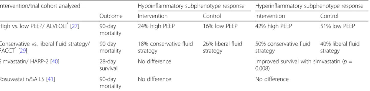
No trials have yet used biologic phenotyping pre-randomization, because bedside testing of biomarkers is not widely available. There is, however, mounting evi-dence that biologic phenotype predicts treatment re-sponse. In retrospective analyses, the hypo- and hyperinflammatory phenotypes discussed above have been observed to have differential treatment responses to several different interventions, including PEEP and fluid management strategies (Table 3) [27, 29]. Subphe-notypic differences in response to simvastatin were also observed in reanalysis of one clinical trial (HARP-2 trial) [40], but not in a similar reanalysis of a separate trial of rosuvastatin for ARDS (SAILS trial) [41]. While it is pos-sible this discrepancy reflects differences in trial design (and the particular statin that was tested), it also high-lights the uncertainty that remains when differential treatment response has been observed only retrospectively.
The use of metabolomics, transcriptomics, and gen-omics for ARDS phenotyping and predictive enrichment is in even earlier stages than proteomic phenotyping. Bos et al. used the “uninflamed” and “reactive” subphe-notypes they had previously identified based on plasma protein biomarkers to test whether there were differ-ences in blood leukocyte gene expression between
Fig. 2The hypoinflammatory and hyperinflammatory
subphenotypes of ARDS are associated with different biomarkers and outcomes. These two distinct subphenotypes have been identified by Calfee et al. in multiple previous ARDS clinical trial cohorts [27,29,40,41].ILinterleukin,bicarbbicarbonate,TNFr1 tumor necrosis factor receptor 1
groups, and found that approximately one-third of genes were differentially expressed. Specifically, there was up-regulation of oxidative phosphorylation genes in the“ re-active”subphenotype, leading the authors to suggest that for patients in this group, interventions focused on this pathway should be explored [42]. While these data cer-tainly merits further investigation, translating biologic associations into effective treatments based on mechan-ism is by no means straightforward. For example, a retrospective analysis of a previous negative clinical trial of recombinant IL-1 receptor antagonist for sepsis found a treatment benefit in the subset of patients withhigher levels of baseline IL-1 receptor antagonist, arguably a completely counterintuitive result [43].
The benefit of biologically tailored precision therapies for ARDS remains theoretical. The translation of the in-sights gained from studying subphenotypes of ARDS into targeted therapies based on mechanism—and com-parison of this precision approach to standard protoco-lized management—is the next frontier in ARDS research.
Beyond ARDS: Subphenotypes in Other Heterogeneous Syndromes
As mentioned earlier, the search for subphenotypes in ARDS is motivated in part by improvements in the treatment of other heterogeneous diseases gained by using a similar approach. Oncologic therapies in par-ticular are increasingly guided by molecular pheno-type, and this strategy has improved survival substantially even in patients with advanced disease. Survival with metastatic melanoma, for example, has improved significantly since the advent of checkpoint inhibitors and therapies targeting the BRAF V600 mu-tation [44]. Asthma therapy has also been changed by the identification of clinically significant subpheno-types: patients with severe, uncontrolled eosinophilic asthma have been found to have fewer exacerbations with monoclonal antibodies aimed at reducing eosino-phil activation [45].
Subphenotypes have also been identified in sepsis, an-other heterogeneous syndrome in the critically ill that has historically been treated with a protocolized ap-proach. Wong et al. have developed a biomarker-based mortality risk model for pediatric sepsis, as well as gene-expression-based subphenotypes of pediatric septic shock. In a retrospective analysis of a cohort of pediatric patients with septic shock, they found that among inter-mediate- and high-risk patients, corticosteroids were as-sociated with a more than tenfold reduction in the risk of a complicated course in one subphenotype but not the other [46]. Similar subphenotyping of adult sepsis is an area of active study. Gårdlund et al. used latent class analysis to identify six distinct subphenotypes of septic shock using clinical data from a previous large clinical trial cohort [47]. Seymour et al. used a different ap-proach (machine learning applied to electronic health record data) and identified four subphenotypes with dif-ferent genetic and inflammatory markers and markedly different mortality rates [48].
Finally, there is a small but growing body of evidence that there are meaningful subphenotypes within the het-erogeneous post-cardiac arrest syndrome, beyond type of arrest and initial post-resuscitation neurologic status. For example, Bro-Jeppesen et al. have reported that IL-6, a marker of systemic inflammation, is correlated with poor prognosis in comatose patients resuscitated from out-of-hospital cardiac arrest [49]. Anderson et al. found that patients with post-resuscitation shock who had a preserved left ventricular ejection fraction (LVEF) had less favorable neurologic outcomes, increased organ fail-ure, and higher mortality compared to patients with de-pressed LVEF [50]. In addition, patients in the preserved LVEF group exhibited a subtype-specific response to early fluid resuscitation, with lower mortality and im-proved neurologic outcomes associated with larger vol-ume fluid resuscitation that was not observed in the group with depressed LVEF. These findings suggest that there are identifiable subphenotypes in post-cardiac ar-rest syndrome, and that subphenotypes may be import-ant drivers of variable treatment response.
Table 3Subphenotype-specific treatment response in the reanalyses of outcomes in four different clinical ARDS trials
Intervention/trial cohort analyzed Hypoinflammatory subphenotype response Hyperinflammatory subphenotype response Outcome Intervention Control Intervention Control High vs. low PEEP/ ALVEOLI*[27] 90-day
mortality
24% high PEEP 16% low PEEP 42% high PEEP 51% low PEEP
Conservative vs. liberal fluid strategy/
FACCT*[29] 90-daymortality 18% conservative fluidstrategy 26% liberal fluidstrategy 50% conservative fluidstrategy 40% liberal fluidstrategy Simvastatin/ HARP-2 [40] 28-day
survival
No difference Improved survival with simvastatin (p= 0.008)
Rosuvastatin/SAILS [41] 90-day mortality
No difference No difference
PEEPpositive end-expiratory pressure;∗pvalue <0.05 for interaction between treatment and subphenotype
Conclusion
The armamentarium of therapies for patients with ARDS remains limited and mortality remains high. While nega-tive results in several large randomized controlled trials of new treatments for ARDS have frustrated many, they have also motivated multiple novel approaches to under-standing the clinical and biologic heterogeneity among ARDS patients. The identification of meaningful ARDS subphenotypes—and the ways in which their outcomes and treatment responses differ—promises prognostic and predictive enrichment for future trials. Prospective evaluation of methods for reliable phenotyping at the point of care is a crucial next step in translating these discoveries into new personalized therapies for ARDS. Ultimately, identification of ARDS subphenotypes may help fulfill the aspiration of precision critical care for ARDS: replacing blunt interventions aimed at all patients who meet diagnostic criteria with therapies tailored to the clinical and biologic profile of each patient.
Acknowledgements Not applicable. Authors’contributions
Both JGW and CSC contributed to the conception and design of this work, drafted and revised the manuscript, and approved the submitted version. Funding
Publication was funded by NHLBI R35 grant (HL140026). The funding agency had no role in the writing of this manuscript.
Availability of data and materials Not applicable.
Ethics approval and consent to participate Not applicable.
Consent for publication Not applicable. Competing interests
CSC receives grant funding from NIH, FDA, DOD, Bayer, Roche/Genentech, and has worked as a consultant for Bayer, Roche/Genentech, CSL Behring, Prometic, and Quark. JGW receives grant funding from NIH.
Author details
1
Department of Emergency Medicine, Stanford University, Palo Alto, CA, USA. 2Department of Medicine, University of California, San Francisco, San Francisco, CA, USA.3Department of Anesthesia, University of California, San Francisco, San Francisco, CA, USA.
References
1. Definition Task Force ARDS, Ranieri VM, Rubenfeld GD, et al. Acute respiratory distress syndrome: the Berlin definition. JAMA. 2012;307:2526–33. 2. Bellani G, Laffey JG, Pham T, et al. Epidemiology, patterns of care, and
mortality for patients with acute respiratory distress syndrome in intensive care units in 50 countries. JAMA. 2016;315:788–800.
3. Thompson BT, Chambers RC, Liu KD. Acute respiratory distress syndrome. N Engl J Med. 2017;377:562–72.
4. FDA. Draft guidance: enrichment strategies for clinical trials to support approval of human drugs and biological products. Available fromhttps:// www.fda.gov/media/121320/download. Accessed 27 Aug 2019.
5. Papazian L, Forel JM, Gacouin A, et al. Neuromuscular blockers in early acute respiratory distress syndrome. N Engl J Med. 2010;363:1107–16. 6. Guerin C, Reignier J, Richard JC, et al. Prone positioning in severe acute
respiratory distress syndrome. N Engl J Med. 2013;368:2159–68. 7. Moss M, Huang DT, Brower RG, et al. Early neuromuscular blockade in the
acute respiratory distress syndrome. N Engl J Med. 2019;380:1997–2008. 8. Nuckton TJ, Alonso JA, Kallet RH, et al. Pulmonary dead-space fraction as a
risk factor for death in the acute respiratory distress syndrome. N Engl J Med. 2002;346:1281–6.
9. Sinha P, Calfee CS, Beitler JR, et al. Physiological analysis and clinical performance of the ventilatory ratio in acute respiratory distress syndrome. Am J Respir Crit Care Med. 2019;199:333–41.
10. Amato MB, Meade MO, Slutsky AS, et al. Driving pressure and survival in the acute respiratory distress syndrome. N Engl J Med. 2015;372:747–55. 11. Calfee CS, Eisner MD, Ware LB, et al. Trauma-associated lung injury differs
clinically and biologically from acute lung injury due to other clinical disorders. Crit Care Med. 2007;35:2243–50.
12. Luo L, Shaver CM, Zhao Z, et al. Clinical predictors of hospital mortality differ between direct and indirect ARDS. Chest. 2017;151:755–63. 13. Liu KD, Glidden DV, Eisner MD, et al. Predictive and pathogenetic value of
plasma biomarkers for acute kidney injury in patients with acute lung injury. Crit Care Med. 2007;35(12):2755–61.
14. McNicholas BA, Rezoagli E, Pham T, et al. Impact of early acute kidney injury on management and outcome in patients with acute respiratory distress syndrome: a secondary analysis of a multicenter observational study. Crit Care Med. 2019;47:1216–25.
15. Liao KM, Chen CW, Hsiue TR, Lin WC. Timing of acute respiratory distress syndrome onset is related to patient outcome. J Formos Med Assoc. 2009; 108:694–703.
16. Zhang R, Wang Z, Tejera P, et al. Late-onset moderate to severe acute respiratory distress syndrome is associated with shorter survival and higher mortality: a two-stage association study. Intensive Care Med. 2017;43:399–407. 17. Schenck EJ, Oromendia C, Torres LK, Berlin DA, Choi AMK, Siempos II.
Rapidly improving ARDS in therapeutic randomized controlled trials. Chest. 2019;155:474–82.
18. Mrozek S, Jabaudon M, Jaber S, et al. Elevated plasma levels of sRAGE are associated with nonfocal CT-based lung imaging in patients with ARDS: a prospective multicenter study. Chest. 2016;150:998–1007.
19. Murray JF, Matthay MA, Luce JM, Flick MR. An expanded definition of the adult respiratory distress syndrome. Am Rev Respir Dis. 1988;138:720–3. 20. Peek GJ, Mugford M, Tiruvoipati R, et al. Efficacy and economic assessment
of conventional ventilatory support versus extracorporeal membrane oxygenation for severe adult respiratory failure (CESAR): a multicentre randomised controlled trial. Lancet. 2009;374:1351–63.
21. Warren MA, Zhao Z, Koyama T, et al. Severity scoring of lung oedema on the chest radiograph is associated with clinical outcomes in ARDS. Thorax. 2018;73:840–6.
22. Constantin JM, Jabaudon M, Lefrant JY, et al. Personalised mechanical ventilation tailored to lung morphology versus low positive end-expiratory pressure for patients with acute respiratory distress syndrome in France (the LIVE study): a multicentre, single-blind, randomised controlled trial. Lancet Respir Med. 2019;7:870–80.
23. Thille AW, Richard JC, Maggiore SM, et al. Alveolar recruitment in pulmonary and extrapulmonary acute respiratory distress syndrome: comparison using pressure-volume curve or static compliance. Anesthesiology. 2007;106:212–7.
24. Walter JM, Wilson J, Ware LB. Biomarkers in acute respiratory distress syndrome: from pathobiology to improving patient care. Expert Rev Respir Med. 2014;8:573–86.
25. Jabaudon M, Blondonnet R, Pereira B, et al. Plasma sRAGE is independently associated with increased mortality in ARDS: a meta-analysis of individual patient data. Intensive Care Med. 2018;44:1388–99.
26. Rogers AJ, Guan J, Trtchounian A, et al. Association of elevated plasma interleukin-18 level with increased mortality in a clinical trial of statin treatment for acute respiratory distress syndrome. Crit Care Med. 2019;47: 1089–96.
27. Calfee CS, Delucchi K, Parsons PE, et al. Subphenotypes in acute respiratory distress syndrome: latent class analysis of data from two randomised controlled trials. Lancet Respir Med. 2014;2:611–20.
28. Delucchi K, Famous KR, Ware LB, et al. Stability of ARDS subphenotypes over time in two randomised controlled trials. Thorax. 2018;73:439–45.
29. Famous KR, Delucchi K, Ware LB, et al. Acute respiratory distress syndrome subphenotypes respond differently to randomized fluid management strategy. Am J Respir Crit Care Med. 2017;195:331–8.
30. Bos LD, Schouten LR, van Vught LA, et al. Identification and validation of distinct biological phenotypes in patients with acute respiratory distress syndrome by cluster analysis. Thorax. 2017;72:876–83.
31. Meyer NJ, Feng R, Li M, et al. IL1RN coding variant is associated with lower risk of acute respiratory distress syndrome and increased plasma IL-1 receptor antagonist. Am J Respir Crit Care Med. 2013;187:950–9. 32. Zhu Z, Liang L, Zhang R, et al. Whole blood microRNA markers are associated
with acute respiratory distress syndrome. Intensive Care Med Exp. 2017;5:38–50. 33. Rogers AJ, Contrepois K, Wu M, et al. Profiling of ARDS pulmonary edema fluid identifies a metabolically distinct subset. Am J Physiol Lung Cell Mol Physiol. 2017;312:L703–9.
34. Sinha P, Delucchi KL, McAuley DF, O’Kane CM, Matthay MA, Calfee CS. Development and validation of parsimonious algorithms to classify ARDS phenotypes. Lancet Respir Med. 2020. https://doi.org/10.1016/S2213-2600(19)30369-8[Epub ahead of print].
35. Prescott HC, Calfee CS, Thompson BT, Angus DC, Liu VX. Toward smarter lumping and smarter splitting: rethinking strategies for sepsis and acute respiratory distress syndrome clinical trial design. Am J Respir Crit Care Med. 2016;194:147–55.
36. Calfee CS, Janz DR, Bernard GR, et al. Distinct molecular phenotypes of direct vs indirect ARDS in single-center and multicenter studies. Chest. 2015;147:1539–48. 37. Gattinoni L, Pelosi P, Suter PM, et al. Acute respiratory distress syndrome
caused by pulmonary and extrapulmonary disease. Different syndromes? Am J Respir Crit Care Med. 1998;158:3–11.
38. Riva DR, Oliveira MB, Rzezinski AF, et al. Recruitment maneuver in pulmonary and extrapulmonary experimental acute lung injury. Crit Care Med. 2008;36:1900–8. 39. Leite-Junior JH, Garcia CS, Souza-Fernandes AB, et al. Methylprednisolone
improves lung mechanics and reduces the inflammatory response in pulmonary but not in extrapulmonary mild acute lung injury in mice. Crit Care Med. 2008;36:2621–8.
40. Calfee CS, Delucchi KL, Sinha P, et al. Acute respiratory distress syndrome subphenotypes and differential response to simvastatin: secondary analysis of a randomised controlled trial. Lancet Respir Med. 2018;6:691–8. 41. Sinha P, Delucchi KL, Thompson BT, et al. Latent class analysis of ARDS
subphenotypes: a secondary analysis of the statins for acutely injured lungs from sepsis (SAILS) study. Intensive Care Med. 2018;44:1859–69.
42. Bos LDJ, Scicluna BP, Ong DSY, et al. Understanding heterogeneity in biological phenotypes of acute respiratory distress syndrome by leukocyte expression profiles. Am J Respir Crit Care Med. 2019;200:42–50.
43. Meyer NJ, Reilly JP, Anderson BJ, et al. Mortality benefit of recombinant human interleukin-1 receptor antagonist for sepsis varies by initial interleukin-1 receptor antagonist plasma concentration. Crit Care Med. 2018;46:21–8. 44. Silva IP, Long GV. Systemic therapy in advanced melanoma: integrating
targeted therapy and immunotherapy into clinical practice. Curr Opin Oncol. 2017;29:484–92.
45. FitzGerald JM, Bleecker ER, Nair P, et al. Benralizumab, an antiinterleukin-5 receptor alpha monoclonal antibody, as add-on treatment for patients with severe, uncontrolled, eosinophilic asthma (CALIMA): a randomised, double-blind, placebo-controlled phase 3 trial. Lancet. 2016;388:2128–41. 46. Wong HR, Atkinson SJ, Cvijanovich NZ, et al. Combining prognostic and
predictive enrichment strategies to identify children with septic shock responsive to corticosteroids. Crit Care Med. 2016;44:e1000–3.
47. Gårdlund B, Dmitrieva NO, Pieper CF, Finfer S, Marshall JC, Thompson BT. Six subphenotypes in septic shock: latent class analysis of the PROWESS shock study. J Crit Care. 2018;47:70–9.
48. Seymour CW, Kennedy JN, Wang S, et al. Derivation, validation, and potential treatment implications of novel clinical phenotypes for sepsis. JAMA. 2019;321:2003–17.
49. Bro-Jeppesen J, Kjaergaard J, Wanscher M, et al. Systemic inflammatory response and potential prognostic implications after out-of-hospital cardiac arrest: a substudy of the Target Temperature Management Trial. Crit Care Med. 2015;43:1223–32.
50. Anderson RJ, Jinadasa SP, Hsu L, et al. Shock subtypes by left ventricular ejection fraction following out-of-hospital cardiac arrest. Crit Care. 2018;22:162.
Publisher’s Note
Springer Nature remains neutral with regard to jurisdictional claims in published maps and institutional affiliations.
![Table 2 The Berlin Definition of ARDS categorizes patients according to the severity of their oxygenation deficit; increasing severity is associated with increased mortality [1]](https://thumb-us.123doks.com/thumbv2/123dok_us/512963.2560499/3.892.84.437.165.270/definition-categorizes-according-oxygenation-increasing-associated-increased-mortality.webp)