eScholarship@UMMS
eScholarship@UMMS
Cancer Concepts: A Guidebook for the
Non-Oncologist Radiation Oncology
2015-08-30
Breast Cancer
Breast Cancer
Maria Giulia Cicchetti
University of Massachusetts Medical School Et al.
Let us know how access to this document benefits you.
Follow this and additional works at: https://escholarship.umassmed.edu/cancer_concepts Part of the Cancer Biology Commons, Neoplasms Commons, Oncology Commons, Radiology Commons, and the Women's Health Commons
Repository Citation Repository Citation
Cicchetti MG, Liebmann J, Chen A, DeBenedectis C, Kurian E. (2015). Breast Cancer. Cancer Concepts: A Guidebook for the Non-Oncologist. https://doi.org/10.7191/cancer_concepts.1019. Retrieved from https://escholarship.umassmed.edu/cancer_concepts/16
Creative Commons License
This work is licensed under a Creative Commons Attribution-Noncommercial-Share Alike 4.0 License.
This material is brought to you by eScholarship@UMMS. It has been accepted for inclusion in Cancer Concepts: A Guidebook for the Non-Oncologist by an authorized administrator of eScholarship@UMMS. For more information, please contact [email protected].
M. Giulia Cicchetti, MD James Liebmann, MD Andrew Chen, MD
Carolynn DeBenedectis, MD Elizabeth Kurian, MD
Summary and Key Points
1. Breast cancer is the the most common non-skin cancer and the second leading cause of cancer death in women (after lung cancer). 2. The most common breast cancer type is ductal carcinoma followed
by lobular carcinoma.
3. Risk factors include family history, breast density, nulliparity, prolonged hormonal exposure, radiation exposure, high alcohol consumption, sedentary lifestyle and aging.
4. Early stage disease is usually asymptomatic.
5. Breast cancer screening lowers the risk of dying from breast cancer. 6. Treatment is multidisciplinary and requires close cooperation
between surgeons, radiation oncologists, and medical oncologists. 7. Breast preservation is an important goal of treatment for early stage
disease. Trials have shown that survival is equal whether a patient chooses mastectomy or lumpectomy with radiation therapy to the breast.
8. The multidisciplinary approach to therapy has improved breast cancer outcome so that most U.S. women are cured.
9. Breast cancer occurs in males but is rare.
Introduction and Etiology
Breast cancer is the most common non-skin cancer in United States (US) women and the second highest cause of cancer deaths. In 2011 the American Cancer Society estimated the annual incidence of invasive breast cancer to be 230,480 (female) and 2,140 (male) with total deaths of 39,520 (female) and 450 (male). An additional 57,650 women were diagnosed with carcinoma in situ of the breast. The lifetime risk of developing breast cancer for U.S. women is 1 in 9. The annual death rate has been declining steadily since 1990 which is attributed to both
improved detection and improvements in treatment.1 With these improvements, the net effect is most US women with breast cancer will be cured of their disease.
Male breast cancer is rare, accounting for 1% of all breast cancers. The etiology is obscure, but there is a familial association with male or female breast cancer. 1.
Risk factors for developing breast cancer in females include:
• Age over 50
• Personal or family history of breast cancer
• Nulliparity
• First child after age 30
• Early menarche
• Increased time between menarche and first child
• Exposure of the breasts to ionizing radiation before age 40
• High alcohol consumption
• Sedentary lifestyle
• Excessive weight gain
• Hormonal exposure
• Breast density
Overall, increased estrogen and progesterone exposure increases breast cancer risk. Smoking does not seem to be a risk factor. Breast cancer is more common in Caucasians, upper socioeconomic classes and people of Jewish descent. Germline mutations of BRCA1 or BRCA2 are present in 5-10% of breast cancer patients. Genetic testing for BRCA1 and BRCA2 is available. A common method to determine a woman’s risk of developing breast cancer, the modified Gail model, is available at the US National Cancer Institute (NCI) web site.
Citation: Cicchetti MG, Liebmann J, Chen A, DeBenedectis C, Kurian E. Breast Cancer. In: Pieters RS, Liebmann J, eds. Cancer Concepts: A Guidebook for the Non-Oncologist. Worcester, MA:
University of Massachusetts Medical School; 2015. doi: 10.7191/cancer_concepts.1019.
This project has been funded in whole or in part with federal funds from the National Library of Medicine, National Institutes of Health, under Contract No. HHSN276201100010C with the University of Massachusetts, Worcester.
Copyright: All content in Cancer Concepts: A Guidebook for the Non-Oncologist, unless otherwise noted, is licensed under a Creative Commons Attribution-Noncommercial-Share Alike License, http://creativecommons.org/licenses/by-nc-sa/4.0/
Screening
The mammographic findings of breast cancer are commonly a mass and/or suspicious calcifications. Mammography routinely includes 2 views of each breast: cranio-caudal (CC) and medial-lateral oblique (MLO). The mammograms are labeled noting the side and the orientation by positioning the markers on the images closest to the axilla. At least 10% of tumors are not seen on mammography, particularly lobular carcinoma in situ (LCIS) and invasive lobular carcinoma.
Early stage breast cancer is usually asymptomatic, making screening all the more important. Screening mammography for women between the ages of 50 and 70 has been shown to both shift diagnosis to an earlier stage and to reduce mortality. However, it is controversial whether the risks of mammography and its cost outweigh the reduction in mortality in women younger than 50.
In 2009, the U.S. Preventative Services Task Force issued revised guidelines for mammographic screening that recommended against screening for women < 50 years old and > 75 years old, recommended against teaching breast self-examination and recommended only biennial screening. Many experts and groups (including the American Cancer Society (ACS), The American College of Surgeons, The American College of Obstetricians-Gynecologists and the American College of Radiologists) oppose these new guidelines. Therefore, despite the new Task Service recommendations, we think it is reasonable for patients of average risk to follow the ACS Guidelines' recommendation of yearly mammograms starting at age 40 and continuing in healthy women with a >10 year life expectancy.
Pathology
The most common form of breast cancer in women is ductal carcinoma of the usual type (Figure 1 a,b) followed by lobular cancer and less frequently by other histologic subtypes (i.e. special types of ductal carcinoma such as adenoid cystic, neuroendocrine, papillary, medullary etc.). Rarely lymphomas, sarcomas and metastasis may occur in the breast. Since male breast tissue does not contain lobules, ductal carcinoma is most common type in men.
Figure 1 a,b. Mastectomy, Invasive Ductal Carcinoma. (a) The carcinoma has
ulcerated the nipple. (b) The cross-section shows the skin with ulceration (adjacent to the ruler) by the cancer which is the solid white area with irregular infiltrating borders. The fatty breast parenchyma appears yellow and the surgical margin has been inked black by the pathologist. University of Massachusetts Medical School, Department of Pathology.
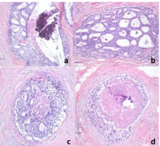
As the name implies, ductal carcinoma arises from the terminal duct and is characterized by the formation of glands (Figure 2).
Figure 2 a,b,c,d. Breast Invasive Ductal Carcinoma. (a) Poorly differentiated.
Notice that it does not form glands and has no similarity to normal breast tissue. (b) Moderately differentiated with a mixture of glands and sheets of cells. (c) Well-differentiated has well-formed glands which resemble normal. Note the cancer invasion of the adjacent fat, and atypical nuclear features noted on high magnification (d) 60x magnification. University of Massachusetts Medical School, Department of Pathology.
Breast ductal adenocarcinoma may be focal or multifocal. The precursor lesion is ductal carcinoma in situ (DCIS) and comprises 15-30% of breast cancers. DCIS can be classified into subtypes. The most common two include DCIS comedo type and noncomedo DCIS. The classic picture of DCIS comedo type is the presence of high grade pleomorphic nuclei contained within the basement membrane with central necrosis. This necrosis is referred to comedo necrosis. The necrotic cell membranes may calcify allowing screening mammography to detect these areas. Calcium, in itself, is not specific for cancer and can be found in benign proliferating glands. The more frequently encountered noncomedo DCIS includes solid, papillary, micropapillary and cribiform types (Figure 3 a, b, c, d). In these types the nuclei appear similar or clonal and can show low or high grade nuclear atypia.
Figure 3 a,b,c,d. Ductal Carcinoma In Situ (DCIS) (10x). (a) Intraductal
calcifications (purple crystalline material) which can be detected by screening mammography. (b) DCIS with a cribiform pattern with rigid “cookie cutter-like” punched out holes. (c) Cribiform DCIS with comedo necrosis. (d) Solid DCIS with comedo necrosis with high grade nuclear atypia. University of Massachusetts Medical School, Department of Pathology.
In contrast, lobular carcinoma tends to be a diffuse process that can involve both breasts at the time of asymptomatic presentation. The histology is characterized by single cells classically seen in a single file line infiltrating into the breast parenchyma (Figure 4 a, b). They can also infiltrate in single file lines around benign residual normal ducts referred to as “targetoid pattern” (Figure 4 c, d). See Figure 2 for comparison with invasive ductal carcinoma and note lobular carcinoma lacks gland formation.
Figure 4 a,b,c,d. Lobular Carcinoma. (a) The malignant cells lack any ductal
formation, but instead line up in a row like a string of pearls (10x). Higher magnification (60x) shows each of the cells in the group are relatively small. (c) Targetoid pattern, low magnification (10x) (d) Targetoid pattern, higher magnification (40x). University of Massachusetts Medical School, Department of Pathology.
The precursor lesion of lobular carcinoma in situ appears as monotonous clonal population with a well-circumscribed basement membrane. It often has the appearance of a bag of marbles with slight bulging of the borders and retains some semblance to lobular configuration (Figures 5 & 6). Unlike DCIS, LCIS does not require wide excision so the pathologic distinction is important.
Figure 5. Normal Breast Lobule (10x). University of Massachusetts Medical
School, Department of Pathology.
Figure 6. Lobular Carcinoma In Situ (10x). University of Massachusetts Medical
School, Department of Pathology.
Work-up and Staging
The work-up for early stage breast cancer includes history and physical, basic labs (cbc, chemistry profile to include liver function tests), and biopsy
for histological and hormonal evaluation. In taking the history, it is important to ask menstrual status, parity and family history. Clinical examination of the axilla can be misleading with about 30% false positive and 30% false negative results.
For invasive cancer, it is important to surgically stage the axilla with either sentinel lymph node sampling or an axillary lymph node dissection. For stage I and II patients, bone scan is not necessary (only 2% yield); for stage III, bone scan is indicated as asymptomatic metastases can be identified in 20-25%. Liver scanning is useful if abnormal LFTs, hepatomegaly or significant weight loss suggestive of hepatic metastases are present. A routine screening mammogram and a positive screening mammogram are shown below (Figures 7 & 8). Ultrasound of a palpable mass and ultrasound guided biopsy are also shown below (Figures 9 & Video 1). MRI may be helpful in complicated cases (Figure 10). It is important to remember that mammograms are screening tools. They have limitations. For example, Figure 11a demonstrates a dense breast that masks the presence of an adenocarcinoma. This mass was better identified by CT, Figure 11b.
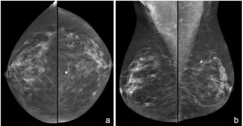
Figure 7 a,b. Screening Mammogram. Standard mammographic views are the
following (a) Craniocaudal (CC) View and (b) Mediolateral Oblique (MLO) View. University of Massachusetts Medical School, Department of Radiology.
Figure 8 a,b. Positive Screening Mammogram. The green arrow points to a
spiculated mass, biopsy proven invasive ductal carcinoma, in the upper inner posterior breast (a) MLO view and (b) compression CC view. University of Massachusetts Medical School, Department of Radiology.
Figure 9 a,b. Ultrasound of a Palpable Mass. (a) Irregular hypoechoic shadowing
right breast mass on ultrasound. (b) Yellow arrows indicate the poorly defined margins. University of Massachusetts Medical School, Department of Radiology.
Video 1. Ultrasound Guided Biopsy. University of Massachusetts Medical School,
Figure 10 a,b,c,d. Contrast Enhanced MRI of the Breast. The green arrow points
to an enhancing mass in the lower outer left breast on subtraction (a) and MIP images (b). The mass demonstrates mixed kinetics (c & d). The kinetic curve (d) shows initial rapid uptake of contrast from 0 to 173% and progressive washout of contrast over time. This pattern of contrast uptake and washout statistically favors a malignancy. University of Massachusetts Medical School, Department of Radiology.
Figure 11 a,b. (a) Standard MLO View. (b) Breast CT of the same patient. Green
arrow points to a biopsy proven adenocarcinoma that is obscured by normal glandular tissue on the routine mammogram. Breast CT was recently approved by the FDA with specific restrictions for use only as a diagnostic imaging tool. This modality does not have FDA approval for screening at this time but it offers exciting avenues of approach in the fight against breast cancer. Images contributed by Drs. Andrew Karellas and Srinivasan Vedantham, University of Massachusetts Medical School, Department of Radiology, from research supported in part by NCI grant, 1R21 CA134128-01A2.
Stage information for Breast Cancer can be found in the AJCC Staging manual and at the NCI- Breast Cancer Treatment (PDQ).2. Figures 12-16 are three-dimensional staging images of breast cancer.
Figure 12. Breast Cancer, T2. University of Massachusetts Medical School,
Department of Radiation Oncology.
Figure 13. Breast Cancer, T2N1 Solitary axillary node. University of
Massachusetts Medical School, Department of Radiation Oncology.
Figure 14. Breast Cancer, T2N2, Matted axillary nodes. University
of Massachusetts Medical School, Department of Radiation Oncology.
Figure 15. Breast Cancer, T2N3, Axillary & internal mammary nodes. University of
Figure 16. Breast Cancer, T4dN3. Inflammatory carcinoma, axillary & internal
mammary nodes. University of Massachusetts Medical School, Department of Radiation Oncology.
Oncologists have long used simple drawings of the body to draw diagrams of the extent of a patient’s disease. These drawings, referred to as staging diagrams, are useful tools to demonstrate extent of disease and facilitate treatment planning by clarifying the thought process. The process of filling out staging diagrams is a good way to learn staging systems. Interactive staging diagrams for breast cancer are available here.
Prognostic Factors
Poor prognostic factors include higher stage (larger tumor sizes and nodal involvement), higher grade, lymphatic vessel invasion (LVI), age < 40, negative estrogen receptor/progesterone receptor (ER/PR) status, and overexpression of human epidermal growth factor receptor 2 (HER2). Figure 17 a & b demonstrate immunohistochemical (IHC) stains for ER and PR.
HER2/neu is a transmembrane tyrosine kinase receptor on chromosome 17q12. Amplification of HER2 is associated with poor prognosis, early relapse, and shorter disease-free survival, therefore accurate detection is necessary for treatment. Figure 18 is an example of IHC staining for
HER2/neu. This is a screening study, since a majority of cases of breast cancer cases scoring HER2 weak positive (2+) are not actually Her2/neu positive. This study requires confirmation by fluorescence in‐situ hybridisation (FISH). Figure 19 a & b show FISH test results for normal breast cells (Figure 19a) and HER2/neu+ cells (Figure 19b).
Figure 17 a,b. Immunohistochemical Stains for the Estrogen and Progesterone
Receptor. For either stain, cells are positive if there is nuclear staining. A standardized pathology report will also include the percentage of positive cells. (a) Low power magnification of ER stain (b) High power of PR stain. University of Massachusetts Medical School, Department of Pathology.
Figure 18. Immunohistochemical Staining for HER2/neu. In contrast to ER/PR
circumferential and strong membranous staining of most cells for a positive interpretation. Any equivocal cases are triaged for fluorescent in-situ hybridization (see next figure). University of Massachusetts Medical School, Department of Pathology.
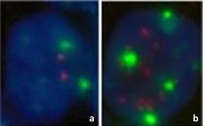
Figure 19 a,b. FISH of Invasive Breast Ductal Adenocarcinoma. (a) Normal cell:
The green signals indicate 2 copies of chromosome 17 and the two red signals are HER2. (b) Breast ductal carcinoma cell with multiple HER2 signals that proportionally exceed the number of chromosome 17 signals, indicates amplification of HER2/neu and can be interpreted as a positive HER2/neu test. University of Massachusetts Medical School, Department of Pathology.
Principles of Treatment
Treatment is multidisciplinary and requires close cooperation between surgeons, radiation oncologists and medical oncologists. Patients also meet with behavioral medicine, and if indicated, plastic surgeons and genetic counselors. Each patient’s individual care is discussed at the Multidisciplinary Breast Cancer Tumor Board, at which time the radiologists and pathologists review the imaging and pathology and a management plan is decided.
Treatment for invasive cancers begins with accurately staging the patient. If metastatic disease is present, systemic therapy becomes the mainstay of treatment. For M0 patients, local therapy and sequencing of therapy
hinge on operability. Locally advanced breast cancers are best served with
preoperative chemotherapy (usually referred to as neoadjuvant
chemotherapy). For early stage disease, stages I and II, surgery is generally employed first, and the choice is between modified radical mastectomy or breast preserving surgery or lumpectomy plus radiation therapy (RT). Six randomized prospective trials comparing mastectomy to breast preserving surgery plus RT have shown that survival is identical between these two choices. Trials have also shown that local failure in the breast is markedly reduced by the addition of radiation therapy over lumpectomy alone. Adjuvant chemotherapy and/or endocrine therapy can further improve local control and also adds a significant survival advantage.
In making the choice between mastectomy and breast preservation with RT, several factors are important. It helps if the patient can be easily followed for early recurrence detection – e.g. films are easy to read and the lesion was readily palpable. Relative contraindications to breast preservation plus RT include pregnancy, multicentric disease, diffuse malignant calcifications throughout the breast, collagen vascular disease, prior RT or persistently positive margins.
Surgery
Surgery is important in establishing diagnosis and removing gross disease. For DCIS, surgery is typically confined to the breast and options include lumpectomy or mastectomy. For invasive disease it becomes important to sample the axilla. If the axilla is clinically negative the patient is a candidate for sentinel lymph node sampling (SNB). If the axilla is involved with tumor then an axillary dissection can be important to both determine the exact number of involved nodes and for local control of the axilla. A current, ongoing research focus concerns the treatment of low volume, clinically occult axillary disease. A recent randomized trial has shown no difference in survival in patients who had a sentinel node biopsy with no further axillary dissection compared with patients who had an axillary dissection in addition to a sentinel node biopsy, regardless of whether the sentinel node was involved with cancer.3 This trial was limited to patients with smaller tumors, clinically uninvolved nodes and all patients received whole breast RT which a subsequent analysis of the RT data demonstrated included axillary RT in a high proportion of patients.4 Another recently published trial randomized patients with clinical N0 disease found on SNB to have one or two cancer containing nodes to
axillary radiotherapy or node dissection and found that while both arms provided excellent and statistically identical axillary control, morbidity was higher in the dissection arm.5. These trials support that axillary
radiotherapy may be a utilized instead of axillary dissection for low volume sentinel node biopsy detected disease.
Radiation Therapy (RT)
RT after surgery kills any cancer cells that could remain in the breast, the chest wall, or nodal basins. RT for early stage disease can allow for breast preserving surgery instead of mastectomy. In Phase III breast trials for DCIS, moderate dose RT has been shown to eradicate residual microscopic disease in the breast. This reduces the chance that the cancer will come back, thus allowing patients the ability to conserve the breast. Likewise, Phase III trials for stage I-II breast cancer have proven that adding RT to lumpectomy dramatically reduces breast cancer recurrence in the breast over lumpectomy alone, and that survival is identical between mastectomy and lumpectomy plus RT. Post-mastectomy RT (PMRT) is indicated for stage III disease (high risk patients with cancers > 5 cm or involving lymph nodes). RT is also used for recurrent breast cancer and in stage IV disease to palliate symptomatic sites. The most common way RT is delivered is via external beam using either photons or electrons. A typical curative course lasts 6 weeks, with daily treatments delivered Monday- Friday, and about 15 minutes is required per treatment. Ongoing trials are assessing the value of shorter course partial breast irradiation. In the palliative setting, RT can be delivered in a much shorter course, typically over 2 weeks.
RT is carefully planned and delivered. Treatment planning CT scans with either 3D or IMRT treatment planning allow precise targeting and minimize dose to adjacent normal structures. Common side effects of RT include temporary fatigue and skin changes that mimic sunburn.
Chemotherapy
Many randomized trials, dating back over four decades and involving tens of thousands of women have definitively shown that post-operative (adjuvant) chemotherapy or hormonal therapy can prevent breast cancer recurrence and lower the risk of dying from breast cancer in women with completely resected Stage I-III disease. The Early Breast Cancer Trialists Collaborative Group (EBCTCG) publishes a large overview and
meta-analysis of drug treatment of early stage breast cancer about every five years. The most recent update6 showed that post-operative chemotherapy lowers the risk of dying from recurrent breast cancer by 38% in women younger than 50 and by 20% in women older than 50. In women with estrogen receptor positive tumors, tamoxifen lowers the risk of death by 31%.
The EBCTCG meta-analyses have helped standardize treatments of patients with early stage breast cancer. However, these analyses may lag behind state-of-the-art treatments. In the last decade, several trials have demonstrated that women with cancers that over-express her-2 derive additional benefit from treatment with the monoclonal antibody trastuzumab.
These studies show that the addition of trastuzumab to standard chemotherapy in the treatment of patients with her-2 positive disease lowers the risk of death from recurrent breast cancer by an additional 33% compared with treatment with chemotherapy alone.
At present, it is standard of care to recommend tamoxifen to any premenopausal woman with an estrogen receptor positive tumor. Postmenopausal women with estrogen receptor positive tumors can also receive tamoxifen, though aromatase inhibitors are more effective than tamoxifen and are largely replacing tamoxifen in this group of women. Chemotherapy will usually be recommended to women with estrogen receptor negative disease, or to patients with lymph nodes involved by tumor, regardless of estrogen receptor status.
Because many patients with early stage breast cancer are cured by surgery alone and because chemotherapy can cause significant side effects, efforts are made to try to define those patients most likely to benefit from chemotherapy. Increasingly, gene expression assays are used to assess tumor tissue both for likelihood of developing recurrent cancer as well as for the probability of benefiting from chemotherapy. It is likely that such assays will become a common component of the overall evaluation of breast cancer patients to help guide treatment decisions.7.Some useful websites include: www.adjuvantonline.com, and www.nccn.org.
Treatment for Locally Advanced Breast Cancer
At the time of diagnosis, 2% to 5% of all breast cancer are locally advanced. This includes inflammatory breast cancer (T4d) which on clinical exam presents as diffuse erythema, warmth, edema (peau d' orange), induration, + palpable mass. The hallmark of inflammatory breast cancer on skin biopsy is dermal lymphatic invasion. Treatment generally consists of induction chemotherapy and, if good response, mastectomy followed by post-mastectomy radiation therapy. If there is only a partial response to chemotherapy with residual skin disease remaining, then radiation therapy followed by mastectomy is employed. This is followed by adjuvant chemotherapy and/or hormonal therapy if indicated.
Treatment for Metastatic Breast Cancer
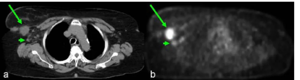
Approximately 40,000 U.S. patients will die of metastatic breast cancer each year. Metastatic breast cancer is incurable, with a median survival of only 2 to 3 years. With newer systemic treatments, perhaps 5 to 10% will live > 10 years, especially those who achieve a complete remission (CR). Predicting for a good response to therapy include good performance status, younger age, and lower tumor burden. Additionally, the natural history of metastatic breast cancer is affected by the initial site of relapse. Patients who relapse initially on their chest wall or in regional lymph nodes typically have a longer life span than patients whose initial relapse is in bones or the lungs. Patients who present with metastatic disease in the liver or brain tend to have a poor prognosis. Imaging modalities used in the diagnosis and monitoring of metastatic disease include PET/CT (Figure 20 a & b) and bone scan (Figure 21a). CT (Figure 21b) is also useful for identification of breast cancer bony metastases as CT has higher resolution, and may aid in assessment of bone integrity and risk for pathologic fracture.
Figure 20 a,b. PET/CT. (a) CT has high spatial resolution for identification of
anatomic structures while (b) PET shows areas of increased metabolic activity. Long arrow points to an invasive ductal carcinoma in the right lateral breast. Short arrow points to metastatic disease to the axillary lymph nodes. University of Massachusetts Medical School, Department of Radiology.
Figure 21 a,b. (a) Bone scan shows heterogeneous radiotracer uptake along the
medial proximal left femur. (b) CT aids with higher spatial resolution showing lytic breast metastasis to the proximal left femur and lower lumber spine. University of Massachusetts Medical School, Department of Radiology.
Conclusion
Management of breast cancer requires a true multidisciplinary management team. As the science develops, both detection techniques and treatment are evolving rapidly. Over the last century, the mortality rate and the late morbidity of treatment and uncontrolled disease have declined dramatically. Hopefully future advances will continue to improve both morbidity and mortality.
Thought Questions
1. Why should patients with early stage (Stages I-III) breast cancer who have a complete resection of their cancer be at risk of relapse?
Your answer:
Expert Answer:
2. If metastatic breast cancer is incurable, why should chemotherapy or hormonal blockade be able to improve survival in early stage disease? Your answer:
3. Survival is identical in patients with early stage disease who are treated with either mastectomy or lumpectomy followed by radiation. Yet there is a risk (<10%) of recurrent breast cancer in the residual breast after radiation. Why would survival in patients who have lumpectomy and radiation be identical to that seen in patients who have mastectomy despite the risk of recurrence in the residual breast? Your answer:
Expert Answer:
Glossary
Adjuvant chemotherapy- Chemotherapy given prior to surgical resection of tumor
Lumpectomy- Surgical procedure to remove breast tumor and a small margin surrounding it
Mastectomy- Total surgical removal of a breast
Neoadjuvant chemotherapy- Chemotherapy given prior to surgical resection of tumor
Nulliparity– The state of never having given birth. Often used (inaccurately) to imply never having been pregnant
References
1. Hsing AW, McLaughlin JK, Cocco P, Co Chien HT, Fraumeni JF Jr.
Risk factors for male breast cancer (United States). Cancer Causes Control. 1998;9(3):269-75.
PubMed Abstract
2. National Cancer Institute. Breast Cancer Treatment- for Health Professionals (PDQ). Stage Information for Breast Cancer. Updated 24 June 2015. Accessed 02 July 2015.
3. Giuliano AE, Hunt KK, Ballman KV, et al. Axillary dissection vs no axillary dissection in women with invasive breast cancer and sentinel
node metastasis: A randomized clinical trial. JAMA.
2011;305(6):569-75.
4. Jagsi R, Chadha M, Moni J, et al. Radiation field design in the
ACOSOG Z0011 (Alliance) trial. J Clin Oncol. 2014;32(32):3600-6.
5. Donker M, van Tienhoven G, Straver ME, et al. Radiotherapy or surgery of the axilla after a positive sentinel node in breast cancer (EORTC 10981-22023 AMAROS): a randomised, multicentre, open-label, phase 3 non-inferiority trial. Lancet Oncol. 2014;15(12):1303-10. 6. Olopade OI, Grushko TA, Nanda R, Huo D. Advances in breast
cancer: pathways to personalized medicine. Clin Cancer Res.
7. Early Breast Cancer Trialists' Collaborative Group (EBCTCG). Effects of chemotherapy and hormonal therapy for early breast cancer on recurrence and 15-year survival: an overview of the randomised trials. Lancet. 2005;365(9472):1687-717.
AJCC staging consists of the disease (cancer site/type), the type of staging or timing of the staging: clinical (c) versus pathologic (p) versus post-neoadjuvant therapy (yp), the stage group (I-IV) and the T (primary site), N (nodal involvement), and M (metastasis) stage categories. Oncologists draw tumor deposits on outlines of body regions, usually with red ink, during the staging of a cancer. The process of drawing helps physicians remember the patient’s stage; drawing a series of stages for a given disease site helps learn the staging system, and makes the AJCC staging table meaningful for visual learners.
In this packet are:
1. Blank staging diagrams to assist with staging the patients you see. Printing out these blank diagrams and staging patients on the diagrams would be a valuable exercise. 2. Case-based interactive staging diagrams to provide a self-assessment of your staging
knowledge. A patient’s extent of disease is described in each case. Write the Type of Staging, Stage Group, T stage, N stage, and M stage in the corresponding blank fields. Clicking on the Expert Answers and Diagrams link will trigger the correct stage and annotated diagram to appear.
The blank diagrams below were created using a CT image set on the Varian Aria Radiation Therapy Treatment Planning System. Images Courtesy of University of Massachusetts Medical School, Department of Radiation Oncology.
This project has been funded in whole or in part with federal funds from the National Library of Medicine, National Institutes of Health, under Contract No. HHSN276201100010C with the University of Massachusetts, Worcester.
AJCC Breast Cancer Staging, 7th Edition is available at:
https://cancerstaging.org/references-tools/quickreferences/Documents/BreastMedium.pdf
Disease:
Breast Cancer
AJCC Breast Cancer Staging, 7th Edition is available at:
https://cancerstaging.org/references-tools/quickreferences/Documents/BreastMedium.pdf
A.
Patient presents with a 2.5 cm mass in the upper outer quadrant of the left breast.Initial physical examination is remarkable for a single, 2x1 cm rock-hard, non-tender, mobile mass in left axilla, consistent with involved lymph node. Patient is otherwise asymptomatic. Needle biopsy of palpable breast mass is positive for ductal carcinoma cells.Disease:
Breast Cancer
Type of Staging Stage T N M
Click here to reveal Expert Answers and Annotated Diagram
breast, and a palpable node in the right supraclavicular fossa. She is otherwise asymptomatic, but has been hiding from her family for many months and has not seen a physician since the birth of her last child 40 years ago. Biopsy reveals grade 1 infiltrating ductal carcinoma in breast. Disease:
Breast Cancer
Type of Staging Stage T N M
Click here to reveal Expert Answers and Annotated Diagram
inner quadrant of the right breast of a 40 year old, asymptomatic nurse. Physical examination is entirely unremarkable. Needle biopsy is not diagnostic, so lumpectomy is performed. Pathology is read as ductal carcinoma-in-situ, with necrosis and high nuclear grade.
Disease:
Breast Cancer
Type of Staging Stage T N M
Click here to reveal Expert Answers and Annotated Diagram
several weeks ago and she felt a small mass under the bruise in the upper inner quadrant. It measures about 1.5 cm, and the rest of the physical examination is normal. Needle biopsy is positive, and lumpectomy and sentinel node procedure is performed. Pathology comes back 1.5 cm invasive ductal carcinoma with 0.4 cm cluster of malignant cells in the sentinel node.
Disease:
Breast Cancer
Type of Staging Stage T N M
Click here to reveal Expert Answers and Annotated Diagram
mass deep in the central right breast, with a 5 cm firm, fixed, mass in the right axilla. She feels fine, and never noticed anything unusual. Biopsy reveals grade 3 infiltrating ductal carcinoma in the axillary mass.
The patient receives 3 cycles of chemotherapy, and the tumor now measures about 2 cm and the axillary mass is no longer palpable. Lumpectomy and sentinel node procedure is performed and pathology reveals a 0.9 cm mass in the central breast specimen and 12 negative lymph nodes.
Disease:
Breast Cancer
Type of Staging Stage T N M
Click here to reveal Expert Answers and Annotated Diagram
mastectomy for high grade ductal carcinoma in the right breast, the tumor measured 4 cm in greatest diameter, the ipsilateral internal mammary node was seen on preoperative MRI and so removed and was positive.
On follow-up now, she complains of back and left shoulder pain, constant, waking her from sleep and morning headache with nausea. MRI of spine reveals tumor in the fourth thoracic vertebra, and bone scan is positive in many areas, including multiple vertebrae and the left proximal humerus. MRI of brain shows an enhancing lesion consistent with metastasis. CT chest demonstrates left lung metastasis and a liver lesion. Disease:
Breast Cancer
Type of Staging __ Stage: ____ T _ N _ M __